Brain Dynamics Underlying the Development of Cognitive Flexibility
←
→
Page content transcription
If your browser does not render page correctly, please read the page content below
Please do not remove this page

Brain Dynamics Underlying the Development of
Cognitive Flexibility
Kupis, Lauren
https://scholarship.miami.edu/discovery/delivery/01UOML_INST:ResearchRepository/12381228840002976?l#13381228830002976

Kupis. (2021). Brain Dynamics Underlying the Development of Cognitive Flexibility [University of Miami].
https://scholarship.miami.edu/discovery/fulldisplay/alma991031583588402976/01UOML_INST:ResearchR
epository

Open
Downloaded On 2021/12/09 11:10:49 -0500

Please do not remove this page
UNIVERSITY OF MIAMI

BRAIN DYNAMICS UNDERLYING THE DEVELOPMENT OF COGNITIVE
                     FLEXIBILITY

                                 By

                           Lauren Kupis

                            A THESIS

                       Submitted to the Faculty
                     of the University of Miami
            in partial fulfillment of the requirements for
                  the degree of Master of Science

                       Coral Gables, Florida

                            August 2021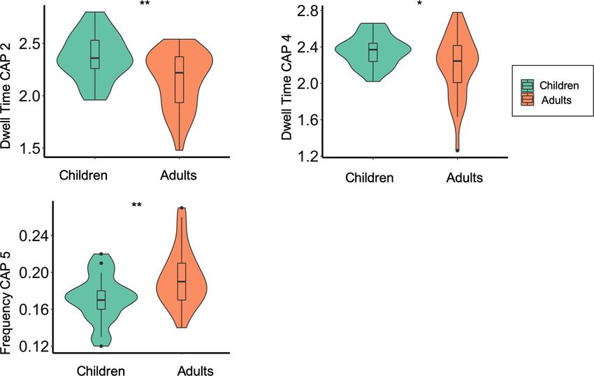
©2021
   Lauren Kupis
All Rights Reserved
UNIVERSITY OF MIAMI

                       A thesis submitted in partial fulfillment of
                           the requirements for the degree of
                                   Master of Science

    BRAIN DYNAMICS UNDERLYING THE DEVELOPMENT OF COGNITIVE
                         FLEXIBILITY

                                     Lauren Kupis

Approved:

Lucina Uddin, Ph.D.                                   Aaron Heller, Ph.D.
Professor of Psychology                               Professor of Psychology

Manish Saggar, Ph.D.                                  Guillermo Prado, Ph.D.
Stanford University                                   Dean of the Graduate School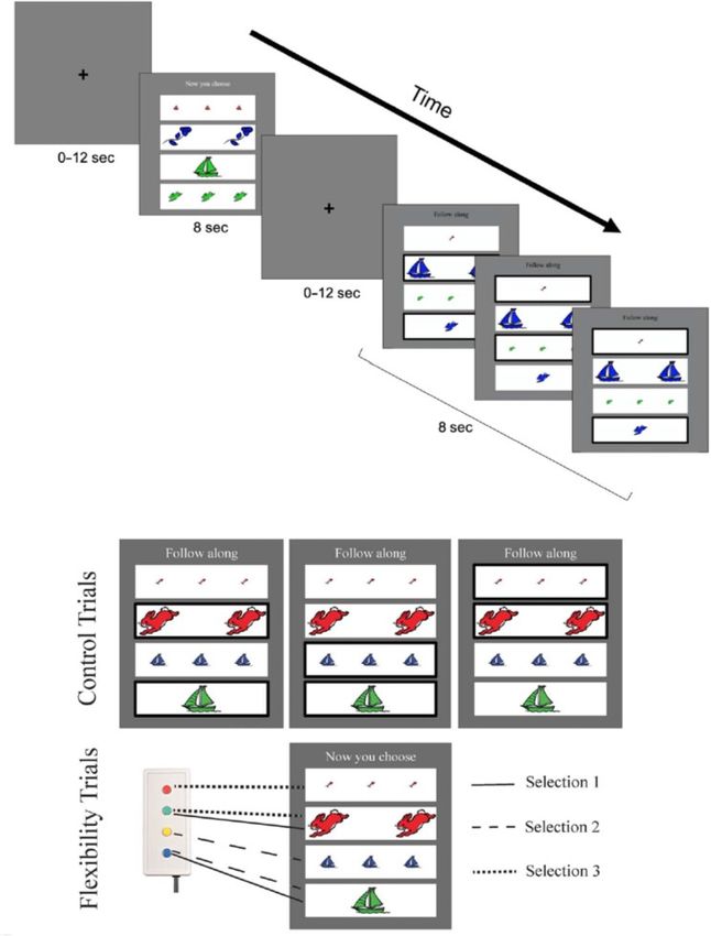
LAUREN KUPIS                                                           (M.S., Psychology)
Brain Dynamics Underlying the Development                                 (August 2021)
of Cognitive Flexibility

Abstract of a thesis at the University of Miami.

Thesis supervised by Professor Lucina Uddin.
No. of pages in text. (68)

       Cognitive flexibility, or the ability to mentally switch according to changing

environmental demands, supports optimal outcomes across development. Despite the

importance of cognitive flexibility for development, little is known regarding the neural

mechanisms underlying it. The goal of the current study was to uncover developmental

differences in the neural systems supporting cognitive flexibility using a functional MRI

task designed to elicit switching mechanisms in both children and adults and by using a

novel method called co-activation pattern (CAP) analysis. The current study examined

neural differences in brain activation and dynamic brain states between children and

adults during a cognitive flexibility task, and the relationships between brain dynamics

and behavior. The CAP analysis revealed that children as compared with adults dwelled

longer in brain states consisting of hybrid brain states consisting of between-network

coupling. Additionally, in both children and adults, more frequent occurrence of a brain

state consisting of coupling between the default and central executive networks was

associated with better cognitive flexibility as measured by the Behavior Rating Inventory

of Executive Function. This study provides the first evidence of the developmental

changes associated with brain dynamic changes during cognitive flexibility and links

brain function with a real-world measure of cognitive flexibility, thereby paving the way
for future research of neurodevelopmental disorders characterized by atypical cognitive flexibility.
TABLE OF CONTENTS

                                                                                                                                  Page

LIST OF FIGURES .................................................................................................................. iv

LIST OF TABLES ................................................................................................................... v

Chapter

     1     BACKGROUND ......................................................................................................... 1
           Cognitive flexibility… ................................................................................................. 1
           Executive functions needed to implement cognitive flexibility… .................................. 6
           Development of cognitive flexibility ............................................................................ 11
           Brain network findings in cognitive flexibility.............................................................. 19
           Specific aims and hypotheses ....................................................................................... 23

     2     METHODS ................................................................................................................. 26
           Behavioral measures ................................................................................................... 27
           Data acquisition .......................................................................................................... 30
           Data preprocessing ...................................................................................................... 30
           Analytic plan............................................................................................................... 32

     3     RESULTS................................................................................................................... 34
           Brain activation........................................................................................................... 34
           Co-activation pattern analysis (CAP)........................................................................... 38
           Age differences in CAPs ............................................................................................. 38
           Brain-behavior relationships with CAPs ...................................................................... 39

     4     DISCUSSION ............................................................................................................. 41
           Brain activation........................................................................................................... 41
           Co-activation pattern analysis (CAP)........................................................................... 43
           Limitations and future directions ................................................................................. 48
           Conclusion .................................................................................................................. 49

FIGURES ................................................................................................................................ 50

REFERENCES ......................................................................................................................... 56

                                                                    iii
LIST OF FIGURES
FIGURE 1…………………………………………………………………………….. 50
FIGURE 2…………………………………………………………………………….. 51
FIGURE 3…………………………………………………………………………….. 52
FIGURE 4…………………………………………………………………………….. 53
FIGURE 5…………………………………………………………………………….. 54
ivLIST OF TABLES
TABLE 1…………………………………………………………………………….. 26
TABLE 2…………………………………………………………………………….. 35
vCHAPTER 1: BACKGROUND
Flexible cognition and behavior are required to adaptively respond to changing
environmental demands. This process is enabled by cognitive flexibility, the mental
readiness to switch to initiate flexible behavioral responses (Dajani & Uddin, 2015).
Cognitive flexibility is a core feature of executive functioning (Diamond, 2013; Logue &
Gould, 2014) and is associated with positive life outcomes including the successful
transition into adulthood (Burt & Paysnick, 2012), resilience to negative life events
(Genet & Siemer, 2011), and better quality of life (Davis et al., 2010). Cognitive
flexibility is also critical for developmental outcomes, including reading and math skills
(Yeniad et al., 2013), social competence (Ciairano et al., 2006), and overall academic
achievement (Titz & Karbach, 2014). Despite the importance of cognitive flexibility
across the lifespan, little is known regarding the neural mechanisms supporting the
development of this ability.
Cognitive flexibility
Examples of cognitive flexibility include the ability to think differently about a
situation, and quickly switch between tasks or strategies to solve a problem. Being
flexible ultimately aids creativity (Lu et al., 2017, 2019), problem-solving (Ionescu,
2012), learning (Kehagia et al., 2010), and resilience to negative life events (Genet &
Siemer, 2011). Cognitive flexibility benefits adaptation via the ability to quickly
transition from different activities or change perspective. Therefore, cognitive flexibility
is an important feature of daily functioning.
Cognitive flexibility is also associated with positive life outcomes including
academic achievement such as reading and math skills (Yeniad et al., 2013), and the
12
successful transition into adulthood (Burt & Paysnick, 2012). Conversely, cognitive
inflexibility may be a risk factor for repetitive or ruminative thoughts (Deveney &
Deldin, 2006; Genet et al., 2013; Whitmer & Banich, 2007), which underlie
psychological disorders such as anxiety, depression, obsessive-compulsive disorder
(OCD), and autism spectrum disorder (ASD) (Keenan et al., 2018; McDougle et al.,
1995; van Steensel et al., 2011). Ultimately, cognitive flexibility is important during
childhood and adolescence because these periods are accompanied by learning,
susceptibility to psychological disorders (e.g., anxiety and depression; (Côté et al., 2009),
and increased substance use (Rose et al., 2019). In all cases, cognitive flexibility may
buffer against negative effects.
Although cognitive flexibility contributes to positive adaptation and learning
across development, the underlying brain regions supporting developmental changes of
cognitive flexibility are not fully understood. Characterizing the brain regions involved in
cognitive flexibility across development may clarify the mechanisms underlying the
cognitive and behavioral changes observed.
The lateral frontoparietal network (FPN) has been found to be important in the
development of executive function broadly (Dajani & Uddin, 2015). Further, flexible
interactions among brain regions and neural networks may contribute to greater cognitive
flexibility (Nomi, Bolt, et al., 2017). However, these neuroimaging findings are primarily
in adults, and reflect the neural processes associated with mature cognitive flexibility. A
more complete understanding of the neural mechanisms underlying cognitive flexibility
may lead to the creation of targeted treatments for neurodevelopmental disorders, the3
updating of classroom curricula, and the creation of individualized preventative
measures for psychological disorders or substance use (Lucina Q. Uddin, 2021).
Terminology
A variety of terminologies exist to describe tasks that have been developed to
study cognitive flexibility. Cognitive flexibility is first considered to be the ‘umbrella’
term describing a cognitive construct that is investigated using ‘set-shifting’ and ‘task
switching’ tasks (Konishi et al., 1998; Monsell, 2003). Relatedly, cognitive flexibility is
referred to as ‘shifting’ in latent models of components of executive function (A. Miyake
et al., 2000).
Task switching and set shifting
Two types of tasks commonly used to assess cognitive flexibility are ‘task
switching’ and ‘set-shifting’ paradigms. Task switching paradigms involve alternating
between tasks, and performance is measured by a ‘switch cost’, or the difference in task
performance (e.g., reaction time) in switching versus non-switching task blocks (Monsell,
2003; Vandierendonck et al., 2010). An example of task switching are rule-switching
tasks, which require participants to switch their response selection (or task) based on the
presented rule (Wendelken et al., 2012). Set-shifting, on the other hand, involves shifting
or switching within a task (Dajani & Uddin, 2015). For example, a commonly used set-
shifting task requires participants to shift attention between color and shape dimensions
and choose the unique attribute (Casey et al., 2004). Although these tasks differ in terms
of switching within versus between tasks, they both are thought to rely on cognitive
flexibility. However, these subtle differences may have important implications in the
neural processes underlying the different types of shifting processes occurring.4
There are different types of task switching and set-shifting paradigms used to
study cognitive flexibility in developmental neuroimaging studies. Broadly, the
categories of these various tasks include: attention-shifting, complex set-shifting, rule-
switching, and performance-based switching tasks. These tasks are often adaptations of
common neuropsychological test batteries that assess cognitive flexibility including the
Dimensional Change Card Sort (DCCS; Zelazo, 2006), the Wisconsin Card Sorting Task
(WCST; Heaton RK et al., 1993), and shifting tasks within the Delis-Kaplan Executive
Function System (D-KEFS; Delis et al., 2001). In the DCCS, usually given to children
between 3 and 5 years, children are presented with two cards (e.g., blue rabbit and red
boat) and are asked to match a series of cards to the two cards based on one dimension
such as color or shape (e.g., blue boat is matched with the blue rabbit) (Zelazo, 2006).
The WCST can be given to individuals from 6.5 to 89 years of age, and requires cards to
be sorted based on one of three dimensions (color, shape, or number) and update the
sorting criteria based on the experimenter’s feedback (Heaton RK, Chelune GJ, Talley
JL, Kay GG, Curtiss G, 1993). Lastly, the D-KEFS consists of several tasks (color-word
interference, card sorting, design fluency, and verbal fluency tasks) that test shifting
abilities.
Functional Magnetic Resonance Imaging cognitive flexibility tasks
Four general categories of fMRI tasks that have been used to study cognitive
flexibility include: attention shifting, complex set-shifting, rule-switching, and
performance-based switching tasks. These tasks range in difficulty in that some have only
one task demand (e.g., attention) or include many demands (e.g., attention, working
memory, and switching). The fMRI cognitive flexibility tasks also differ greatly in the5
type of switching required (e.g., attention versus rule-based switching). The following
section describes in more detail these categories of tasks.
Attention Shifting
In attention shifting tasks, participants shift attention between stimuli dimensions
(e.g., color, shape) within a trial. This type of task has been primarily utilized in
developmental studies, as it is generally easy for young children to understand the simple
instructions (Casey et al., 2004; Dirks et al., 2020; Morton et al., 2009; Yerys et al.,
2015). Attention-shifting tasks are often an adaptation of the DCCS test battery (Ezekiel
et al., 2013; Morton et al., 2009), and therefore, can be given to children as young as 3
years (Hanania & Smith, 2010).
Complex Set-Shifting
In complex set-shifting tasks, participants are still required to shift within a trial,
but utilize more cognitive abilities than simply shifting attention (Yasumura et al., 2015).
One example of a complex set-shifting task is the flexible item selection task (FIST;
Jacques & Zelazo, 2001). Participants are presented with three cards and are instructed to
select 2 cards that match on one dimension (e.g., number, color, size) and then shift by
selecting another matching pair based on a different dimension (Jacques & Zelazo, 2001).
Moreover, the FIST is similar to the DCCS and WCST, but can vary in difficulty by
including more abstract/complex dimensional shifts, which can be further altered by
increasing dimensions or shifts within a trial (Dajani et al., 2020; Dick, 2014).
Additionally, other cognitive processes such as working memory may be recruited to
complete the task depending on the number of switches needed to complete the trial.
Recruitment of other cognitive processes such as working memory and inhibition in6
addition to shifting are thought to support cognitive flexibility (Dajani & Uddin, 2015),
and may better represent real-world scenarios. Because the FIST can be altered to make it
more difficult, and may be more representative of real-world scenarios, children tend to
perform less well compared with adults (Dick, 2014). Therefore, complex set-shifting
tasks may require greater cognitive demands to complete the task.
Rule-Switching
Rule-switching tasks require participants to flexibly respond to a stimulus based
on the given rule (e.g., respond based on color or direction) (Wendelken et al., 2012).
Similar to complex set-shifting tasks, rule switching tasks sometimes require higher order
processes (i.e., working memory) when participants must mentally maintain the rules
(Dajani & Uddin, 2015).
Performance-Based Switching
Performance-based switching tasks typically involve: 1) an adaptation of the
WCST, where participants have to switch between three possible response rules (Crone et
al., 2008), and 2) a probabilistic task (Hauser et al., 2015; van den Bos et al., 2012),
where participants have to switch their stimulus choice based on probabilistic feedback
(e.g., choosing stimulus A receives positive feedback 80% of the time whereas choosing
B results in only 20% positive feedback).
Executive functions needed to implement cognitive flexibility
Cognitive flexibility is a key aspect of executive functioning, or the higher order
processes that enable goal-oriented behaviors. However, other executive functions may
be involved in cognitive flexibility, and are thought to be distinct yet correlated
constructs (Akira Miyake & Friedman, 2012). The executive functions involved in7
cognitive flexibility processes may include working memory and inhibition, along with
other processes such as salience detection and attention (Dajani & Uddin, 2015).
Common behavioral cognitive flexibility tasks, including the Wisconsin Card Sorting
Test (WCST; Heaton RK, Chelune GJ, Talley JL, Kay GG, Curtiss G, 1993), the
Dimensional Card Sort (DCCS; Zelazo, 2006), and various tasks of the Delis Kaplan
Executive Functioning (D-KEFS; Delis et al., 2012), require multiple executive functions
to complete the tasks (Coulacoglou & Saklofske, 2017; Lange et al., 2016; A. Miyake et
al., 2000). For example, during the WCST, participants classify cards differing on color,
shape, or number of designs. Participants need to be able to orient their attention to
salient features of the cards, utilize working memory to recall previously used responses
(Lange et al., 2016), inhibit prior responses (Diamond et al., 2005), and ultimately switch
their response. Therefore, it is theorized that cognitive flexibility is orchestrated in
accordance with other crucial executive functions of working memory and inhibition
(Diamond et al., 2005), and salience/attention processes (Chen et al., 2016).
Salience detection
Salience detection is the process of determining which stimuli are salient and
subsequently drawing attention to the stimuli (Lucina Q. Uddin, 2015). Salience detection
is supported by the mid-cingulo insular network (M-CIN; Salience network) (Lucina Q.
Uddin et al., 2019), including the anterior insula (AI), dorsal anterior cingulate cortex
(dACC), and subcortical and limbic structures (Seeley et al., 2007). The M-CIN
integrates external sensory information and internal emotional and body states that are
used to guide behavior. The M-CIN additionally plays a role as a flexible hub, facilitating
brain network interactions, and allowing salient information to be processed and8
responded to (Sridharan et al., 2008). Specifically, the posterior and mid-insula integrate
and transmit interoceptive signals, whereas the AI orchestrates the brain network
dynamic interactions (Dajani & Uddin, 2015). The flexible variability in the M-CIN is
thought to be an important contributor to flexible behavior, and it has been previously
shown to predict individual differences in cognitive flexibility (Chen et al., 2016).
Bottom-up/top-down attention
Attention processes can be partitioned into dorsal and ventral pathways in the
brain, contributing to goal-oriented (top-down) and stimulus-driven (bottom-up) attention
(Chica et al., 2011). The dorsal-attention network (DAN), comprised of the intraparietal
sulcus (IPS), superior parietal lobule, and frontal eye fields (FEF), supports top-down
processing, whereas the ventral attention network (VAN) comprised of the ventrolateral
prefrontal cortex (vlPFC) and temporoparietal junction, supports bottom-up processing
(Corbetta et al., 2008). The DAN is therefore important for spatial orienting of attention
and feature-based attention (Vossel et al., 2014), useful in cognitive flexibility tasks such
as during attention-shifting.
Working memory
Working memory, or the ability to mentally maintain information needed for an
ongoing task or process (Oberauer, 2019), is largely supported by the lateral-frontal
parietal network (L-FPN; central executive network) or areas of the dorsolateral PFC,
vlPFC, premotor and parietal cortices (Thomason et al., 2009). The L-FPN broadly
supports goal-directed systems and control of information flow in the brain (Niendam et
al., 2012). Working memory is an important aspect in cognitive flexibility tasks, and a
potential confounder if not properly accounted for. The Flexible Item Search Task (FIST)9
is a cognitive flexibility task that requires participants to match stimuli based on a shared
dimension and flexibly switch to make a new dimensional match (Dick, 2014). Further,
as more matches are required, greater working memory is needed to prevent repeating
prior matches. Greater working memory load is also indicative of task performance,
primarily in children as they typically perform worse on tasks requiring high working
memory loads, compared with older children and adults (Cowan et al., 2010). However,
in the FIST, working memory was found not to impact cognitive flexibility performance
when the working memory load was age-appropriate. Overall, working memory is an
important aspect of performing cognitive flexibility tasks, but age-related confounds need
to be accounted for.
Inhibition
Inhibition is regarded as an important function for cognitive flexibility (Davidson
et al., 2006) because prior tasks or responses need to be inhibited prior to switching.
Inhibition is most commonly supported by the brain regions of the right vlPFC (Aron et
al., 2014), AI, and the inferior frontal junction (IFJ) (Aron & Poldrack, 2006). The right
vlPFC plays a role primarily in response inhibition (Sebastian et al., 2016), the rAI plays
a role in detecting behaviorally relevant events and an important region in the M-CIN,
and the IFJ plays a role in detecting behaviorally relevant stimuli (Sebastian et al., 2016).
Inhibition therefore allows for the previous task set to be inhibited prior to switching.
Isolating cognitive flexibility from other executive functions in neuroimaging studies
A challenge with studying cognitive flexibility involves isolating cognitive
flexibility from other core executive functions (i.e., inhibition and working memory).
Difficulty in isolating the process may impact our understanding of the brain regions10 underlying cognitive flexibility across development. Executive functions enable goal- oriented behaviors and extant literature suggests core executive functions are independent constructs yet highly related (A. Miyake et al., 2000). Therefore, isolating executive functions from each other becomes difficult, and maybe impossible, because many of these processes are related and in some cases may even depend on each other. For example, many of the flexibility tasks described above involve working memory to keep the task rules in mind, and inhibition to prevent repetition of previously answered responses. These more complex tasks, however, are thought to be more representative of real-world applications as real world situations integrate multiple processes (e.g., visual, sensorimotor, attention, salience, various executive functions) (Matusz et al., 2019; Bottenhorn et al., 2019). Additionally, in adult studies of the neural substrates underlying cognitive flexibility, there are brain regions activated solely during cognitive flexibility tasks and brain regions activated across tasks of executive functions. Because cognitive flexibility tasks sometimes involve many executive functions, and has been seen to impact the findings in adult fMRI cognitive flexibility studies, studies during childhood and adolescence may not always be testing pure cognitive flexibility. However, study of ‘pure’ cognitive flexibility may not be representative of real-world switching (Bottenhorn et al., 2019). Because of this, multiple considerations need to be made when assessing the results from the developmental fMRI studies of cognitive flexibility including the type of task used, executive functions required to complete the task, and developmental stage of the participants since different components of executive function have different developmental trajectories.
11
Development of cognitive flexibility
Behavioral Findings
Cognitive flexibility and its associated executive functions exhibit different
developmental trajectories across childhood (Anderson, 2002). In general, these skills
begin to emerge during preschool in children as young as 3 years (Hughes, 1998).
Cognitive flexibility skills in preschool children represent lower order forms, such as
shifting, evidenced by children’s ability to perform well on shifting tasks such as the
DCCS (Diamond et al., 2005). Further, integration of other executive functions such as
working memory and inhibition with cognitive flexibility are not yet fully developed in
preschool-aged children, revealed by poor performance on tasks requiring the integration
of various executive functions (Diamond et al., 2005). For example, 4.5 year-old children
successfully perform the DCCS task but perform poorly on the WCST (Chelune & Baer,
1986; Dick, 2014), suggesting cognitive flexibility can be altered by task difficulty, and
higher order flexibility takes longer to develop. Through elementary school age,
cognitive flexibility takes a protracted development compared with other executive
functions (Davidson et al., 2006), and reaches similar behavioral performance as adults
by 10 years of age (Chelune & Baer, 1986; Dick, 2014; Rosselli & Ardila, 1993; Welsh
et al., 1991). However, cognitive flexibility skills continue to develop into adolescence
and adulthood (Anderson, 2002), peaking between 21 and 30 years (Cepeda et al., 2001).
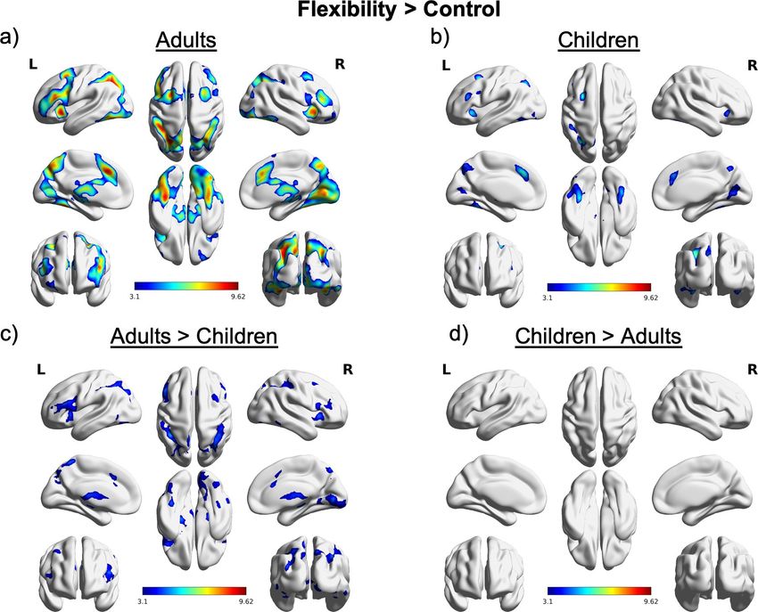
Brain Regions in neurotypical adults
Greater insight into the neurodevelopmental processes associated with cognitive
flexibility can be gleaned from studies of the adult brain. Findings from functional MRI
studies of adults using age-adjusted versions of cognitive flexibility tasks have shown12
core brain regions and networks underlying cognitive flexibility (Dajani et al., 2016), as
well as task-dependent brain regions (C. Kim et al., 2011; Chobok Kim et al., 2012)
(Figure 3). Brain regions found to be involved in core cognitive flexibility across all task
types include the ventrolateral prefrontal cortex (vlPFC), dorsolateral PFC (dlPFC),
anterior cingulate cortex (ACC), right anterior insula (AI), inferior frontal junction (IFJ),
premotor cortex, inferior and superior parietal cortices, inferior temporal cortex, occipital
cortex, and subcortical structures such as the caudate and thalamus (Dajani & Uddin,
2015; Chobok Kim et al., 2012; Niendam et al., 2012). Brain regions involved in
cognitive flexibility further differentiate depending on the type of task (C. Kim et al.,
2011; Chobok Kim et al., 2012). More abstract switching recruits anterior-PFC regions;
moderately abstract switching recruits mid-PFC; and constrained switching recruit
posterior-PFC regions (C. Kim et al., 2011). Results from Kim et al., (2012) meta-
analysis further revealed region-specific activation during attention-shifting tasks in the
dorsal portion of the premotor cortex, and during set-shifting tasks in the frontopolar
cortex. Overall, the findings from adult neuroimaging studies of cognitive flexibility
reveal brain regions associated with cognitive flexibility that may differentiate depending
on the task.
Neural correlates in developmental cohorts
Developmental neuroimaging studies suggest cognitive flexibility takes a
protracted developmental trajectory, as paralleled in behavioral findings. A prolonged
development among the brain regions supporting cognitive flexibility, mainly the
frontoparietal network (Barber & Carter, 2005; Cole & Schneider, 2007), is also observed
in structural findings suggesting the gray matter in those regions mature later than other13
regions (Casey et al., 2000; Giedd et al., 1999). Similarly to adult findings, certain brain
regions are commonly activated in children across switching tasks among the
frontoparietal network such as the dlPFC and the pre-SMA (Morton et al., 2009;
Wendelken et al., 2012), and the basal ganglia (Casey et al., 2004; Crone, Wendelken, et
al., 2006; Rubia et al., 2006). However, activation among the frontoparietal network and
insula increases in activation strength across development and most strongly in adults,
suggesting as these regions develop, cognitive flexibility strengthens (Rubia et al., 2006;
Taylor et al., 2012; Wendelken et al., 2012).
Attention-shifting
Cognitive flexibility can also be observed differentially depending on the
switching/shifting task, as observed in adult studies of cognitive flexibility (Chobok Kim
et al., 2012). Attention-shifting, shifting attention between stimuli dimensions (e.g., color,
shape), has been primarily studied across development (Casey et al., 2004; Dirks et al.,
2020; Morton et al., 2009; Yerys et al., 2015). Children and adults share common brain
activation regions during attention-shifting including the superior parietal cortex, dlPFC,
IFJ, pre-SMA region (Morton et al., 2009), and the caudate nucleus (Casey et al., 2004).
Key developmental differences between children and adults are also seen during
attention-shifting. In Morton et al., (2009) children (11-13 years) had unique activation
among the R superior frontal sulcus, whereas adults had unique activation in the L
superior parietal cortex and R thalamus. This finding implies children may have different
switching strategies compared to adults resulting in differing brain activation patterns. In
another study, Casey et al., (2004) observed more prefrontal and parietal regions in adults
compared to children (7-11 years) suggesting greater recruitment of these cortical regions14
across development. In a study of typically developing (TD) children compared with
children with autism spectrum disorder (ASD), utilizing the same task from Casey et al.,
(2004), TD children (7-12 years) had brain activation in the L posterior supramarginal
gyrus/angular gyrus during shift trials (Dirks et al., 2020). The discrepancies in attention-
shifting findings may be due to small participation sample sizes and the shifting task,
previously shown to be a poor indicator of cognitive flexibility (Dirks et al., 2020).
Set-shifting
Complex set-shifting tasks require different brain regions to complete the task
compared with lower level shifting tasks. Attention-shifting studies primarily utilize the
DCCS, shown to be relatively easy for children at young ages, but the WCST (set-
shifting) appears to be more difficult for children, and in some cases adults (Dajani et al.,
2020; Dick, 2014). Shifting tasks that require greater cognitive flexibility include more
abstract/complex dimensional shifts or rules requiring a greater number of dimensions or
switches (Dajani et al., 2020; Dick, 2014). Further, the requirements during the shift are
also more complex and require using more cognitive abilities (Yasumura et al., 2015).
In one study that examined set-shifting abilities in children using the WCST task,
children had activation in the right insula, a region important for switching between brain
networks to enable flexible behavior (Menon & Uddin, 2010), with increasing activation
with age (Taylor et al., 2012). Only one study in adults has used the Flexible Item
Selection Task (FIST), a complex version of a set-shifting task that requires subjects to
abstract a matching dimension and switch flexibly to a new matching dimension (Dajani
et al., 2020; Dick, 2014). Although the FIST has only been studied in adults, behavioral15
evidence reveals age-related changes at least until 10 years of age, suggesting age-related
neural differences in set-shifting at least until 10 years (Dick, 2014).
Rule-switching
Rule-switching is another cognitive flexibility task that requires higher order
processes particularly in rule switching that requires working memory to mentally
maintain the rules (Wendelken et al., 2012). The earliest study found adolescents had
similar brain activation as adults during rule-switching, among regions of the pre-
SMA/SMA (Crone, Donohue, et al., 2006). In a recent rule-switching study, the authors
utilized a task that required participants to switch flexibly from one task rule to another
(Wendelken et al., 2012). In children (8-13 years) and adults, brain regions of the L
dlPFC, L PPC, and pre-SMA regions were involved in rule-switching trials. The dlPFC
and SMA were similarly activated as in other cognitive flexibility studies. Further, there
were key developmental differences seen between children and adults in their brain
activations during the task. Adults had greater activation overall and among the pre-
SMA, PMC, and L PPC. Children also had regions more activated than adults among the
right inferior parietal lobe and the cingulate gyrus. Additionally, children engaged the L
superior temporal gyrus and the R middle temporal gyrus, in the switch trials compared
with repeating trials. However, this pattern of activation was not seen in adults,
suggesting rule switching may create greater cognitive demands and may be more
difficult for children compared to adults. Further, children had more initial dlPFC
activation driven by the previous trial rule, suggesting children are more likely to
maintain previous rules when it is no longer relevant and may have difficulties with
switching to a new rule set (Wendelken et al., 2012).16
Performance-based switching
Performance-based switching studies have revealed many behavioral and
neurodevelopmental differences between children and adults (Crone et al., 2008; Hauser
et al., 2015; van den Bos et al., 2012). The type of tasks used to test performance-based
switching involve an adaptation of the WCST, where participants had to spatially switch
between three possible response rules (Crone et al., 2008) and a probabilistic task
(Hauser et al., 2015; van den Bos et al., 2012), where participants had to switch their
stimulus choice based on probabilistic feedback (e.g., choosing stimulus A receives
positive feedback 80% of the time whereas choosing B results in only 20% positive
feedback). The insula appears to be an important contributor to performance-switching as
indicated by activation during the WCST adapted task and in adolescents, where greater
activation occurred in the anterior insula for reward prediction errors prior to switching
(Crone et al., 2008). This is also in line with findings in a meta-analysis of developmental
executive functions, where the right anterior insular cortex was shown to have age-related
involvement among inhibition, switching, and working memory (Houdé et al., 2010).
Other regions were important during performance-switching including the DLPFC,
mPFC, ACC, striatum, vmPFC, amygdala, L PCC, L Putamen, R precentral gyrus, L
SFG, and L IPL. The mPFC was commonly observed across the three developmental
studies, consistent with reports of the role of the PFC in cognitive flexibility (Rougier et
al., 2005).
Overview of neurodevelopmental findings
The neurodevelopmental studies of cognitive flexibility provide initial insight into
the developmental processes surrounding switching/shifting, an ability important for life17
outcomes. The regions commonly activated in children during switching/shifting across
task types include the dlPFC and the pre-SMA/SMA. The dlPFC is one region of the
frontoparietal network involved in switching in adults. The dlPFC is primarily thought to
be involved with working memory (Thomason et al., 2009), therefore children may be
using working memory to complete the tasks. However, in cognitive flexibility tasks with
higher loads of working memory, children typically perform less accurately than adults
(Thomason et al., 2009; Wendelken et al., 2012), suggesting the integration of working
memory and switching processes are not fully developed during childhood. The pre-
SMA/SMA was also discovered to be activated in children across multiple developmental
cognitive flexibility studies. Activation of the pre-SMA is commonly seen in set-shifting
tasks (Barber & Carter, 2005) in adults and broadly during executive functioning tasks
(Duncan & Owen, 2000). The Pre-SMA is overall thought to be involved in task-set
reconfiguration, suppression of previous responses, and error likelihood estimation
(Morton et al., 2009).
The developmental neuroimaging studies reviewed also provided insight into the
regions that were not commonly activated in children during cognitive flexibility tasks.
For example, only one study found evidence of activation of the IFJ in both children and
adults (Morton et al., 2009). The IFJ was found to be implemented across various
cognitive flexibility tasks in adults (Dajani et al., 2020; C. Kim et al., 2011). The IFJ was
additionally found to coordinate switches among brain regions during cognitive
flexibility (Dajani et al., 2020). Specifically, the IFJ gets activated first, and leads to
activations in other regions involved in cognitive flexibility. Since this region was not18
commonly activated in cognitive flexibility studies with children, it suggests the ability to
coordinate switching undergoes developmental changes.
Another set of brain regions found to be commonly observed in adults is the M-
CIN (dACC, AI, insula). There were mixed findings surrounding the M-CIN among the
children studies with some instances with increased activation of the insula in children
(Ezekiel et al., 2013; Mogadam et al., 2018; Rodehacke et al., 2014) and conversely some
instances of decreased or no activation of the insula during switching (Crone, Donohue,
et al., 2006; Crone et al., 2008; Dibbets et al., 2006; Dirks et al., 2020; Nelson et al.,
2007; Taylor et al., 2012; Wendelken et al., 2012). The M-CIN supports coordination
among large-scale networks (M-FPN/DMN and L-FPN/CEN) and is thought to enable
flexible behavior (Lucina Q. Uddin et al., 2015). The mixed findings regarding the M-
CIN brain regions in children may be due to the various tasks (i.e., attention-shifting vs.
performance-switching) or varying age groups across the studies (i.e., from 3 years to 17
years). In most cases, children and adolescents were separately grouped, and generally
adolescents had more insula activation than children, suggesting an age-related increase
of activation of the insula (Taylor et al., 2012) associated with better behavioral
performance (Chen et al., 2016). The studies reviewed provide valuable insight into the
development of cognitive flexibility but there are still many questions to be answered,
including how the brain organization of these brain regions enable cognitive flexibility
and changes across development.19
Brain network findings in cognitive flexibility
Brain networks enabling flexibility
A growing body of literature has begun to explore the dynamic brain network
topology that enables cognitive flexibility and flexible behavior. Prior evidence reveals
adaptive behavior is supported by flexible network interactions among the L-FPN and M-
CIN (Braun et al., 2015; Cohen et al., 2014; Cole et al., 2013; Shine et al., 2016). The L-
FPN and M-CIN have the most flexible connections with other brain regions compared
with other brain networks (Cocuzza et al., 2020; Mattar et al., 2016). The medial
frontoparietal network (M-FPN or default network) appears to have the greatest
flexibility using static FC methods (Cole et al., 2013), where the FC is averaged across
the duration of the scan, but recent advanced dynamic FC methods that account for time-
varying FC across the duration of the scan, reveal the M-CIN has flexible connections
(Nomi et al., 2016). Specifically, the right dAIC of the M-CIN acts as a switcher hub,
facilitating flexible interactions between the M-FPN and L-FPN (Nomi et al., 2016;
Sridharan et al., 2008; L. Q. Uddin et al., 2011). The temporal flexibility of the M-CIN
has also been shown to predict individual differences in cognitive flexibility in adults
(Chen et al., 2016).
Functional/Dynamic connectivity cognitive flexibility
Brain network associations, identified using FC or correlations between separate
brain regions (Eickhoff & Müller, 2015), have revealed network interactions in cognitive
flexibility not otherwise seen using brain activation methods. One study evaluated the FC
during a resting-state fMRI scan to task performance on a set-shifting task performed
outside of the scanner. Greater posterior cingulate cortex/precuneus (M-FPN)
connectivity with the ventromedial striatopallidum (basal ganglia) was correlated with
fewer total errors on the set-shifting task, suggesting the relationship between the M-FPN20
and basal ganglia is important for cognitive flexibility (Vatansever et al., 2016).
However, another study found evidence for state-dependent FC relationships between the
L-FPN and M-FPN such that greater L-FPN and M-FPN connectivity during task-state is
associated with cognitive flexibility, whereas greater L-FPN and M-FPN connectivity
during resting-state relates to poorer performance (Douw et al., 2016). Together, these
studies reveal the importance of considering resting- or task-states when investigating
brain network relationships with cognitive flexibility and considering the role of the M-
FPN in cognitive flexibility.
A recent fMRI study examined the neural correlates of cognitive flexibility in
adults using the FIST (Dajani et al., 2020; Dick, 2014). A variety of brain regions were
activated in flexibility versus control comparisons including the M-FPN and M-CIN,
such as the lIFJ extending into the left IFG, dlPFC, FEFs, AI, dACC/pre-SMA and IPL
(Dajani et al., 2020). The regions activated by the flexibility versus control comparisons
were also analyzed using FC network analyses and an extended unified structural equal
modeling approach to estimate ROI to ROI directed network connectivity (Dajani et al.,
2020). They found only the lIFJ was directly activated and other activations seen were
due to their functional connections with the lIFJ. The lIFJ therefore was hypothesized to
be the first region to activate in response to engagement with cognitive flexibility and
leads to engagement among other regions of the prefrontal, parietal and cerebellar
regions. Therefore, the lIFJ modulates cognitive flexibility due to its role in updating task
sets during switching (Chobok Kim et al., 2012). Dajani et al., (2020) additionally found
roles for the dlPFC in maintaining information such as during working memory tasks, the
AI and dACC of the M-CIN as being an important connection to the lIFJ during cognitive21
flexibility, and the lAG of the M-FPN in task performance as reported in the literature
(Douw et al., 2016; Vatansever et al., 2016).
Development
A network approach has also provided greater insight to the brain related changes
associated with cognitive flexibility across development. In a study using the DCCS, an
attention-shifting task, age-related differences were seen among the FC of the lPFC with
the anterior cingulate, inferior parietal cortex, and the ventral tegmental area, showing
stronger activation in adults compared with children (12 years mean age) (Ezekiel et al.,
2013). Children had greater FC between the frontal pole and insula. Together, these
findings suggest children may utilize a different cognitive strategy to implement
attention-shifting as shown by the connection among the frontal pole and insula, and may
develop FC among the lPFC later (Ezekiel et al., 2013).
Brain network flexibility is also shown to change across development, and reflects
changes in cognitive functioning. In children (8-15 years), brain signal variability
increases with age and corresponds to a reduction in behavioral variability and greater
accuracy (McIntosh et al., 2008). Across the lifespan, brain signal variability decreases
across most regions of the brain, but these decreases are offset by increases in select brain
regions including the anterior insula of the M-CIN (Nomi, Bolt, et al., 2017). There are
also age-related changes in dwell time and frequency of certain brain states, and increases
in the number of brain state transitions during task-elicited states across development
(Hutchison & Morton, 2015), suggesting the brain becomes more flexible with age (9
years to 32 years). Additionally, in separate study with a sample of 7-16 year old
children, there were age related increases in temporal variability of FC among the M-22
CIN, L-FPN, and M-FPN (Marusak et al., 2017). The same study also found highly
variable connections between the L-FPN and M-FPN, indicating these regions integrate
early in life (Marusak et al., 2017). Additional work found the functional organization of
the whole brain in children (9-12 years) during resting-state is similar to adults, and
variability in network organization among the L-FPN and M-CIN can be observed in late
childhood (Le et al., 2020). This finding aligns with behavioral evidence suggesting
children reach similar behavioral performance as adults on set-shifting tasks around 10
years of age (Dick, 2014). The dynamic coordination within the brain and primarily
among large-scale neurocognitive networks support the development of cognitive
flexibility.
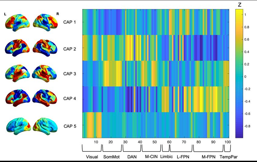
Co-activation Pattern Analysis (CAP)
Although the studies reviewed provide insight into the dynamic network changes
seen across development and their relationship with cognitive performance, the methods
used rely on either ‘static’ methods (i.e., FC), where the time series is averaged across the
fMRI scan (Biswal et al., 1995), or time-varying methods (i.e., dynamic FC), where the
moment to moment changes in FC is captured (Chang & Glover, 2010). FC methods in
particular do not capture time-varying representations of the brain. Further, dFC relies on
the ‘sliding window’ approach (Chang & Glover, 2010) to capture FC changes across a
fixed window length (Preti et al., 2017). FC and dFC methods, although valuable, rely on
many assumptions and arbitrarily collapse data into time and space. Novel dynamic
methods such as co-activation pattern (CAP) analysis (Liu et al., 2013) are increasingly
utilized (Kupis et al., 2020, 2021) because they capture time-varying brain state
alterations not otherwise observed using static methods and in some instances reveal23
more brain and behavior relationships compared with static methods (Lurie et al., 2020).
For the first time, this study aims to examine the neural correlates of cognitive flexibility
across development using a novel brain dynamic method and link brain function during a
cognitive flexibility task with behavior.
Specific aims and hypotheses
Aim 1: To understand the neural bases of cognitive flexibility in neurotypical children
and adults during task elicited brain states.
Aim1a. To examine brain activation responses in children compared with adults
during a flexible item selection task that requires flexible switching between stimulus
dimensions (Jacques & Zelazo, 2001).
Aim1b. To examine dynamic brain state metrics (dwell time, frequency, and
transitions) in children compared with adults during a flexible item selection task that
requires flexible switching between stimulus dimensions (Jacques & Zelazo, 2001).
Hypotheses: Children will have reduced activation in regions previously associated with
flexibility in adults (IFJ, dACC, Cerebellum, AI), but not in regions of the dlPFC, L PPC,
and pre-SMA. The IFJ, dACC, cerebellum, and AI are previously shown to be activated
in adults during cognitive flexibility tasks (Dajani et al., 2020; Dajani & Uddin, 2015;
Chobok Kim et al., 2012). Conversely, the dlPFC, L PPC, and pre-SMA have been
shown to be as strongly activated in children when compared to adults in cognitive
flexibility tasks (Crone, Donohue, et al., 2006; Crone et al., 2008; Morton et al., 2009;
Wendelken et al., 2012).
Children will additionally exhibit longer dwell times and less frequently occurring
brain states associated with brain dynamics during the cognitive flexibility task, and24 have fewer transitions during flexibility trials. Previous research shows children have longer dwell times (Ryali et al., 2016) and less frequent occurrences of certain brain states during rest fMRI (Kupis et al., under review). Additionally, children are hypothesized to have fewer transitions in brain network configurations compared with adults due to growing evidence demonstrating brain state transitions increase across development and during task states (Hutchison & Morton, 2015). Aim 2: To investigate developmental differences in behavioral measures of flexibility (BRIEF) (Dajani et al., 2020; Gioia G. A., Isquith P. K., Guy S. C., Kenworthy L.., 2000; Roth R. M., Isquith P. K., Gioia G. A.., 2005) in children and adults and behavioral associations (reaction time, accuracy, BRIEF) with dynamic brain state metrics during the cognitive flexibility task. Hypotheses: Children will have higher (poorer) scores on the BRIEF shift scales compared with adults. Children will also have less brain activation in the IFJ, dACC, and AI associated with poorer (lower) scores on the BRIEF shift scales. Prior studies found the IFJ to be an important hub for cognitive flexibility tasks in adults (Dajani et al., 2020; Chobok Kim et al., 2012), indicating this region may undergo the greatest developmental changes to support switching mechanisms. Additionally, the dACC and AI of the M-CIN is hypothesized to be less activated and associated with behavioral performance and the BRIEF due to a growing body of literature suggesting this area has the most age-related changes during development during cognitive flexibility tasks (Crone et al., 2008; Hauser et al., 2015; Rubia et al., 2006; Taylor et al., 2012). Additionally, there is prior evidence showing the flexibility within the M-CIN predicts individual differences in cognitive flexibility in adults (Chen et al., 2016). Therefore, it is
25

hypothesized that lower activation in the dACC and AI will be associated with poorer

cognitive flexibility behavior in children.

       Secondly, using CAP analysis, children will have reduced brain dynamics as

measured by dwell time, frequency of occurrence, and transitions during the cognitive

flexibility task, associated with poorer behavioral measures of flexibility compared with

adults. Specifically, longer dwell times, less frequently occurring states, and fewer

transitions is predicted to be associated with poorer performance on the cognitive task

and lower BRIEF scores. Many prior studies have observed increased brain flexibility

with age corresponding with greater task performance (Burzynska et al., 2015; Hutchison

& Morton, 2015; McIntosh et al., 2008).
CHAPTER 2: METHODS
Participants included 32 adults (19-46 years) and 25 typically developing (TD)
children (7-12 years) recruited from the University of Miami and the wider Miami
community (Table 1). Exclusionary criteria included 1) less than 2 usable task runs and
2) incidental findings. Subjects additionally underwent a visual Quality Control
inspection and were excluded if they had one or more visually identifiable artifacts
including but not limited to: excessive motion, ringing, blurring, ghosting, wrapping,
signal loss, and head coverage. All participants were right-handed as determined by self
or parental report, with no history of psychological disorders.
Table 1
Children (N = 25) Adults (N = 32) p-value
Age (year) 10.20 ± 1.50 (7 - 12) 24.78 ± 6.46 (19 - 46) < .001
Sex 16M 9F 17M 15F .409
Ethnicity 16 (Not 15 (Not .053
Hispanic/Latino) 8 Hispanic/Latino) 11
(Hispanic/Latino) 1 (Hispanic/Latino) 6
(NR) (NR)
Race 15 (White) 0 (Black 16 (White) 0 (Black or .002
or African American) African American) 0
0 (Asian) 6 (More (Asian) 0 (More than
than one race) 3 one race) 4 (Other) 12
(Other) 1 (NR) (NR)
Mean FD (mm) .21 ± .11 (.07 - .47) .09 ± .03 (.05 - .15) < .001
BRIEF Shift T 44.84 ± 6.97 (36 - 45.19 ± 9.87 (39 - 77) .882
score 63)
Mean ± sd (minimum - maximum)
2627
Note: M, male; F, female; Mean FD, mean framewise displacement; NR, not
reported; BRIEF, Behavior Rating Inventory of Executive Function.
Participant preparation for MRI
Children participants viewed a 5-minute videotape of a child volunteer
undergoing a fMRI scan to familiarize them of the procedure before their scan date. All
participants underwent a training procedure the day of their scheduled scan. Participants
were trained on the FIST/cFIST by an experimenter on a computer desktop and practiced
a computer-based task and fMRI adapted task before undergoing a mock MRI training.
During the mock MRI, participants completed the same fMRI task. Scanner training was
followed by functional and structural brain imaging along with a task refresher before the
first task run in the scanner. For more details of the experimental procedure see Dajani et
al., 2020.
Behavioral measures
Behavior Rating Inventory of Executive Function (BRIEF)
For children, the Behavior Rating Inventory of Executive Function-2 (BRIEF-2)
was completed by parents (Gioia G. A., Isquith P. K., Guy S. C., Kenworthy L.., 2000).
The BRIEF-2 is a 86-item parent report questionnaire used to assess executive function
and organizational skills in children. The adults were administered the BRIEF-A, the
adult version of the BRIEF. The BRIEF-A is a 75-item self-report measure that assesses
executive function and organizational skills in adults (Roth et al., 2005). Both the BRIEF-
2 and BRIEF-A include a shift subscale and T scores were examined as shifting skills are
a measure of cognitive flexibility (Dajani & Uddin, 2015). T scores at or above 70 are
considered clinically elevated.You can also read