Recent advance in surface modification for regulating cell adhesion and behaviors
←
→
Page content transcription
If your browser does not render page correctly, please read the page content below
Nanotechnology Reviews 2020; 9: 971–989
Review
Shuxiang Cai, Chuanxiang Wu, Wenguang Yang*, Wenfeng Liang, Haibo Yu, and Lianqing Liu
Recent advance in surface modification for
regulating cell adhesion and behaviors
https://doi.org/10.1515/ntrev-2020-0076
received September 01, 2020; accepted September 20, 2020
1 Introduction
Abstract: Cell adhesion is a basic requirement for ancho- Cell adhesion is critical in life systems, ranging from organ-
rage-dependent cells to survive on the matrix. It is the isms to individual cells. It plays an important role in
first step in a series of cell activities, such as cell diffusion, cell communication, cell regulation, organ formation, and
migration, proliferation, and differentiation. In vivo, cells are tissue maintenance [1–5]. As a complex dynamic process,
surrounded by extracellular matrix (ECM), whose physical cell adhesion consists of adsorption of proteins to the sur-
and biochemical properties and micromorphology may af- face and the expression of specific peptide sequences. In
fect and regulate the function and behavior of cells, causing vivo, cells are surrounded by extracellular matrix (ECM).
cell reactions. Cell adhesion is also the basis of communica- ECM is a three-dimensional (3D) network structure com-
tion between cells and the external environment and plays posed of proteoglycans, glycosaminoglycans, adhesion pro-
an important role in tissue development. Therefore, the sig- teins, and fibrin. It provides a wide range of biochemical
nificance of studying cell adhesion in vitro has become in- and mechanical signals for cells and affects a variety of cell
creasingly prominent. For instance, in the field of tissue behaviors [6–8]. Cells adhere to specific surfaces through
engineering and regenerative medicine, researchers have integrins, and those that do not adhere usually die. ECM
used artificial surfaces of different materials to simulate contains proteins recognized by integrin and other cellular
the properties of natural ECM, aiming to regulate the beha- receptors (such as arginine-glycine-aspartic acid [RGD]
vior of cell adhesion. Understanding the factors that affect ligands, fibrinogen, vitamin c protein, collagen, and fibro-
cell behavior and how to control cell behavior, including cell nectin [FN]). These ligands regulate cell physiological pro-
adhesion, orientation, migration, and differentiation on arti- cesses, including adhesion, migration, growth, secretion,
ficial surfaces, is essential for materials and life sciences,
gene expression, and apoptosis, which are triggered by
such as advanced biomedical engineering and tissue engi-
ECM [9,10]. Cell adhesion is also associated with a range
neering. This article reviews various factors affecting cell
of pathological diseases [11], such as arthritis, cancer, os-
adhesion as well as the methods and materials often used
teoporosis, and atherosclerosis. The adhesion of cancer
in investigating cell adhesion.
cells is usually lower than that of normal cells, resulting
in the destruction of tissue structure [12,13]. This morpho-
Keywords: surface modification, cell adhesion, cell beha- logical feature is generally considered a sign of malignant
viors tumors. Research on cell adhesion in vitro has been widely
concerned in the fields of cell biology, biomedicine, and
tissue engineering [14,15], covering biomaterials with im-
* Corresponding author: Wenguang Yang, Department of Robotics plantable sensors, artificial bone and tooth replacement,
Engineering, School of Electromechanical and Automotive skin regeneration, organ transplantation, and so on. Ex-
Engineering, Yantai University, Yantai 264005, China, ploring how to control the behavior of cells on artificial
e-mail: ytu_yangwg@163.com
surfaces is the key to many biomedical and biotechnological
Shuxiang Cai, Chuanxiang Wu: Department of Robotics Engineering,
School of Electromechanical and Automotive Engineering, Yantai applications. In recent years, researchers have devoted
University, Yantai 264005, China themselves to creating structures close to natural ECM to
Wenfeng Liang: Department of Mechanical and Electronic regulate the gene expression of cells in vitro, thus regulating
Engineering, School of Mechanical Engineering, Shenyang Jianzhu cell adhesion, activity, proliferation, and differentiation.
University, Shenyang 110016, China
Surfaces that control cell adhesion are also arousing more
Haibo Yu, Lianqing Liu: State Key Laboratory of Robotics, Shenyang
Institute of Automation, Chinese Academy of Sciences, Shenyang and more interest, and various materials and surfaces have
110016, China been prepared to mimic natural ECM [16].
Open Access. © 2020 Shuxiang Cai et al., published by De Gruyter. This work is licensed under the Creative Commons Attribution 4.0
International License.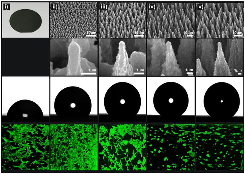
972 Shuxiang Cai et al.
Although the aforementioned methods have been sum-
marized by other researchers, there is an urgent need for
a comprehensive and systematic review of engineering-
facilitated surface-modified cell adhesion techniques. On
that account, this review focuses on recent advances in
surface-modified techniques related to cell adhesion. In
Section 2, we present a series of factors influencing surface
properties related to cell adhesion. Section 3 is about sur-
face-modified methods are demonstrated in detail, followed
Section 4 which discusses about how they work and how
they are used in real-world situations. Finally, in Section 5,
we summarize the current challenges and future directions
of surface-modified methods in relation to cell adhesion.
Figure 1: Cell morphology of bone marrow formed along grooves and
influenced by surface roughness (reproduced from ref. [27]).
2 Factors influencing surface effects on cell adhesion and growth but in a positive
way. For example, Zhao et al. found that the number of
properties related to cell MG63 cells on titanium discs with submicron surface
adhesion roughness is lower than that on flat nanostructures [26].
On the other hand, nano-roughness is considered to be the
closest to natural tissue morphology, and it is an ideal
2.1 Effect of substrate surface topography factor that has a positive effect on cell adhesion, growth,
on cells and maturation. For example, in human venous endothe-
lial cells, increasing the roughness of the biomaterial sur-
Substrate surface topography has been proved to affect face at the nanometer scale can enhance cell adhesion and
cell adhesion [17–19]. Biological tissues in the body have growth on the rough surface.
a variety of surface morphological features such as fibers,
pores, and pits. Various kinds of micromorphological fea-
tures have a specific effect on cell behavior, which we call 2.1.2 Micropores on substrate surface
“contact guidance” [20,21]. Related studies have shown
that micromorphology mainly affects the morphology of On the substrate of cell growth, the micropore mor-
whole cells, while nano-morphology mainly regulates the phology on the surface is another crucial factor affecting
subcellular sensing mechanism [22]. cell adhesion, migration, proliferation, and differentia-
tion. Pore size is the main factor affecting cell adhesion
[28–30]. Previous studies have shown that nanoscale
2.1.1 Roughness of substrate surfaces pores are prone to the formation of collagen fibers and
ECM (Figure 2). Larger pores affect cell seeding, distribu-
Surface roughness is an important factor affecting cell ad- tion, migration, and further neovascularization in vivo.
hesion behavior [23–25]. Some early studies have found Murphy et al. studied the effect of the surface pore size
that surface roughness may affect cell adhesion behavior of polycarbonate on cell adhesion and differentiation
regardless of the cell type and matrix materials (Figure 1). [31]. The adhesion of MG63 human osteoblasts to a mem-
Depending on the irregularities of the material surface, brane 0.2–8 μm in aperture was observed. Studies have
surface roughness can be divided into several different shown that MG63 cells adhere better on the surface of the
grades: macroscopic roughness, microscopic roughness, membrane with pores 0.2–1 μm in size; while on the
submicron surface roughness, and nanometer roughness. membrane with larger micropores (3.0–8.0 μm), the cells
Different surface roughness has different effects on the are spherical with a small amount of filling and foot. In
cells. Macroscopic roughness has little effect on cell adhe- addition, the cells grown on the membrane with pores
sion behavior, because cells have enough space to spread 5.0–8.0 μm in size have a higher degree of differentiation
and grow between macroscopic irregularities. Surface and reach the highest degree of differentiation on pores
roughness on micron and submicron scales has dual 8 μm in size. Hatano et al. found that, compared with a
Recent advance in surface modification for regulating cell adhesion and behaviors 973
Figure 2: The relationship is shown between mean pore size and cell
attachment. Cells cultured on scaffold with pore size of 325 μm shows
the highest percentage of cell adhesion (reproduced from ref. [31]).
smooth surface, osteoblasts attached and proliferated
more effectively on a rough surface (0.81 μm in aperture)
[32]. Various experiments have shown that nanopores on
the substrate can affect cell adhesion and activity [33].
However, the size of nanopores should be within the ap-
propriate range. For example, cells on a smooth surface
will form clamps at the edges, which obstruct the spread Figure 3: Cell adhesion and morphology were controlled by col-
of nutrients, hinder the removal of cell waste, and da- lagen-I and stiffness of substrate (reproduced from ref. [36]).
mage the functions of these cells. Pores with a large size
will reduce the adhesion of cells. Cell adhesion usually increases with matrix hardness. For
example, when mesencgymal stem cells (MSCs) adhere
to type I collagen-modified polyacrylamide (PAAM) gels,
2.2 Effect of substrate surface physical adhesion is marked with parslin. The study also showed
properties on cells that NIH3T3 fibroblasts were more dispersed and adhered
better on the harder collagenous type I-coated PAAM gel
2.2.1 Mechanical properties of the substrate (7.69 kPa), and that the cell survival rate after centrifugation
was >80%, while the softer gel (2.68 kPa) had a cell survival
In vivo, most tissue cells, such as soft brain tissue and hard rate of about 30% only.
bone tissue cells, adhere to fiber ECM with different hard- In addition, the mechanical properties of the sub-
ness and elasticity [34]. The stiffness of ECM in vivo ranges strate surface have extremely important effects on cell
from about 0.1 kPa (brain tissue) to about 100 GPa (bone structure and protein expression [37,38]. Some studies
tissue) [35]. The composition of collagen and elastin in have found that the essential condition for the formation
ECM determines the stiffness and elasticity of fiber ECM. of fibroblast actin stress fibers is that elastic modulus is
Living cells generally perceive the mechanical properties greater than 2,000 Pa. On the contrary, neutrophils seem
of ECM by applying forces and detecting the resulting to be insensitive to changes in stiffness in a large range.
gaps and respond to ECM by regulating local adhesion This indicates that either the mechanical sensing uses the
structure, cytoskeleton tissues, and the overall state. The internal stiffness of the cell as the standard or the signal
stiffness of ECM affects cell activities ranging from gene of cadherin in cell-to-cell contact takes precedence over
transcription, through cytoskeletal remodeling, to intercel- the signal of the cell–matrix adhesion complex.
lular interactions (Figure 3). Engler et al. prepared polyacry-
lamide (PA) gels with different mechanical properties and
studied the relationship between the spread area of smooth 2.2.2 Wettability of substrate surfaces
muscle cells and the elastic modulus of matrix [36]. The
results showed that the elastic modulus of PA gel matrix The wettability (hydrophobicity and hydrophilicity) of cell
had a significant effect on cell expansion and adhesion. adhesion surfaces can affect surface protein adsorption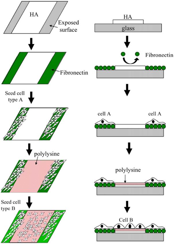
974 Shuxiang Cai et al.
and cell adhesion [39,40]. According to previous studies, 2.3.1 Surface energy of substrate
cells are more likely to adhere to hydrophilic surfaces. For
example, Wei et al. found that the adhesion of osteoblasts Surface energy is regarded as a measure of the unsatu-
decreased when the contact angle increased from 0° to rated bond energy caused by the hanging bond of the
106° [41]. When the contact angle is between 60° and surface material [46,47]. It can affect the activity of cells.
80°, the adhesion of fibroblasts is the highest. The wett- For example, when the polymer surface comes into con-
ability of a surface is greatly affected by the surface func- tact with a biological fluid, serum protein adsorption and
tional groups, the surface roughness of the material, and cell adhesion depend on the energy of the polymer sur-
so on. On the other hand, the super-hydrophilic matrix face. A large number of early studies have reported the
surface (with a contact angle of less than 5°) and the relationship between cell adhesion and matrix surface
super-hydrophobic surface (with a contact angle of more free energy. The surface with high free energy can im-
than 150°) are unconducive to cell attachment and growth. prove cell adhesion and spreading, while that with low
This may be due to the fact that the wettability of the matrix free energy can inhibit cell behavior [48,49]. Surface
surface affects the type, conformation, and binding strength energy can be changed by plasma treatment. For ins-
of the proteins adsorbed from the culture medium, which tance, Ozcan et al. demonstrated that the surface free
further influences cell attachment. If the surface is too hy- energy of poly(methyl methacrylate) (PMMA) membrane
drophobic, the proteins in the ECM (such as FN, vitrein, was enhanced by oxygen plasma, and that fibroblasts and
collagen, and laminin) are adsorbed in a denatured state, serum proteins were cultured on PMMA membrane [50].
and their geometry becomes unsuitable for cell binding. A Syromotina et al. modified poly(3-hydroxybutyrate) (P3HB)
highly hydrophilic surface inhibits the binding of these films first by oxygen plasma and then by ammonia plasma
cell adhesion mediators, thus hindering cell adhesion be- and found that, after plasma treatment, the surface free
havior. Interestingly, surface wettability exerts different energy of the modified P3HB film significantly increased
effects on the adhesion of different types of cells. For (oxygen-modified P3HB = 53.5 ± 0.9 mN/m and ammonia-
example, Wei et al. brushed grafted polyhexamethyldisi- modified P3HB = 57.4 ± 0.9 mN/m) [51]. However, compared
loxane (PHMDSO) on the substrate to prepare PHMDSO with the untreated and the oxygen-plasma-treated P3HB,
with different surface wettability (from hydrophobic to the fibroblasts on the ammonia plasma-treated P3HB mem-
super-hydrophilic) [41]. It was found that, with the enhan- brane adhered and proliferated well. It can be found that
cement of the hydrophilicity of the polymer surface, more once a polymer surface is treated by different plasma, cell
fibroblasts could adhere and spread widely on the surface. adhesion is stronger on the surface with a larger polar com-
Using some hydrophilic materials, Filová et al. found that ponent of surface free energy. In addition, the surface can
the absolute amount of ECM molecules mediating cell ad- affect the number of proteins adsorbed on it through con-
hesion was smaller than that of the more hydrophobic trolling its wettability. Jordi Comelles et al. studied the be-
surfaces formed by octadiene [42]. However, more cells havior of cells on polymer matrix and glass (Figure 4) [52]. A
adhered to more hydrophilic materials. This may suggest linear trend was observed and it was found that serum pro-
that the number of ECM molecules affecting the adhesion teins were preferentially adsorbed on the surface with low
process is not the key factor , but the spatial conformation energy.
of the adsorption molecules mediates cell adhesion.
2.3.2 Surface charge of substrates
The charge property of cell adhesion surfaces is also an
2.3 Cells adhesion influenced by chemical important factor affecting cell adhesion [53,54]. A large
properties of substrate surfaces number of studies have found that cells adhere to positively
charged surfaces rather than negatively charged ones. Sur-
Previous studies have concluded that the chemical proper- face charge can change cell behavior through the chemical
ties of the material surface can change cell adhesion be- functional groups of polymer materials. Lee et al. prepared
havior [43–45]. ECM in vivo provides a variety of chemical polyethylene (PE) surfaces (–COOH, –CH2OH, –CONH2 and
information to cells to guide their behavior. The chemical –CH2NH2 groups) with different functional groups to study
properties that affect cell adhesion mainly include surface their effects on cell behavior [55]. The results showed that
energy, surface charge, and bioactive factors. the adhesion of hamster ovary cells to the functional group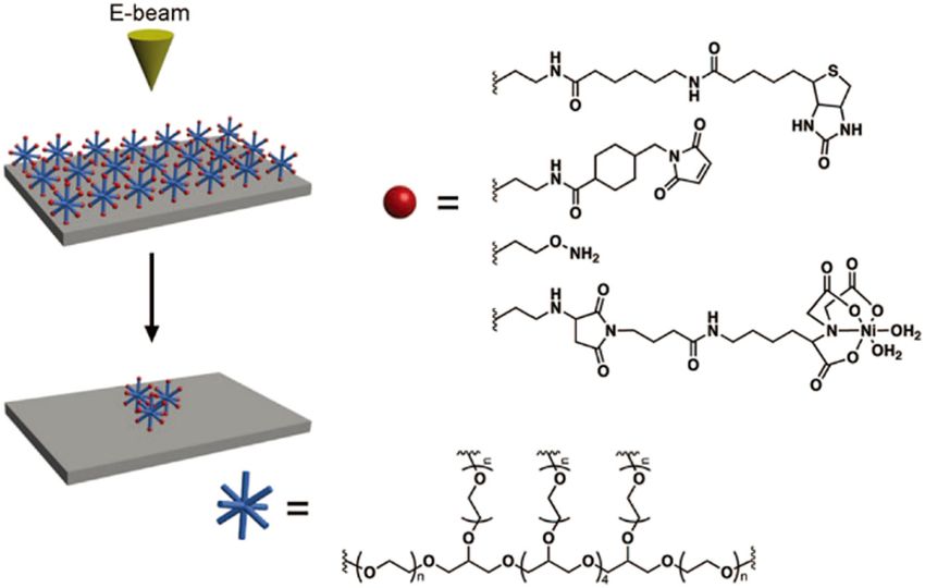
Recent advance in surface modification for regulating cell adhesion and behaviors 975
Figure 4: Total surface energy of different materials (reproduced
from ref. [52]).
Figure 5: Surface modified by immobilized ligands which act as
grafted surface was higher than that in the control surface. agonists of the ECM. Cells cannot adhere to the substrate with
The adhesion, growth, and expansion rate of cells were nonimmobilized ligands leading to apoptosis (reproduced from
optimal on polar surfaces and positively charged surfaces ref. [62]).
(amino grafted PE). On the other hand, surface charge can
regulate protein adsorption, directly bind to integrin, and
tripeptide component of Arg–Gly–Asp. Many biomole-
produce specificity, thus controlling cell adhesion. The-
cules are used to modify material surface to improve ad-
venot et al. mentioned that the incorporation of negative
hesion. For example, planting adhesion-active proteins
charge might promote the adsorption of proteins, thus pro-
on scaffolds, such as type I collagen, vitreous laminin,
moting cell adhesion and reaction [56]. Abarrategi et al.
FN, and laminin, can significantly promote cell adhesion.
reported that the surface with different charge functional
Liu found that the surface modification of type I collagen
groups (–CH3, –OH, –COOH, and –NH2 groups) regulated
scaffold could not only promote cell adhesion and pro-
the adsorption of FN and the direct binding of integrins
liferation but also significantly facilitate the differentia-
and found that the specific trend of adhesion of MC3T3
tion of stem cells into osteoblasts (Figure 5). However,
osteoblasts to the surface of FN coating would be OH >
surface protein modification also has many disadvan-
COOH═NH2 > CH3 [57]. This cellular behavior may be
tages, such as poor protein degradability and difficulty
modulated by the more favorable geometric conformation
in extraction. In this context, the use of RGD sequence as
of vitrein and FN on positively charged surfaces. Interest-
a modification material has emerged as an alternative.
ingly, the negatively charged –COOH group has a dual
RGD is a site that can be specifically recognized by
effect on cell material adhesion. The –COOH is a high-
cells, and the surface modified by RGD can significantly
polarity group that can adjust the wettability of the material
increase cell adhesion. In addition, immobilization of
surface to a level suitable for cell adhesion. However, Bet et
some growth factors, such as bone morphogenetic pro-
al. reported that the introduction of negatively charged
tein (BMP) (an acidic polypeptide) and basic fibroblast
carboxyl into the adhesion matrix reduced the adhesion
growth factor, on material surface can also play an im-
of human erythroleukemia cells to type I collagen matrix
portant role in regulating osteoblast adhesion.
in vitro [58].
2.3.3 Bioactive molecules on the substrate surface
3 Surface-modified techniques for
Biomolecules in body fluids, such as vitronectin, fibri- cell adhesion
nogen, and FN, are immediately adsorbed on material
surface to form a protein layer, which is conducive to In order to further understand the mechanism of cell-sur-
cell adhesion [59–61]. Cells connect with these ECM face interactions, various methods have been developed
proteins through a specific RGD sequence, which is a to change cell adhesion surfaces, as shown in Table 1.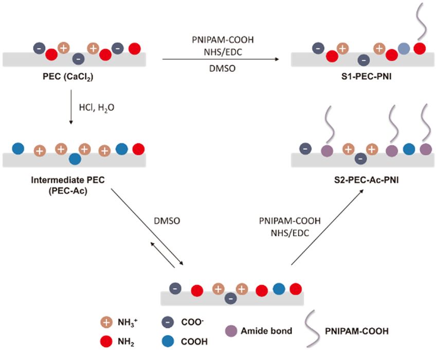
976 Shuxiang Cai et al.
Table 1: Methods of creating cell adhesion surfaces
Techniques Production methods Advantages Disadvantages
SAMs Ordered molecular structures formed by Higher order and orientation Need a specific and
the adsorption of an active substance on specially treated solid
a solid surface in a dilute solution surface
Polymer brush Macromolecular structure composed of Significantly improved the Complex process and easy
polymer chains with one end tightly performance of the substrate, to lose material’s activity
grafted on a curved surface or plane showing different properties as
environmental conditions change
Layer-by-layer Alternate deposition of interacting Controlled layered structures, Depend on centrifugation,
assembly species on a substrate with an simple, inexpensive, and rapid difficult-to-scale, low-
intervening rinsing step following each procedures throughput assembly
deposition
Photolithography Using various sources of energy, such as High precision Complex operation, high-
ultraviolet light, electron beam, and cost equipment
laser, to create patterns on substrate
surface
Electrospun fibers Under high-voltage bias, the polymer Porous fiber structure, high precision High-pressure conditions,
solution or melt is attracted to the of orientation control easy to mechanical
material substrate by static electricity deformation
Spin coating Four basic steps: deposition, rotation, No coupling process variables, high- Low material utilization
rotation, and evaporation cost performance, energy saving, and rate, constant waste
little pollution
3D bio-printing The 2D patterned polymer layer is High precision and speed Shear force, low-cell
surface modified with computer-aided survival rate
imaging technology, while the 3D
patterned polymer layer is assembled
from the bottom up and printed with
liquid biological materials
3.1 Self-assembled monolayers (SAMs) and instance, self-assembled monolayers require the presence of
polymer brush mercaptan on the substrate (only for precious metals or si-
lanes) in order to deposit them. The manufacture of polymer
SAMs were first reported by Zisman in 1946 [63,64]. They brushes requires complex preparations.
are spontaneous molecular assemblies formed by the ad-
sorption of a solution or gas phase to a solid or liquid
surface [65]. When molecules in a solution or gas phase are 3.2 Layer-by-layer (LbL) assembly
adsorbed and spontaneously organized into a single layer
on the surface, a self-assembled monolayer is formed. Poly- The LbL deposition method was first proposed by Moeh-
ethylene glycol, protein, and deoxyribonucleic acid (DNA) wald, Decher, and Lvov 20 years ago [69]. This method
samples are commonly used surface-mount polymer sam- uses self-organized polyelectrolytes alternately adsorbed
ples (Figure 6). Polymer brushes can improve the mechan- on the material surface to form polyelectrolyte multilayer
ical and chemical properties of the surface and introduce (PEM) films [70]. One outstanding advantage of PEM
functional groups into the surface. The free movement of films is that they can maintain the biological activity of
the end of the polymer chain in the solution can endow biomolecules and may provide a large number of bio-
the substrate surface with different micromorphologies and molecules. Their growth and internal structure can be ad-
mechanical properties, thus affecting the behavior of cells. justed by controlling the process parameters. PEM films
An important application of polymer brushes is to make can be prepared in aqueous environment under mild con-
responsive switchable surfaces. For example, thermosensi- ditions, which is a great advantage in the use of biopoly-
tive polyisopropylacrylamide (PNIPAM) brushes are cellular mers and bioactive molecules. Therefore, the LbL compo-
adhesives above lower critical solution temperature (LCST) nents are widely used to regulate these parameters, with
and cellular inert below LCST [66,67]. However, these two the purpose of controlling the adhesion behavior of dif-
surface modification methods have some shortcomings. For ferent cells. Croll et al. put forward antiadhesive LbL films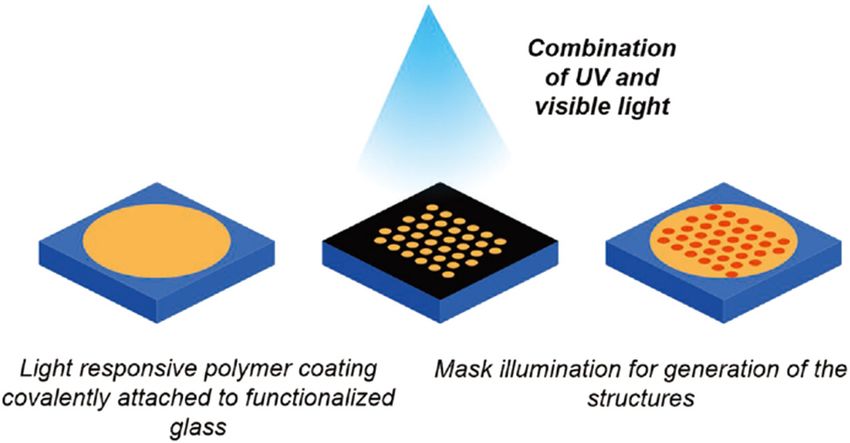
Recent advance in surface modification for regulating cell adhesion and behaviors 977
Figure 6: The PNIPAM-COOH was grafted on chitosan’s amines for cell adhesion via an NHS/EDC coupling (reproduced from ref. [68]).
composed of high-molecular-weight hyaluronic acid (HA) PL is to transfer the pattern on the mask to a photosensi-
and chitosan (CHI). Upon covalent grafting of collagen tive photoresist on the substrate and, after chemical
(COL) IV on their top, the films switched to cytophilic to treatment, leave the exposure pattern on the surface
NIH-3T3 fibroblasts [71]. Kinnane et al. proposed an alter- below the photoresist, or deposit new materials (such
native strategy based on “click” chemistry [72]. First, they as proteins and polymers) on the surface with the desired
employed poly(ethylene glycol) (PEG)-acrylate polymers pattern. The radiation energy types commonly used in PL
with alkyne or azide groups and constructed low-fouling are white light, ultraviolet light, X-ray, electrons, ions,
multilayers through the click-LbL technique. After that, and so on [78,79]. Ultraviolet light is the most widely
they “clicked” an RGD peptide onto the low-fouling films used, but the wavelength used during irradiation limits
to promote the adhesion and proliferation of the epithelial its resolution. Therefore, in a research environment with
cells. The coupled effects of grafting RGD peptide on low- more flexible requirements and patterns, electron beam
fouling, biopolymer-based CHI/HA films were also studied lithography (EBL) is the most common choice.
by Chua et al. and shown to promote the adhesion and EBL is a maskless lithography technology that scans a
proliferation of osteoblasts [73]. The assembly technolo- beam of electrons through a surface covered by a resist film
gies used to assemble such films form five distinct cate- sensitive to these electrons, thus depositing energy on the
gories: (i) immersive, (ii) spin, (iii) spray, (iv) electro- resist film in the desired manner [80]. One of the main
magnetic, and (v) fluidic assembly [74]. These assembly differences between EBL and PL is that the former uses a
technologies affect both the process properties and the much shorter focused electron beam as energy source and
resultant material properties (Figure 7). is exposed to photoresist, so EBL has a stronger anti-inter-
ference ability. Other main features of EBL include ultra-
high resolution (below 5 nm) nano-features, a wide range
3.3 Lithographic surface modification of applications (applicable to a variety of materials), and
techniques availability for preparing a variety of patterns (Figure 8). Its
disadvantages are low speed, complex operation, costly
Photolithography (PL) is the most mature technology EBL equipment, and frequent maintenance.
used to manufacture precise and complex pattern sur- Soft lithography (SL) is a micro-nano processing tech-
faces with resolution up to hundreds of nanometers nology based on elastic seal printing [82,83]. Elastic seals
[76,77]. In general, to prepare a surface pattern using with patterned relief structures are used to generate978 Shuxiang Cai et al.
microscope [84]. Because of its ability to control a large
number of processes by using sharp probes in contact with
the nanometer area of the sample surface, the lithography
technology has been widely used. For instance, atomic force
microscopy (AFM), pen-immersion nano-lithography, poly-
mer pen-type lithography (PPL), and pen-immersion nano-
displacement lithography can be used to produce poly-
mer patterns with nano-resolution and high registration.
Scanning probe lithography (SPL) uses scanning tips to gen-
erate patterns on ultrahigh-resolution surfaces. One feature
shared by all scanning probe-based technologies is the use
of sharp scanning probes to produce local modifications on
the surface. It is well-known that AFM is embraced by the
most current SPL methods. Compared with other technolo-
gies such as EBL, SPL is advantageous in that it is a single-
step process with resolutions up to 10 nm. Most SPL writing
processes are “direct writes” in nature, and no additional
development steps are required to produce the desired pat-
terns. SPML combines nanoscale feature size, low technical
requirements, and the ability to handle soft matter from
small organic molecules to proteins and polymers. In addi-
tion, the scanning probe microscope can detect surface char-
acteristics of atomic resolution. Compared with the beam-
based method, the imaging and patterning in SPL are ortho-
gonal, that is, the imaging process does not affect the writing
structure, nor does it involve partial writing operations.
Figure 7: Schematic diagram of HA-PLL deposition on substrate. The Extreme ultraviolet (EUV) irradiation is another litho-
HA surface was firstly deposited on substrate to prevent cell adhe- graphy technology [85]. The photons of EUV are high-
sion. Then the PLL was applied to the surface and converted HA energy and low-wavelength photons, ranging from 10
surface to cell and protein adhesive(reproduced from ref. [75]). to 124 eV, corresponding to 124 to 10 nm in wavelength.
Photons in this energy range can destroy multiple bonds
of polymer materials and introduce microscopic and
nanoscale structures by direct lithography. Because the
patterns and structures with feature sizes from 30 nm to EUV photons have limited penetration depth, they can be
100 μm. The process of preparing polydimethylsiloxane used for surface modification of polymers without chan-
(PDMS) seals usually involves pouring PDMS prepolymers ging the volume properties of the treated materials and
onto patterned molds (Figure 9), which are usually made by are often used to optimize the roughness of polymer
PL or EBL. After curing, they are stripped from the molds. materials. For example, polytetrafluoroethylene (PTFE),
The surface of these protruding nano- or micro-embossed a hydrophobic polymer material, is widely used in tissue
seals can carry the printed material, which is then trans- regeneration. This material has inherent low surface
ferred to the substrate surface. Therefore, SL is a simple, energy, which makes it chemically inert to a large extent
high-throughput, and inexpensive technology. It provides and very suitable for passive structural applications.
a convenient, effective, and low-cost method for the forma- However, its disadvantage is also obvious, that is, the
tion and fabrication of micro-nanostructures and is neces- surface is incompatible with cell adhesion and easily
sary in the large-scale production of patterned polymer affected by pathological conditions, such as peeling off
surfaces. However, there are still many problems with SL, the surface of vascular grafts. In order to modify the
such as how to use elastic materials to reproduce impres- highly stable surface of PTFE, the EUV irradiation in
sion images with high precision, and how to control the the presence of nitrogen is used to increase surface
deformation and distortion of elastic materials. roughness. The average surface roughness of a polymer
Scanning probe microscopy-based lithography (SPML) treated with EUV is more than 4 times higher than that of
is a lithography technology based on a scanning probe an untreated one, which enhances the hydrophobicity ofRecent advance in surface modification for regulating cell adhesion and behaviors 979
Figure 8: End-functionalized eight-arm PEG polymers were cross-linked in specific patterns using electron beam lithography for protein
patterning (reproduced from ref. [81]).
Figure 9: Soft lithography uses elastomeric stamp to replace hard stamp in traditional lithography to fabricate arrays of microstructures
onto hydrogel surfaces (reproduced from ref. [82]).
PTFE and significantly improves the adhesion and mor- polylactic acid micropore structure prepared by 3D fused
phology of L929 fibroblasts. wire, so as to enhance the functionalization ability of the
In practical applications, especially in the fields of implant material. The direct laser writing technique with fs
biomedicine and tissue engineering, surface modification laser pulses (temporal pulse width r = 300 fs, λ = 1,030 nm,
is an indispensable process to improve the compatibility or repetition rate v = 25 kHz) permits the creation of features
biocompatibility of biomaterials. Femtosecond laser pro- with great reproducibility and does not require a clean
cessing is a common surface pattern and construction room. The laser-induced microfeatures improve the sur-
technology [86,87]. For example, the surface is modi- face roughness of the PLA construct and enhance cell ad-
fied through producing grooves and micropores on the hesion in relation to cell types used for cell growth [88].980 Shuxiang Cai et al. 3.4 Electrospun fibers 3.5 Spin coating Electrospinning is driven by the free charge on the sur- Spin coating is a simple and high-throughput technique face or inside of the polymer liquid [89,90]. The polymer for depositing large-area polymer films on the substrate solution or melt is electrostatically drawn to the material surface [94]. Film thickness can be controlled with high substrate under high-voltage bias. Electrospinning can precision. Currently spin coating is the main technology produce fibers with 10 nm to several microns on the sub- for the preparation of micron and nanometer organic strate. Chen et al. studied the effect of the shape and size photosensitive films (Figure 11). Spin coating consists of of electrospun polycaprolactone (PCL) fibers on the adhe- four basic steps: deposition, rotation, rotation, and eva- sion properties of fibroblasts [91]. The results showed that poration. Seen from maturity, spin coating has many the cell adhesive rate of scaffolds with a higher specific advantages in coating operation, and its biggest advan- surface area increased significantly and that nanofibers tage is that there are no coupled process variables. By could promote cell adhesion better than microfibers. One changing the rotation speed or switching to the photore- outstanding advantage of electrospinning is that it can sist with different viscosity, it is easy to change film thick- produce porous fabrics which can enhance the adsorption ness and obtain a film with uniform thickness. However, of proteins and provide a larger specific surface area the most prominent disadvantages of spin coating are for the application of more binding sites on cell mem- low material efficiency and insufficient utilization. brane receptors. Therefore, electrospinning is often used to create 3D scaffolds with high porosity and spatial con- nectivity. Another advantage is that it can achieve precise alignment control to produce oriented nanofiber networks 3.6 3D bio-printing through adjusting the electric field and the substrate direc- tion [92]. However, the shape of densely deposited low- 3D scaffold is a common tool for cell culture [96]. The porosity fibers hinders the effective penetration of cells. To common methods of making scaffolds include molding, overcome these limitations, salting out and low-tempera- pore-forming agent leaching, gas foaming, fiber bonding, ture electrospinning are developed to increase the pore freeze-drying, solvent casting, and so on. However, the size [93]. LbL electrospinning is utilized to form a thick preparation of 3D polymer scaffolds by these traditional multilayer fiber platform composed of nanofibers and mi- methods usually results in randomly distributed geo- crofibers (Figure 10). However, these approaches still lead metry that seriously limits cell adhesion and diffusion. to mechanical deformation during transport. Therefore, an Recently, some new methods for fabricating scaffolds ideal engineering fiber platform should have high porosity have been put forward, such as selective laser sintering, to provide better nutrient diffusion and inward cell growth melt deposition molding, and stereoscopic lithography as well as reasonable complex structures to mimic natural technology. The 3D bio-printing surface modification of tissues and maintain mechanical properties. 2D patterned polymer layers is conducted by micro-/ Figure 10: Soft lithography uses elastomeric stamp to replace hard stamp in traditional lithography to fabricate arrays of microstructures onto hydrogel surfaces (reproduced from ref. [82]).
Recent advance in surface modification for regulating cell adhesion and behaviors 981
is the most important property. This chapter introduces
several common biomaterials, as shown in Table 2.
4.1 Silicon
Silicon has special semiconductor properties and is often
used as a material for implanting electronic devices in
vivo. Silicon has been shown to increase the biological
activity of various materials without affecting their mec-
hanical properties or inducing cytotoxicity. Up to now, a
series of manufacturing technologies have been applied
to the surface characteristics of silicon on the micro- and
nanoscale [97]. It is found that surface-modified silicon
can significantly improve cell adhesion. However, the
biocompatibility of silicon in vivo is very poor, and the
biological stability is also insufficient, so silicon is often
covered with surface coating materials in application to
improve biocompatibility. Porous silicon is a nontoxic
Figure 11: Biodegradable polymer film was coated on Mg samples and biodegradable biomaterial with great application
using magnesium by spin coating, and cells were cultured with these potential. Surface modification can not only control the
different samples for 1, 4 and 7 days (reproduced from ref. [95]).
degradation rate but also promote cell adhesion. Low
et al. modified the surface of porous silicon by ozone oxi-
dation, silanization, collagen, and serum coating [98]. The
nano-manufacturing methods to create 3D structures. adhesion of rat pheochromocytoma (PC12) and human
Another method is to use the bottom-up assembly of 2D lens epithelial cells to these surfaces was studied. It was
patterned polymer layers. The 3D structures are fabri- found that the two cell lines had more adhesion to col-
cated by assembling 2D patterned polymer layers from lagen coating and aminosilanized porous silicon, while
the bottom up. It is a relatively advanced manufacturing cell adhesion on the surface of ozonation and polyethy-
method that is widely used for preparing 3D tissue struc- lene glycol silanization was poor. In addition, silicon sub-
tures for medical and tissue engineering. It creates cell strate is also commonly used as a model scaffold to study
patterns by accurately printing live cells, biochemicals, the effect of 3D micro-/nano-morphology and surface en-
and biomaterials LbL. One outstanding advantage of this ergy on cell adhesion and growth. Ranella et al. studied
method is that the printed cells can remain alive. The the adhesion and activity of fibroblasts on highly rough 3D
technology also has drawbacks, i.e., the shear force and silicon (Si) surfaces with gradient roughness ratio and wett-
the impact force of droplets during the spraying process ability (Figure 12) [99]. The results showed that cell adhe-
affect the activity of the printed cell fluid. At the same sion had nothing to do with surface chemical composition
time, the printed cells or molecules must remain liquid or wettability and was better on the silicon surface with less
before printing and must be cured immediately after roughness, indicating that the adhesion of fibroblasts was
printing to maintain a viscoelastic state. This liquid-to- not monotonically dependent on surface energy.
solid transition, which must be prevented from damaging
cells, bioactive factors, or other particles, also poses con-
siderable challenges to the development of 3D printing.
4.2 Metals
A large number of metal materials are used in tissue
4 Materials for cell adhesion engineering and regenerative medicine, such as human
titanium alloy dental implant, titanium alloy bone trans-
Cells respond differently to different materials. On the plantation, and stainless steel used in heart scaffold
surface of cell culture, the biocompatibility of materials [100–102]. Therefore, studying cell adhesion on metal982 Shuxiang Cai et al.
Table 2: Several common materials for cell adhesion
Materials Advantages Disadvantages Application
Silicon Increase biological activity, improve Poor biocompatibility, unstable Bone tissue engineering
cell adhesion
Metal High strength, toughness, fatigue Inadequate long-term security Hard tissues such as bones and teeth that
resistance, easy processing and and reliability need to bear higher loads and stents for
forming interventional treatment
Polymer Biocompatibility, biodegradability, and High production cost and Organ repair and transplantation
low toxicity complex synthesis process
Nanotubes Special and excellent performance in Carbon nanotubes are poorly Adsorption materials
mechanics, electricity, magnetism, and dispersed and difficult to process
optics
Bioceramics Biocompatibility, low toxicity Low toughness and strength Dental restoration materials, artificial hips,
and other artificial bones, tooth roots,
joints, bolts, etc.
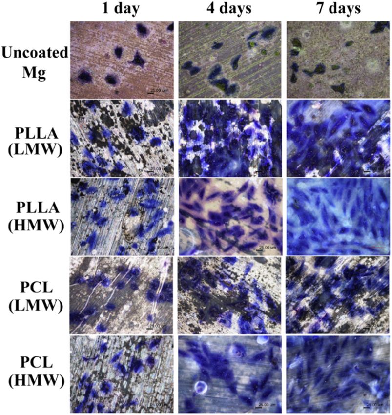
surface in vitro is of great interest. Because the metal surface also affects cell behavior (Figure 13). For example,
surface has high surface energy, it often enhances cell titanium surface with random nano-morphology can pro-
adhesion. J. Hallab et al. used the jet impact method to mote the adhesion of vascular endothelial cells. There-
determine the adhesion strength of 3t3mc fibroblasts to fore, more and more optical techniques, such as laser
metal and polymer and identified the colonization of ablation, are used to change the surface properties of
3t3mc fibroblasts on hs25 (a cobalt-based implant alloy,
metal materials, thus modifying the adhesion properties of
astmf75), 316l stainless steel, ti-6al-4v (a titanium implant
cells on metal surfaces. Many deficiencies in metal mate-
alloy), commercial pure tantalum (ta), PTFE, silicone
rials, including osteolysis, edema, thromboembolism, endo-
rubber (SR), and high-density polyethylene [49]. The
results showed that the adhesion strength of cells to the thelial overgrowth, infection, tumor, and other adverse
metal material was about 5 times higher than that of reactions, are often caused by fatigue corrosion (such as
the tested polymer material. At the same time, many wear and metal ion dissolution). This points out the direc-
studies have found that the nano-morphology of metal tion of metal material improvement in the future.
Figure 12: Cell adhesion was regulated by controlling the roughness and wettability of 3D micro/nano silicon structures (reproduced from
ref. [99]).Recent advance in surface modification for regulating cell adhesion and behaviors 983
surface of chemical groups or carry out pattern modifica-
tion to improve biocompatibility and bioactivity [105].
Many studies have shown that surface modification plays
an important role in cell and tissue reactions. In addition,
the application of thin films is widely regarded as a
promising method to change the surface properties of
biomaterials. Thin films are widely used in tissue engi-
neering, mainly for their simple processing process, low
production cost, and unique ability to control various
physical and chemical properties. They are usually used
in dental and orthopedic implants, biodegradable scaf-
folds, and osseointegration of biomimetic materials in the
fields of tissue engineering and biomedicine.
4.3.3 3D polymer scaffolds
Cells in vivo are surrounded by ECM, a 3D natural struc-
ture. For the purpose of maximizing the simulation of the
Figure 13: Enhanced adhesion of osteoblasts was observed on environment around cells in vivo, many 3D biological scaf-
nanometer particles of alumina compared to conventional metals folds have been developed in tissue engineering. These
(reproduced from ref. [101]). biological scaffolds affect cell behavior in a 3D way, which
offers the most suitable culture tool for the real growth
4.3 Polymer environment of cells. Pore size, porosity, shape specificity,
binding with natural tissue, degradation according to
4.3.1 Polymer gels tissue formation rate, and cost-effectiveness are important
factors in the development of scaffolds. The chemical and
Polymer gels are classified into physical gels and che- mechanical properties of 3D polymer scaffolds made of
mical gels [103]. Silicon-based elastomer PDMS gel is micropores, microfibers, or nanofibers are similar to those
more common. As a low-cost elastomer material, PDMS of natural tissues. Scaffolds with different softness and
gel is characterized by simple preparation process, low hardness have different applications. For instance, stents
cost, and good biocompatibility. Because it can provide a with higher hardness are often used in bone regeneration;
wide range of stiffness values for living cell culture, it is the stents used for bladder, vein, and artery regeneration
widely used to simulate ECM and in tissue engineering are mostly soft and elastic materials.
and regenerative medicine. In addition, hydrogel has
great application potentials in the 3D cell culture and
tissue regeneration medicine. By virtue of its unique si- 4.3.4 Nanotubes
milarity to natural ECM in composition and structure as
well as its important role in cell proliferation and sur- Nanotubes prepared from various materials have been
vival, hydrogel is often the main candidate material for widely used in tissue engineering. They can improve the
engineering tissue scaffolds. mechanical or conductive properties of the substrate or
form specific micromorphologies on the surface. Carbon
nanotubes are most widely used in all materials [106,107].
4.3.2 Polymer thin films Because of their good mechanical, physical, and chemical
properties, they present a great application potential in
Surface bioactivity (or biocompatibility) is an important the fields of tissue engineering and regenerative medicine.
property of materials, so it is very important to control the Lovat et al. functionalized carbon nanotubes, prepared
surface properties of biomaterials and to ensure their layered carbon nanotubes, directly introduced hippo-
biocompatibility and bioactivity. Organic films and coat- campal neurons into the surface of carbon nanotubes,
ings, especially polymer films, are most commonly used and compared them with glass matrix [108]. The results
in biomaterials, because they can be combined on the showed that cell adhesion, dendritic elongation, and984 Shuxiang Cai et al.
Figure 15: Neonatal hippocampal neuron adhesion (b) and survival
were boosted by purified multiwalled carbon nanotubes (MWNTs)
deposited on substrate (a) (reproduced from ref. [110]).
Figure 14: Light-responsive polymer was coated on glass substrate
and hierarchically structures were generated by mask illumination relatively dense. This dense ceramic surface will affect the
(reproduced from ref. [104]). recognition of cells to the material, and even affect the ad-
hesion, spreading, proliferation, differentiation, and func-
nerve signal transmission were stronger on the surface of tional expression of cells on material surface. In vitro studies
conductive nanostructures than in the control group. have shown that the treatment of material surface and the
Titanium is also frequently used in biomaterials, thanks introduction of viscous protein molecules conducive to cell
to its good biocompatibility and common uses of bio- recognition can increase cell adhesion and proliferation.
medical implants. Oh et al. have shown that the adhesion Bioceramic materials also have some shortcomings, such
and differentiation characteristics of human bone marrow as the inadequate strength and toughness of the materials.
mesenchymal stem cells can be controlled by growing tita- However, bioceramics has a broad development prospect
nium nanotubes with different diameters [109]. The beha- because of its biocompatibility and almost nontoxicity.
vior of cells on nanotubes was observed by electron micro-
scope. It was found that the change in nanotube size had a
significant effect on cell characteristics (Figure 14). Most 4.3.6 Dynamic materials
cells adhered to the 30 nm nanotubes but did not differ-
entiate. Larger diameter nanotubes of 70–100 nm signifi- As is well-known, the ECM around the cells in the body is
cantly promoted the morphological elongation of cells. a dynamic 3D structure, and the cells live in the ecolo-
gical environment [113]. The culture surfaces are used
in many in vitro experiments only for the purpose of
4.3.5 Bioceramics studying cell behavior on a static basis, and the proper-
ties of the substrate surface remain unchanged, which is
Calcium phosphate ceramics (CPCs) is a kind of tunable incapable of reflecting the dynamic characteristics of
bioactive materials widely used in bone tissue repair and ECM. In this context, studying dynamic surfaces can
reinforcement [111]. They have the surface properties of more accurately restore the real living environment of
supporting osteoblast adhesion/proliferation (i.e., bone cells [114]. There are also materials that can achieve the
conduction) and stimulating new bone formation (i.e., response of the material surface by changing the pH,
bone induction). Hydroxyapatite, tricalcium phosphate, temperature, light, and other external stimuli. We call
amorphous calcium phosphate, and biphasic calcium this type of material a “stimulus response” or “smart”
phosphate have inorganic composition and crystal struc- material. Examples in this regard include temperature-
ture similar to those of natural bones, so they are the responsive polymers modified with nano-thick PNIPAAm
most commonly used bioceramics in orthopedic surgery. graft layer, photoresponsive polymers modified with nitro-
In recent years, with the research and development of benzyl ester derivatives, UV-mediated rigid modulated
tissue engineering, porous CPCs, as a kind of bone tissue hydrogels, and dynamically controllable cell culture
engineering cell scaffolds, have been widely used in the surfaces. This provides a way for us to understand
repair and replacement of bone defects [112]. At present, how cell functions and fate are affected by continuous
many methods of preparing porous ceramics have been changes in cell niche. In addition, a dynamic surface
developed, such as organic foam impregnation method, also shows better bionic performance than a static one.
foaming method, and adding pore-forming agent (Figure In fact, based on their unique advantages, dynamic
15). These methods usually need to be formed by high-tem- surfaces are also used in in vitro diagnosis, intelligent
perature sintering (>1,100°C), and the ceramics obtained is robot skin, and other intelligent systems. So far, theRecent advance in surface modification for regulating cell adhesion and behaviors 985
dynamic properties of materials are mainly limited to longtime. For instance, Jeon, Hojeong et al. used direct-
simple physical and chemical properties and unable to write ablation lithography to fabricate nanocrater for
completely simulate complex situations in the body. directing cell migration and organization [88]. While na-
Therefore, the most advanced bionic design standards noscale craters could modulate cell adhesion, the super
should be introduced to develop “more intelligent” clean experimental environment is required. Once the fab-
dynamic surfaces. ricated surface was contaminated with dust, nanoscale
craters are easier to be damaged. To overcome these chal-
lenges, materials with natural cell-adhesive properties are
expected to be developed, and these materials can be used
5 Conclusion and prospective for cell adhesion without any treatment.
Furthermore, it appeared that the combination of na-
Surface modification for cell adhesion and regulating cell noscale and microscale topographies could be superior to
behaviors, which could gain a fundamental understan- using each single-size scale alone for cell adhesion. It is
ding of how cells respond to these structures, is vital for generally known that the microstructures of biological
a broad applications in cells research, drug discovery, and interface and tissues exist, ranging from nanometer to
tissue engineering. For instance, PEG diacrylate (PEGDA), micrometer scale, and these two-size scaled microstruc-
a common hydrogel, was widely used for tissue engi- tures have an impact on biological functions. Researchers
neering owing to its tunable mechanical properties and have found that the elongation of endothelial cells was
biocompatibility. However, the native PEGDA film shows enhanced once nanofibrous matrices deposited on the
bio-inertness which means that cells cannot adhere to the surface of the micrograting substrates [115]. On the other
surface of this film, thereby limiting its applications. In hand, combination of different technologies is the trend of
order to solve this problem and fabricate cell-friendly surface modification technologies. Lithographic techni-
PEGDA film, researchers have attempted to modify the phy- ques were usually used to change the physical properties
sical or biochemical properties of PEGDA film for cell adhe- of surface. Combination with other biochemical methods
sion. One of the most effective and common methods to including LbL deposition method will bring considerable
achieve this effect is functionalized with proteins such as improvement in cell adhesion.
FN-derived arginine–glycine–aspartic acid–serine (RGDS) Overall, the significance of studying cell adhesion
sequence to form PEG-RGDS conjugation. Within this con- and a comprehensive understanding of cell substrate in-
text, a general overview of surface modification for cell teractions in vitro have become increasingly prominent
adhesion was introduced. Influence factors of surface prop- such as in the field of tissue engineering and regenerative
erties for cell adhesion were firstly presented. Then surface medicine. Although significant challenges abound, the
modification methods were demonstrated through working method and materials for surface modification should
principles and functions for cell adhesion. Finally, a large be improved continuously to make exciting contributions
number of material surfaces including silicon, metals, to fundamental biology.
polymer, and ceramics, which could be modified for cell
adhesion, were summarized. However, new challenges Acknowledgements: The authors wish to acknowledge
need to be addressed in future studies. the funding provided by the National Natural Science
More accurate and sophisticated 3D surface modifi- Foundation of China (Project No. 61803323 and 61973224)
cation are required. Generally, cells cultured on plain 2D and Natural Science Foundation of Shandong Province
plate were studied in vitro. However, in vivo, cells were (Project No. ZR2019BF049).
usually viable in 3D structures. The existing surface-trea-
ting methods including lithographic techniques mostly Conflict of interest: The authors declare no conflict of
confined to modify the 2D surface. With the future of tech- interest regarding the publication of this paper.
nological developments, cell adhesive features were ex-
pected to accommodate the more complex and hierarch-
ical structures.
Smart and adaptable materials are also necessary to References
be developed for surface modification. Although surface
can be modified by methods mentioned above, the mor- [1] Deng J, Zhao C, Spatz JP, Wei Q. Nanopatterned adhesive,
phology, physicochemical, and biological properties may stretchable hydrogel to control ligand spacing and regulate
be failure or changed and hard to be maintained for a cell spreading and migration. ACS Nano. 2017;11:8282–91.986 Shuxiang Cai et al.
[2] Kukumberg M, Yao Y, Goh SH, Neo DJ, Yao JY, Yim EK. [20] Hu J, Hardy C, Chen C, Yang S, Voloshin AS, Liu Y. Enhanced
Evaluation of the topographical influence on the cellular cell adhesion and alignment on micro-wavy patterned sur-
behavior of human umbilical vein endothelial cells. Adv faces. PLoS one. 2014;9(8):e104502.
Biosyst. 2018;2:1700217. [21] Thakar RG, Chown MG, Patel A, Peng L, Kumar S, Desai TA.
[3] Cavalcanti-Adam EA. Building nanobridges for cell adhesion. Contractility-dependent modulation of cell proliferation and
Nat Mater. 2019;18:1272–3. adhesion by microscale topographical cues. Small.
[4] Langhe RP, Gudzenko T, Bachmann M, Becker SF, 2008;4:1416–24.
Gonnermann C, Winter C, et al. Cadherin-11 localizes to focal [22] Nguyen AT, Sathe SR, Yim EK. From nano to micro:
adhesions and promotes cell-substrate adhesion. Nat Topographical scale and its impact on cell adhesion, mor-
Commun. 2016;7:10909. phology and contact guidance. J Phys: Condens Matter.
[5] Yang W, Sun L, Cai S, Chen Y, Liang W, Zhou P, et al. 2016;28:183001.
Dynamically directing cell organization via micro-hump [23] Singh AV, Ferri M, Tamplenizza M, Borghi F, Divitini G,
structure patterned cell-adhered interfaces. Lab a chip. Ducati C, et al. Bottom-up engineering of the surface
2020;20(14):2447–52. roughness of nanostructured cubic zirconia to control cell
[6] Biehl JK, Yamanaka S, Desai TA, Boheler KR, Russell B. adhesion. Nanotechnology. 2012;23:475101.
Proliferation of mouse embryonic stem cell progeny and the [24] Amani H, Arzaghi H, Bayandori M, Dezfuli AS,
spontaneous contractile activity of cardiomyocytes are Pazokitoroudi H, Shafiee A, et al. Controlling cell behavior
affected by microtopography. Dev Dyn. 2009;238:1964–73. through the design of biomaterial surfaces: A focus on
[7] Bigerelle M, Anselme K. Statistical correlation between cell surface modification techniques. Adv Mater Interfaces.
adhesion and proliferation on biocompatible metallic mate- 2019;6:1900572.
rials. J Biomed Mater Res Part A. 2005;72:36–46. [25] Yang W, Yu H, Li G, Wang Y, Liu L. Facile modulation of cell
[8] Agmon G, Christman KL. Controlling stem cell behavior with adhesion to a poly(ethylene glycol) diacrylate film with in-
decellularized extracellular matrix scaffolds. Curr OpSolid corporation of polystyrene nano-spheres. Biomed
State Mater Sci. 2016;20:193–201. Microdevices. 2016;18:107.
[9] Diener A, Nebe B, Lüthen F, Becker P, Beck U, Neumann HG, [26] Zhao G, Zinger O, Schwartz Z, Wieland M, Landolt D,
et al. Control of focal adhesion dynamics by material surface Boyan BD. Osteoblast-like cells are sensitive to submicron-
characteristics. Biomaterials. 2005;26:383–92. scale surface structure. Clin Oral Implant Res.
[10] Seo CH, Furukawa K, Montagne K, Jeong H, Ushida T. The 2006;17:258–64.
effect of substrate microtopography on focal adhesion ma- [27] Deligianni DD, Katsala ND, Koutsoukos PG, Missirlis YF.
turation and actin organization via the rhoa/rock pathway. Effect of surface roughness of hydroxyapatite on human bone
Biomaterials. 2011;32:9568–75. marrow cell adhesion, proliferation, differentiation and
[11] Parsons JT, Horwitz AR, Schwartz MA. Cell adhesion: detachment strength. Biomaterials. 2001;22:87–96.
Integrating cytoskeletal dynamics and cellular tension. Nat [28] O’Brien FJ, Harley BA, Waller MA, Yannas IV, Gibson LJ,
Rev Mol Cell Biol. 2010;11:633–43. Prendergast PJ. The effect of pore size on permeability and
[12] Shin J, Kim HN, Bhang SH, Yoon J, Suh K, Jeon NL, et al. cell attachment in collagen scaffolds for tissue engineering.
Topography-guided control of local migratory behaviors and Technol Health Care. 2007;15:3–17.
protein expression of cancer cells. Adv Healthc Mater. [29] O’Brien FJ, Harley BA, Yannas IV, Gibson LJ. The effect of pore
2017;6:1700155. size on cell adhesion in collagen-gag scaffolds. Biomaterials.
[13] Park J, Kim D, Levchenko A. Topotaxis: A new mechanism of 2005;26:433–41.
directed cell migration in topographic ecm gradients. [30] Schmitt S, Hummer J, Kraus S, Welle A, Grosjean S,
Biophysical J. 2018;114:1257–63. Hankeroos M, et al. Tuning the cell adhesion on biofunctio-
[14] Iulian A, Cosmin S, Aurora A. Adhesion aspects in nalized nanoporous organic frameworks. Adv Funct Mater.
biomaterials and medical devices. J Adhes Sci Technol. 2016;26:8455–62.
2016;30:1711–15. [31] Murphy CM, Haugh MG, O’Brien FJ. The effect of mean pore
[15] He Q, Wang RK. Analysis of skin morphological features and size on cell attachment, proliferation and migration in
real-time monitoring using snapshot hyperspectral imaging. collagen-glycosaminoglycan scaffolds for bone tissue
Biomed Opt Express. 2019;10:5625–38. engineering. Biomaterials. 2010;31:461–6.
[16] Khalili AA, Ahmad MR. A review of cell adhesion studies for [32] Hatano K, Inoue H, Kojo T, Matsunaga T, Tsujisawa T,
biomedical and biological applications. Int J Mol Sci. Uchiyama C, et al. Effect of surface roughness on prolifera-
2015;16:18149–84. tion and alkaline phosphatase expression of rat
[17] Zhou J, Zhang X, Sun J, Dang Z, Li J, Li X, et al. The effects of calvarial cells cultured on polystyrene. Bone. 1999;25:
surface topography of nanostructure arrays on cell adhesion. 439–45.
Phys Chem Chem Phys. 2018;20:22946–51. [33] Jeon H, Koo S, Reese WM, Loskill P, Grigoropoulos CP,
[18] Anselme K, Ploux L, Ponche A. Cell/material interfaces: Healy KE. Directing cell migration and organization via
Influence of surface chemistry and surface topography on nanocrater-patterned cell-repellent interfaces. Nat Mater.
cell adhesion. J Adhes Sci Technol. 2010;24:831–52. 2015;14:918–23.
[19] Ventre M, Natale CF, Rianna C, Netti PA. Topographic cell [34] Yang Y, Wang K, Gu X, Leong KW. Biophysical regulation of
instructive patterns to control cell adhesion, polarization and cell behavior-cross talk between substrate stiffness and
migration. J R Soc Interface. 2014;11:20140687. nanotopography. Eng (Beijing, China). 2017;3:36–54.You can also read