The extracellular matrix in the kidney: a source of novel non-invasive biomarkers of kidney fibrosis?
←
→
Page content transcription
If your browser does not render page correctly, please read the page content below
Genovese et al. Fibrogenesis & Tissue Repair 2014, 7:4
http://www.fibrogenesis.com/content/7/1/4

 REVIEW                                                                                                                                         Open Access

The extracellular matrix in the kidney: a source of
novel non-invasive biomarkers of kidney fibrosis?
Federica Genovese1*, Alba A Manresa1, Diana Julie Leeming1, Morten Asser Karsdal1 and Peter Boor2,3,4*

  Abstract
  Interstitial fibrosis is the common endpoint of end-stage chronic kidney disease (CKD) leading to kidney failure. The
  clinical course of many renal diseases, and thereby of CKD, is highly variable. One of the major challenges in
  deciding which treatment approach is best suited for a patient but also in the development of new treatments is
  the lack of markers able to identify and stratify patients with stable versus progressive disease. At the moment renal
  biopsy is the only means of diagnosing renal interstitial fibrosis. Novel biomarkers should improve diagnosis of a
  disease, estimate its prognosis and assess the response to treatment, all in a non-invasive manner. Existing markers
  of CKD do not fully and specifically address these requirements and in particular do not specifically reflect renal
  fibrosis. The aim of this review is to give an insight of the involvement of the extracellular matrix (ECM) proteins in
  kidney diseases and as a source of potential novel biomarkers of renal fibrosis. In particular the use of the protein
  fingerprint technology, that identifies neo-epitopes of ECM proteins generated by proteolytic cleavage by proteases
  or other post-translational modifications, might identify such novel biomarkers of renal fibrosis.
  Keywords: Kidney fibrosis, Biomarkers, Extracellular matrix, Matrix metalloproteinases

Review                                                                               due to the need of long and expensive clinical trials, as the
Renal fibrosis is the principal pathological process under-                          currently used clinical endpoints require long study dura-
lying the progression of chronic kidney disease (CKD) and                            tions and a large number of patients [3]. The development
finally leading to end-stage renal disease (ESRD). For pa-                           of novel, non-invasive, fibrosis-specific biomarkers, reflect-
tients progressing to ESRD the mortality levels exceed                               ing morphological tissue changes at early stages and pre-
those of some malignancies. This devastating condition is                            dicting the evolution of renal fibrosis, would be of great
not only a major problem for the lives of patients, but also                         importance. Such biomarkers would facilitate clinical
an economic burden for the health system.. The US Renal                              studies with experimentally established drugs targeting
Data System, USRDS 2013 Annual Data Report estimated                                 profibrotic molecules and could identify patients that need
that 14% of the adult population in the USA had CKD and                              to be treated at the right moment.
the costs for CKD patients older than 65 reached over                                  The PubMed database was searched to identify arti-
$ 45 billion [1]. Patients with ESRD require lifelong dialysis                       cles on renal fibrosis using the following keywords:
and the only possible treatment is kidney transplant.                                renal fibrosis, extracellular matrix (ECM), CKD, bio-
   Renal and in particular interstitial fibrosis is a common                         markers, collagen, proteoglycans, glomerular basement
feature of CKD, regardless of the etiology of the primary                            membrane, mesangium and matrix metalloproteinase
disease. Interstitial fibrosis is the strongest indicator of                         (MMP), as Medical Subject Headings (MeSH). The refer-
disease progression, even when the primary disease is of                             ence lists of identified papers were also used for further
glomerular origin [2]. Therapies for renal fibrosis with                             search. Each author further selected key publications
proven efficacy in clinical settings currently do not exist.                         based on their personal knowledge on the topic of bio-
The challenge in finding anti-fibrotic therapies is partly                           markers for renal fibrosis. Only full-text articles written
                                                                                     in English were included and the focus was placed on
* Correspondence: fge@nordicbioscience.com; pboor@ukaachen.de                        studies published within the last three years.
1
 Nordic Bioscience, 2730 Herlev, Denmark
2
 Nephrology and Immunology, RWTH Aachen University, Aachen, North
Rhine-Westphalia, Germany
Full list of author information is available at the end of the article

                                       © 2014 Genovese et al.; licensee BioMed Central Ltd. This is an Open Access article distributed under the terms of the Creative
                                       Commons Attribution License (http://creativecommons.org/licenses/by/2.0), which permits unrestricted use, distribution, and
                                       reproduction in any medium, provided the original work is properly credited. The Creative Commons Public Domain
                                       Dedication waiver (http://creativecommons.org/publicdomain/zero/1.0/) applies to the data made available in this article,
                                       unless otherwise stated.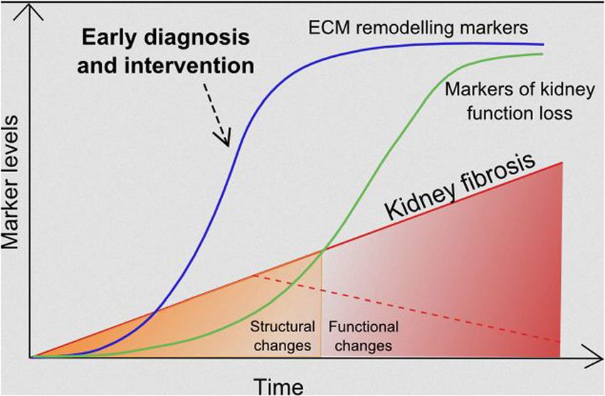
Genovese et al. Fibrogenesis & Tissue Repair 2014, 7:4                                                                                     Page 2 of 14
http://www.fibrogenesis.com/content/7/1/4

Mechanisms of renal fibrosis                                                    Renal fibrosis is the result of a failed wound healing
Renal fibrosis, that is, the accumulation and dysregulated                    process that occurs after an initial insult. The patho-
remodelling of ECM, can affect all major compartments                         physiology of renal fibrosis can be divided into four
of the kidney being termed glomerulosclerosis in the                          phases: 1) cellular activation and injury phase or priming;
glomeruli, tubulointerstitial fibrosis in the tubulointersti-                 2) fibrogenic signalling phase or activation; 3) fibrogenic
tium and arterio- and arteriolosclerosis in the vascula-                      phase or execution; and 4) destructive phase or progres-
ture. At a certain point, virtually all renal cells are                       sion. Figure 1 describes the different phases of tubular
involved in fibrosis [4]. The description of the cellular                     interstitial fibrosis and some of the cells and molecules
and molecular mechanisms of kidney fibrosis is beyond                         that intervene in the process. These phases can be best
the scope of this review and has already been thoroughly                      studied and differentiated in animal models, in which a
discussed by others [5-7]. We will focus on the mecha-                        disease stimulus is often applied at a single time-point so
nisms related to ECM accumulation and remodelling in                          that the injury and the progression are synchronized. In
renal fibrosis as a potentially relevant source of novel                      most, if not all, human diseases this is not the case and, to
biomarkers for renal fibrosis.                                                a variable and yet not defined extent, all phases can be

 Figure 1 Progression of renal interstitial fibrosis. Fibrogenesis starts with an initial tissue injury that causes inflammation as the physiological
 host defense response. When this response becomes uncontrolled and sustains itself with continuous production of chemotactic cytokines,
 inflammation does not resolve and can create the optimal microenvironment for tissue fibrogenesis.
Genovese et al. Fibrogenesis & Tissue Repair 2014, 7:4                                                              Page 3 of 14
http://www.fibrogenesis.com/content/7/1/4

observed at the same time. Various mediators of renal              is under evaluation [18]. At the moment, there are no
fibrosis have been described, such as the prototypical             specific molecular imaging modalities for renal fibrosis.
profibrotic molecules transforming growth factor beta 1              Serological and urinary markers can rapidly change
(TGF-β1) and platelet-derived growth factor (PDGF),                following a physiological or pathological event, and are
which will not be discussed in detail here [8,9]. Among            therefore dynamic. Here we will discuss established, de-
the effectors causing a pathological matrix accumula-              veloping and potential serological and urinary markers
tion, plasminogen activator inhibitor-1 (PAI-1), which is          of renal fibrosis.
induced by TGF-β, was shown to modulate fibrosis via
effects on cell migration, matrix turnover and macrophage          Chronic kidney disease (CKD) biomarkers
infiltration [10]. The role of this effector in kidney fibrosis    In the last decade, there has been intense interest and
has been described elsewhere [11]. Even though many                effort in finding novel predictive biomarkers for the
cell types in the kidney are able to produce ECM, (myo-)           diagnosis and prognosis of CKD. Several molecules in-
fibroblasts in the interstitium and mesangial cells in the         volved in kidney function, signalling and structure have
glomeruli are considered the main cellular mediators of            been evaluated as potential markers for CKD [19]. The
interstitial fibrosis and glomerulosclerosis, respectively         only markers currently accepted and used in clinical
[2,12]. In the kidney, myofibroblasts can originate from           practice for the diagnosis and prognosis of CKD are
different sources, the most important being resident               markers of loss of kidney function. The most widely
interstitial fibroblasts in the cortex and pericytes in the        used are the estimated glomerular filtration rate (eGFR)
medulla. Other sources seem to contribute to a lesser              [20], serum creatinine, blood urea nitrogen (BUN) [21]
and varying extent to the pool of myofibroblasts and in-           and albuminuria or proteinuria [22]. Cystatin C [23] and
clude endothelial cells (via endothelial-to-mesenchymal            β-trace protein [24] have been proposed as an alternative
transition), tubular epithelial cells (via epithelial-to-          to creatinine to estimate the GFR. These markers indicate
mesenchymal transition) and fibrocytes [2,12,13].                  impaired renal function but have no disease specificity,
   Fibronectin is the first ECM protein that is deposited          and detectable changes in their concentration come after
in fibrogenesis [14]. It activates integrins, functions as a       the biological changes in the organ causing the functional
fibroblast chemoattractant and co-localizes with collagen          impairment.
formation. This triggers the production of a large variety           Molecules involved in inflammation or in signalling
of ECM proteins, discussed below [7]. The synthesis,               leading to the onset of fibrosis have been studied as pos-
deposition and degradation of different ECM proteins,              sible markers for renal fibrosis. Some of these molecules
their post-translational modifications, together with the          belong to the panel of urinary biomarkers proposed by the
induction of proteases and protease inhibitors and other           Predictive Safety Testing Consortium (PSTC) for the de-
ECM remodelling enzymes (for example tissue transglu-              tection of drug-induced kidney toxicity [25]. Even though
taminase) contribute to the development of irreversible            the purpose of these markers is to detect an acute re-
fibrosis [7].                                                      sponse to the injury, some are also being evaluated as early
                                                                   markers of CKD and progression towards ESRD. These
                                                                   molecules include: C-reactive protein (CRP), tumor necro-
Diagnosis of renal fibrosis                                        sis factor receptor II (TNFRII), TGF-β1 and pentraxin-3
At present, kidney biopsy is the only method to detect             as cytokines involved in the development of CKD; asym-
renal fibrosis. It is an invasive procedure with possible          metric dimethylarginine (ADMA) as a marker of endo-
complications. The extent of interstitial fibrosis in kidney       thelial dysfunction and consequent kidney damage [26];
biopsy is most often reported in a semi-quantitative man-          fibroblast growth factor-23 (FGF-23), adiponectin and
ner and has several intrinsic limitations, mainly due to           apolipoprotein A-IV as metabolic factors involved in the
sampling error and to intra- and inter-observer variability        regulation of kidney metabolism; and gamma-glutamyl
[15]. Imaging techniques, such as ultrasound, can show             transpeptidase (GGT) as molecules involved in oxida-
signs of corticomedullary differentiation, which is a sen-         tive stress, which can contribute to CKD pathogenesis.
sitive but not specific marker of CKD; it can moreover             Endostatin, the N-terminal portion of collagen type
show the size of the kidneys, the presence of cysts and            XVIII, is a potent anti-angiogenic factor which has been
solid lesions, urinary obstruction or scars but it cannot          recently evaluated as a marker of CKD. A significant
diagnose the presence of ongoing interstitial or glom-             elevation of endostatin in plasma of patients with CKD
erular fibrogenesis [16]. Another imaging technique that           following disease severity compared to controls without
is attracting increasing interest is the magnetic resonance        CKD was observed [27]. Except FGF-23, all the other
elastography (MRE), already used in the hepatic field to           markers are not kidney-specific and require further
detect liver fibrosis [17]. MRE can non-invasively sample          evaluation in larger clinical cohorts to confirm their
tissue stiffness in vivo, and its possible use in renal fibrosis   potential (reviewed thoroughly elsewhere [19]). FGF-23
Genovese et al. Fibrogenesis & Tissue Repair 2014, 7:4 Page 4 of 14 http://www.fibrogenesis.com/content/7/1/4 is currently one of the most promising markers for In the search for specific biomarkers of kidney fibrosis, CKD. This phosphaturic hormone is increased in serum the ECM proteome is a large source of new potential in a physiological adaptation to the hyperphosphatemia targets. Only a few of these proteins have been analyzed that arises when the GFR decreases below 25 ml/min/ as diagnostic and prognostic markers, despite their in- 1.73 m2 [28]. Several studies demonstrated the potential volvement in renal fibrosis that has been proven. A good of FGF-23 as a marker of mortality in dialysis patients biomarker should reflect the presence of renal fibrosis [29], initiation of chronic dialysis [30], CKD progression and be linked to an outcome (decline in eGFR, renal [31,32], cardiovascular disease [30], cardiovascular mor- failure, death). The ideal biomarker should be detected tality [32] or all-cause mortality [30,32]. non-invasively and should be able to predict the pro- Kidney-specific molecules are more likely to specifically gression of the disease and/or the response to a treat- reflect renal injury. Such molecules include podocyte- ment in a more sensitive and specific manner compared specific proteins nephrin, podocin and podocalyxin as to standard parameters. urinary markers of glomerular damage [33,34]. Following The following sections give an overview on involvement the same rationale, neutrophil gelatinase-associated lipoca- of renal ECM proteins and proteases in renal fibrosis and lin (NGAL) [35], kidney injury molecule-1 (KIM-1) [36,37], their potential utility as diagnostic or prognostic markers N-acetyl-beta-D-glucosaminidase (NAG) and liver-type of renal fibrosis. fatty acid-binding protein (L-FABP) [38-41] can be markers of tubular damage, as these are proteins expressed in the The extracellular matrix (ECM) of the kidney tubules that can be released in serum or urine following The ECM is a very dynamic, highly charged structure tubular damage. Both NGAL and KIM-1 are well-known which acts both as a support structure for the cells and as markers of acute kidney injury and their potential as diag- an active component in cell signalling [46]. It is composed nostic and prognostic markers of CKD has been evaluated of collagens, glycoproteins and elastin molecules which in various studies and reviewed in detail elsewhere [19]. form a complex network interacting with each other and NGAL was shown to be increased in serum and/or urine of with the surrounding cells. Proteases, for example MMPs patients suffering from different kidney diseases, for ex- and their inhibitors, are responsible for maintaining the ample in patients with IgA nephropathy (IgAN), various equilibrium between formation and degradation of ECM glomerulonephritis, autosomal dominant polycystic kidney proteins. In the kidney cortex, the ECM is present in ana- disease (ADPKD), pediatric lupus nephritis and CKD from tomically distinct areas with different functions depending a range of etiologies, and to differentiate between CKD on its molecular components: stages [19,42]. NGAL might be a good marker for tubuloin- terstitial injury in CKD, and might identify progression of 1. in the glomeruli the disease. It has to be mentioned though, that the results a. glomerular basement membrane are not consistent in all studies [35,43,44]. Urinary KIM-1 b. Bowman’s capsule levels were associated with the outcome of incident CKD c. mesangial ECM or rapidly declining kidney function in the Multi-Ethnic 2. in the tubulointerstitium Study of Atherosclerosis (MESA) cohort [44]. Other studies a. tubular basement membrane (in part segment- [41,43,44] showed a good potential for KIM-1 as a diag- specific) nostic marker for CKD and even as a marker of efficacy of b. peritubular capillary basement membrane intervention. However, as for many other markers, con- c. interstitial ECM firmation in long-term observational studies using larger 3. in larger vessels populations is still required [19,43]. First hints suggest that a. within the vessels (lamina elastica interna and cytokeratin 18, which can be released into urine and circu- externa) lation following renal epithelial cell death, might also be a b. around the vessels (adventitia of arteries and novel marker of CKD. Serological and urinary concentra- veins) tion levels of total cytokeratin 18 measured in CKD pa- tients could separate patients with advanced CKD from Medullary interstitial ECM is physiologically more patients with mild disease and healthy controls [45]. All prominent compared to the cortical interstitial ECM, these molecules have been evaluated for their association steadily increasing in quantity in the direction from with impaired kidney function, but they are not directly outer to inner medulla/papilla. The functional consequence linked to fibrosis, that is, to the deposition and remodel- of this difference is yet unclear. The hilar region, renal pel- ling of ECM. The tubular damage markers are not com- vis (for example suburothelial basement membrane) and pletely specific for the kidneys, as many of these proteins renal capsule are also composed of ECM. The contribu- are also involved in other diseases, as for example NGAL tion and remodelling of ECM in these specific anatomical (also known as lipocalin-2) in the liver. locations in renal fibrosis are not well-studied.
Genovese et al. Fibrogenesis & Tissue Repair 2014, 7:4                                                          Page 5 of 14
http://www.fibrogenesis.com/content/7/1/4

  The next paragraphs describe the proteins that compose      animal studies as a marker of glomerular sclerosis and
the renal ECM in healthy state and those involved in the      interstitial fibrosis [8,54].
onset of fibrosis, with particular focus on those that can       Elevated urinary concentration levels of collagen type IV
be a source of new biomarkers. A comprehensive list of        have been associated with the decline of renal function in
experimental evidence for ECM proteins being involved in      patients with type 1 [55] and type 2 diabetes [56,57], but
renal fibrosis, derived from both pre-clinical and clinical   also in non-diabetic nephropathies, such as membranous
studies, is included in Additional file 1: Table S1.          nephropathy and anti-neutrophil cytoplasmic antibody
                                                              (ANCA)-associated glomerulonephritis [58]. Specifically,
Glomerular basement membrane (GBM)                            type 1 diabetic nephropathy (DN) patients with elevated
The glomerular basement membrane (GBM) is thicker             urinary collagen type IV to creatinine ratio (T4C) but
compared to most other basement membranes. It contains        normal albumin to creatinine ratio (ACR) declined more
four main macromolecules: laminin, collagen type IV,          rapidly in eGFR than patients with normal T4C [55]. In
nidogen and heparan sulphate proteoglycans. The main          type 2 diabetic patients, increased collagen type IV urine
function of the GBM is to act as a charge- and size-          excretion was associated with the severity of morpho-
selective filtration barrier between the vascular system      logical alterations in fibrosis, albeit no direct relationship
and the urinary space.                                        with the content of collagen type IV in the kidney could
  Laminin is secreted as an αβγ heterotrimer (α5, β2          be observed [56]. Another study in type 2 diabetic patients
and γ1 laminins are present in the mature GBM [47]),          with normoalbuminuria and microalbuminuria found
which forms a network required to maintain the base-          an inverse correlation between urinary collagen type IV
ment membrane integrity. Mutations of the laminin             excretion and the outcome annual decline of eGFR, but
genes can lead to kidney diseases, for example mice with      no correlation with progression to advanced diabetic
a hypomorphic mutation in the gene for the laminin α5         nephropathy was found [57]. In a study on biopsy-proven
subunit develop polycystic kidney disease [48]; a null        membranous nephropathy and ANCA-associated glomer-
mutation of the gene for the laminin α4 subunit can           ulonephritis [58] elevated levels of urinary collagen type
cause progressive glomerular and tubulointerstitial fibro-    IV were correlated with urinary proteins, urinary NAG
sis [49]; and truncation or severe missense mutations in      and selectivity index. Different results were observed in a
the gene for the laminin β2 subunit can cause Pierson         urinary peptidome study performed in type I diabetic pa-
syndrome, characterized by premature death from renal         tients. Patients with progressive early function decline
failure [47].                                                 showed a decreased expression of fragments of collagen
  Collagen type IV is composed by three α chains that         type IV (α1 chain) compared with control subjects with
fold in a triple helix and, by binding with other collagen    stable renal function [59]. These results suggest that
type IV molecules, form the meshwork conformation             urinary collagen type IV might be a promising additive
typical of the basement membrane. The α3(IV), α4(IV)          biomarker in patients with diabetic nephropathy and
and α5(IV) chains are the most expressed in the adult         further clinical studies are eagerly awaited.
GBM [50]. Mutations in the gene for the α5 chain of              The transmembrane collagen type XVII has been
collagen type IV cause the X-linked Alport syndrome in        recently identified in the GBM. Its deficiency causes
humans, a rare genetic disease characterized by progres-      effacement of podocyte foot processes, therefore it
sive glomerular injury. Mutations in the genes for the α3     might be involved in the attachment of the podocyte to
and α4 chains can cause autosomal recessive and auto-         the GBM [60]. Nidogen 1 and 2 bind to collagen type
somal dominant Alport syndrome and thin basement              IV and laminin separately. Although nidogens have a
membrane nephropathy. Collagen type IV is also the tar-       role in the basement membrane formation, experimen-
get of two autoimmune diseases affecting the kidney:          tal evidence showed that they are not strictly required
Goodpasture’s syndrome and Alport post-transplantation        for GBM formation [61]. Agrin is the major heparan
disease. Both diseases are characterized by autoantibodies    sulphate proteoglycan of the GBM in healthy kidneys,
attacking the GBM and causing rapidly progressive glom-       while perlecan is an abundant component of other
erulonephritis [47]. Knock-out mice for the gene for the      basement membranes [62]. Perlecan expression levels
α3 chain and for the α5 chain of collagen type IV are         are increased in the glomeruli of IgAN patients and
widely used as murine models of autosomal and X-linked        correlate with a lower urinary albumin excretion, sug-
Alport syndrome, respectively [51,52]. Increased collagen     gesting that perlecan could be a marker of slower pro-
type IV expression was described in chronic transplant        gression of the disease, and therefore of better outcome
nephropathy using immunohistochemistry [53]. The              [63]. Perlecan and agrin, as all the heparan sulphate
distribution of up-regulated collagen type IV was uni-        proteoglycans, have highly negatively charged glycosami-
form in the GBM, in the mesangium and in the intersti-        noglycans (GAGs), assumed to contribute to the negative
tium. Collagen type IV was also used in experimental          charge of the basement membrane [61]. Interestingly,
Genovese et al. Fibrogenesis & Tissue Repair 2014, 7:4 Page 6 of 14
http://www.fibrogenesis.com/content/7/1/4
several studies showed that lack of perlecan and agrin Collagen type I accumulates in fibrotic glomeruli,
does not lead to proteinuria, even though it affects the tubulointerstitial space and arterial walls in pathological
negative charge of the GBM [64-66]. conditions, co-localizing with decorin and biglycan [79].
Collagen type I accumulation in fibrosis, as many other
Mesangial ECM ECM molecules, is both due to decreased degradation
The mesangial ECM provides structural support for the and elevated synthesis [14,69]. Urinary proteome ana-
glomerular capillary convolute, connecting with the extra- lyses could differentiate DN patients from healthy indi-
glomerular mesangium at the vascular pole. It has a role viduals and patients with other chronic kidney diseases
in cell-matrix signalling in a bidirectional manner. Dysreg- [80]. Among the proteins differentially expressed, frag-
ulation of this cell-matrix signalling plays a role in a wide ments of collagen type I were significantly less present
range of glomerular diseases [67], such as IgAN [68] and in the urine of DN patients. The authors suggested that
DN [69]. Mesangial ECM differs substantially from GBM, this indicated a decreased collagen proteolysis, probably
and its composition allows larger molecules to pass to the due to cross-linking rendering the collagens resistant to
mesangium. In physiological conditions its major compo- proteolytic cleavage or to increased protease inhibitor
nents are fibronectin, collagen type IV (α1 and α2 chains, expression [80].
but not α3 and α5), collagen type V, laminin A, B1 and B2, In physiological conditions collagen type III is normally
chondroitin sulphate and heparan sulphate proteoglycans expressed at low levels in the interstitium, and it is un-
(perlecan, collagen type XVIII and bamacan, but not detectable in glomeruli. However, during fibrosis the ex-
agrin) and nidogen [47,67]. The small proteoglycans dec- pression levels are increased in the interstitium and in the
orin, biglycan, fibromodulin and lumican are weakly glomeruli, as shown by immunohistochemical analysis on
expressed in the mesangial matrix and rather localized in human renal biopsies using antibodies against the collagen
the tubular interstitium [70]. Under pathological condi- type III N-terminal pro-peptide (PIIINP) [81].
tions, decorin and biglycan were shown to be up-regulated PIIINP was detected in high concentrations in urine and
in glomeruli [63]. Consistently, elevation of collagen type serum of patients with various renal diseases [81-83].
IV was also reported in several studies with humans and Urinary PIIINP (and collagen IV) levels were elevated in
rodent models where protein localization and both protein patients with various nephropathies and correlated with
and mRNA levels were assessed [69,71-73]. the extent of interstitial fibrosis in kidney biopsies [81].
Typical scar collagen type I is de novo expressed in The urinary PIIINP to creatinine ratio (uPIIINP/Cr) was
glomerulosclerosis, and by inhibiting its accumulation, a evaluated in kidney transplant patients and correlated
reduction in the extent of glomerulosclerosis was obtained with the extent of interstitial fibrosis [82]. Furthermore,
in a model of DN [74]. MMPs play an important role in in another study on patients with different CKD stages
the homeostasis of the mesangial matrix, for example subjected to kidney biopsy, uPIIINP/Cr correlated with
alterations on MMP function were shown to be linked serum creatinine, eGFR and CKD stage as well as with
to light chain diseases [75]. the extent of fibrosis evaluated in the biopsies [83].
At the moment it is unclear whether glomerular ECM Elevation of collagen type V and VI in kidney fibrotic
(either GBM or mesangium) might provide specific bio- tissue has been reported in various studies [69,84].
markers of glomerular injury. Conversely, decreased concentrations of collagen type
V (α1 chain) were observed in a urinary peptidome of
patients with type 1 DN with early renal function decline
Interstitial ECM [59]. Although different types of collagen are highly up-
The renal interstitial matrix is normally composed by regulated in renal fibrosis, so far only PIIINP and collagen
collagen type I, III, V, VI, VII and XV, both sulphated type IV have been analyzed as potential biomarkers. Both
and non-sulphated glycosaminoglycans, glycoproteins and are among the most promising specific markers reflecting
polysaccharides. During fibrosis, the formation of scar renal fibrosis.
tissue in the interstitial space is the result of the excessive
accumulation of ECM components. Glycoproteins
Fibronectin is an adhesive glycoprotein involved in the
Collagens organization of the ECM. Its accumulation is one of the
Collagens constitute the main structural element of the first events during renal fibrosis [14]. It was shown to
interstitial ECM, providing tensile strength, regulating be up-regulated in many animal models and in human
cell adhesion, support, chemotaxis, cell migration and CKD [69,71,73,78,85].
tissue development [76]. Collagen type I and III are Thrombospondin-1 (TSP-1) is an adhesive glycoprotein
known to be deposited in early stages during renal involved in fibroblast proliferation and migration [86]. It
fibrosis [73,77,78]. was shown to be up-regulated before the disease onset,Genovese et al. Fibrogenesis & Tissue Repair 2014, 7:4 Page 7 of 14 http://www.fibrogenesis.com/content/7/1/4 and correlated with the degree of tubulointerstitial fibrosis by interacting with hyaluronan [89,95]. In healthy renal in three different rat models of renal fibrosis. TSP-1 was tissue, versican is found expressed in the tubulointersti- observed to be transiently expressed at early fibrosis tium and the blood vessels, but not in the glomeruli [96]. stages, suggesting a possible role as a mediator of intersti- In patients with different proteinuric nephropathies, versi- tial fibrosis via activation of TGF-β [69,86]. can expression was increased in areas with marked tubu- Proteoglycans are a subgroup of glycoproteins with a lointerstitial fibrosis, suggesting that versican may have an high content of carbohydrates, which fill the majority of important role during CKD progression [97]. Most of the renal extracellular interstitial space. They have a wide the above mentioned glycoproteins undergo significant variety of functions, such as hydration, force-resistance regulation during kidney fibrosis. Still, no data exist on and growth factor binding [87]. The latter is important in their potential as renal biomarkers. renal fibrosis as proteoglycans act as a reservoir of profi- brotic growth factors, such as the latent forms of TGF-β Matrix metalloproteinases (MMPs) and other proteases or FGF-2 [5]. The enzymes playing a central role in matrix remodelling Decorin, biglycan and fibromodulin are small leucine- are metalloproteinases. Metalloproteinases are synthesized rich proteoglycans (SLRPs), which act as potent regulators in kidneys and have an important function in maintaining of TGF-β [62,88]. Decorin and biglycan also have an the homeostasis of the ECM. The main families of metallo- important role in collagen fibrillogenesis. In healthy proteinases are MMPs, a disintegrin and metalloproteinase adult renal tissue, decorin and biglycan are expressed in (ADAM) proteins and a disintegrin and metalloproteinase the tubulointerstitium and weakly in the glomeruli [69]. with thrombospondin motifs (ADAMTS). The role of AD- However, during progressive renal scarring an increased AMs and ADAMTS is only starting to emerge and will expression of decorin and biglycan was observed in vari- not be discussed here [98-103]. Serine proteases (plasmin ous experimental models of renal injury and in humans and cathepsin G) and cysteine proteases (cathepsins B, H [79,84,85,89-91]. Specifically, in the unilateral ureteral ob- and L) can also contribute to the degradation of ECM struction (UUO) model, tubular biglycan up-regulation components at neutral pH [104]. was observed before macrophage infiltration, indicating that biglycan could act as an initiator and regulator of MMPs and tissue inhibitors of metalloproteinases (TIMPs) inflammation in the kidney [89]. Biglycan and decorin MMPs are zinc-dependent enzymes involved in ECM expression was also found to be highly up-regulated in remodelling, which play a central role in tissue homeo- the glomeruli of IgAN patients, indicating a potential stasis. There are 23 MMPs in humans [105] and at least role in this glomerular disease [63]. Decorin has well- ten of them are expressed in the kidney (MMP-1, -2, -3, known anti-fibrotic properties: it neutralizes TGF-β activity -9, -13, -14, -24, -25, -27 and -28) [106]. MMP-12 was by interfering with its signalling; it exerts an anti-apoptotic thought not to be expressed in the kidney even though activity on tubular epithelial and endothelial cells; and can some experimental results in an animal model suggest induce fibrillin-1 expression, by binding the insulin-like the opposite [107]. MMPs were hypothesized to be anti- growth factor type I (IGF-I) receptor [92]. Its use as an fibrotic due to their function as ECM degradation en- anti-fibrotic molecule has been shown in a rat model of zymes. Increasing evidence suggests that MMPs have a glomerulonephritis [93]. more complex role in renal fibrosis [4,108,109]. For ex- Hyaluronan is a high molecular weight glycosaminogly- ample, MMP-9-mediated degradation of collagens creates can formed by the repetition of disaccharides composed collagen fragments, which possess chemotactic properties by N-acetylglucosamine and glucuronic acid [62]. It has for neutrophils and are able to stimulate MMP-9 produc- the ability to bind to a variety of proteoglycans and to cell tion. Apart from their action on ECM components, MMPs receptors acting as a signalling molecule. In a healthy hu- are also known to modulate growth factors and their re- man kidney, hyaluronan is very little expressed. However, ceptors (TGF-β, FGF-R1), adhesion molecules (integrins during progressive kidney disease, it accumulates in the and cadherins) [109], cytokines and chemokines. Conse- cortical interstitium and may potentiate interstitial inflam- quently, MMPs are involved in several processes aside mation by stimulating the recruitment of monocytes to from ECM remodelling, such as destruction of the base- the interstitial space [5]. Elevated levels of hyaluronan in ment membrane, angiogenesis, cell migration and cell renal tissue were reported in several kidney diseases in apoptosis, some being pro- and some anti-fibrotic de- both rat models, for example ischemia-reperfusion injury, pending on the context [4,109,110]. MMPs are inhibited and human diseases, for example DN, renal transplant permanently by degradation or temporarily by tissue in- rejection and kidney stone formation [94]. hibitors of metalloproteinases (TIMPs). A balance be- Versican is a chondroitin sulphate proteoglycan, the tween MMP and TIMP activity is essential for ECM largest member of the modular proteoglycans, with an homeostasis [109]. Among the four TIMPs that have important role in maintaining the integrity of the ECM been identified in vertebrates, TIMP-1, -2 and -3 are
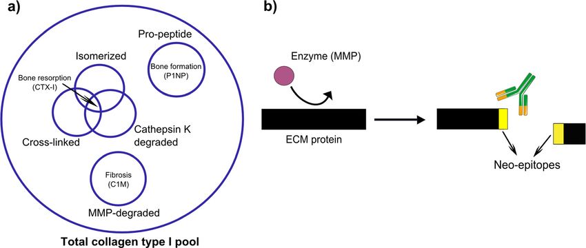
Genovese et al. Fibrogenesis & Tissue Repair 2014, 7:4                                                                            Page 8 of 14
http://www.fibrogenesis.com/content/7/1/4

expressed in the kidney [106]. Increased mRNA and                        In many cases the findings are based on up- or down-
protein levels of TIMP-1 were reported in several human                  regulated expression of MMP genes, which do not ne-
and rodent models of different renal diseases, suggesting                cessarily translate into an increased presence of active
that TIMP-1 might be involved in the early events during                 proteases. This is the main limitation in the use of
the progression of renal diseases [5,73,109]. TIMP-2 has                 MMPs and TIMPs as markers of renal fibrosis. Given
also been shown to be elevated in various rat models of                  the functional complexity of the MMPs, it is likely that
renal disease [73]. The exact localization and temporal                  they themselves might not be suitable biomarkers of
expression of MMPs in the human kidney is still not                      renal fibrosis.
completely understood [108]. Most of the data on MMP
expression derive from animal models of kidney dis-                      Protein fingerprint technology
eases (Additional file 1: Table S1). MMP-2 and MMP-9                     A highly regulated equilibrium between synthesis and
are known to be involved in the proteolysis of collagen                  degradation of ECM proteins is required to maintain
type IV, which accumulates in the basement membranes,                    tissue homeostasis. A disruption of this equilibrium is
for example in early stages of DN. MMP-2 and MMP-9                       at the base of pathological processes such as fibrosis
expression and activity were up-regulated in different ani-              [104]. The measurement of the ECM remodelling rate,
mal models of renal fibrosis [69,91,109,111,112], but were               represented by end-products of ECM proteins in the
decreased in cases of DN in both humans and rats                         biological fluids [116], can give an indication on the dis-
[69,113]. Changes in MMP-2 and MMP-9 activity might                      ease activity and progression. The peptides generated by
therefore influence the ECM composition causing renal                    specific protein degradation by MMPs or other proteases
damage at early stages of DN [106]. However, another                     involved in a specific disease provide a unique fingerprint
study showed that urinary levels of MMP-9, together with                 for a particular disease [117]. This approach is called pro-
collagen type IV, were elevated in type 2 DN patients with               tein fingerprint (Figure 2a). Compared to measurement of
macroalbuminuria [114]. MMP-3 expression and activity                    the intact/whole protein, the measurement of such modi-
during DN was decreased in both humans and rats [69].                    fied ‘fingerprint’ peptides are likely to be more sensitive
MMP-7 is not expressed in healthy human kidneys but                      markers of pathology. This is because only the action
was found in epithelial cells and atrophic tubules in pa-                of a specific protease (or other post-translational mod-
tients with ADPKD and in a mouse model of acute renal                    ifications) on a specific protein that is accumulated in
tubule injury and chronic progressive renal fibrosis [115].              a particular diseased tissue can generate the new N- or
Some of the contrasting results, particularly in regards to              C-terminal, namely the neo-epitope.
MMP-2 and MMP-9, can be explained by the impossibility                      The peptides originating from the protease-mediated
to distinguish between the active and the inactive form                  degradation of the ECM may be small enough to be re-
of the protease with the commercially available assays.                  leased in circulation or urine. There they can be detected

 Figure 2 Neo-epitope markers for ECM remodelling. a) Neo-epitopes of collagen type I generated by different post-translational modifications
 provide more information than the measurement of total collagen type I. b) Formation of detectable neo-epitopes generated by cleavage of
 ECM proteins by specific proteases. ECM, extracellular matrix.
Genovese et al. Fibrogenesis & Tissue Repair 2014, 7:4 Page 9 of 14
http://www.fibrogenesis.com/content/7/1/4
by antibodies raised specifically to react against the monitoring the efficacy of the treatment with statins in an
neo-epitope (Figure 2b). Other post-translational modifi- experimental model of liver fibrosis [132]. C3M was ele-
cations, for example isomerization, citrullination, glycosyla- vated in urine of mice treated with bleomycin to induce
tion and cross-linking can also originate from neo-epitopes skin fibrosis compared to the controls, showing a potential
to be used for protein fingerprint [118], but will be not use of this marker in skin fibrosis [125].
discussed here. Markers reflecting ECM remodelling can Clinical studies showed that the markers BGM, elastin
not only identify and quantify a pathological process MMP-generated neo-epitope fragment (ELM) C1M, C3M,
within the organ of interest, but can potentially describe C4M C5M, collagen type VI MMP-generated neo-epitope
the disease activity. This might for example help to segre- fragment (C6M), Pro-C3 and P4NP 7S were associated
gate the patients that progress faster with the disease. with portal hypertension in patients with cirrhosis, reflect-
Markers reflecting the disrupted ECM turnover might de- ing the degree of liver dysfunction [123]. A marker of
tect tissue modifications, which happen in the first stages MMP-mediated versican degradation (VCANM) was el-
of the disease when the pathological process can possibly evated in plasma of patients suffering from different
still be reversed. cardiovascular diseases [130]. Promising clinical results
As outlined above, surprisingly very little data exists were also obtained in lung fibrosis: the previously men-
on the use of ECM, the principal underlying structure of tioned ELM [124] C1M, C3M, C4M, C5M and C6M
fibrotic tissue, as a source of biomarkers of renal fibrosis. [121] could separate patients affected by chronic obstruct-
Such biomarkers could identify the early modifications ive pulmonary disease (COPD) and idiopathic pulmonary
that lead to renal fibrosis and could allow early treatment, fibrosis (IPF) from healthy individuals in a small observa-
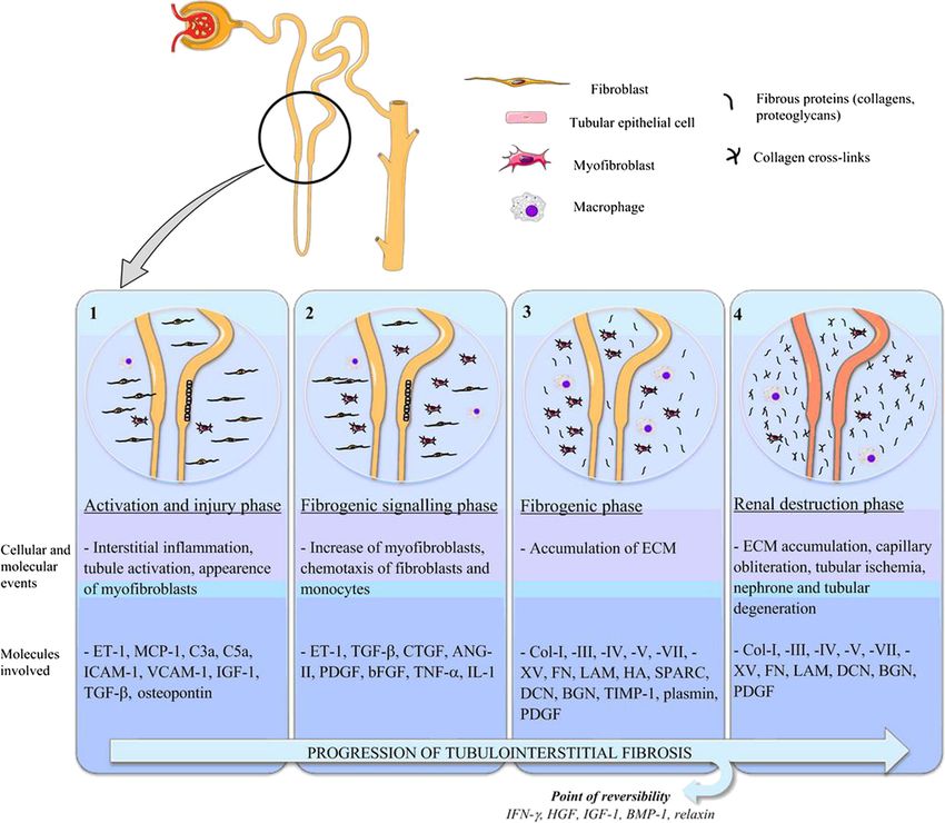
helping in the resolution of fibrosis. Figure 3 illustrates the tional cohort.
possible advantages of markers of structural changes over As the mechanisms of kidney, liver and lung fibrosis
markers of loss of kidney function. share common features and involve similar ECM proteins,
Neo-epitopes of different types of collagen (type I, II, the successful biomarkers identified in these pre-clinical
III, IV, V and VI collagen), proteoglycans (biglycan and and clinical studies are also likely to prove valuable in
versican) and elastin have already proven to be biomarkers renal fibrosis, as a first study in kidney patients suggests.
of connective tissue diseases, such as osteoarthritis [119] Plasma levels of P4NP 7S were significantly associated
or organ fibrosis, in both animal models and clinical with mortality in ESRD patients undergoing hemodialysis
studies [120-130]. [122]. Specifically, the patients in the highest quartile of
Experimental evidence in well-characterized animal P4NP 7S plasma levels had an increased risk of death
models showed that neo-epitope fragments of collagen compared to the patients in the other quartiles. The high
type I (C1M), III (C3M), IV (C4M) and V (C5M), biglycan plasma levels of this marker were considered a sign of ac-
(BGM) MMP-mediated degradation, and of collagen type celerated systemic fibrosis in ESRD patients with the worst
III (Pro-C3), IV (P4NP 7S) and V (P5NP) formation were prognosis. These results confirm the high value of collagen
markers of liver fibrosis [120,126-129,131]. These markers type IV as a prognostic marker in kidney diseases demon-
(except BGM) also showed a promising potential for strated by the previously described studies. The before
mentioned results were obtained in urine and using an
assay based on polyclonal antibodies, while in this study,
an assay using a specific monoclonal antibody for the α1
chain of the P4NP 7S domain of collagen type IV was used
to detect collagen type IV in plasma [133].
The main limitation of this technique in kidneys is that
neo-epitope peptides coming from organs other than
kidneys can also contribute to the pool of neo-epitopes
detected in serum or plasma. Urine is a more suitable
matrix to find protein fragments originating in the kid-
ney. However, the detection of protein fragments in
urine can be biased by the altered GFR during the late
stages of CKD: lower or higher levels of the markers
Figure 3 Biomarkers of ECM remodelling may identify cannot be a result of lower or higher remodelling, but of
molecular processes occurring in the early phases of impaired excretion. Furthermore, the urinary concen-
fibrogenesis, giving the opportunity for early intervention in tration of the markers can be altered by non-selective
stages in which the disease is still reversible. The development proteinuria in proteinuric kidney disease. The picture is
of fibrosis is schematically indicated as linear for simplicity. ECM,
further complicated by the frequent presence of co-
extracellular matrix.
morbidities affecting other organs in the presence ofGenovese et al. Fibrogenesis & Tissue Repair 2014, 7:4 Page 10 of 14
http://www.fibrogenesis.com/content/7/1/4
kidney diseases, or even causing kidney diseases in the PIIINP to creatinine ratio; UUO: Unilateral ureteral obstruction; VCANM: MMP-
first place. mediated versican degradation fragment.
The challenge to identify a disease- and/or organ-specific
Competing interests
and sensitive biomarker for renal fibrosis might be met FG, MK and DL are full-time employees at Nordic Bioscience, Herlev,
by narrowing the selection of neo-epitopes to ECM pro- Denmark. Other authors have no competing interests.
tein or protein isoforms that are most exclusively
expressed in kidneys and the action of a protease whose Authors’ contributions
FG, AM, MK and DL conceived and designed the review. FG, AM and PB
expression is up-regulated specifically during the patho- carried out the literature research and drafted the manuscript. PB, DL and
genesis of renal fibrosis. MK critically revised the manuscript for important intellectual content. All
authors read and approved the final manuscript.
Conclusions
Acknowledgements
The identification of reliable biomarkers for early diagno- This work was supported by research grants TP25 and Q1 of the SFB/Transregio
sis and prognosis of renal fibrosis is of paramount import- 57 of the German Research Foundation (Deutsche Forschungsgemeinschaft,
ance. The perfect biomarker for kidney fibrosis should be DFG) ‘Mechanisms of organ fibrosis’, BO 3755/1-1 of the DFG and 2012_A216 of
the Else-Kröner Fresenius Stiftung (EKFS), all to PB.
non-invasive, specific, involved in the mechanisms of fi-
brosis, with low (or no) background in healthy individuals Author details
1
and able to reflect treatment effects. Several molecules im- Nordic Bioscience, 2730 Herlev, Denmark. 2Nephrology and Immunology,
RWTH Aachen University, Aachen, North Rhine-Westphalia, Germany.
plicated in the mechanisms of fibrosis have been proposed 3
Institute of Pathology, RWTH Aachen University, Aachen, North
as biomarkers, but none of them have been validated and Rhine-Westphalia, Germany. 4Institute of Molecular Biomedicine, Comenius
accepted in clinical practice yet. In this review we have University, Bratislava, Slovakia.
proposed a new perspective, introducing the possible Received: 21 November 2013 Accepted: 27 February 2014
use of ECM protein fingerprint as a source of novel bio- Published: 28 March 2014
markers for renal fibrosis.
References
1. U.S. Renal Data System, USRDS 2013 Annual Data Report: Atlas of Chronic
Additional file Kidney Disease and End-Stage Renal Disease in the United States, National
Institutes of Health, National Institute of Diabetes and Digestive and Kidney
Additional file 1: Table S1. Pre-clinical and clinical experimental Diseases. Bethesda, MD; 2013.
evidence of involvement of extracellular matrix (ECM) protein and 2. Barnes JL, Glass WF: Renal interstitial fibrosis: a critical evaluation of the
proteases in kidney disease [14,53,55-59,63,69-73,77-79,83-86,90,94,97, origin of myofibroblasts. Contrib Nephrol 2011, 169:73–93.
99-101,112,113,115,134-154]. 3. Friedman SL, Sheppard D, Duffield JS, Violette S: Therapy for fibrotic
diseases: nearing the starting line. Sci Transl Med 2013, 5:167sr1.
4. Boor P, Sebekova K, Ostendorf T, Floege J: Treatment targets in renal
Abbreviations fibrosis. Nephrol Dial Transplant 2007, 22:3391–3407.
ACR: Albumin to creatinine ratio; ADAM: A disintegrin and metalloproteinase; 5. Eddy AA: Molecular basis of renal fibrosis. Pediatr Nephrol 2000,
ADAMTS: A disintegrin and metalloproteinase with thrombospondin motifs; 15:290–301.
ADMA: Asymmetric dimethylarginine; ADPKD: Autosomal dominant 6. Boor P, Ostendorf T, Floege J: Renal fibrosis: novel insights into
polycystic kidney disease; ANCA: Anti-neutrophil cytoplasmic antibody; mechanisms and therapeutic targets. Nat Rev Nephrol 2010, 6:643–656.
BGM: MMP-generated neo-epitope fragment of biglycan; BUN: Blood urea 7. Liu Y: Cellular and molecular mechanisms of renal fibrosis. Nat Rev
nitrogen; C1M: MMP-generated neo-epitope fragment of collagen type I; Nephrol 2011, 7:684–696.
C3M: MMP-generated neo-epitope fragment of collagen type III; C4M: MMP- 8. Boor P, Konieczny A, Villa L, Kunter U, Van Roeyen CR, LaRochelle WJ,
generated neo-epitope fragment of collagen type IV; C5M: MMP-generated Smithson G, Arrol S, Ostendorf T, Floege J: PDGF-D inhibition by CR002
neo-epitope fragment of collagen type V; C6M: MMP-generated neo-epitope ameliorates tubulointerstitial fibrosis following experimental
fragment of collagen type VI; CKD: Chronic kidney disease; COPD: Chronic glomerulonephritis. Nephrol Dial Transplant 2007, 22:1323–1331.
obstructive pulmonary disease; CRP: C-reactive protein; DN: Diabetic 9. Boor P, Floege J: Chronic kidney disease growth factors in renal fibrosis.
nephropathy; ECM: Extracellular matrix; eGFR: Estimated glomerular filtration Clin Exp Pharmacol Physiol 2011, 38:441–450.
rate; ELM: MMP-generated neo-epitope fragment of elastin; ESRD: End-stage 10. Fogo AB: Renal fibrosis: not just PAI-1 in the sky. J Clin Invest 2003,
renal disease; FGF: Fibroblast growth factor; FGF-R1: Fibroblast growth factor 112:326–328.
receptor 1; GAG: Glycosaminoglycan; GBM: Glomerular basement membrane; 11. Malgorzewicz S, Skrzypczak-Jankun E, Jankun J: Plasminogen activator
GFR: Glomerular filtration rate; GGT: Gamma-glutamyl transpeptidase; inhibitor-1 in kidney pathology (Review). Int J Mol Med 2013, 31:503–510.
IgAN: IgA nephropathy; IGF: Insulin-like growth factor; IPF: Idiopathic 12. Boor P, Floege J: The renal (myo-)fibroblast: a heterogeneous group of
pulmonary fibrosis; KIM-1: Kidney injury molecule 1; L-FABP: Liver-type fatty cells. Nephrol Dial Transplant 2012, 27:3027–3036.
acid-binding protein; MESA: Multi-Ethnic Study of Atherosclerosis; 13. Lebleu VS, Taduri G, O’Connell J, Teng Y, Cooke VG, Woda C, Sugimoto H,
MeSH: Medical Subject Headings; MMP: Matrix metalloproteinase; Kalluri R: Origin and function of myofibroblasts in kidney fibrosis. Nat
MRE: Magnetic resonance elastography; NAG: N-acetyl-beta-D- Med 2013, 19:1047–1053.
glucosaminidase; NGAL: Neutrophil gelatinase-associated lipocalin; 14. Eddy AA: Molecular insights into renal interstitial fibrosis. J Am Soc
NIH: National Institutes of Health; P4NP 7S: Collagen type IV fragment Nephrol 1996, 7:2495–2508.
belonging to the 7S domain; P5NP: Collagen type V pro-peptide; PAI- 15. Farris AB, Colvin RB: Renal interstitial fibrosis: mechanisms and evaluation.
1: Plasminogen activator inhibitor-1; PDGF: Platelet-derived growth factor; Curr Opin Nephrol Hypertens 2012, 21:289–300.
PIIINP: Collagen type III N-terminal pro-peptide; Pro-C3: Propeptide of 16. Goldsmith D, Jayawardene S, Ackland P: ABC of Kidney Disease. 2nd edition.
collagen type III; PSTC: Predictive Safety Testing Consortium; SLRP: Small Chichester: John Wiley & Sons, Ltd; 2013.
leucine-rich proteoglycan; T4C: Collagen type IV to creatinine ratio; 17. Kirk GD, Astemborski J, Mehta SH, Spoler C, Fisher C, Allen D, Higgins Y,
TGF-β1: Transforming growth factor beta 1; TIMP: Tissue inhibitors of Moore RD, Afdhal N, Torbenson M, Sulkowski M, Thomas DL: Assessment of
metalloproteinase; TNFR: Tumor necrosis factor receptor; uPIIINP/Cr: Urinary liver fibrosis by transient elastography in persons with hepatitis C virusGenovese et al. Fibrogenesis & Tissue Repair 2014, 7:4 Page 11 of 14
http://www.fibrogenesis.com/content/7/1/4
infection or HIV-hepatitis C virus coinfection. Clin Infect Dis 2009, glucosaminidase in comparison to alpha 1-microglobulin as a marker in
48:963–972. evaluating tubular dysfunction in glomerulonephritis patients. Clin Chim
18. Korsmo MJ, Ebrahimi B, Eirin A, Woollard JR, Krier JD, Crane JA, Warner L, Acta 2000, 297:93–102.
Glaser K, Grimm R, Ehman RL, Lerman LO: Magnetic resonance 40. Kamijo A, Kimura K, Sugaya T, Yamanouchi M, Hikawa A, Hirano N, Hirata Y,
elastography noninvasively detects in vivo renal medullary fibrosis Goto A, Omata M: Urinary fatty acid-binding protein as a new clinical
secondary to swine renal artery stenosis. Invest Radiol 2013, 48:61–68. marker of the progression of chronic renal disease. J Lab Clin Med 2004,
19. Fassett RG, Venuthurupalli SK, Gobe GC, Coombes JS, Cooper MA, Hoy WE: 143:23–30.
Biomarkers in chronic kidney disease: a review. Kidney Int 2011, 41. Nielsen SE, Sugaya T, Hovind P, Baba T, Parving HH, Rossing P: Urinary
80:806–821. liver-type fatty acid-binding protein predicts progression to nephropathy
20. O’Callaghan C: The Renal System at a Glance. 3rd edition. Chichester: John in type 1 diabetic patients. Diabetes Care 2010, 33:1320–1324.
Wiley & Sons, Ltd; 2009. 42. Malyszko J, Malyszko JS, Bachorzewska-Gajewska H, Poniatowski B, Dobrzycki
21. Waikar SS, Bonventre JV: Can we rely on blood urea nitrogen as a S, Mysliwiec M: Neutrophil gelatinase-associated lipocalin is a new and
biomarker to determine when to initiate dialysis? Clin J Am Soc Nephrol sensitive marker of kidney function in chronic kidney disease patients
2006, 1:903–904. and renal allograft recipients. Transplant Proc 2009, 41:158–161.
22. D’Amico G, Bazzi C: Pathophysiology of proteinuria. Kidney Int 2003, 43. Shlipak MG, Day EC: Biomarkers for incident CKD: a new framework for
63:809–825. interpreting the literature. Nat Rev Nephrol 2013, 9:478–483.
23. Newman DJ, Thakkar H, Edwards RG, Wilkie M, White T, Grubb AO, Price CP: 44. Peralta CA, Katz R, Bonventre JV, Sabbisetti V, Siscovick D, Sarnak M, Shlipak
Serum cystatin C measured by automated immunoassay: a more MG: Associations of urinary levels of kidney injury molecule 1 (KIM-1)
sensitive marker of changes in GFR than serum creatinine. Kidney Int and neutrophil gelatinase-associated lipocalin (NGAL) with kidney
1995, 47:312–318. function decline in the Multi-Ethnic Study of Atherosclerosis (MESA).
24. Hoffmann A, Nimtz M, Conradt HS: Molecular characterization of Am J Kidney Dis 2012, 60:904–911.
beta-trace protein in human serum and urine: a potential diagnostic 45. Roth GA, Lebherz-Eichinger D, Ankersmit HJ, Hacker S, Hetz H, Vukovich T,
marker for renal diseases. Glycobiology 1997, 7:499–506. Perne A, Reiter T, Farr A, Hörl WH, Haas M, Krenn CG: Increased total
25. Bonventre JV, Vaidya VS, Schmouder R, Feig P, Dieterle F: Next-generation cytokeratin-18 serum and urine levels in chronic kidney disease.
biomarkers for detecting kidney toxicity. Nat Biotechnol 2010, 28:436–440. Clin Chim Acta 2011, 412:713–717.
26. Ravani P, Tripepi G, Malberti F, Testa S, Mallamaci F, Zoccali C: Asymmetrical 46. Bosman FT, Stamenkovic I: Functional structure and composition of the
dimethylarginine predicts progression to dialysis and death in patients extracellular matrix. J Pathol 2003, 200:423–428.
with chronic kidney disease: a competing risks modeling approach. 47. Chen YM, Miner JH: Glomerular basement membrane and related
J Am Soc Nephrol 2005, 16:2449–2455. glomerular disease. Transl Res 2012, 160:291–297.
27. Chen J, Hamm LL, Kleinpeter MA, Husserl F, Khan IE, Chen CS, Liu Y, Mills 48. Shannon MB, Patton BL, Harvey SJ, Miner JH: A hypomorphic mutation in
KT, He C, Rifai N, Simon EE, He J: Elevated plasma levels of endostatin are the mouse laminin alpha5 gene causes polycystic kidney disease.
associated with chronic kidney disease. Am J Nephrol 2012, 35:335–340. J Am Soc Nephrol 2006, 17:1913–1922.
28. Jonsson KB: The role of fibroblast growth factor 23 in renal disease. 49. Abrass CK, Hansen KM, Patton BL: Laminin alpha4-null mutant mice
Nephrol Dial Transplant 2005, 20:479–482. develop chronic kidney disease with persistent overexpression of
29. Gutiérrez OM, Mannstadt M, Isakova T, Rauh-Hain JA, Tamez H, Shah A, platelet-derived growth factor. Am J Pathol 2010, 176:839–849.
Smith K, Lee H, Thadhani R, Jüppner H, Wolf M: Fibroblast growth factor 50. Fischer E, Mougenot B, Callard P, Ronco P, Rossert J: Abnormal expression
23 and mortality among patients undergoing hemodialysis. N Engl J Med of glomerular basement membrane laminins in membranous
2008, 359:584–592. glomerulonephritis. Nephrol Dial Transplant 2000, 15:1956–1964.
30. Kendrick J, Cheung AK, Kaufman JS, Greene T, Roberts WL, Smits G, 51. Rheault MN, Kren SM, Thielen BK, Mesa HA, Crosson JT, Thomas W, Sado Y,
Chonchol M: FGF-23 associates with death, cardiovascular events, and Kashtan CE, Segal Y: Mouse model of X-linked Alport syndrome. J Am Soc
initiation of chronic dialysis. J Am Soc Nephrol 2011, 12:1913–1922. Nephrol 2004, 15:1466–1474.
31. Isakova T, Xie H, Yang W, Xie D, Anderson AH, Scialla J, Wahl P, Gutierrez 52. Hudson BG, Tryggvason K, Sundaramoorthy M, Neilson EG: Alport’s
OM, Steigerwalt S, He J, Schwartz S, Lo J, Ojo A, Sondheimer J, Hsu CY, Lash syndrome, Goodpasture’s syndrome, and type IV collagen. N Engl J Med
J, Leonard M, Kusek JW, Feldman HI, Wolf M, Chronic Renal Insufficiency 2003, 348:2543–2556.
Cohort (CRIC) Study Group: Fibroblast growth factor 23 and risks of 53. Kawase T, Shimizu A, Adachi E, Tojimbara T, Nakajima I, Fuchinoue S,
mortality and end-stage renal disease in patients with chronic kidney Sawada T: Collagen IV is upregulated in chronic transplant nephropathy.
disease. JAMA 2011, 305:2432–2439. Transplant Proc 2001, 33:1207–1208.
32. Wolf M: Update on fibroblast growth factor 23 in chronic kidney disease. 54. Boor P, Celec P, Behuliak M, Grancic P, Kebis A, Kukan M, Pronayova N,
Kidney Int 2012, 82:737–747. Liptaj T, Ostendorf T, Sebekova K: Regular moderate exercise reduces
33. Wang G, Lai FM, Lai KB, Chow KM, Li KT, Szeto CC: Messenger RNA advanced glycation and ameliorates early diabetic nephropathy in
expression of podocyte-associated molecules in the urinary sediment of obese Zucker rats. Metabolism 2009, 58:1669–1677.
patients with diabetic nephropathy. Nephron Clin Pract 2007, 55. Morita M, Uchigata Y, Hanai K, Ogawa Y, Iwamoto Y: Association of urinary
106:c169–c179. type IV collagen with GFR decline in young patients with type 1
34. Kanno K, Kawachi H, Uchida Y, Hara M, Shimizu F, Uchiyama M: Urinary diabetes. Am J Kidney Dis 2011, 58:915–920.
sediment podocalyxin in children with glomerular diseases. Nephron Clin 56. Okonogi H, Nishimura M, Utsunomiya Y, Hamaguchi K, Tsuchida H, Miura Y,
Pract 2003, 95:c91–c99. Suzuki S, Kawamura T, Hosoya T, Yamada K: Urinary type IV collagen
35. Bolignano D, Lacquaniti A, Coppolino G, Donato V, Campo S, Fazio MR, excretion reflects renal morphological alterations and type IV collagen
Nicocia G, Buemi M: Neutrophil gelatinase-associated lipocalin (NGAL) expression in patients with type 2 diabetes mellitus. Clin Nephrol 2001,
and progression of chronic kidney disease. Clin J Am Soc Nephrol 2009, 55:357–364.
4:337–344. 57. Araki S, Haneda M, Koya D, Isshiki K, Kume S, Sugimoto T, Kawai H, Nishio Y,
36. Lim AI, Tang SC, Lai KN, Leung JC: Kidney injury molecule-1: more than Kashiwagi A, Uzu T, Maegawa H: Association between urinary type IV
just an injury marker of tubular epithelial cells? J Cell Physiol 2013, collagen level and deterioration of renal function in type 2 diabetic
228:917–924. patients without overt proteinuria. Diabetes Care 2010, 33:1805–1810.
37. van Timmeren MM, van den Heuvel MC, Bailly V, Bakker SJ, van Goor H, 58. Furumatsu Y, Nagasawa Y, Shoji T, Yamamoto R, Iio K, Matsui I, Takabatake
Stegeman CA: Tubular kidney injury molecule-1 (KIM-1) in human renal Y, Kaimori JY, Iwatani H, Kaneko T, Tsubakihara Y, Imai E, Isaka Y, Rakugi H:
disease. J Pathol 2007, 212:209–217. Urinary type IV collagen in nondiabetic kidney disease. Nephron Clin Pract
38. Kern EF, Erhard P, Sun W, Genuth S, Weiss MF: Early urinary markers of 2011, 117:c160–c166.
diabetic kidney disease: a nested case–control study from the Diabetes 59. Merchant ML, Perkins BA, Boratyn GM, Ficociello LH, Wilkey DW, Barati MT,
Control and Complications Trial (DCCT). Am J Kidney Dis 2010, 55:824–834. Bertram CC, Page GP, Rovin BH, Warram JH, Krolewski AS, Klein JB: Urinary
39. Holdt-Lehmann B, Lehmann A, Korten G, Nagel H, Nizze H, Schuff-Werner P: peptidome may predict renal function decline in type 1 diabetes and
Diagnostic value of urinary alanine aminopeptidase and N-acetyl-beta-D- microalbuminuria. J Am Soc Nephrol 2009, 20:2065–2074.You can also read