Palindromes in DNA-A Risk for Genome Stability and Implications in Cancer - MDPI
←
→
Page content transcription
If your browser does not render page correctly, please read the page content below
International Journal of
               Molecular Sciences

Review
Palindromes in DNA—A Risk for Genome Stability and
Implications in Cancer
Marina Svetec Miklenić and Ivan Krešimir Svetec *

                                            Faculty of Food Technology and Biotechnology, University of Zagreb, Pierottijeva 6, 10000 Zagreb, Croatia;
                                            mmiklenic@pbf.hr
                                            * Correspondence: iksvetec@pbf.hr; Tel.: +385-1483-6016

                                            Abstract: A palindrome in DNA consists of two closely spaced or adjacent inverted repeats. Certain
                                            palindromes have important biological functions as parts of various cis-acting elements and protein
                                            binding sites. However, many palindromes are known as fragile sites in the genome, sites prone to
                                            chromosome breakage which can lead to various genetic rearrangements or even cell death. The
                                            ability of certain palindromes to initiate genetic recombination lies in their ability to form secondary
                                            structures in DNA which can cause replication stalling and double-strand breaks. Given their
                                            recombinogenic nature, it is not surprising that palindromes in the human genome are involved in
                                            genetic rearrangements in cancer cells as well as other known recurrent translocations and deletions
                                            associated with certain syndromes in humans. Here, we bring an overview of current understanding
                                            and knowledge on molecular mechanisms of palindrome recombinogenicity and discuss possible
                                            implications of DNA palindromes in carcinogenesis. Furthermore, we overview the data on known
                                            palindromic sequences in the human genome and efforts to estimate their number and distribution,
                                            as well as underlying mechanisms of genetic rearrangements specific palindromic sequences cause.

                                  Keywords: DNA palindromes; quasipalindromes; palindromic amplification; palindrome-mediated

                                            genetic recombination; carcinogenesis
Citation: Svetec Miklenić, M.; Svetec,
I.K. Palindromes in DNA—A Risk for
Genome Stability and Implications in
Cancer. Int. J. Mol. Sci. 2021, 22, 2840.
                                            1. Introduction
https://doi.org/10.3390/ijms22062840
                                                 As the exploration of various genomes, including our own, moves forward thanks
Academic Editor: Miroslav Chovanec          to ever more advanced and more easily available techniques, it is becoming clear that
                                            the genome is much more than the assembly of genes. The architecture of the genome
Received: 15 February 2021                  is complex and wondrous, with sequences that play various important roles, but also
Accepted: 8 March 2021                      those, such as DNA palindromes, that create fragile sites (i.e., sites prone to potentially
Published: 11 March 2021                    lethal chromosome breakage), thus endangering genome stability. As a response to the
                                            breakage (double-strand break—DSB) in DNA, the cell employs various repair mechanisms
Publisher’s Note: MDPI stays neutral        which can ultimately result in genetic rearrangements. First, in Section 2 of this review, we
with regard to jurisdictional claims in     explore the recombinogenic nature of palindromic sequences. We explain why palindromes
published maps and institutional affil-     are recombinogenic and what is the current understanding of molecular mechanisms
iations.                                    underlying palindrome recombinogenicity in eukaryotic cells. In Section 3, we focus on
                                            the role DNA palindromes play in carcinogenesis. Palindromic amplification of genes is
                                            one of the hallmarks of cancer cells and is often linked to poor treatment prognosis. We
                                            discuss the mechanisms of de novo palindrome formation and palindromic amplification
Copyright: © 2021 by the authors.           in cancer cells, as well as possible roles preexisting DNA palindromes and short inverted
Licensee MDPI, Basel, Switzerland.          repeats might have in these processes. Finally, in Section 4, we overview the available data
This article is an open access article      on palindrome number and distribution in eukaryotic genomes and discuss the newest
distributed under the terms and             experimental evidence regarding mechanisms underlying recurrent genetic rearrangements
conditions of the Creative Commons          instigated by known palindromic sequences in the human genome.
Attribution (CC BY) license (https://
creativecommons.org/licenses/by/
4.0/).

Int. J. Mol. Sci. 2021, 22, 2840. https://doi.org/10.3390/ijms22062840                                             https://www.mdpi.com/journal/ijms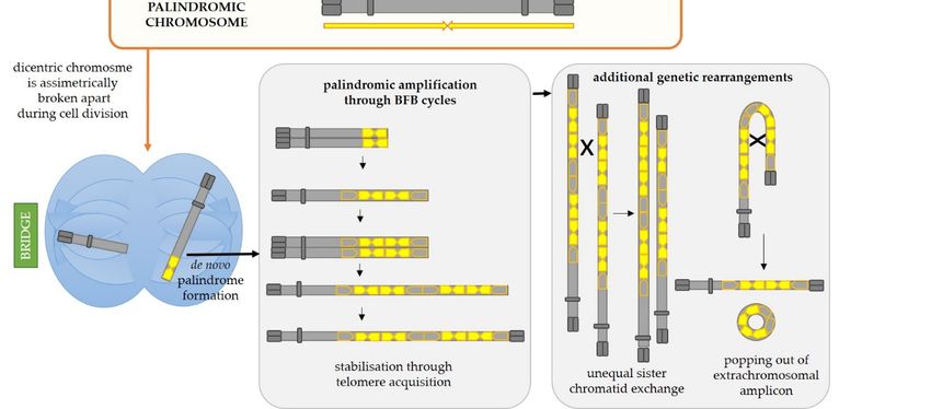
Int. J. Mol. Sci. 2021, 22, 2840                                                                                                   2 of 18

                                   2. The Recombinogenic Nature of Palindromic Sequences
                                   2.1. DNA Palidromes Can Form Secondary Structures
                                         A palindrome in DNA is a sequence consisting of two identical or highly similar
                                   inverted repeats which are either adjacent to one another or separated by a spacer region
                                   (Figure 1a). If the repeats (also called the palindrome arms) are identical and have no
                                   spacer in between, the palindrome is referred to as perfect. The term quasipalindrome can
                                   be used to refer to a non-perfect palindrome. Palindromes are found in genomes of all
                                   species investigated so far and they often play important roles as binding sites for homod-
                                   imeric proteins, parts of promoters, replication origins or other regulatory sequences [1].
                                   However, many of the discovered palindromes have no known biological function and
                                   can be relatively long (from several dozen to several hundred base pairs). If a palindrome
                                   is of sufficient length, intrastrand base pairing can occur and this results in formation of
                                   secondary structures in DNA (Figure 1b). In the single-stranded DNA, a hairpin structure
                                   forms, while in the double-stranded DNA, a cruciform structure consisting of two hairpins,
                                   one in each strand, forms. Each hairpin consists of a stem comprised of complementary
                                   paired inverted repeats and a loop. The loop either consists of bases within the spacer,
                                   if such region is present in a specific palindrome, or of four–six bases which lie in the
                                   center of symmetry of the two inverted repeats and cannot be complementarily paired
                                   due to the rigidity of the DNA strand [2,3]. It is considered that a hairpin structure can
                                   occur in the single-stranded lagging strand during DNA replication. A cruciform structure
                                   forms in dsDNA by gradual extrusion which begins at the center of the palindrome. After
                                   denaturation of a short region of DNA at the center, intrastrand base pairing occurs and
                                   a small proto-cruciform forms which can further extrude into a larger cruciform [2,4,5].
                                   Conditions which lead to cruciform extrusion have been extensively studied in vitro [6–9]
                                   and theoretical kinetic models of cruciform extrusion were postulated [10,11]. These ex-
                                   periments show that cruciform extrusion is thermodynamically unfavorable, and in those
                                   early studies, it was debated if extrusion is even possible in vivo. However, processes such
 Int. J. Mol. Sci. 2021, 22, x     as transcription and replication can increase the density of negative supercoils in DNA. 3 of 19
                                   The added torsion can be released through cruciform extrusion and this is considered to be
                                   a mechanism driving extrusion in vivo [12–14].

                                       (a)                                                            (b)
       Figure1.1.Palindromic
      Figure      Palindromicsequences
                               sequencesandandsecondary
                                               secondarystructures.
                                                           structures.(a)
                                                                       (a)Types
                                                                           Typesofofpalindromic
                                                                                      palindromicsequences.
                                                                                                   sequences.AAperfect
                                                                                                                perfectpalindrome
                                                                                                                        palindrome
       consists  of two identical inverted repeats  adjacent to one another. A  quasipalindrome    can contain mismatches
      consists of two identical inverted repeats adjacent to one another. A quasipalindrome can contain mismatches between  between
       the two  inverted  repeats and/or a central spacer region. (b) Palindromic   sequences can  form secondary  structures
      the two inverted repeats and/or a central spacer region. (b) Palindromic sequences can form secondary structures due    duetoto
       intrastrand base pairing. In ssDNA, the hairpin structure, consisting of a stem and a loop, is formed. Two hairpins, one
      intrastrand base pairing. In ssDNA, the hairpin structure, consisting of a stem and a loop, is formed. Two hairpins, one
       across from the other in a dsDNA, together constitute a cruciform structure.
      across from the other in a dsDNA, together constitute a cruciform structure.
                                    2.2. Molecular Mechanisms of Palindrome Recombinogenicity
                                         Molecular mechanisms of palindrome recombinogenicity have been investigated in
                                    model organisms Escherichia coli and Saccharomyces cerevisiae as well as in human cells.
                                    While in E. coli the recombinogenicity of palindromes seems to be exclusively replication-
                                    related [22–24], in eukaryotic cells, both hairpin formation during replication and cruci-
                                    form formation not related to replication seem to be at play [25–27]. The mechanisms of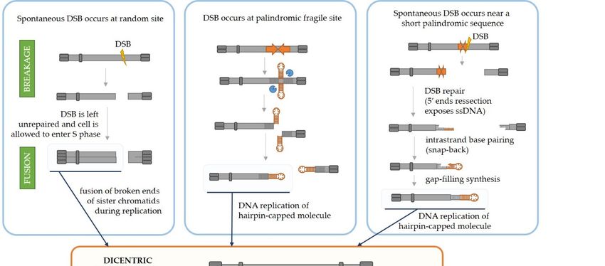
Int. J. Mol. Sci. 2021, 22, 2840 3 of 18
Palindromes which have the potential to form secondary structures in DNA present a
risk for genome stability since these structures can result in double-strand DNA breaks
(DSBs) which in turn lead to genetic recombination, potentially resulting in translocations,
deletions or gene amplifications with various consequences for the cell and organism. A
specific palindrome is more likely to lead to DSB and subsequent recombination (i.e., is
more recombinogenic) if the likelihood for the secondary structure formation is higher
and if the structure is more stable once it forms. Thus, palindromes consisting of longer
inverted repeats sharing a higher degree of sequence similarity and having a shorter or
absent spacer present a higher risk for the genome stability. These principles have not only
been experimentally proven for palindromes in model organisms [5,15–20] but also noted
for palindromes with varying spacer length and arm length polymorphisms within the
human population [21].
2.2. Molecular Mechanisms of Palindrome Recombinogenicity
Molecular mechanisms of palindrome recombinogenicity have been investigated in
model organisms Escherichia coli and Saccharomyces cerevisiae as well as in human cells.
While in E. coli the recombinogenicity of palindromes seems to be exclusively replication-
related [22–24], in eukaryotic cells, both hairpin formation during replication and cruciform
formation not related to replication seem to be at play [25–27]. The mechanisms of DSB
Int. J. Mol. Sci. 2021, 22, x
formation and processing of palindromic sequences in eukaryotic cells are shown in
4 of 19
Figure 2 and explained in detail in Sections 2.2.1 and 2.2.2.
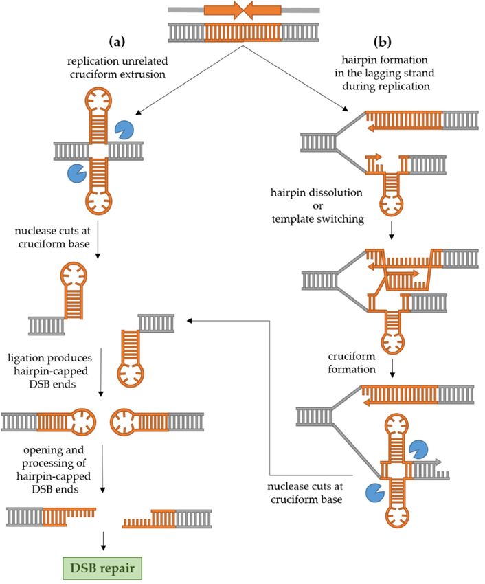
Figure 2. Mechanisms of palindrome recombinogenicity. (a) Independently from DNA replication,
Figure 2. Mechanisms of palindrome recombinogenicity. (a) Independently from DNA replication, a
a cruciform structure can extrude in the dsDNA. The cruciform is cut by a structure-specific endo-
cruciform structure
nuclease, after can extrude
which ligation producesinclosed
the dsDNA. The cruciform
hairpin-capped is break
double-strand cut by(DSB)
a structure-specific
ends. endonucle-
Suchafter
ase, ends must
whichbe ligation
opened and processedclosed
produces into conventional DSB endsdouble-strand
hairpin-capped which can undergo repair.
break (DSB) ends. Such ends
(b) During replication, especially if the replication machinery is impaired, a hairpin can form in
must be opened and processed into conventional DSB ends which can undergo repair. (b) During
the ssDNA of the lagging strand, causing replication stalling. The hairpin can be dissolved by hel-
replication, especially
icase, or, alternatively, if theswitching
template replication machinery
allows isthe
synthesis of impaired, a hairpin
nascent strand using can form in the ssDNA of the
the leading
strand as a template. This produces a nascent complementary strand containing
lagging strand, causing replication stalling. The hairpin can be dissolved by helicase, a hairpin as well, or, alternatively,
across the hairpin in the lagging strand. Again, the resulting cruciform can be cut by a structure-
template switchingfollowed
specific endonuclease, allowsbysynthesis
formation of ofhairpin-capped
the nascent DSB strand
ends,using the leading
their processing and strand as a template.
subsequent
This producesDSB repair.
a nascent complementary strand containing a hairpin as well, across the hairpin in
the lagging strand. Again, the resulting cruciform can be cut by a structure-specific endonuclease,
2.2.2. Replication-Dependent Palindrome Recombinogenicity
followed by formation of hairpin-capped DSB ends, their processing and subsequent DSB repair.
During DNA replication, the palindromic sequence can result in the formation of a
hairpin in the single-stranded lagging strand which can cause replication stalling and a
double-strand break. The stalling of the replication fork at a palindrome consisting of two
inverted Alu repeats separated by a 12 bp spacer was demonstrated in E. coli, yeast and
primate fibroblasts in the work by Voinegau et al. [26]. Interestingly, when the same re-
peats were separated by a longer 52 bp spacer, frequent stalling was observed only in E.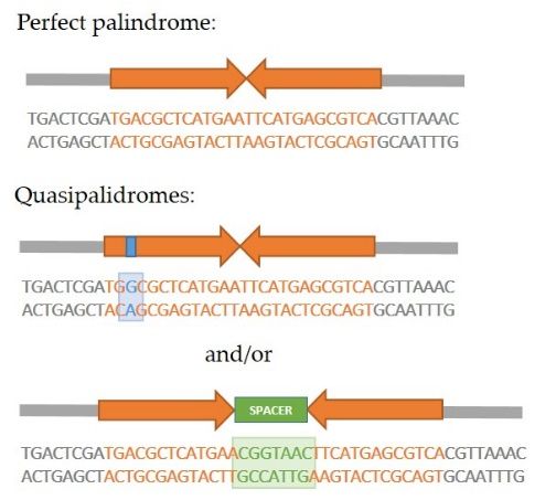
Int. J. Mol. Sci. 2021, 22, 2840                                                                                             4 of 18

                                   2.2.1. Replication-Independent Palindrome Recombinogenicity
                                         The cruciform structure in dsDNA can be cleaved at the base by a structure-specific
                                   endonuclease. Since the base of the cruciform resembles a Holliday junction, the likely
                                   candidates for cruciform cleavage are resolvases. This idea is in accordance with the
                                   fact that the cruciform is efficiently cut by bacterial resolvase RuvC as well as T4 DNA
                                   endonuclease VII in vitro [7,28]. In a culture of human cells, Inagaki et al. [29] used two
                                   non-replicating plasmids carrying two palindromes originating from the human genome
                                   (PATRRs, palindromic AT-rich repeats) to demonstrate that the cruciform structure is rec-
                                   ognized and cut exclusively by Gen1 resolvase, and not by Mus81-Eme1 or Slx1–Slx4. In
                                   yeast, however, the cruciform-cutting endonuclease remains elusive [5]. Experimental
                                   evidence suggests that the two nicks created by the action of resolvase are usually lig-
                                   ated in vivo, since replication of DNA with an unprocessed hairpin-capped end leads to
                                   large palindromic duplication [25,30]. Such ends need to be processed into conventional
                                   DSB in order to be repaired. In human cells, the hairpin-capped DSB ends are opened
                                   by Artemis [29], while in yeast, the Sae2 endonuclease together with the MRX complex
                                   processes the ends [25]. Once hairpin-capped ends are processed, the conventional DSB can
                                   undergo repair. In yeast, the predominant mechanism of DSB repair is homologous recom-
                                   bination, while in human cells, the break will most often be repaired by non-homologous
                                   end joining (NHEJ) [31].

                                   2.2.2. Replication-Dependent Palindrome Recombinogenicity
                                         During DNA replication, the palindromic sequence can result in the formation of a
                                   hairpin in the single-stranded lagging strand which can cause replication stalling and a
                                   double-strand break. The stalling of the replication fork at a palindrome consisting of two
                                   inverted Alu repeats separated by a 12 bp spacer was demonstrated in E. coli, yeast and
                                   primate fibroblasts in the work by Voinegau et al. [26]. Interestingly, when the same repeats
                                   were separated by a longer 52 bp spacer, frequent stalling was observed only in E. coli, but
                                   not in eukaryotic cells. This is in line with the idea that replication stalling occurs because a
                                   hairpin is formed in a single-stranded region of the lagging strand. Namely, in prokaryotes,
                                   the Okazaki fragment initiation zone is at least 1500 bp long, while in eukaryotes, it is
                                   limited to 100–200 bp [32]. Therefore, an increase in spacer length reduces the likelihood
                                   that a central region of a palindrome which can form a hairpin will be contained within
                                   the ssDNA. However, in case DNA replication is in some way impaired (e.g., slowed
                                   because of deficiency in one of the proteins of the replication machinery), the ssDNA could
                                   potentially become abnormally long which can result in otherwise stable repeats to become
                                   unstable and recombinogenic [19,27,33]. For example, the fragile site 2 in yeast which
                                   consists of two large inverted repeats (Ty elements, each 6 kb long) separated by a large
                                   280 bp spacer is completely stable in wild-type yeast but becomes prone to DSB and genetic
                                   rearrangements when expression of DNA polymerase α is reduced [33].
                                         Zhang et al. [27] also demonstrated that the frequency of DSB and recombination
                                   instigated by a palindrome introduced in the yeast genome is increased in strains which
                                   lack or have a transient reduction in expression of proteins involved in DNA replication
                                   (including Pol2 and Pol3 which are catalytic subunits of DNA polymerases ε and δ, respec-
                                   tively, and others such as endonuclease Rad27), proteins involved in cell cycle arrest during
                                   replication, maintaining telomere stability, and proteins belonging to the Sgs1-Top3-Rmi1
                                   complex. Interestingly, when the ends of a DSB instigated by a palindromic sequence in
                                   cells with reduced quantities of Pol2 or Pol3 were analyzed, it was found that they are also
                                   hairpin-capped, just as in the case of the cruciform resolution described above. Moreover,
                                   in this case, the MRX-Sae2 complex was also required for opening and processing hairpin-
                                   capped ends into a canonical DSB which could undergo repair. Based on these results,
                                   the authors proposed a model shown in Figure 2b. They postulated that in conditions of
                                   a slower replication, a hairpin can form in the lagging strand which can be dissolved by
                                   the Sgs1-Top3-Rmi1 complex. If the hairpin is not dissolved, a Rad51-mediated template
                                   switching occurs and the 30 end of the nascent strand is elongated using the leading strand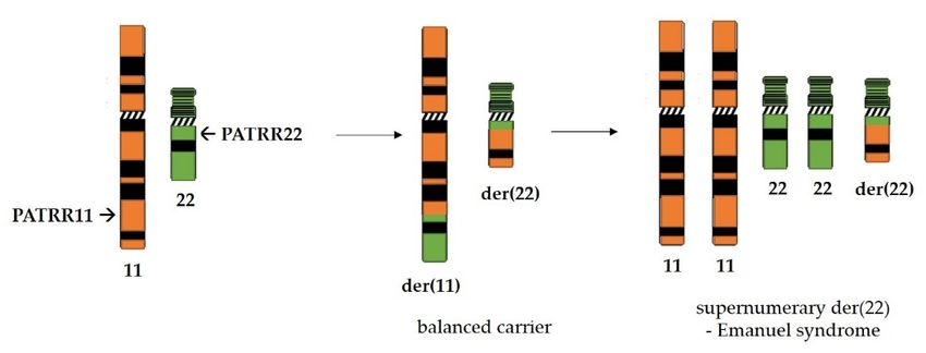
Int. J. Mol. Sci. 2021, 22, 2840                                                                                             5 of 18

                                   as a template for synthesis, which results in formation of a cruciform. This proposal is
                                   supported by the fact that in rad51 and rad54 mutants (proteins important for 30 end
                                   invasion and pairing with the homologous sequence in conditions of slowed replication),
                                   the frequency of DSB and the recombination rate are again reduced. The cruciform is then
                                   recognized and cut by a putative endonuclease in yeast and hairpin-capped ends of a DSB
                                   are produced as described above.

                                   3. DNA Palindromes and Genetic Instability in Cancer Cells
                                   3.1. Palindromic Amplifications in Cancer
                                        Genetic instability resulting in various DNA rearrangements is one of the hallmarks of
                                   cancer cells. It is not uncommon for cancer cells to feature amplifications of genes or large
                                   genomic regions. This results in an altered level of gene expression, often with an adverse
                                   effect on the disease outcome since such tumors can become more aggressive and more
                                   proliferative (e.g., amplification of HER2 oncogene in breast cancer, [34]) or resistant to
                                   drugs (e.g., amplification of TYMS gene conferring resistance to 5-flourouracil in colorectal
                                   cancer, [35]). Multiple studies [36,37] demonstrated a link between a specific pattern of
                                   amplified regions in the cancer cell genome and a certain cancer pathophysiology. For
                                   example, in their study in breast cancer samples, Hicks et al. [37] identified a specific
                                   amplification pattern confined to a single chromosome arm. This pattern consists of
                                   multiple closely spaced amplified regions (amplicons which they named “firestorms”),
                                   often with deletions in the regions between them. Hence, copy numbers of some genes are
                                   increased, while other genes are deleted, resulting in loss of heterozygosity. They showed
                                   that even a single region featuring the “firestorm” pattern in the breast cancer genome
                                   that is otherwise relatively quiet is linked to poor prognosis. Interestingly, such studies
                                   often identify recurrent amplifications (i.e., amplifications encompassing same genomic
                                   regions found in different patients) which can help to better predict disease progression
                                   and outcome and guide treatment decisions in clinics. Growing effort is being invested
                                   into understanding cancer genomics. The Cancer Genome Atlas (TCGA) program [38] has
                                   so far molecularly characterized over 20,000 primary cancers and matched normal samples
                                   across 33 cancer types. The program uses microarrays for determining copy number
                                   variations, methylation and protein expression as well as high-throughput sequencing for
                                   characterization of DNA and RNA. Amplified genomic regions (cytobands) are determined
                                   for various cancer types. Tanaka and Watanabe [39] summarized data from TCGA on
                                   recurrently amplified regions from six epithelial tumor types (breast, colorectal, lung
                                   squamous, lung adenocarcinoma, prostate and ovarian). They found, just to outline one
                                   example, that an 800-kb-long region, 8q24.21, which carries the MYC oncogene is amplified
                                   across all these tumors. On the other hand, tumor type-specific amplified regions were
                                   also found. They concluded that tumor type-specific amplified regions often carry lineage-
                                   specific transcription factors, while regions amplified across several cancer types often carry
                                   genes involved in fundamental processes responsible for cell proliferation. Undoubtedly,
                                   these recurrent events are, on the one hand, under a selective pressure since deletion or
                                   overexpression of certain genes (“driver genes”) can confer advantage to a tumor cell. On
                                   the other hand, however, it is likely that certain regions of the genome are predisposed to
                                   such rearrangements due to the presence of specific DNA sequences, such as palindromes.

                                   3.1.1. Palindromic Amplification in Cancer Can Arise as a Result of Iterative
                                   Break-Fusion-Bridge Cycles
                                         The pattern of DNA amplification characterized by multiple closely spaced amplified
                                   regions can be explained by breakage-fusion-bridge (BFB) cycles, first discovered in maize
                                   by Barbara McClintok [39–43] and illustrated in Figure 3. Tumor cells often carry deficien-
                                   cies in one or more tumor suppressor genes important for DNA repair and DNA damage
                                   sensing as well as deficiencies in checkpoint systems which should lead the cell into cell
                                   cycle arrest and apoptosis if DNA damage or mitotic dysfunction exists [44]. Hence, if the
                                   cell is impaired in one or several of these vital processes, a double-strand break could be left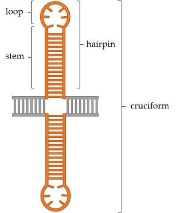
Int. J. Mol. Sci. 2021, 22, 2840                                                                                          6 of 18

                                   unrepaired and the cell could be allowed to progress into the S phase of the cell cycle. At
                                   that point, the two segments of a broken chromosome are replicated, and the resulting four
                                   DNA strands can either fuse correctly to produce two identical chromosomes or incorrectly,
                                   thus producing one null-centric and one dicentric palindromic chromosome (Figure 3,
                                   central panel). During the cell division, the dicentric chromosome will be broken by the
                                   forces of the mitotic spindle pulling centromeres in the opposite directions. The anaphase
                                   bridges are often observed in tumor cells undergoing genetic rearrangements [45]. Each
                                   daughter cell will inherit a segment of the dicentric chromosome, but one of them will
                                   most likely inherit a somewhat larger segment than the other (depending on the position
                                   of the chromosome break during cell division) which will thus have a palindrome at its
                                   end. Such broken chromosomes are then a substrate for the next BFB cycle in daughter
                                   cells. This can go on for many cycles, ultimately resulting in accumulation of multiple
                                   palindromic gene amplifications at breakpoints. Such chromosome might finally be sta-
                                   bilized by telomere addition [46], but processes such as intrachromosomal homologous
                                   recombination between the acquired repeats and unequal sister chromatid exchange could
                                   drive further rearrangements and thus gene copy number gains or reductions [46–49].
                                   Moreover, intrachromosomal recombination between the acquired DNA repeats can eas-
                                   ily result in the popping-out of an extrachromosomal circular amplicon (named double
                                   minute chromosomes, DMs), a type of amplification event that is commonly detected in
                                   cancer cells but almost never in normal cells [50,51]. It is often the case that such DMs
                                   harbor oncogenes which are amplified either solely on the DM or both on the DM and
                                   on the chromosome. Mathematical modeling indicates that the oncogene copy number
                                   and genetic heterogeneity would be increased more effectively on DMs than chromosomal
                                   amplification, and this might contribute to accelerated evolution in cancer [52]. Hence, it
                                   is not surprising that the existence of DMs is also linked to poor prognosis and shorter
                                   survival for patients with various cancer types [51]. Additionally, certain complex DMs
                                   carrying DNA from various distant loci can arise during a process of chromothripsis (greek
                                   thripsis—breaking into pieces)—a single catastrophic event in which a chromosome arm is
                                   fragmented into pieces which are subsequently randomly reconnected [53].

                                   3.1.2. Relatively Short Palindromes in the Genome Can Facilitate the Initiating Event of
                                   Palindromic Amplification through the Fold-Back Priming Mechanism
                                         Furthermore, the initiation of palindromic gene amplifications in cancer cells can
                                   also be explained by the fold-back priming mechanism (Figure 3, right panel). Namely,
                                   if a chromosome break occurs in the vicinity of an existing palindromic sequence in the
                                   genome, the processing of broken ends by DNA repair machinery (50 end resection, [54])
                                   will expose the palindromic sequence in a single-stranded form. This will result in a
                                   fold-back, i.e., intrastrand base pairing and hairpin formation. The 30 -end can then serve
                                   as a primer for gap-filling DNA synthesis which will result in a sealed hairpin-capped
                                   DNA. If left unopened and unprocessed (by MRX-Sae2 in yeast or Artemis in human
                                   cells), the replication of such chromosome will result in a large palindromic duplication
                                   (palindromic chromosome). It has been shown in yeast [55–57] and mammalian cells [58]
                                   that palindromic duplication can be mediated by relatively short inverted repeats which is
                                   consistent with this model. Butler et al. [55] demonstrated that induction of DSB by HO
                                   endonuclease on a yeast minichromosome next to a short 42 bp inverted repeat efficiently
                                   produced a palindromic chromosome. A similar result was obtained in Chinese hamster
                                   ovary cells [58] by induction of DSB with I-SceI endonuclease next to a 79 or 229 bp inverted
                                   repeat. Furthermore, by analyzing breakpoints at the center of palindromic duplication,
                                   Deng et al. [59] demonstrated that inverted repeats as short as 5–9 bp separated by 3–12 bp
                                   spacer sequences can drive large palindromic duplications in Mre11- or Sae2-deficinet yeast
                                   cells. Their results also indicate that a large proportion of snap-back events are prevented
                                   or removed by RPA (Replication Protein A), the ssDNA-binding protein which normally
                                   prevents annealing of very short homologous sequences as well as removing secondary
                                   structures from ssDNA [60]. The deficiency in RPA in mre11 or sae2 mutants increases the
                                   frequency of palindromic duplications by 1000-fold. Moreover, it appears that, besides
Int. J. Mol. Sci. 2021, 22, 2840 7 of 18
small loop hairpins forming in close proximity (within 50 bp) to a DSB, a large loop hairpin
after an extensive DSB end resection can be formed in yeast [61]. Although they are likely
Int. J. Mol. Sci. 2021, 22, subsequently
x processed through different pathways using different enzymes, both7types
of 19 of
hairpins can result in a gross chromosomal rearrangement and gene amplification.
Figure 3. Mechanisms responsible for initiation of de novo palindrome formation followed by subsequent palindromic
Figure 3. Mechanisms responsible for initiation of de novo palindrome formation followed by subsequent palindromic
amplifications and additional genetic rearrangements typical for cancer cells. On the left upper panel, the initial steps of
amplifications the
andbreakage-fusion-bridge
additional genetic rearrangements
(BFB) mechanismtypical are shown.for cancer
If a DSBcells.
is leftOn the left and
unrepaired upper
the panel,
DNA isthe initial steps
subsequently of the
repli-
cated, the broken
breakage-fusion-bridge ends of sister chromatids
(BFB) mechanism are shown.can If fuse
a DSB to form two
is left palindromic
unrepaired chromosomes,
and the DNA isone of which will be
subsequently dicentric. the
replicated,
broken ends ofMiddle and right upper panels depict alternative mechanisms for formation of a palindromic dicentric chromosome, an
sister chromatids can fuse to form two palindromic chromosomes, one of which will be dicentric. Middle and
initiating event for palindromic amplification. In the middle panel, a DSB is mediated by a secondary structure-forming
right upper panels
palindrome in the genome.mechanisms
depict alternative As explained in fordetail
formation
in Section of2,athis
palindromic dicentric chromosome,
results in hairpin-capped an unprocessed,
DSB ends. If left initiating event
for palindromictheamplification. In the
replication of the middle panel, ahairpin-capped
centromere-proximal DSB is mediated by a results
molecule secondary
in an structure-forming
dicentric chromosome. palindrome
On the upper in the
right panel, the fold-back priming mechanism is depicted. If a DSB occurs in the vicinity
genome. As explained in detail in Section 2, this results in hairpin-capped DSB ends. If left unprocessed, the replicationof a palindrome or short inverted
repeats, the DSB repair, which normally involves 5′ end resection, will expose the repeats in the ssDNA form. This can
of the centromere-proximal hairpin-capped
allow for intrastrand moleculeand
base pairing (fold-back) results in an 3′-end
the folded dicentric chromosome.
can then On the
serve as a primer for upper right
gap-filling DNApanel,
syn- the
fold-back priming mechanism
thesis. The resultingis hairpin-capped
depicted. If a molecule
DSB occurs in the
can also vicinity
lead of a palindrome
to occurrence of a dicentricorchromosome
short inverted
uponrepeats, the DSB
DNA replica-
tion. Regardless
repair, which normally involves 50initial
of the mechanismwill
end resection, of formation,
expose the the palindromic
repeats in the dicentric
ssDNA chromosome
form. This cancan
be asymmetrically bro-
allow for intrastrand
ken during cell division and one of the resulting chromosomes will carry a de novo formed palindrome (palindromic
0 -end
base pairing (fold-back) and the folded 3 can then serve as a primer for gap-filling DNA synthesis.
duplication). Additional BFB cycles can result in palindromic amplifications of certain genomic regions and such chromo-
The resulting
hairpin-cappedsome
molecule
could atcan also
some lead
point to occurrence
become stabilized ofby atelomere
dicentric chromosome
addition, uponiterations
thus breaking DNA replication.
of BFB cyclesRegardless
(lower middle of the
panel).
initial mechanism of However,
formation, additional genetic rearrangements
the palindromic and gene copy
dicentric chromosome cannumber gains or losses broken
be asymmetrically can ariseduring
throughcellunequal
division
sister chromatid exchange and intrachromosomal recombination between the acquired repeats which can result in the
and one of the resulting chromosomes will carry a de novo formed palindrome (palindromic duplication). Additional
popping out of an extrachromosomal amplicon (double minute chromosome), also a typical feature of cancer cells.
BFB cycles can result in palindromic amplifications of certain genomic regions and such chromosome could at some point
become stabilized by telomere addition, thus breaking iterations of BFB cycles (lower middle panel). However, additional
genetic rearrangements and gene copy number gains or losses can arise through unequal sister chromatid exchange and
intrachromosomal recombination between the acquired repeats which can result in the popping out of an extrachromosomal
amplicon (double minute chromosome), also a typical feature of cancer cells.Int. J. Mol. Sci. 2021, 22, 2840 8 of 18
3.1.3. Longer Palindromes in the Genome Are Fragile Sites Which Can Lead to
Palindromic Duplication
Moreover, if a palindrome present in the genome is long enough to have the potential
to extrude into a cruciform structure, the resolution of the cruciform by an endonucle-
ase also leads to the formation of hairpin-capped chromosome ends and potentially to
palindromic chromosome formation (Figure 3, middle panel). This was demonstrated by
Lobachev et al. [25] using two human Alu repeats separated by a 12 bp spacer inserted into
a yeast chromosome. In the absence of Mre11, Xrs2, Rad50 (MRX complex) or Sae2, a large
palindromic duplication occurs after DNA replication. Additionally, it was shown in yeast
that palindromes can cause replication stalling [26], potentially leading to a double-strand
break during replication [27], as depicted in Figure 2 (right). Further, if the stall persists,
the replication fork can collapse and lead to a DSB or restart erroneously, leading to ge-
netic rearrangements [62–64]. An interesting study identified genomic features in human
and mouse genomes which highly depend on the ATR checkpoint kinase to maintain
stability [65]. Namely, ATR is activated upon DNA polymerase stalling which results in
stabilization of intermediates at stalled replication forks and prevents progression into the
M phase. Sequences for which the stability is highly dependent on ATR, and thus most
likely to stall replication, are those which can form secondary structures including some
microsatellites, inverted retroelements and AT-rich palindromes and quasipalindromes.
Ultimately, regardless of the mechanism of formation, any dicentric chromosome
could be broken apart by the mitotic spindle during cell division and initiate BFB cycles.
A dicentric chromosome could also be produced upon telomere shortening and fusion of
different chromosomes or sister chromatids with dysfunctional telomeres, as is the case
in cells with deficient shelterin (protein complex responsible for telomere maintenance)
lacking both p53 and retinoblastoma (RB) tumor suppressors [66,67]. Additionally, a
palindromic duplication results in a new genomic fragile site since it produces a palindrome
long enough to extrude into a cruciform structure or form a hairpin during DNA replication.
3.2. Challenges in Decyphering the Initiating Event Responsible for Palindromic Amplifications
in Cancer
When it comes to various types of tumors isolated from patients, it can be difficult
to pinpoint the initial mechanism which led to gene amplifications since the initial am-
plification is followed by subsequent rearrangements. Thus, it is difficult to determine
whether the initial breakpoint and the molecular mechanism which led to subsequent
rearrangements are common in a certain locus in various tumor types or not. Several
interesting studies have aimed to unravel the origins of gene amplifications in human tu-
mor samples. Tanaka et al. [68] developed a microarray-based approach for genome-wide
analysis of palindrome formation (GAPF). Briefly, total genomic DNA from the sample
is first digested with a restriction enzyme and then heat denaturated. The renaturation
is performed under conditions (rapid cooling, presence of NaCl) which favor intrastrand
over interstrand base pairing. Therefore, DNA palindromes quickly “snap back” and
renaturate while the rest of the genome remains single-stranded. Next, the ssDNA is
degraded with S1 nuclease, and the remaining dsDNA is digested with selected restriction
enzymes, ligated with terminal linkers and amplified by PCR using Cy5-labelled linker-
specific primers. In parallel, the DNA standard for competitive hybridization is isolated
from normal human fibroblasts, subjected to the same procedure as the DNA from the
tumor sample, except for the denaturation step, and labelled with Cy3. The standard and
the sample DNAs are cohybridized on a human spotted cDNA microarray. The GAPF
technique was later modified to reduce the detection of false positives and coupled with
next-generation sequencing [69]. Using this technique to analyze samples from colorectal
and breast cancer cell lines and primary medulloblastomas, the authors found [68,70] a
frequent occurrence of de novo palindromes. Interestingly, in colorectal cancer cell line
Colo320DM and breast cancer cell line MCF7, common loci containing DNA palindromes
were detected, indicating that these regions might be susceptible to chromosomal breaks
and palindrome formation. Guenthoer et al. [71] used the same GAPF technique coupledInt. J. Mol. Sci. 2021, 22, 2840 9 of 18
with gene copy number analysis to look into commonly amplified regions involved in
breast tumorigenesis (chromosome arms 8q and 11q) and found that formation of palin-
dromes de novo coincided with copy number gains and with amplicon breakpoints. These
results point to the palindrome-associated mechanism of gene amplification. Moreover,
in similar subsets of breast cancers featuring similar characteristics, similar amplified loci
were found, implicating that preexisting fragile sites might initiate amplification.
Narayanan et al. [30] used yeast as a eukaryotic model organism to select and analyze
gross chromosomal rearrangements resulting from hairpin-capped breaks occurring at
the inverted human Alu repeats. The recovered rearrangements resembled those typically
found in cancer cells, including palindromic gene amplifications and terminal deletions,
highlighting the potential role of existing inverted repeats in the human genome in tumori-
genesis. All of these studies strongly point to the conclusion that a de novo palindrome
formation in cancer cells is not necessarily a random event and that, once it occurs, it serves
as a platform for subsequent gene amplifications.
4. DNA Palindromes in the Human Genome
4.1. Bioinformatics in Quest for DNA Palindromes
The effort to estimate the abundance and distribution of DNA palindromes in genomes
greatly relies on the use of bioinformatics tools to analyze genome sequence data. However,
when reading such reports, one has to be aware of the specificities of each palindrome-
counting algorithm which might result in a great number of palindromic sequences that are
inherently recombinogenic to be overlooked or underrepresented. Moreover, the cloning of
palindrome-containing genome fragments could be difficult or unsuccessful. In addition,
some palindromes maintained on BACs (bacterial artificial chromsomes) in E. coli could be
lost during propagation. Nonetheless, this does not diminish the value and importance of
such studies. Most recently, Ganapathiraju et al. [72] constructed a catalog of human DNA
palindromes and analyzed variations in these palindromic sequences in 1000 sequenced
human genomes. Their algorithm starts palindrome counting by finding an 8 bp perfect
palindrome and then extends the span of the sequence to each side, allowing a maximum of
four mismatches between the palindrome arms. In other words, it counts only near-perfect
palindromes without a spacer. Through counting, they found that the human genome
contains around 13 million palindromes shorter than 40 bp, about 180,000 longer than 40 bp
and 718 palindromes longer than 200 bp, with palindromes 8 and 20 bp in length being the
most abundant. As shown in previous studies on the human genome [73], palindromes
are mostly located in the intronic and intergenic regions, probably because extensive
intrastrand base paring within mRNA molecules could have negative repercussions on pro-
cesses of transcription and translation. Palindrome avoidance of coding regions was also
demonstrated in yeast [74]. Strawbridge et al. [75] also found that closely spaced inverted
repeats in the yeast S. cerevisiae genome are clustered in intergenic regions, with clustering
being more pronounced in 30 than in 50 flanks of genes. Moreover, they noticed that repeats
capable of forming cruciform structures are rare in the yeast genome. Further, in accor-
dance with earlier studies on the human [73] genome and genomes of yeasts [74,76], the
authors found long palindromes are highly AT-rich. Additionally, Ganapathiraju et al. [72]
analyzed the GWAS catalog SNPs (single nucleotide polymorphisms) to see if any of the
catalogued SNPs are found within palindrome sequences, possibly making them longer or
more perfect. Interestingly, disease risk-associated SNPs were 14 times more likely to be
found within the palindromic regions than in other regions. Furthermore, in their earlier
study [77], the same research group used a similar approach to compare the 69 serum-
normal and tumor genomes originating from The Cancer Genome Atlas with 1000 human
genomes as a control. The aim of this study was to computationally identify differences in
palindrome occurrence on chromosomes 8 and 11 between these two groups of genomes.
Interestingly, they found that, in multiple cases, palindromes which happen to be near
genes involved in breast carcinogenesis were among those which differed the most in
cancer compared to normal cell genomes. Additionally, several other groups worked onInt. J. Mol. Sci. 2021, 22, 2840 10 of 18
palindrome-counting algorithms which are publicly available [78,79]. Moreover, efforts
are being made to overcome other obstacles in identifying DNA palindromes in genomes.
For example, it has been demonstrated that a palindromic sequence present in the human
genome is much better preserved in the case the human genome fragment was cloned
before sequencing in yeast Saccharomyces cerevisiae rather than bacteria E. coli [80].
4.2. From Short Interspersed Elements (SINEs) to Segmental Duplications—Possibilities for
Palindrome Occurrence in the Human Genome
Given the great number of various repeated sequences in the human genome, it is
not surprising that they can be found in various relations to one another, such as direct
tandem repeats as well as closely spaced inverted repeats, i.e., palindromes. Alu repeats, a
type of primate-specific short interspersed element (SINE), are one of the most abundant
repeated elements in the human genome. They were named Alu because they usually
feature a recognition site for the AluI restriction endonuclease [81]. They make up for
about 11% of the human genome and count more than one million copies per haploid
genome [82]. One Alu element is about 300 bp long, and based on their divergence,
they are divided into subfamilies [83]. Alu repeats are often found at breakpoints of
genetic rearrangements and are linked to genetic instability [84,85], gene copy number
variations [86] and various diseases [87]. The potency of inverted Alu repeats to instigate
palindrome-mediated genetic instability has been demonstrated by Lobacev et al. [19]
in yeast. In the human genome, Alu-mediated rearrangements can occur as a result
of recombination between various Alus throughout the genome and through various
molecular mechanisms (homologous recombination, single-strand annealing, template
switching, etc.), and in some cases, they are likely to be palindrome-mediated. However,
what portion of Alu-mediated rearrangements involves palindrome instability remains to
be investigated. The same is true for various other repeated elements in the human genome
which together constitute around 50% of the human DNA content [82].
Additionally, the human genome harbors large segmental duplications which make
up for about 5% of the genome and are together clustered in about 400 regions. Segmental
duplications arose during primate evolution and maintain high sequence identity between
duplicated regions [88]. For example, palindromes P1–P8 on the male-specific section of
chromosome Y span dozens of genes, many of which are essential for spermatogenesis.
It is considered that these palindromes have an important evolutionary purpose since
they allow intrachromsomal recombination in an otherwise non-recombining chromosome.
Intrachromosomal gene conversion can protect against deleterious mutations, but also
having an extra copy of these genes can enhance the adaptive evolution of chromosome
Y [89]. However, recombination between palindrome arms can result in formation of isodi-
centric chromosome Y [90] and recombination between different Y-specific palindromes can
result in massive deletion [91,92], all known causes of a range of sex-linked reproductive
disorders, including relatively common spermatogenic failure [93]. Other segmental dupli-
cations have also been identified as fragile sites as well. Segmental duplication KFTAP-1
on chromosome 17, which is often found as a boundary for palindromic amplification of
the ERBB2 gene (amplified in up to 30% of breast tumors as well as stomach, bladder and
esophageal cancers), has been shown to be susceptible to DSBs in normal human cells and
even more so if the exonuclease activity of DNA repair protein Mre11 is disabled [94].
4.3. PATRRs and Other Known Palindromes in the Human Genome
Perhaps the most thoroughly studied palindromes in the human genome are PA-
TRRs (palindromic AT-rich repeats). These sequences are near-perfect palindromes that
are quite long—several hundred base pairs with more than 90% of base pairs being AT.
They do not share any significant degree of homology between them, yet, not surpris-
ingly given their palindromic nature, they are often sites of chromosome breakage and
genetic rearrangements [95]. Palindromes PATRR11 (approximately 450 bp long) and
PATRR22 (approximately 595 bp long), located on chromosomes 11 and 22, respectively,
are involved in the most common recurrent non-Robertsonian translocation in the humanInt. J. Mol. Sci. 2021, 22, 2840 11 of 18
genome (Figure 4) which is the underlying cause of Emanuel syndrome [96]. Reciprocal
translocation with breakpoints in these palindromic sequences produces balanced carriers
which are, for the most part, healthy. They do have an increased risk of developing breast
cancer [97], as well as infertility issues and recurrent pregnancy losses. However, if the
small derivative of chromosome 22 produced by translocation is passed on alongside the
ci. 2021, 22, x normal chromosome set, the zygote is viable. Unfortunately, such children 12 of
of 19
balanced
carriers suffer from Emanuel syndrome which is characterized by a range of psychical
disorders involving heart and kidney function as well as mental retardation [98].
Reciprocal
Figure 4. Figure translocation
4. Reciprocal between palindromes
translocation betweenPATRR11 on chromosome
palindromes PATRR11 11onand PATRR22 on11chromosome
chromosome and 22 in
the human genome produces
PATRR22 a generally
on chromosome healthy
22 in balanced
the human carrier.produces
genome However,aoffspring
generallywho inherit
healthy the supernumerary
balanced carrier.
derivativeHowever,
der(22) alongside thewho
offspring normal chromosomal
inherit set suffer from
the supernumerary Emanuel syndrome.
derivative der(22) alongside the normal chro-
mosomal set suffer from Emanuel syndrome.
Since the de novo translocations between PATRR11 and PATRR22 occur recurrently
at similar breakpoints, Kurahashi et al. [99] were able to design a translocation-specific
Since the de novo translocations between PATRR11 and PATRR22 occur recurrently
PCR assay to investigate the origins of translocation. The PCR assay at the single-molecule
at similar breakpoints,
detection Kurahashi et al. [99]
level was performed were able
on sperm of mento with
design a translocation-specific
a normal karyotype (46+XY). Approx-
PCR assay to investigate
imately 1 inthe origins
10,000 DNA ofaliquots
translocation. The PCR
were positive assay
for the at the single-molecule
translocation t(11;22) PCR product,
detection level indicating
was performedde novoon sperm of men
translocation in spermwith[100].
a normal karyotype
However, using the(46+XY). Ap-
same methodology,
proximately 1 in 10,000 DNA aliquots were positive for the translocation t(11;22) PCR
the authors found no evidence of de novo t(11;22) translocation in blood and cheek swab
cells from
product, indicating de novothe same men, nor in
translocation in sperm
lymphoblastoid cell linesusing
[100]. However, and cultured
the same fibroblasts.
meth- This
points to the conclusion that de novo t(11;22) translocations
odology, the authors found no evidence of de novo t(11;22) translocation in blood and occur during gametogenesis.
Moreover, although a too small number of female oocytes are available to perform similar
cheek swab cells from the same men, nor in lymphoblastoid cell lines and cultured fibro-
testing, there was some indication that occurrence of those de novo t(11;22) transloca-
blasts. This points to the conclusion that de novo t(11;22) translocations occur during gam-
tions might be sperm-specific. Ohye et al. [101] analyzed eight cases of offspring carrying
etogenesis. Moreover,
derivativealthough
chromosomea too 22small
andnumber
their parents.of female
Due to oocytes are availablebetween
the polymorphism to per- PATRR
form similar testing, there
sequences was some
originating indication
from the mother that occurrence
and father, theyofwere
those detonovo
able t(11;22)
determine that in all
translocations might be sperm-specific. Ohye et al. [101] analyzed eight cases of
eight cases, the de novo t(11;22) translocation was of paternal origin. The authors proposed offspring
carrying derivative chromosome
that sperm specificity22does
and exist,
their parents.
and it could Duebetoexplained
the polymorphism between
by a translocation occurring
PATRR sequences due to the palindrome-mediated
originating from the mother instability during
and father, DNA
they werereplication. Namely, there is a
able to determine
that in all eight great
cases,difference
the de novo in the number
t(11;22) of cell divisions
translocation was(and thus DNAorigin.
of paternal replications) in pre-meiotic
The authors
gametogenic cells in males and females—around 150 divisions until adulthood and an
proposed that sperm specificity does exist, and it could be explained by a translocation
additional 23 each year in spermatogenesis vs. 22 divisions throughout the female lifetime
occurring due to the palindrome-mediated instability during DNA replication. Namely,
in oogenesis. However, although the frequency of mutations caused by replication errors
there is a greatas difference
well as theinfrequency
the number of someof cell divisions translocations
non-recurrent (and thus DNA replications)
in sperm in with
does increase
pre-meiotic gametogenic cells in males
age in men [102–104], this is and
not thefemales—around 150 divisions
case for de novo t(11;22) until [105].
translocation adult-
hood and an additional In another line of research, the same research group demonstrated that,the
23 each year in spermatogenesis vs. 22 divisions throughout if there is
female lifetimesufficient
in oogenesis.
negative However,
superhelicityalthough
of DNA,the frequency
PATRRs of mutations
can extrude caused
into a cruciform by cloned
when
replication errorson non-replicative
as well as the plasmids
frequency in human
of some cells [106,107]. Further,
non-recurrent the most recent
translocations experimental
in sperm
evidence by Correl-Tash et al. [108] demonstrated that PATRRs
does increase with age in men [102–104], this is not the case for de novo t(11;22) translo- can extrude into a cruciform,
cation [105]. leading to a higher frequency of DSBs in both mitotic and meiotic cells. They used sister
chromatid exchanges (SCEs) observed in the metaphase chromosome spread using a
In another line of research, the same research group demonstrated that, if there is
sufficient negative superhelicity of DNA, PATRRs can extrude into a cruciform when
cloned on non-replicative plasmids in human cells [106,107]. Further, the most recent ex-
perimental evidence by Correl-Tash et al. [108] demonstrated that PATRRs can extrude
into a cruciform, leading to a higher frequency of DSBs in both mitotic and meiotic cells.Int. J. Mol. Sci. 2021, 22, 2840 12 of 18
florescent microscope in samples where PATRR regions were labeled with florescent probes.
Since SCEs indicate sites at which DSBs occur and are repaired, they can be used as an
indirect measure of genetic instability [109,110]. In mitotic cells (lymphocytes) of normal
individuals, SCEs colocalize with PATRRs significantly more often than with a control
region of the genome. Moreover, when DNA associated with DSB repair proteins (Rad51,
NBS1 and γH2AX) was pooled by chromatin immunoprecipitation (ChIP), the qPCR
analysis showed an increase in PATRR sequences in relation to various other control DNA
regions. In both experiments, when DNA from balanced t(11;22) carriers was analyzed,
no increase in SCEs frequency or DSB repair protein coprecipitation was detected. This
is in accordance with the fact that after reciprocal translocation, the recombined PATRR
sequences are no longer palindromic and thus no longer pose a threat to genome stability.
The same ChIP-qPCR assay but using the cruciform-binding 2D3 antibody demonstrated an
increased association between PATRRs and cruciform structures. Moreover, using the same
methodology, they showed that PATRRs can result in cruciform formation, leading to an
increase in DSB frequency and subsequent repair in spermatogenic cells, but this increase
is not higher than in mitotic cells. Although it appears that the likelihood of PATRR-related
cruciform extrusion and DSB formation is equal in mitotic and meiotic cells, a de novo
PATRR-mediated translocation as a result of DSB repair is typically not found in mitotic
cells [95]. Exceptionally, a case report described a woman with a mosaic karyotype, with
64% of normal cells and 36% of cells with t(11;22), indicating that translocation occurred
in a post-zygote mitotic cell [111]. The explanation for most de novo PATRR-mediated
translocation occurrences possibly lies in some specific DSB response or repair mechanism
during gametogenesis.
Moreover, using the immunofluorescence labeling method and 2D3 cruciform bind-
ing antibody, Feng et al. [112] demonstrated the presence of cruciforms in mice growing
oocytes. Interestingly, cruciform foci were detected in different phases of oocyte growth
but were no longer present in fully grown oocytes (characterized by silencing of tran-
scription activities, chromatin condensation and formation of a Hoechst-positive ring-like
structure surrounding the nucleolus). Cruciform foci colocalized with PARP1 (poly(ADP-
ribose)-polymerase, a protein involved in DNA damage sensing (mainly of single-stranded
breaks), which was previously shown to bind hairpin loops [113], but not with γH2A.X,
indicating that in normal oocytes, a cruciform-induced DSB is probably rare. Additionally,
Feng et al. [112] analyzed data from The Genotype-Tissue Expression Project (GTEx, [114])
to see the expression pattern for PATRR11- and PATRR22-related transcripts in various
human tissues. They found that the nearest transcript to PATRR11 (located about 1 kb
downstream of PATRR11) is specifically expressed in liver and testes and that PATRR22 is
located in the intron of a gene also specifically expressed in testes. Since DNA transcription
induces a significant amount of negative superhelicity, they proposed that the transcription
might be a driving process for cruciform extrusion. To further strengthen this hypothesis,
they treated a transcriptional active growing oocyte with RNA-polymerase II inhibitor
α-amanitin and noted a sharp decrease in the number of cruciform foci in treated cells.
Although palindromes PATRR11 and PATRR22 are the longest and thus the most
recombinogenic, as well as the most extensively studied, the human genome harbors a
dozen PATRRs. Palindromes PATRR8 (approximately 350 bp long) and PATRR17 (approxi-
mately 200 bp long), located on chromosomes 8 and 17, respectively, are also involved in
constitutional translocations with PATRR22 as a partner. PATRR17 is located in the intron
of the NF1 gene, and a t(17;22) translocation which led to inactivation of the NF1 gene
was found in several patients with neurofibromatosis type 1 [115,116]. Recurrent t(8;22)
translocation and inheritance of supernumerary derivative chromosome der(22)t(8;22)
are linked to a syndrome characterized by ear and extremity abnormalities, in addition
to mild mental retardation [117]. A translocation event between PATRR8 and PATRR11
has also been detected in sperm of healthy males [117]. Furthermore, the constitutional
rearrangement between PATRR3 and PATRR8, t(3;8)(p14.2;q24.1), was shown to segregate
with renal cell carcinoma in two families. Both PATRR8 and PATRR3 are located withinInt. J. Mol. Sci. 2021, 22, 2840 13 of 18
introns of genes [118]. Non-recurrent translocations involving PATRRs on chromosomes 4,
1, 3 and 9 were also detected [119–122].
Additionally, there are examples where investigation of underlying molecular causes
of various conditions in humans leads to the discovery of a palindrome in the genome
clearly capable of instigating DNA rearrangements. In three unrelated families in Mex-
ico and China, congenital generalized hypertrichosis (excessive hair growth all over the
body) was linked to insertions mediated by a 180 bp palindrome located on chromosome
X [123,124]. Likewise, a 160 bp palindrome located 30 kb upstream of the β-globin gene
has been implicated in deletions leading to (γδβ)0 -thalassemia [125,126].
5. Concluding Remarks
Throughout this review, we tried to summarize the current state of knowledge on
various aspects and consequences of compromised genome stability due to the presence of
recombinogenic palindromic sequences. Although occasionally it appears that the research
in this filed progresses more slowly than in some other areas due to the difficulties in the
cloning and sequencing of DNA palindromes, recent advancements in our understanding
of palindrome-instigated genome instability described in this review show that this subject
holds the interest of researchers. Future research in this field will, on the one hand, continue
to be focused on molecular mechanisms and protein players involved in palindrome
recombinogenicity. It is important to understand when, why and under which conditions a
certain palindromic sequence, which was stably replicated and inherited through numerous
cell divisions, initiates genetic recombination, possibly with devastating consequences for
the cell and organism. When it comes to cancer cells, genetic rearrangements are often
abundant and complex, so it can be difficult to unravel the initiating and subsequent chain
of events leading to a certain genotype. Undoubtedly, palindromic gene amplifications are
an important mechanism involved in carcinogenesis. However, palindromes and inverted
repeats preexisting in the genome could have a much greater role in initiating recurrent
recombination events during carcinogenesis than is currently appreciated. Therefore, it is
important to continue the deep sequencing efforts to fill the gaps in the human genome
sequence which likely harbor multiple recombinogenic palindromic sequences, but also to
analyze as much individual cancer genomes as possible and link them to specific cancer
pathophysiology and treatment outcomes. Hopefully, with enough data analyzed, patterns
will emerge which can improve diagnostics as well as the choice of appropriate treatment,
but perhaps which can also uncover markers signifying elevated risk even before the
disease onset.
Funding: This research received no external funding.
Conflicts of Interest: The authors declare no conflict of interest.
Abbreviations
BAC bacterial artificial chromosome
BFB breakage-fusion-bridge
DSB double-strand break
GAPF genome-wide analysis of palindrome formation
GWAS genome-wide association study
NHEJ non-homologous end joining
PATRR palindromic AT-rich repeat
SINE short interspersed element
SNP single nucleotide polymorphism
TCGA The Cancer Genome Atlas
References
1. Brázda, V.; Laister, R.C.; Jagelská, E.B.; Arrowsmith, C. Cruciform structures are a common DNA feature important for regulating
biological processes. BMC Mol. Biol. 2011, 12, 33. [CrossRef]You can also read