Ion Channel Targeting with Antibodies and Antibody Fragments for Cancer Diagnosis - MDPI
←
→
Page content transcription
If your browser does not render page correctly, please read the page content below
antibodies
Review
Ion Channel Targeting with Antibodies and Antibody
Fragments for Cancer Diagnosis
Claudia Duranti and Annarosa Arcangeli *
 Department of Experimental and Clinical Medicine, Section of Internal Medicine, University of Florence,
 50134 Firenze, Italy; claudia.duranti@unifi.it
 * Correspondence: annarosa.arcangeli@unifi.it; Tel.: +39-055-2751283

 Received: 21 March 2019; Accepted: 20 May 2019; Published: 24 May 2019

 Abstract: The antibody era has greatly impacted cancer management in recent decades. Indeed, antibodies
 are currently applied for both cancer diagnosis and therapy. For example, monoclonal antibodies are the
 main constituents of several in vitro diagnostics, which are applied at many levels of cancer diagnosis.
 Moreover, the great improvement provided by in vivo imaging, especially for early-stage cancer diagnosis,
 has traced the path for the development of a complete new class of antibodies, i.e., engineered antibody
 fragments. The latter embody the optimal characteristics (e.g., low renal retention, rapid clearance,
 and small size) which make them ideal for in vivo applications. Furthermore, the present review focuses
 on reviewing the main applications of antibodies and antibody fragments for solid cancer diagnosis,
 both in vitro and in vivo. Furthermore, we review the scientific evidence showing that ion channels
 represent an almost unexplored class of ideal targets for both in vitro and in vivo diagnostic purposes.
 In particular, we review the applications, in solid cancers, of monoclonal antibodies and engineered
 antibody fragments targeting the voltage-dependent ion channel Kv 11.1, also known as hERG1.

 Keywords: antibodies; cancer diagnostics; in vivo imaging; engineered antibody fragments; hERG1

1. Introduction
     Antibodies have become common and essential research instruments over the last fifty years,
providing highly specific and versatile tools for a wide array of experimental applications in many
fields. Furthermore, monoclonal antibodies (mAbs) and, more recently, recombinant antibodies have
gained clinical applications for diagnosis and therapy of different diseases, including cancer [1,2].
The translation of antibodies from basic research into the clinic has therefore significantly changed
the prognosis for different classes of human cancers. The great success of the clinical application
of antibodies mainly relies on the high versatility of these biological molecules. Indeed, antibodies
combine the specificity of antigen targeting with, e.g., the possibility to be easily conjugated with various
molecular or chemical agents, thus improving their pharmacological efficacy. Targeted therapeutic
strategies have recently raised the possibility of tailoring cancer treatment to an individual patient after
assessing the peculiar molecular characteristics of the tumor under treatment. Such refined diagnostic
assessments can be achieved using antibody molecules. The fusion between diagnostic imaging
techniques and therapeutic intervention is important to address cancer heterogeneity [3]. The term
“theranostics” was hence coined to describe a molecular tool having both diagnostic and therapeutic
applications [4]. Moreover, several platforms linking a diagnostic tool, often represented by an antibody,
with a defined therapeutic compound have been developed and marketed. Such “companion
diagnostics” are embodying an indispensable part of personalized cancer medicine [5].
     The present review focuses on reviewing the main applications of mAbs for cancer diagnosis
in vitro. Moreover, we address how the technology of engineering antibody molecules, and in particular
the possibility of developing antibody fragments, is greatly impacting on in vivo molecular imaging,

Antibodies 2019, 8, 33; doi:10.3390/antib8020033                              www.mdpi.com/journal/antibodies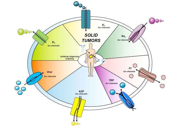
Antibodies 2019, 8, 33                                                                                2 of 21

for diagnostic applications in solid cancers. We also provide strong evidence that ion channels are
relevant molecular devices in cancer establishment and progression, and that can be exploited for
either in vitro or in vivo cancer diagnosis. In particular, the diagnostic and prognostic applications,
in solid cancers, of mAbs and antibody fragments targeting the voltage-dependent ion channel Kv 11.1,
also known as hERG1, are thoroughly discussed.

2. Antibody-Based Cancer Diagnostics
     Solid cancer diagnosis is currently based on imaging techniques (e.g., Computer-Assisted
Tomography, Magnetic Resonance Imaging, etc.), laboratory assays (e.g., tests for circulating tumor
markers such as the carcinoembryonic antigen) and the pathological evaluation of either biopsies or
surgical specimens. The latter can take advantage of either biomolecular techniques or antibody-based
immunohistochemistry (IHC) to provide further insights for patients’ prognostic stratification and
therapeutic choice. The number and type of techniques available to allow physicians to detect and
diagnose cancer had significant changes in the last years. In fact, more accurate and reproducible
imaging techniques have been developed and applied to the clinical setting. Moreover, novel cancer
biomarkers have been identified to improve diagnosis and prognosis. In this scenario, antibodies
represent key devices for both in vitro and in vivo diagnosis, since they can specifically recognize
specific cancer biomarkers in tissues and body fluids. In particular, while mAbs represent good
molecular tools to detect cancer biomarkers in vitro, in tissue specimens, their use in vivo is hindered
by several concerns (see Section 3.2) and are progressively being substituted by antibody fragments [6].
     Hereafter, the main antibody-based in vitro and in vivo techniques for cancer diagnosis
are reported.

2.1. In Vitro Cancer Diagnostics
       Solid cancer diagnosis in vitro is now routinely improved by the detection of clinically validated
biomarkers through IHC on paraffin-embedded tissue slides. After antibody binding to the specific
antigen, the target region can be visualized by an enzyme-linked (e.g., horseradish peroxidase) or
a fluorescent dye, a radioactive tracer or a colloidal gold reagent. The positivity of the tumor for a given
marker is hence evaluated, applying predetermined cutoffs. New IHC techniques have improved both
the optical resolution and the sensitivity of detection, mainly through the use of amplification procedures,
despite the risks of false-positive and false-negative staining [6]. Some “in vitro diagnostics” (IVD)
based on antibodies (and the related IHC technique) have been clinically validated and are currently
applied in the clinical practice (see Table 1). mAbs can also be utilized as “companion diagnostics”,
i.e., diagnostics that can be associated with the use of a particular treatment, either a small molecule
or a therapeutic antibody. The path to companion diagnostics started in 1998 with the approval of
the therapeutic humanized mAb Trastuzumab, which was paralleled by the simultaneous approval of
a diagnostic test, the HercepTest. Some of the approved companion diagnostics are reported in Table 1.
       An emerging application of antibodies for cancer diagnostic and prognostic purposes relies on the
use of “omics” data [7]. However, both IHC and omics strategies display some disadvantages, since
they can hardly address tumor heterogeneity. Furthermore, tumor biopsies are not always able to reveal
the overall antibody binding or the overall expression of the biomarker. Both hindrances have been
recently addressed through the so called “liquid biopsy”, aimed at the detection of either circulating,
cell-free, DNA or of circulating tumor cells (CTCs). CTCs detection can help to monitor a patient’s
condition, including the tracking of the genomic evolution of the tumor. CTC studies are accomplished
through microfluidics platforms, and a new approach includes the use of EpCAM expression levels
for the capture of CTCs. Besides anti-EpCAM antibodies, other antibodies targeting specific tumor
biomarkers are now under development to improve microfluidic-based CTC analyses [8].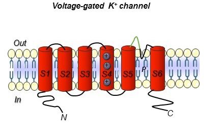
Antibodies 2019, 8, 33                                                                                                                                                                                 3 of 21

      Table 1. Antibody-based in vitro diagnostics (IVDs) which are already approved by the FDA (Federal Drug Administration) and/or EMA (European Medicine Agency)
      and used for cancer diagnosis. CTA, Cancer-testis antigen; CEA, carcinoembryonic antigen; PSMA, prostate-specific membrane antigen; TAG-72, tumor-associated
      glycoprotein 72; PDL-1, programmed death-ligand 1; HER 2, human epidermal growth factor receptor 2; EGFR, epidermal growth factor receptor; ALK, anaplastic
      lymphoma kinase.

      IVD Commercial Name             Manufacturer             Antigen    Antibody Format                Tumor Type                Diagnostic Significance       Possibility of Companion Diagnostic
                                                                                                   Colorectal cancer/tumor
           Humaspect®                Organon Teknica            CTA       Humanized mAb                                                                                         NA
                                                                                                            detection
                                                                                                   Colorectal cancer/tumor         Tumor marker, Prognostic
            CEA-scan®                Immunomedics               CEA      Murine Fab fragment                                                                                    NA
                                                                                                            detection                     marker
                                                                                               Prostate adenocarcinoma/tumor
           ProstaScint®                  Cytogen               PSMA         Murine mAb                                                Prognostic marker                         NA
                                                                                                            detection
                                                                                                 Small-cell lung cancer/tumor
       Verluma® (Diagnostic)   Boehringer Ingelheim, NeoRx     CD-20     Murine Fab fragment                                                                                    NA
                                                                                                            detection
                                                                                                    Colorectal and ovarian
            OncoScint®                   Cytogen               TAG-72       Murine mAb                                                                                          NA
                                                                                                    cancer/tumor detection
                                                                                                      Non-small-cell lung                                                          Yes
     PD-L1 IHC 22C3 pharmDx      Dako North America Inc.       PDL-1        Murine mAb
                                                                                                    cancer/tumor detection                                      Keytruda (pembrolizuma)—BLA 125514
      VENTANA PD-L1(SP142)                                                                      Non-small-cell lung cancer and                                                     Yes
                               Ventana Medical Systems, Inc.   PDL-1         Rabbit mAb
            Assay                                                                              urothelial cancer/tumor detection                              Tecentriq (atezolizumab)—NDA 761034/S012
                                                                                                                                                                                   Yes
                                                                         Murine mAb, (clone        Colorectal cancer/tumor
      Dako EGFR pharmDx Kit      Dako North America, Inc.       EGFR                                                                                               Erbitux (cetuximab)—BLA 125084
                                                                              2-18C9)                     detection
                                                                                                                                                                  Vectibix (panitumuma)—BLA 125147
      PATHWAY anti-Her2/neu                                                                                                                                                        Yes
                               Ventana Medical Systems, Inc.    HER2         Rabbit mAb         Breast cancer/tumor detection
               (4B5)                                                                                                                                             Herceptin (trastuzumab)—BLA 103792
       Bond Oracle HER2 IHC                                                    mAb                                                 Tumor marker, Prognostic                        Yes
                                     Laica Biosystem            HER2                               Breast cancer detection
              System                                                        (CB11 clone)                                                  marker                 Herceptin (trastuzumab)—BLA 103792
                                                                                                                                   Tumor marker, Prognostic                        Yes
            HercepTest              Dako Denmark A/S            HER2         Rabbit mAb            Breast cancer detection
                                                                                                                                          marker                 Herceptin (trastuzumab)—BLA 103792
                                                                                                                                                                                   Yes
    VENTANA ALK (D5F3) CDx                                                                     Non-small-cell lung carcinoma       Tumor marker, Prognostic        Zykadia (ceritinib)—NDA 205755
                               Ventana Medical Systems, Inc.    ALK          Rabbit mAb
           Assay                                                                                        detection                         marker                   Xalkori (crizotinib)— NDA 202570
                                                                                                                                                                   Alecensa (alectinib)—NDA 208434
Antibodies 2019, 8, 33 4 of 21
2.2. In Vivo Cancer Diagnostics: Molecular Imaging
The accuracy that allows antibodies to precisely identifying their targets has stimulated their
application also for in vivo imaging, thus improving the diagnostic imaging approaches currently used.
For example, the 18F-Fluoridexyglucose-Positron Emission Tomography (FDG-PET) imaging represents
an invaluable diagnostic and prognostic tool to detect high levels of glycolytic activity, a sign of malignant
transformation. However, FDG-PET is not free of false negatives (e.g., tumors with more indolent
growth or dependent on different metabolic pathways) or false positives, as in the case of infection or
inflammation [9]. Notably, the majority of imaging probes actually in use to detect cancer can also detect
inflammation, thus leading to significant numbers of potential false positives. Hence, using antibodies
for in vivo imaging may lead to a versatile approach which allows more accurate diagnosis, staging and
hence disease management. Some practical examples of mAbs recognizing cancer specific biomarkers
that are approved by the FDA and/or EMA and are currently used in the clinical setting are shown in
Table 2A. Among these, ProstaScint is a mAb used in prostate cancer patients as a diagnostic imaging
agent to detect nodal metastases “pre-prostatectomy”, or recurrence in post prostatectomy patients with
a rising prostate-specific antigen (PSA). The therapeutic mAbs, cetuximab and trastuzumab, are also used
as in vivo tracers. In particular, the dual-labeled (111 In-DTPA)n-trastuzumab-(IRDye800)m is capable
of tracking HER2 overexpression in breast cancer patients [10]. cetuximab has been repurposed for
fluorescent imaging and is in phase I and phase II clinical trials for malignant glioma and pancreatic
cancer imaging and fluorescence-guided surgery with IRDye-800CW [11].
Table 2. Antibodies for in vivo use in tumor imaging diagnostics approved by the FDA and/or EMA
and present on the market. CEA, carcinoembryonic antigen; Tc, technetium; In, indium.
A) Monoclonal
Antibodies
Commercial Antibody Conjugated
Company Antigen Tumor Application
Name Format Probe
Capromab
7E11-C5.3, 100-kDa 111 In
pendetide Cytogen Prostate carcinoma
mouse IgG1 glycoprotein
(ProstaScint)
Votumumab 88BV59, human Altered Colorectal, ovarian
Intracel “
(HumaSPECT) IgG3 cytokeratins and breast carcinoma
Ibritumomab
2B8, mouse Non-Hodgkin
tiuxetan Spectrum Pharms CD20 “
IgG1 lymphoma
(Zevalin)
Tositumomab B1, mouse
SmithKline Beecham “ “ “
(Bexxar) IgG2a
Human-murine
Head and neck
chimeric
squamous cell
Cetuximab Cetuximab-IRDye800CW monoclonal EGFR IRDye800CW
carcinoma, pancreatic
antibody
cancer
(mAb)
Humanized
(111 In-DTPA)n-trastuzumab-(IRDye
Trastuzumab monoclonal HER2 “ Breast cancer
800CW)
antibody
B) Engineered
Antibody
Fragments
IMMU-4,
Arcitumomab 99m Tc Colorectal and ovarian
Immunomedics mouse IgG1 CEA
(CEA-Scan) carcinoma
Fab’
Nofetumomab NR-LU-10, Small-cell and
40-kDa
merpentan Boehringer Ingelheim mouse IgG2b “ non-small-cell lung
glycoprotein
(Verluma) Fab carcinoma
Bectumomab LL2, mouse 99m Tc
Immunomedics CD22 “
(LymphoScan) IgG2a Fab’
Igovomab OC125, mouse 111 In
CIS Bio International CA125 Ovarian cancer
(Indimacis-125) IgG1 F(ab’)2
Although antibodies have definitely improved cancer diagnostics through their application in IVD kits,
the use of mAbs as molecular imaging tools for in vivo diagnostics still needs further improvements.Antibodies 2019, 8, 33 5 of 21
In other words, antibodies need to be designed differently for their use as diagnostics in vitro and for their
application as in vivo imaging agents. When injected in vivo, in fact, whole antibodies display long serum
half-lives (1–3 weeks), which are favorable if they are envisaged to be applied as therapeutics, thanks to the
enhancement of the exposure of target tissues to the antibody. Moreover, the effector domain (crystallizable
fragment, Fc) often exerts biological activities which are essential for the therapeutic functions of the whole
antibody molecule [12]. However, such characteristics are drawbacks for the use of such molecules as
imaging2019,
Antibodies agents, as several
8, x FOR days are required to obtain a good signal-to-noise ratio, and a biological 6activity
PEER REVIEW of 23
is undesirable for an imaging agent.
Many of
Many of the
the disadvantages
disadvantagesofof whole antibody
whole molecules
antibody have have
molecules been overcome thanks to
been overcome antibody
thanks to
antibody engineering, producing smaller and highly versatile molecules [13], which maintain theof
engineering, producing smaller and highly versatile molecules [13], which maintain the specificity
mAbs butof
specificity allow
mAbs higher
but tumor penetration
allow higher tumorand shorter clearance
penetration times,clearance
and shorter both optimal characteristics
times, both optimal for
diagnostic purposes.
characteristics for diagnostic purposes.
3.3.Antibody
AntibodyFragments
Fragmentsfor
forCancer
CancerDiagnostics
Diagnostics
Generally, the
Generally, the preservation
preservationofof thethe
variable fragment
variable (Fv) domain
fragment in engineered
(Fv) domain antibody antibody
in engineered fragments
preserves the antigen binding. Conversely, the absence of the Fc region abolishes
fragments preserves the antigen binding. Conversely, the absence of the Fc region abolishes the the immune interactions
mediated
immune by the complement
interactions mediated and
bybytheother effectors. The
complement andelimination of the FcThe
by other effectors. region also blocksofrecycling
elimination the Fc
through the path of the neonatal Fc receptor (FcRn), facilitating the contrast and
region also blocks recycling through the path of the neonatal Fc receptor (FcRn), facilitating the thus the visualization
of targeted
contrast and tissues
thus the through rapid clearance
visualization from
of targeted the blood
tissues through [13].rapid
Smaller fragments
clearance from allow the use
the blood [13].of
radionuclides
Smaller thatallow
fragments decaythe
rapidly
use of(for example through
radionuclides labeling
that decay with(for
rapidly 18F), thus resulting
example throughinlabeling
reduced
radiation exposure. Furthermore, the use of antibodies with a reduced molecular
with 18F), thus resulting in reduced radiation exposure. Furthermore, the use of antibodies with weight (below ~60 kDa)
a
speeds up the elimination through the renal clearance [14]. The main structural
reduced molecular weight (below ~60 kDa) speeds up the elimination through the renal clearance characteristics, molecular
weight
[14]. Theandmain
renal clearance
structuralof characteristics,
different engineered antibodyweight
molecular fragments are renal
and shownclearance
in Figure 1 of
anddifferent
described
in the following paragraph.
engineered antibody fragments are shown in Figure 1 and described in the following paragraph.
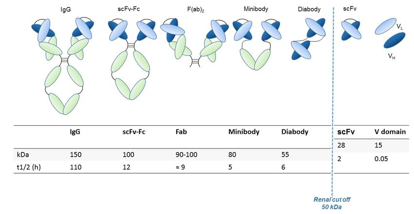
Figure 1. Engineered antibody formats. Different antibody formats are reported in the figure, showing
Figure 1. Engineered
their different antibody
size compared formats.
to intact IgG.Different antibody formats
Ig, Immunoglobulin; are reported
scFv, single-chain in thefragment;
variable figure,
showing their different size compared to intact IgG. Ig, Immunoglobulin; scFv, single-chain
V domain, variable domain. In the figure, the size (KDa) of each different antibody format and their variable
fragment;
half-livesV(tdomain, variable domain. In the figure, the size (KDa) of each different antibody format
1/2 , i.e., time needed to eliminate half of the molecule from circulation) are also reported.
and their half-lives (t1/2, i.e., time needed to eliminate half of the molecule from circulation) are also
3.1. reported.
Antibody Fragments: Characteristics and Development
A comparison of the pharmacokinetics and target specificities between intact and fragmented
3.1. Antibody Fragments: Characteristics and Development
antibodies has been performed, and the results show that recombinant antibodies retain the antigen
A comparison of the pharmacokinetics and target specificities between intact and fragmented
antibodies has been performed, and the results show that recombinant antibodies retain the antigen
specificity of the intact Igs from which they derive advantageous characteristics regarding tumor
penetrance and retention. Furthermore, antibody fragments have several formats which are reported
in Figure 1: scFv-Fc (≈ 100 KDa), minibodies (Mb; scFv-CH3 dimers, ≈ 80 kDa), F(ab’)2 and Fab (≈ 50
KDa), bispecific antibodies (≈ 55-60 KDa) and scFv (≈ 25 KDa) [12]. The F(ab) fragment is an antibodyAntibodies 2019, 8, 33 6 of 21
specificity of the intact Igs from which they derive advantageous characteristics regarding tumor
penetrance and retention. Furthermore, antibody fragments have several formats which are reported in
Figure 1: scFv-Fc (≈ 100 KDa), minibodies (Mb; scFv-CH3 dimers, ≈ 80 kDa), F(ab’)2 and Fab (≈ 50 KDa),
bispecific antibodies (≈ 55-60 KDa) and scFv (≈ 25 KDa) [12]. The F(ab) fragment is an antibody
structure that still binds to antigens but is monovalent and lacks the Fc portion. An antibody digested
by the enzyme papain yields two F(ab) fragments of about 50 kDa each and an Fc fragment [15].
Another class of antibody fragments is based on single-domain antibodies.
Among sdAbs, the so called nanobodies merit attention. Nanobodies (~15kDa) are fragments of
heavy single-variable heavy chains (VHH s), originated from antibodies found in the camelids. The small
size of these antibodies allows them to bind to epitopes to which intact molecules cannot access [16]
and makes them appropriate for applications where an extremely short time of clearance is desired.
The latter is also determined by the size, the charge and by the presence/absence of conjugated parts.
Fragments with weights lower than the renal threshold (~60 kDa) are eliminated through the kidneys,
while larger molecules are instead cleared through the liver. Nanobodies can be used in diagnostics
both at the initial stages for the detection of the tumor itself and then to evaluate the expression
of a target for therapeutic intervention or for monitoring of the disease. This class of molecules
can be coupled with various nanocarriers (e.g., iron oxide nanoparticles, silica nanoparticles, gold
nanostructures, and carbon nanomaterials), which could be suitable for anticancer drug delivery [17].
From the diagnostic point of view, two important classes of antibody fragments are the scFvs,
single-chain variable fragments, and the bispecific antibodies. scFvs have a molecular weight around
25 kDa and are composed of VH and VL chains, joined via a flexible peptide linker. The first scFv
molecules were developed in 1988 and represent the smallest functional VH –VL domains of an antibody
necessary for the high-affinity binding of an antigen. Peptide linkers are fundamental for the assembly
of functional scFv antibodies, as they join the VH and VL chains and usually vary from 10 to 25
amino acids in length and typically include hydrophilic amino acids. The most common linker is the
decapenta-peptide (Gly4Ser)3. The variable regions can be connected in either the VH-linker-VL (most
common) or VL-linker-VH orientation. In any case, such orientations can affect expression efficiency,
stability and antigen binding activity [18].
Another class of antibodies which has gained great attention is that of bispecific antibodies, among
which single-domain diabody (scDb) is the most versatile format. The first work on bispecific antibody
generation was published in 1961 and described the production of chimeric antibodies containing two
different antigen-binding sites simultaneously. Such molecules are capable of binding two different
antigens at the same time, thus allowing the recognition of two different targets which might be crucial
in the diagnostic setting. scDbs conjugate the bispecificity with the characteristics (low molecular
weight, high tissue penetration, and good clearance times) of antibody fragments [19].
3.2. Applications of Antibody Fragments for in Vivo Imaging
Recent examples of antibody fragments used as in vivo diagnostics combined encompass several
of the aforementioned formats even though this field offers great space for improvement as few of
such molecules are actually used for in vivo imaging diagnostic applications. Among those few are
many Fab fragments for HER2 (human epidermal growth factor receptor 2) targeting and the use of
124 I-PSCA (Prostate Stem Cell Antigen)-specific minibody in order to assess the response to prostate
cancer treatment using enzalutamide [9]. Moreover, F(ab’)2 and Fab fragments radiolabeled with
111 In (Indio111) and minibodies and diabodies labeled with 89Zr (Zirconium 89) have been used for
imaging with SPECT (Single Photon Emission Computed Tomography) or PET (Positron Emission
Tomography) in small animals targeting the prostate-specific membrane antigen (PSMA). The great
interest in fragments of antibodies is demonstrated by several papers using nanobodies [20,21] and
affibodies [22]. So far, preclinical studies have been performed to give insights into the biochemical
and biophysical features of several antibody fragments as imaging agents [23,24]. Ogasawara and
colleagues demonstrated that antibodies specific for phosphatidylserine can be a valuable tool to assessAntibodies 2019, 8, 33 7 of 21
cell death in response to treatment [25]. Direct imaging with antibodies could also offer a suitable
technique to determine the development of resistance to therapy. Li and colleagues [9] focused on
MET receptor, using various antibody fragments derived from an anti-MET antibody to obtain images
of non-small-cell lung cancer xenografts from cell lines resistant to targeted anti-EGFR therapy due to
the overexpression of MET.
Finally, bispecific radioimmunoconjugates (bsRICs) capable of binding to HER2 and EGFR were
developed for both therapy and diagnostic applications and tested in preclinical models. The aim of
these studies was to better decipher the mechanisms underlying trastuzumab resistance related to the
overexpression of EGFR in patients with HER2-positive carcinomas. These bsRICs are composed of the
trastuzumab Fab (ligand for HER2) linked to EGF (ligand for EGFR) via a long spacer (polyethylene
glycol). Such antibodies show good specificity and low uptake in normal organs except for the
kidneys [26]. We report, in Table 2, the main monoclonal and engineered antibody fragments which
are used in vivo and have already been approved by the FDA and/or EMA.
4. Ion Channels in Cancer
Ion channels are membrane proteins which, besides controlling cell excitability and ionic and fluid
homeostasis, are emerging to be particularly relevant in cancer [27]. In particular, being mainly present
on the plasma membrane of cancer cells, ion channels can mediate the cross-talk between tumor cells and
the tumor microenvironment to drive different features of neoplastic progression (e.g., cell proliferation
and survival, cell invasiveness, and pro-angiogenic programs) [28]. What is more, ion channels represent
one of the rare druggable molecular classes and are increasingly recognized as novel and valuable
molecular targets for antineoplastic therapy [29]. Some ion channel modulators, previously used in not
oncological settings, are currently in clinical trials for cancer treatment (https://clinicaltrials.gov/).
Ion channels are involved in tumor progression through different mechanisms. For example,
+
K channels allow uncontrolled tumor cell proliferation by setting the membrane potential (Vm) to
rather depolarized values. The Ca2+ -dependent K+ (KCa ) channels can couple Vm to variations of the
intracellular Ca2+ concentration ([Ca2+ ]i ). The latter is a finely tuned process that involves both plasma
membrane (e.g., ORAI1 and TRPC1 channels) and intracellular (e.g., STIM1) proteins. In several
types of cancer cells, [Ca2+ ]i regulates critical cellular processes such as gene expression and motility.
Another channel type likely relevant to cancer is the volume-regulated anion channel VRAC, formed
by a multimeric assembly of LRRC8A–LRRC8E proteins. The regulatory effects of VRAC on cellular
volume (in particular in the process called apoptotic volume decrease (AVD)) play a crucial role in
cancer progression and metastasis, as well as in drug resistance.
Another anion channel frequently upregulated in cancer is the Ca2+ -activated Cl- channel
TMEM16A, also called ANO1. Voltage-gated sodium (Nav ) channels are often expressed de novo
in carcinomas. Besides contributing to tumor “electrical excitability”, they can regulate intracellular
Na+ concentrations [Na]i and in turn activate Na+ -driven exchangers. In any case, Nav are mainly
involved in the triggering of cell invasiveness and metastatic spread. The Transient Receptor Potential
(TRP) channels are also dysregulated in cancer, where they can also operate in conjunction with growth
factor receptors: TRPC1 binds to the Fibroblast Growth Factor Receptor (FGFR1) and drives cell
proliferation by a modulation of bFGF-triggered Ca2+ signals; the TRP Ankyrin 1 (TRPA1) channel
binds to FGFR2 in lung adenocarcinoma cells, and activates the metastatic process thanks to this
strict binding. Some cancer-related ion channels operate in a non-canonical way: Kv 11.1 (hERG1),
for example, is strictly associated with the β1 subunit of integrin adhesion receptors in tumors,
and stimulates peculiar intracellular signaling pathways that regulate the metastatic process [30].
Besides the plasma membrane, ion channels are present in intracellular organelles of tumor cells,
such as mitochondria, where they play a central role in the regulation of either metabolic state or
apoptosis. The expression and role of ion channels in cancers has been extensively reviewed by us and
other authors. Hence we refer to extensive reviews and related references for further details on this
topic [27–31]. The main ion channel types expressed in solid cancers are depicted in Figure 2 and listedAntibodies 2019, 8, 33 8 of 21
in Table 3. A concise picture of the functional aspects of ion channels and a focus on the structural
Antibodies 2019, 8, x FOR PEER REVIEW 9 of 23
features of voltage-gated ion channels are shown in Box 1 and Figure 3.
Figure 2. Ion channels topology expressed in solid cancers. The main ion channels and their structures
are reported.
Figure 2. Ion These represent
channels the main
topology proteins
expressed expressed
in solid in solid
cancers. cancers.
The main ion Kchannels
ir , inward-rectifier
and their
potassium
structures channel.
are reported. These represent the main proteins expressed in solid cancers. Kir,
inward-rectifier potassium channel.
Table 3. Main ion channels expressed in solid cancers, with their role in tumor biology. Ion channels
are indicated using the HGNC (HUGO Gene Nomenclature Committee) classification. Cancers are
Table 3. Main ion channels expressed in solid cancers, with their role in tumor biology. Ion channels
indicated using the acronyms as follows: BC, breast cancer; PDAC, pancreatic cancer; CRC, colorectal
are indicated using the HGNC (HUGO Gene Nomenclature Committee) classification. Cancers are
cancer; LC, lung cancer; HC, head cancer; PC, prostate cancer; EC, esophageal cancer; GC, gastric cancer.
indicated using the acronyms as follows: BC, breast cancer; PDAC, pancreatic cancer; CRC, colorectal
Firstly, voltage-gated potassium channels are reported, followed by calcium-activated potassium
cancer; LC, lung cancer; HC, head cancer; PC, prostate cancer; EC, esophageal cancer; GC, gastric
channels. For each channel, it has been indicated whether it is an early biomarker (eb), which allows
cancer. Firstly, voltage-gated potassium channels are reported, followed by calcium-activated
early detection of the cancer in a noninvasive way and thus the secondary prevention of the cancer;
potassium channels. For each channel, it has been indicated whether it is an early biomarker (eb),
a prognostic biomarker (pb), which is a clinical or biological characteristic that provides information on
which allows early detection of the cancer in a noninvasive way and thus the secondary prevention
the likely course of the disease and gives information about the outcome of the patient; or a tumor
of the cancer; a prognostic biomarker (pb), which is a clinical or biological characteristic that provides
marker (tm), which are proteins that can be elevated by the presence of one or more types of cancer.
information on the likely course of the disease and gives information about the outcome of the
patient;
Name
or a tumor marker (tm), which
Tumor Type Role in Tumor Biology
Exploitation
are proteins that can be elevated by thefor
presence of one or
Reference
more types of cancer. Diagnostic Purposes
Potassium
BC, EC, PDAC, Modulation of cell cycle and Exploitation
KCNH1 Tumor proliferation
Role in tumor tm,for
pm [32]
CRC
Name diagnostic Reference
KCNH2 type
Reviwed in detail biology
in Table 4 purposes
Tumor progression, Metastatic
KCNA3 PC, PDAC, CRC tm, pm [33–35]
Potassium spreading
KCNA5 “ “ tm [36]
KCNQ1 LC BC, EC,Hypoxia Resistance
Modulation of cell tm [37]
KCNQ5 CRC
KCNH1 PDAC, Cell proliferation
cycle and tm, pm tm [32] [38]
Modulation ofproliferation
CRC cell cycle and
KCNMA1 BC, PC tm, pm [39,40]
proliferation, Cell proliferation
Modulation Reviwed
of cell cycle and Cell
in detail in
KCNN4 BC, PDAC
KCNH2 tm, pm [41,42]
proliferation
Table 4
KCNC4 CRC “ tm [43]
PC, PDAC, Tumor progression,
KCNA3 tm, pm [33–35]
CRC Metastatic spreading
KCNA5 “ “ tm [36]
KCNQ1 LC Hypoxia Resistance tm [37]Antibodies 2019, 8, 33 9 of 21
Table 3. Cont.
Exploitation for
Name Tumor Type Role in Tumor Biology Reference
Diagnostic Purposes
Modulation of cell cycle and Cell
KCNJ3 BC, PDAC tm, pm [44]
proliferation
Modulation of cell cycle and
KCNK5 BC tm, pm [45]
proliferation
KCNK9 BC, CRC “ pm [46,47]
Sodium
Cell proliferation and
SCN5A BC, CRC tm [48,49]
invasiveness
Migration and metastatic
SCN9A PC, LC tm, pm [50,51]
spreading
Calcium
CACNA2D BC Cell proliferation NA [52]
CACNA1H PC “ tm [53]
CACNA EC, CRC Cell proliferation, Cell invasion tm [54,55]
CACNA2D3 GC, HC Tumor suppression tm [56,57]
ATP2C1 BC Cell proliferation tm, pm [58]
ATP2B2 “ “ tm [59]
ORAI1 BC, PC Cell invasion, Cell survival tm “
Cell proliferation and
ORAI3 BC, LC tm [60]
invasiveness
Chloride
Cell proliferation and
ANO1 BC, PDAC tm [61,62]
invasiveness
Cell proliferation and
CLCA1 CRC tm [63]
invasiveness
CLCA2 BC Tumor suppression tm [64]
CRC Cell differentiation tm [65]
CLCA4 “ Tumor suppression tm [65]
Migration and metastatic
CLIC1 CRC, GC spreading, Cell proliferation, tm [66]
apoptosis, invasiveness
CLIC3 PDAC Cell survival tm [67]
TRP
TRPM8 BC, PC, PDAC “ tm, pm [68–70]
Cell proliferation and
TRPM7 BC, PDAC tm [71,72]
invasiveness
TRPA1 LC Cell survival tm, pm [73]
Cell proliferation, Migration and
TRPC1 BC, PC, LC metastatic spreading AND Cell tm [74,75]
survival
TRPC3 BC, LC Cell proliferation, Cell survival tm [76]
TRPC4 LC Cell proliferation, Cell survival tm [77]
TRPC6 LC, EC “ tm, eb, pm [78,79]
TRPV1 PDAC Cell proliferation tm, pm [80]
Migration and metastatic
TRPV4 BC tm [81]
spreading
TRPV6 PC Reduction of cell growth tm [82]Antibodies 2019, 8, 33 10 of 21
Table 4. Different human solid tumors in which the Kv11.1 (hERG1) ion channel is expressed,
enlightening pre-clinical and clinical aspects in which it is involved. The different types of cancers have
been indicated using acronyms as follows: Head&Neck, HNSCC; Oral squamous cell carcinoma, OSCC;
Glioblastoma Multiforme, GBM; NB, neuroblastoma; BC, breast cancer; PDAC, pancreatic cancer; P.
NET, pancreatic neuroendocrine tumor; CRC, colorectal cancer; LC, lung cancer; HC, head cancer; PC,
prostate cancer; EC, esophageal cancer; GC, gastric cancer; BE, Barrett’s Esophagus; EC, endometrial
cancer; OC, ovarian cancer; ML, melanoma; OSR, osteosarcoma.
Tumor Type hERG1 Involvement in Cancer Biology Aspects References
Signaling pathway Consequences of hERG1
Effect in vitro
affected blockade in vivo
HNSCC HNSCC: Migration OSCC: Sphingosine 1-phosphate
NA [83,84]
OSCC Invasiveness (S1P) receptors
GBM Proliferation, Ki67 Vegf NA [85]
Reduction of mean tumor
weight in mice treated
NB Cell cycle regulation NA [86]
with hERG1 and hERG1b
inhibitor ZC88
Proliferation (small-cell
LC NA NA [87]
lung cancer (SCLC))
Proliferation Block of local growth and
PDAC EGF-R signaling pathway [88–90]
Migration Invasiveness of metastatic spread
Invasiveness Angiogenesis Akt, NFkB, HIF-1/2α, Block of local growth and
CRC [91–94]
Metastasis VEGFHIF-1/2α of metastatic spread
BE, EC NA NA NA [95,96]
Block of local growth
Cell proliferation Combined activity of
GC Apoptosis AKT, pAKT, HIF2α, VEGF hERG1 blockers and [97–100]
VEGF-A secretion anti-VEGF-A antibodies
(Bevacizumab)
P. NET NA NA NA [101]
Induction of cell
senescence Ras-dependent
BC Activation of p21/waf DNA damage Block of metastatic spread [102,103]
transcription Actin assembly
Metastasis
EC NA NA NA [104]
OC Proliferation NA NA [105–107]
Proliferation MAP kinase/c-fos
ML NA [108,109]
Migration pathway.
Proliferation, Migration,
OSC PI3K/Akt/NFkB NA [110]
Apoptosis
Antibodies 2019, 8, x FOR PEER REVIEW 13 of 23
Figure 3. Voltage-gated K+ ion channel topology. The six transmembrane segments S1–S6 are reported.
The S5 and
Figure 3. S6 domain are connected
Voltage-gated with a topology.
K+ ion channel loop that control
The sixselectivity. In green,
transmembrane the loopS1–S6
segments portion are
towards which
reported. Thethe
S5 hERG1-mAb
and S6 domainwasaredeveloped is with
connected highlighted
a loop (see
that the following
control paragraphs).
selectivity. Both
In green, the the
loop
N-portion
and C- terminus
towards are intracellular.
which the hERG1-mAb was developed is highlighted (see the following
paragraphs). Both the N- and C- terminus are intracellular.
5. Development of Antibodies Towards Ion Channels
Ion channels include a very broad collection of structural and functional proteins. Such variety
makes their targeting with antibodies a fascinating job. However, the design of antibodies against
these structurally complex proteins is often challenging (Figure 2 and Box 1). In particular, theAntibodies 2019, 8, 33 11 of 21
Box 1. Insights into ion channel main structure features.
Focus on Ion channels
Functional features:
• Ion channels are proteins with conformations that can switch between ‘closed’, ‘open’ and
inactivated/desensitized states (the gating process).
• Voltage-gated channels have intrinsic voltage-dependency; that is, their conformation is controlled by Vm. In
addition, proteins which act as auxiliaries modulate accessory properties.
- Na+, Ca2+ and unselective cation channels tend to produce cell depolarization when open.
- K+ channels tend to hyperpolarize the cell.
- The effect of Cl- channels is more unstable, because [Cl]i can greatly vary between different cell types.
• Many genes encoding mammalian voltage-gated channels are known. Nevertheless, when describing
cellular currents, it is still habit to use the classic nomenclature. This defines broad functional features which
account for different gene products that generate ion currents with similar (although not identical)
properties.
Structural features of the voltage-gated channel superfamily:
• Voltage-gated K+ channels are formed by four subunits surrounding a central pore. Each subunit is formed by
six transmembrane segments (S1–S6). The N- and the C- terminus are intracellular. The S5 and S6 segments
are connected by a pore loop, the latter controlling ion selectivity. The voltage-dependence is governed by
the S1–S4 domains (VSD, voltage sensor domain).
• The intracellular domains contain consensus sequences for phosphorylation and the N-terminus determines
interactions with other subunits or regulatory proteins.
• This pattern is also shared by Na+ and Ca2+ channels, except that the four elements that surround the pore are
not independent subunits, but are repeated domains of a continuous polypeptide; each domain is
homologous to a K+ channel subunit.
Box 1. Insights into ion channel main structure features.
5. Development of Antibodies Towards Ion Channels
Ion channels include a very broad collection of structural and functional proteins. Such variety
makes their targeting with antibodies a fascinating job. However, the design of antibodies against these
structurally complex proteins is often challenging (Figure 2 and Box 1). In particular, the presence of
short, poorly accessible extracellular loops (Figure 3) makes the identification of antibodies targeting ion
channels from the extracellular side very complex work [111]. Overall, when developing an antibody
towards an ion channel, several characteristics of the protein along with the difficulties in protein
expression and manipulation, as well as in screening, must be taken into account [112]. Considering
the above-mentioned issues, the development of monoclonal antibodies against ion channels still
remains a challenge, justifying why only very few antibodies (Table 5) against those ion channels that
are expressed in solid tumors have been developed so far [113].
The only example of an antibody targeting a cancer-related ion channel (the purinergic receptor
P2X7) which has recently entered into the clinic with the potential to be approved as a first-generation
therapy is BIL010t. BIL010t is a polyclonal antibody that targets a conformational epitope of the
channel in its non-functional form (nfP2X7, Biosceptre). Given the hurdles faced in the aforementioned
1Antibodies 2019, 8, 33 12 of 21
development of the antibody, some agonist antibodies against the same antigen were also developed.
One of them is capable of inducing the cell death of P2X7-positive T cells, hence offering the possibility
of a potential application for onco-immunotherapy [114]. Fully human antibodies targeting the Orai1
protein were raised through the immunization of the “Xenomouse” using U2OS cells overexpressing
human Orai1 as immunogens. One of them was able to impair cell proliferation in peripheral blood
human T lymphocytes [115,116]. In 2014, a well-conceived and simple strategy to isolate functional
antibodies targeting the voltage-dependent Na+ channel, Nav1.7, was published [117]. To this purpose,
mice immunization was performed using a peptide (VELFLADVEG) located in the loop between the
S3 and S4 helices in domain II (Box 1 and Figure 3). Although almost sixteen different antibodies were
isolated against TrpA1, all showing high immunogenicity, their therapeutic potential was considered
poor due to their lack of potency. A mAb against Eag1 was isolated immunizing mice with a fusion
protein composed by residues 374–452 of the E3 loop between the S5–S6 transmembrane segments
(i.e., the only scarce extracellular portion which might be exposed and thus targetable by an antibody).
The latter was fused to the C-terminal tetramerization domain of the channel (residues 872–932) [118].
The molecule was able to inhibit Eag1 currents in HEK cells transfected with the channel and gave
good results for the in vivo imaging of tumor xenografts but lacked biological activities. To induce
apoptosis in Eag1-positive tumor cells, an anti-Eag1 scFv derived from the aforementioned mAb was
joined to the tumor necrosis factor-related apoptosis-inducing ligand (TRAIL) [119]. This antibody,
scFv62-TRAIL, was demonstrated to be a potential tool to overcome resistance to drugs. Overall,
such findings demonstrate the possible application of these antibodies in cancer diagnosis as well as
for targeted cancer therapy and theranostics [120].
Table 5. Ion channel-targeting antibody-based tools developed or under development. P2X7, ionotropic
ATP-gated receptors; Eag-1, ether-à-go-go-1.
Target Ion Channel Type Antibody Format Assay Reference
Cell-binding assays, whole-cell
P2X7 Ligand-gated mAb patch clamp and recognition of [114]
native P2X7
Calcium Cell-binding assays, store-operated
Orai1 release-activated mAb calcium influx, and [115]
channel NFAT-dependent luciferase activity
Calcium ELISA cell-binding assays, calcium
Orai1 release-activated mAb peptide based flux, Orai1 internalization, and [116]
channel T-cell proliferation
Transient receptor Cell-binding and radioactive
TrpA1 mAb [73]
potential channel calcium uptake assay
ELISA using purified sensor domain
Nav1.7 Voltage-gated mAb [117]
protein and whole-cell patch clamp
ELISA and SPR, whole-cell patch
Eag-1 Voltage-gated mAb [119]
clamp
Voltage-gated K+ ELISA and SPR, whole-cell patch
hERG1 mAb [121]
channel clamp and IHC
ELISA and SPR, IHC ex vivo,
“ scFv [122]
in vivo imaging
Voltage-gated K+
hERG1/β1 scDb In vivo tumor targeting [123]
channel
6. Ion Channels in Cancer Diagnostics: The Story of Kv 11.1/hERG1
One of the ion channels that is over-expressed and deregulated in human cancers is the
voltage-dependent K+ channel, Kv 11.1, also known as hERG1. In humans, hERG1 is physiologically
expressed only in selected tissues: cardiac myocytes (where it contributes to the repolarizing potassium
current IKr ), pancreatic beta cells and neuronal cells of some selected areas of the CNS [49]. Since its
first discovery in 1995 [86,124], hERG1 has been shown to be aberrantly expressed in human cancers ofAntibodies 2019, 8, 33 13 of 21
different histogenesis. In cancer cells, hERG1 modulates the main cancer-related intracellular signaling
pathways (FAK, ERK, AKT, NFkB, HIF-α, small GTPases, etc.) and hence drives many characteristics of
neoplastic progression. Some examples, related to solid cancers, are reported in Table 5. Overall, many
data were obtained both in vitro and in vivo, supporting the notion that hERG1 can be considered
a novel cancer biomarker.
6.1. Development of Anti-hERG1 Antibodies
While only a few examples of antibodies targeting cancer-related ion channels [113] are detectable
in literature so far, our research group has developed a mAb directed against hERG1, which turned
out to be applicable for diagnostic purposes through IHC [88,91]. The hERG1-mAb was developed
through the immunization of Balb/c mice following the Hybridoma Technology methodology and
using a 14-amino acid synthetic peptide which encompasses the extracellular S5-P loop of the protein
(highlighted in green in Figure 3). The specific sequence is EQPHMDSRIGWLHN. One out of the
positive clones obtained from cell fusion, clone A12, showed the best performances in biological assays
and was thus patented (patent Ref. n◦ FI2006A000008). Thanks to the use of this antibody, strong
scientific evidence has been provided demonstrating that hERG1 represents a novel cancer biomarker
in patients with both solid cancers and hematologic malignancies [30,125]. A summary of the main
clinical evidence obtained so far in solid cancer, especially those arising from the gastrointestinal tract,
are detailed below.
6.2. Evidence for hERG1 Being a Novel Tumor Biomarker for in Vitro Diagnostics (IVD)
The hERG1-mAb has given encouraging results in different clinical studies, when more than
1500 human tumor samples were analyzed through IHC (see Table 5), reaching a high diagnostic
and prognostic value for surgeons and clinical oncologists. The same antibody (and its engineered
derivative described in paragraph 6.3) may have another clinical application in the endoscopic setting
to detect hERG1 in pre-cancerous or cancerous lesions of GI tracts. In fact, hERG1 is over-expressed
in Barrett’s esophagus (BE), a precursor lesion for Esophageal Adenocarcinoma (EA), while absent
in normal esophageal mucosa [95] and can identify patients with higher probability to malignant
progression towards EA [97]. In other words, the hERG1 biomarker could identify high-risk BE patients
and might be exploited for endoscopic surveillance of BE patients, thus allowing an early EA diagnosis.
hERG1 is also highly expressed in primary Gastric Cancer (GC): a study performed on 508
surgical samples showed a hERG1 immunoreactivity in 69% of cases, with a statistically significant
negative prognostic impact in early-stage GC and in precancerous lesions (gastric metaplasias and
dysplasias) [96]. In particular, hERG1 expression in gastric metaplastic/dysplastic lesions could
determine an innovative prognostic marker of progression towards GC of the intestinal histotype.
Much work has been done evaluating hERG1 expression in colorectal cancer (CRC). In the
early stages, (TNM stage I and II) CRC hERG1 associates with Glut-1, VEGF-A, CA-IX, and EGFR,
and behaves as an independent negative prognostic factor. In metastatic CRC (TNM stage IV), hERG1
represents a factor of positive response to anti-angiogenesis therapy (bevacizumab) [126]. In particular,
hERG1-positive patients have a lower risk to progress during bevacizumab treatment. hERG1 can
hence be proposed as a prognostic biomarker to identify patients to be treated with antiangiogenic
agents, both in first- and second-line treatments.
In Pancreatic Ductal Adenocarcinoma (PDAC), hERG1 is expressed in roughly 60% of surgically
resectable (TNM stages II and III) cases. By using our hERG1-mAb and applying a double scoring system,
based on both signal intensity and percentage of labeled cells, a high hERG1 scoring was significantly
associated with worse prognosis, both in the univariate and multivariate analysis. These results thus
indicate hERG1 as an independent prognostic factor of worse prognosis in PDAC [88].
Finally, similar data were obtained by Pointer and colleagues [127], using the same mAb developed
by our group. The authors concluded that hERG1 can be considered a potential Glioblastoma Multiforme
(GBM) survival marker, since patients whose tumor was positive for hERG1 had a shorter survivalAntibodies 2019, 8, 33 14 of 21
compared to hERG1-negative cases. In addition, hERG1 behaved as a positive biomarker of therapy
response, since those patients whose tumor was hERG1 positive and were treated with chemotherapy
plus a hERG1 blocker (for the treatment of co-morbidities) had a longer survival compared to patients
not treated with a hERG1 blocker. This finding led the authors to conclude that already approved hERG1
blockers might be considered as adjuvant therapy in high hERG1-expressing GBM patients [127].
All the above-mentioned results were obtained through IHC, using the anti-hERG1 mAb developed
by us. Such a tool was hence very important to propose hERG1 as a potential prognostic marker.
The translation potential of such data was corroborated by the possibility of detecting hERG1 in vivo,
after its labeling with Alexa-680. In preclinical mouse models, the labeled mAb was able to identify
hERG1-expressing PDAC tumors either in PDAC xenografts or in transgenic mice that develop tumor
in the pancreas due to the expression of mutated Kras and Trp53 in pancreatic ductal cells [128].
Although, the anti-hERG1 mAb showed valuable proof of concept for in vivo use in preclinical mouse
models, the antibody has been extensively implemented and has given promising results as an in vivo
imaging tool after its engineering in the scFv format [122] (see below).
6.3. Targeting hERG1 for Molecular Imaging
Moving from the monoclonal antibody, we have developed a single-chain variable fragment
antibody, anti-hERG1scFv. The antibody was mutagenized, substituting a phenylalanine residue in the
third framework of the VH domain with a cysteine residue. The resulting scFv–hERG1–Cys showed
much higher stability and protein yield, with better affinity and more advantageous binding kinetics,
compared to the parental anti-hERG1scFv. The scFv–hERG1–Cys properly bound the native hERG1
antigen expressed on cells, was stable in serum, and displayed a fast pharmacokinetic profile (half-life
of 3.1 h) once injected intravenously in nude mice. Moreover, no general toxicity or cardiac toxic
effects were detected. The in vivo distribution of an Alexa Fluor 750 conjugated scFv–hERG1–Cys
showed a good tumor-to-organ ratio, ideal for visualizing hERG1-expressing tumor masses in vivo.
Such findings allowed us to state that the scFv–hERG1–Cys possesses features which make it a suitable
tool for application in cancer molecular imaging ([122], patent Ref: 102017000083637).
The scFv was further developed in order to produce a bispecific antibody in the format of scDb,
directed against the hERG1–β1 complex, which is a macromolecular complex formed between hERG1
and β1 integrins which selectively occurs in cancers [102]. Such an antibody, once tested through IHC
on both CRC and PDAC paraffin-embedded samples, confirmed its specificity for hERG1/β1 complex.
Overall, the scDb–hERG1–β1 antibody could be used as a potential new treatment for cancer patients
and as an early molecular diagnostic marker, thus configured as one of the first examples of companion
diagnostics targeting ion channels ([123], unpublished data).
7. Conclusions and Future Perspectives
Cancer diagnosis has been greatly affected by antibody application. The advent of a completenew
class of antibodies, represented by recombinant antibodies with smaller sizes but retained specificities,
has increased the possible uses of such a class of proteins for both cancer diagnosis and even for
a theranostic approach to cancer. Considering possible novel biomarkers, ion channels are emerging
as a new class of proteins and potential novel cancer biomarkers, since they are highly expressed in
cancers and are involved in cancer establishment and progression. So far, due to the high complexity
of such proteins, only a few antibodies have been developed against ion channels. Hence, there is still
a lack of appropriate ion channel mAbs described in the literature and applied either in preclinical
or in clinical trials. For these reasons, the example we propose in the present review regarding the
antibodies developed against hERG1 by our group will also be remarkably interesting for market
opportunities and new targets in the global clinical pipeline.Antibodies 2019, 8, 33 15 of 21
8. Patents
In the present review, we have extensively reviewed the work accomplished using the antibodies
patented under the following patent references, n◦ FI2006A000008, patent Ref: 102017000083637. It is
worth noting that the anti-hERG1 antibody developed by the University of Florence was licensed to
MCK Therapeutics.
Author Contributions: Conceptualization, writing—original draft preparation, review and editing, C.D. and A.A.
Funding: This research was funded by AIRC 2015 grant n IG 15627, AIRC 2018 grant n IG 21510 and Omiterc
Project “Applicazione degli OMIcs dalla biopsia solida alla biopsia liquida per una TERapia personalizzata del
Cancro.” Bando FAS 2014 to AA.
Acknowledgments: The authors want to acknowledge Elena Lastraioli and Jessica Iorio for their contribution in
drafting Table 4. The authors also want to acknowledge Giovanni Navalesi.
Conflicts of Interest: A.A. is co-founder of MCK Therapeutics, a spin off of the University of Florence that owns
the licence for patent n◦ FI2006A000008 and patent Ref: 102017000083637.
References
1. Zhang, X.; Soori, G.; Dobleman, T.J.; Xiao, G.G. The application of monoclonal antibodies in cancer diagnosis.
Expert Rev. Mol. Diagn. 2014, 14, 97–106. [CrossRef]
2. Scott, A.M.; Allison, J.P.; Wolchok, J.D. Monoclonal antibodies in cancer therapy. Cancer Immun. 2012, 12, 14.
[PubMed]
3. Matsuhashi, N.; Takahashi, T.; Matsui, S.; Tanahashi, T.; Imai, H.; Tanaka, Y.; Yamaguchi, K.; Yoshida, K.
A novel therapeutic strategy of personalized medicine based on anti-epidermal growth factor receptor
monoclonal antibodies in patients with metastatic colorectal cancer. Int. J. Oncol. 2018, 52, 1391–1400.
[CrossRef] [PubMed]
4. Moek, K.L.; Giesen, D.; Kok, I.C.; de Groot, D.J.A.; Jalving, M.; Fehrmann, R.S.N.; Lub-de Hooge, M.N.;
Brouwers, A.H.; de Vries, E.G.E. Theranostics Using Antibodies and Antibody-Related Therapeutics.
J. Nucl. Med. 2017, 58 (Suppl. 2), 83S–90S. [CrossRef]
5. Olsen, D.; Jørgensen, J.T. Companion diagnostics for targeted cancer drugs—Clinical and regulatory aspects.
Front. Oncol. 2014, 4, 105. [CrossRef]
6. Ivell, R.; Teerds, K.; Hoffman, G.E. Proper application of antibodies for immunohistochemical detection:
Antibody crimes and how to prevent them. Endocrinology 2014, 155, 676–687. [CrossRef]
7. Finotello, F.; Eduati, F. Multi-Omics Profiling of the Tumor Microenvironment: Paving the Way to Precision
Immuno-Oncology. Front. Oncol. 2018, 8, 430. [CrossRef] [PubMed]
8. Su, Y.; Shi, Q.; Wei, W. Single cell proteomics in biomedicine: High-dimensional data acquisition, visualization,
and analysis. Proteomics 2017, 17, 1600267. [CrossRef] [PubMed]
9. Li, K.; Tavaré, R.; Zettlitz, K.A.; Mumenthaler, S.M.; Mallick, P.; Zhou, Y.; Marks, J.D.; Wu, A.M. Anti-MET
immunoPET for non-small cell lung cancer using fully human antibody fragments. Mol. Cancer Ther. 2014,
13, 2607–2617. [CrossRef] [PubMed]
10. Korb, M.L.; Hartman, Y.E.; Kovar, J.; Zinn, K.R.; Bland, K.I.; Rosenthal, E.L. Use of monoclonal
antibody-irdye800cw bioconjugates in the resection of breast cancer. J. Surg. Res. 2014, 188, 119–128.
[CrossRef] [PubMed]
11. Day, K.E.; Sweeny, L.; Kulbersh, B.; Zinn, K.R.; Rosenthal, E.L. Preclinical comparison of near-infrared-labeled
cetuximab and panitumumab for optical imaging of head and neck squamous cell carcinoma.
Mol. Imaging Biol. 2013, 15, 722–729. [CrossRef] [PubMed]
12. Holliger, P.; Hudson, P.J. Engineered antibody fragments and the rise of single domains. Nat. Biotechnol.
2005, 23, 1126–1136. [CrossRef] [PubMed]
13. James, M.L.; Gambhir, S.S. A molecular imaging primer: Modalities, imaging agents, and applications.
Physiol. Rev. 2012, 92. [CrossRef]
14. Knowles, S.M.; Wu, A.M. Advances in immuno-positron emission tomography: Antibodies for molecular
imaging in oncology. J. Clin. Oncol. 2012, 30, 3884–3892. [CrossRef] [PubMed]
15. Arslan, M.; Karadağ, D.; Kalyoncu, S. Protein engineering approaches for antibody fragments: Directed
evolution and rational design approaches. Turk. J. Biol. 2019, 43, 1–12. [CrossRef] [PubMed]You can also read