The Effects of Chemotherapeutics on the Ovarian Cancer Microenvironment
←
→
Page content transcription
If your browser does not render page correctly, please read the page content below
cancers
Review
The Effects of Chemotherapeutics on the Ovarian
Cancer Microenvironment
Mark A. Eckert, Carlos Orozco                   , Jason Xiao, Melissa Javellana          and Ernst Lengyel *

                                           Department of Obstetrics and Gynecology/Section of Gynecologic Oncology, University of Chicago,
                                           Chicago, IL 60637, USA; meckert@bsd.uchicago.edu (M.A.E.); seifer121@gmail.com (C.O.);
                                           Jason.Xiao@uchospitals.edu (J.X.); Melissa.Javellana@uchospitals.edu (M.J.)
                                           * Correspondence: elengyel@uchicago.edu

                                           Simple Summary: Cancer cells are the target of most approved therapies. A growing body of
                                           evidence suggests that these agents have important roles in modulating the biology of host cells
                                           and their interactions with cancer cells, including blood vessels, fibroblasts, immune and fat cells,
                                           among others. This review provides an overview of potential roles of commonly used therapeutics
                                           in the tumor microenvironment, with a focus on cancer-associated fibroblasts. This includes an
                                           emphasis on therapies commonly used for the treatment of high-grade serous ovarian cancers
                                           (e.g., platinum, taxanes, PARP inhibitors, and anti-angiogenic agents). In vitro, in vivo, and clinical
                                           studies are included, and perspectives offered on how to best interpret the influence of therapeutics
                                           on normal cells.

                                           Abstract: High-grade serous ovarian cancer (HGSOC) is characterized by a complex and dynamic
                                           tumor microenvironment (TME) composed of cancer-associated fibroblasts (CAFs), immune cells,

                                    endothelial cells, and adipocytes. Although most approved therapies target cancer cells, a growing
                                           body of evidence suggests that chemotherapeutic agents have an important role in regulating the
Citation: Eckert, M.A.; Orozco, C.;
                                           biology of the diverse cells that compose the TME. Understanding how non-transformed cells respond
Xiao, J.; Javellana, M.; Lengyel, E. The
Effects of Chemotherapeutics on the
                                           and adapt to established therapeutics is necessary to completely comprehend their action and develop
Ovarian Cancer Microenvironment.           novel therapeutics that interrupt undesired tumor–stroma interactions. Here, we review the effects of
Cancers 2021, 13, 3136. https://           chemotherapeutic agents on normal cellular components of the host-derived TME focusing on CAFs.
doi.org/10.3390/cancers13133136            We concentrate on therapies used in the treatment of HGSOC and synthesize findings from studies
                                           focusing on other cancer types and benign tissues. Agents such as platinum derivatives, taxanes, and
Academic Editors: Paul J. Higgins          PARP inhibitors broadly affect the TME and promote or inhibit the pro-tumorigenic roles of CAFs by
and Ralf-Peter Czekay                      modifying the bidirectional cross-talk between tumor and stromal cells in the tumor organ. While
                                           most chemotherapy research focuses on cancer cells, these studies emphasize the need to consider all
Received: 29 April 2021
                                           cell types within the tumor organ when evaluating chemotherapeutics.
Accepted: 7 June 2021
Published: 23 June 2021
                                           Keywords: tumor microenvironment; stroma; cancer-associated fibroblasts; ovarian cancer; cancer
                                           therapy; carboplatin; taxanes; PARP inhibitors; review
Publisher’s Note: MDPI stays neutral
with regard to jurisdictional claims in
published maps and institutional affil-
iations.
                                           1. Introduction
                                                 High-grade serous ovarian cancer (HGSOC) is the most lethal gynecologic malignancy.
                                           It is diagnosed at an advanced stage in 75% of women, which substantially contributes
Copyright: © 2021 by the authors.
                                           to the poor five-year survival of less than 50%. Despite aggressive treatment with cy-
Licensee MDPI, Basel, Switzerland.
                                           toreductive surgery and platinum/taxane-based chemotherapy, 70–85% of patients with
This article is an open access article
                                           HGSOC will experience a recurrence [1]. Furthermore, the chemotherapeutic agents used
distributed under the terms and            during treatment can elicit serious off-target effects, including fatigue, myelosuppression,
conditions of the Creative Commons         and neuropathy [2]. A small but growing body of literature has identified important
Attribution (CC BY) license (https://      biological effects of chemotherapeutics on the non-transformed cells of the tumor microen-
creativecommons.org/licenses/by/           vironment (TME) [3]. Several chemotherapeutic agents can regulate cancer-associated
4.0/).                                     fibroblasts (CAFs) by modifying their biology in ways that either enhance or restrain

Cancers 2021, 13, 3136. https://doi.org/10.3390/cancers13133136                                             https://www.mdpi.com/journal/cancers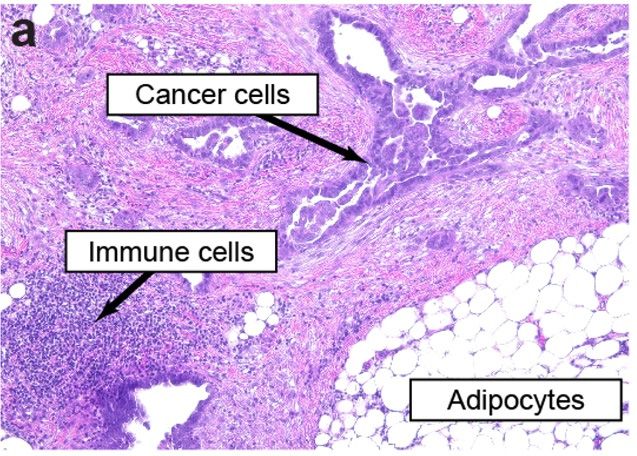
Cancers 2021, 13, 3136 2 of 17
Cancers 2021, 13, x 2 of 18
their pro-tumorigenic behavior. Dissecting these changes will help us better understand
microenvironment (TME) [3]. Several chemotherapeutic agents can regulate cancer-asso-
how chemotherapy influences non-malignant host cell populations to modulate disease
ciated fibroblasts (CAFs) by modifying their biology in ways that either enhance or re-
progression, therapeutic response,
strain their pro-tumorigenic behavior.and side effects
Dissecting of therapy.
these changes will help us better under-
stand how chemotherapy influences non-malignant hostcells
The TME is a multicellular network of tumor and host-derived
cell populations cells,dis-
to modulate including
CAFs, fibroblasts,therapeutic
ease progression, myofibroblasts, adipocytes,
response, endothelial
and side effects cells, and immune cells, embed-
of therapy.
ded inThea distinct
TME is extracellular
a multicellularmatrix
network(ECM; Figure
of tumor 1).and
cells Thehost-derived
host cells oftencells,comprise
includingthe most
significant proportion
CAFs, fibroblasts, of cells within
myofibroblasts, the tumor
adipocytes, organ. As
endothelial oneand
cells, of the most abundant
immune cells, em- cellular
bedded in a distinct
components extracellular
of the TME, CAFs matrix
are of (ECM; Figure
particular 1). Theinhost
interest tumorcellsprogression
often comprise [4]. CAFs
the most significant proportion of cells within the tumor organ. As one
encompass a highly dynamic populations of activated fibroblasts with pleiotropic functions of the most abun-
dantshape
that cellulartumor
components of the
behavior TME,
and CAFs are
engage in aofcomplex
particularnetwork
interest inoftumor progression
cross-talk with cellular
and non-cellular components of the TME. Their rich secretome supports with
[4]. CAFs encompass a highly dynamic populations of activated fibroblasts plei-
interactions with
otropic functions that shape tumor behavior and engage in a complex network of cross-
multiple cell types in the TME to induce tumor growth, epithelial-to-mesenchymal transi-
talk with cellular and non-cellular components of the TME. Their rich secretome supports
tion (EMT), angiogenesis, immunosuppression, and ECM remodeling. These elongated
interactions with multiple cell types in the TME to induce tumor growth, epithelial-to-
cells do not express
mesenchymal epithelial,
transition endothelial, or
(EMT), angiogenesis, leukocyte markers,
immunosuppression, and andECM generally
remodel- maintain
stable
ing. These elongated cells do not express epithelial, endothelial, or leukocyte markers, and reactive
genomes that lack mutations found in nearby tumor cells [5]. In addition,
oxygen
generally species
maintainsecreted by tumorthat
stable genomes cells alter
lack CAF metabolism
mutations found in nearby andtumor
induce oxidative
cells [5]. In stress,
autophagy, and upregulation
addition, reactive oxygen species ofsecreted
glycolyticby enzymes
tumor cells[6]. Although
alter most studies
CAF metabolism and in-support a
pro-tumorigenic
duce oxidative stress,role autophagy,
for CAFs inand theupregulation
TME, other of studies haveenzymes
glycolytic found compelling
[6]. Althoughevidence
mostin
that, studies
some support
situations a pro-tumorigenic
or model systems, role CAFs
for CAFs in thetumor
restrain TME, progression
other studies[7–9].have Several
found compelling
markers have been evidence
used to that, in some situations
characterize distinctorsubtypes
model systems,
of CAFs, CAFswithrestrain
smooth tu- muscle
mor progression
actin (αSMA) and [7–9]. Several markers
fibroblastic activation have been used
protein (FAP) to among
characterize distinct
the most subtypes
widely used. Other
of CAFs, such
markers, with assmooth muscle actingrowth
platelet-derived (αSMA)factorand fibroblastic
receptor beta activation
(PDGFR-β),proteinvimentin,
(FAP) cave-
among the most widely used. Other markers, such as platelet-derived growth factor re-
olin 1, CD10, GPR77, and tenascin C, are also useful to resolve organ-specific CAF subtypes.
ceptor beta (PDGFR-β), vimentin, caveolin 1, CD10, GPR77, and tenascin C, are also useful
Several comprehensive reviews addressing CAF origins, functions, and subtypes can be
to resolve organ-specific CAF subtypes. Several comprehensive reviews addressing CAF
found elsewhere [4,5,8].
origins, functions, and subtypes can be found elsewhere [4,5,8].
Figure 1. Cellular heterogeneity and bidirectional communication in the TME. (a) Representative
Figure 1. Cellular heterogeneity and bidirectional communication in the TME. (a) Representative
image of a high-grade serous ovarian cancer metastasis to the omentum exemplifies the diverse
image of a high-grade serous ovarian cancer metastasis to the omentum exemplifies the diverse
ecosystem of tumors that includes cancer cells, immune cells, CAFs, adipocytes, endothelial cells,
and other cells embedded in an ECM. CAFs and endothelial cells are present throughout the TME.
(1:100) (b) Cross-talk between cell types in the ECM involves bidirectional signaling between tumor
and stromal cell types that enforce a pro-tumorigenic microenvironment.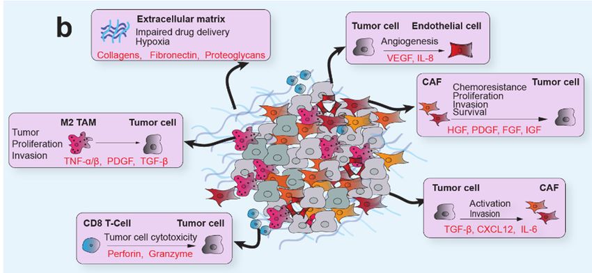
Cancers 2021, 13, 3136                                                                                            3 of 17

                              Advanced HGSOC is treated with an aggressive combination of chemotherapy and
                         surgery. Depending on tumor volume at presentation, patients with localized or low-
                         volume metastatic disease undergo surgery followed by six cycles of chemotherapy. Pa-
                         tients with high-volume metastatic disease first receive three cycles of carboplatin and pacli-
                         taxel prior to surgery, with three additional cycles after surgery (“neoadjuvant chemother-
                         apy”). A debulking surgery that leaves behind no visible disease confers the most survival
                         benefit to the patient, and the choice of treatment sequence is weighted to maximize the
                         benefit and minimize the morbidity of surgery [10,11]. Unfortunately, recurrence rates are
                         high, being greater than 70%, and long-term survival is infrequent, with only 15% of women
                         with advanced stage cancer surviving 7–10 years [12]. An important determinant of overall
                         survival in HGSOC is persistent susceptibility of the disease to platinum-based agents [13].
                         Recurrence therapy is chosen based on the interval from the last platinum agent, overall
                         health, acquired toxicities, germline/somatic mutations, and other factors. Second-line
                         or maintenance treatments can involve anti-angiogenics, anti-metabolites, PARP (poly
                         ADP-ribose polymerase) inhibitors, topoisomerase inhibitors, and occasionally radiation
                         therapy [14–18]. The diverse treatment options used in patient care and clinical trials
                         highlight the relevance of understanding their potential impact on the non-transformed
                         components of the TME.
                              In this review, we assess experimental data that examines how commonly used ovarian
                         cancer chemotherapeutics, including platinum derivatives, taxanes, PARP inhibitors, and
                         anti-angiogenics, alter the function of genetically normal cells in the TME. Although we
                         focus on therapies used in the treatment of ovarian cancer, we incorporate evidence from
                         other histological types of cancer to broadly understand therapy-induced changes in CAF
                         biology and their impact on modifying cancer progression across different tumor types. The
                         challenges in understanding these complex interactions highlight the need for improved
                         model systems that recapitulate the heterogeneity of the TME. Single-cell or compartment-
                         resolved approaches, combined with high-fidelity preclinical models of cancer treatment,
                         will be essential to unravel the biological and clinical relevance of these effects.

                         2. Alkylating Agents (Cisplatin and Carboplatin)
                               Alkylating agents, including cisplatin and carboplatin, act by forming DNA adducts
                         and DNA strand cross links which lead to DNA breakage or cross-linking [19]. If a cell
                         cannot repair the lesion, RNA synthesis and DNA replication stall, and the cell undergoes
                         apoptosis. The most common and most efficient primary therapeutic regimen for HGSOC
                         is carboplatin in combination with paclitaxel every three weeks for a total of six cycles [20].
                         Cisplatin can be used with equivalent efficacy, particularly in patients with myelosuppres-
                         sion that cannot be overcome with carboplatin dose reduction but is associated with a
                         worse overall side effect profile [21]. Platinum compounds cross the fibroblast cell mem-
                         brane via the CTR1 and CTR2 copper membrane transporters [22,23]. CAFs, in particular,
                         express less CTR1 than adjacent normal fibroblasts and cancer cells, which contributes to
                         their generally chemoresistant phenotype [24].
                               Platinum agents have been found to alter the CAF secretome, inducing the secretion
                         of protease inhibitors, cytokines, and miRNA-containing exosomes. In primary esophageal
                         squamous cell cancer (ESCC), cisplatin-treated CAFs secreted high levels of plasminogen
                         activator inhibitor-1 (PAI-1), which subsequently promoted cancer cell proliferation and
                         protection from cisplatin-induced apoptosis via inhibition of caspase-3 and activation of
                         AKT and ERK1/2 pathways [25]. Clinical analysis of 49 ESCC patients indicated that those
                         with high expression of PAI-1 in CAFs had significantly worse progression-free survival
                         (PFS). Masuda et al. further found that in vitro inhibition of PAI-1 in lung CAFs increased
                         cancer cell apoptosis and reduced CAF α-SMA expression. Treatment of a co-culture
                         system with PAI-1 inhibitors consequently increased the efficacy of cisplatin killing cancer
                         cells [26].
                               Multiple studies have found that platinum-based agents can regulate the cytokine
                         profile of CAFs. Lung CAFs treated with cisplatin upregulated IL-11 expression in a time-
Cancers 2021, 13, 3136 4 of 17
and dose-dependent manner, promoting pro-survival STAT3 signaling in lung cancer
cells [27,28]. In a longitudinal study of ovarian cancer patients from whom tumor tissue
was collected before and after chemotherapy, levels of IL-6 were elevated in αSMA+
stromal cells following platinum treatment [29]. In vitro treatment of CAFs from primary
human ovarian cancer with cisplatin enhanced their chemoprotective properties in an
IL-6-dependent manner. Systematic profiling of cytokines upregulated by HGSOC CAFs
following cisplatin treatment found significant elevation of CCL5, IL-8, and MIP [30].
Ovarian cancer cells treated with CCL5 showed decreased apoptosis when exposed to
cisplatin, which was consistent with the finding that platinum-resistant patient samples
expressed elevated CCL5 levels compared to platinum-sensitive samples. Moreover, CAFs
may respond differently than normal fibroblasts to platinum agents, as was observed
when oxaliplatin treatment upregulated sDTK, IL-17A, and TGF-β in CAFs but not in
normal fibroblasts [31]. In colorectal cancer patient samples, IL-17A level was also found
to increase following chemotherapy.
In addition to growth factors and cytokines, CAFs also secrete exosomes containing
RNA molecules that may influence the gene expression profile and behavior of cancer
and normal cells in the TME [32]. Cisplatin- or paclitaxel-treated gastric CAFs secreted
exosomes containing high levels of miR-522, which suppressed ferroptosis in cancer cells
and promoted chemo-resistance [33]. Although miR-522 expression in primary CAFs was
not elevated, treated CAF exosomes contained nearly a four-fold higher level of miR-522,
indicating a preferential enrichment of miR-522 in exosomes. Another miRNA, miR-
196a, was upregulated in exosomes from cisplatin-treated head-and-neck cancer (HNC)
CAFs, which ultimately enhanced the proliferation and chemo-resistance of HNC cells via
targeting CDKN1B and ING5 [34].
Platinum-based interventions may also contribute to transforming normal fibroblasts
into CAFs through increasing the expression of CAF-related markers such as FAP and
α-SMA, altering the metabolic activity of normal fibroblasts, and inducing some aspects of
senescence. Co-culture experiments with bladder cancer suggest that cisplatin accelerates
normal fibroblasts’ transition to CAFs and increased the expression of α-SMA and FAP [24].
In immortalized human foreskin fibroblasts, cisplatin and carboplatin treatment increased
L-lactate production and glucose consumption [35]. Using glycolytic flux analysis, the
authors found that platinum treatment elevated fibroblast glycolysis and reduced oxygen
consumption rates, indicating a metabolic switch. A secreted fibroblast factor, the ECM,
and metabolism are linked in a report describing a novel role for the ECM protein collagen
(COL) 11A1.The CAFs secrete COL11A1 that binds to the cancer cells through a discoidin
receptor, which leads to upregulation of fatty acid oxidation, enabling the cancer cells to
withstand carboplatin treatment [36].
Because some features of the CAF phenotype are similar to the senescence-associated
secretory phenotype (SASP), it may be significant that platinum treatment has also been
found to induce senescence and autophagy in CAFs. Treatment of human lung fibroblasts
with platinum agents caused a prematurely senescent phenotype, evidenced by elevated
p53 expression, loss of the membrane gap junction protein connexin 43 (Cx43), and mor-
phological changes including flattening, filopodia extensions, and cytoplasmic vacuole
formation [37]. Similarly, cisplatin treatment of normal oral fibroblasts and foreskin fibrob-
lasts upregulated senescence markers and increased α-SMA expression [38]. An important
consideration when interpreting these studies is that carboplatin and cisplatin may exert
distinct effects on CAFs due to differences in uptake, bioactivity, and mechanism [39–41].
For example, carboplatin, but not cisplatin, was found to augment the glycolytic reserve,
upregulate senescence and CAF markers, and promote HIF, SMAD, and STAT signaling in
immortalized fibroblasts [35].
As CAFs comprise a highly heterogeneous cell population [42], CAF subpopulations
may respond differently to platinum-based therapies. Indeed, Su et al. demonstrated
that CD10+ GPR77+ CAFs associated with poor prognosis also exhibited docetaxel and
cisplatin resistance compared to CD10− GPR77− CAFs [43]. When challenged with cisplatin,Cancers 2021, 13, 3136 5 of 17
CD10+ GPR77+ breast cancer CAFs demonstrated significantly lower levels of apoptosis
and growth inhibition. Furthermore, not all studies have found that platinum agents
have tumor-promoting effects on the TME. In a study focused on lung cancer, cisplatin
attenuated the ability of CAFs to promote the adhesion and invasion of cancer cells and
reduced cancer cell AKT and NF-κB signaling via an unidentified paracrine mechanism [44].
In vivo experiments demonstrated that while co-implantation of CAFs and lung cancer
cells increased tumor volume by over seven-fold, co-implantation with cisplatin-treated
CAFs was comparable to implantation of cancer cells alone. Mesothelial cell-derived CAFs
in the omental TME decreased cisplatinum sensitivity of HGSOC cells through secretion of
fibronectin, which induced the PI3K pathway in the cancer cells [45].
Fully elucidating the mechanistic effects of therapeutic agents on CAFs will require
understanding CAF heterogeneity and how therapies might balance cell subpopulations.
It is important to note that all these studies indicate that some chemotherapy effects are
tumor-promoting.
The effects of alkylating agents on other cellular components of the TME also de-
serve further attention. Notably, many studies demonstrate alkylating agents cause
bone marrow toxicity, myelosuppression, and inhibition of the self-renewal capacity of
hematopoietic stem cells [46–48]. Cisplatin has multifactorial roles in promoting a tumor-
suppressive immune response, including increasing the range of antigen recognition,
enhancing macrophage tumoricidal ability, promoting Th1 cytokine secretion, and regu-
lating the recruitment of M1 macrophages, T regulatory cells, and CD8+ T cells [49–52].
In adipocytes, cisplatin may increase lipolysis while impeding lipogenesis [53]. Platinum
treatment of endothelial cells induces features associated with angiogenesis, including
dose-dependent decreases in migration and upregulation of ICAM-1, VEGF, and several
cytokines such as IL-1 and IL-6 [54–57]. Independently, CAFs can promote the leakiness of
blood vessels through the secretion of microfibrillar-associated protein (MFAP) 5 which
binds to integrin receptors present on endothelial cells [58]. All of these effects on other
cell types in the TME may also have the potential to regulate the behavior of CAFs.
3. Paclitaxel and Docetaxel
Paclitaxel, a plant alkaloid derived initially from the yew tree, is a cornerstone of
upfront and recurrent treatment of HGSOC. Taxanes act during the M phase of the cell cycle
by binding to intracellular microtubules to promote their assembly and stabilization, thus
disrupting mitosis and leading to cell death [19]. Docetaxel is a therapeutically equivalent
choice with a lower risk of peripheral neuropathy [59]. Taxanes can be used in the recurrent
setting in both platinum-sensitive and -resistant patients [60].
There is emerging evidence that, in many situations, CAFs cooperate with tumor cells
to enhance resistance to anticancer treatments [61]. In response to chemotherapy, CAFs
may secrete cytokines, metabolites, and exosomes to potentiate stemness, metabolic repro-
gramming, and pro-survival signaling in tumor cells to orchestrate chemo-resistance [62].
Co-culture of taxane-treated normal lung fibroblasts with non-small-cell lung carcinoma
tumor cells led to increased paclitaxel resistance of cancer cells, suggesting that paclitaxel
may promote chemo-resistance via paracrine signaling [63]. Taxane-based chemotherapeu-
tics may also regulate other aspects of the CAF phenotype by transcriptionally attenuating
pro-tumorigenic paracrine signaling. Treatment of primary human breast cancer CAFs with
docetaxel led to significant decreases in the expression of CXCL2, MMP1, IL-8, FF1, and
CXCR7, among other cytokines. Co-culture experiments revealed that docetaxel-treated
CAFs promoted the adhesion, invasion, and proliferation of MDA-MB-231 breast cancer
cells [64]. A study of primary breast CAFs treated with docetaxel found that upregulation
of MMP-1 and collagen IV was important for mediating chemo-resistance through ECM
remodeling [65]. Caution should be applied to the interpretation of any in vitro or ex
vivo experiments, however, as one study found divergent effects of paclitaxel treatment
on primary CAFs compared to intact tissue sections, with increased apoptosis of CAFs
observed in the ex vivo model system [66].Cancers 2021, 13, 3136 6 of 17
Several studies have suggested that taxanes may also regulate the behavior of normal
fibroblasts and promote some aspects of the CAF phenotype. In an approach using label-
free, quantitative proteomics, treatment of benign foreskin fibroblasts led to profound
changes in energy metabolism, autophagy, senescence, myofibroblastic differentiation, and
expression of inflammatory markers [67]. This included increased expression of common
CAF markers such as α-SMA, fibronectin, and vimentin and upregulation of interleukin
6 (IL-6) and STAT3 signaling. In parallel to the metabolic reprogramming that occurs in
CAFs [4,68,69], taxane treatment of dermal fibroblasts increased glycolysis, autophagy, and
pro-inflammatory signaling [35,67].
In contrast, studies investigating the effects of taxanes on normal fibroblasts in non-
cancer conditions have found evidence of anti-fibrotic effects. In a model of extrahepatic
bile duct fibrosis, treatment of human gallbladder myofibroblasts with paclitaxel led to
decreased autocrine TGFβ-1 signaling and reduced collagen 1 production associated with
fibrosis [70]. In another study examining renal interstitial fibrosis, paclitaxel treatment led
to decreased SMAD signaling and suppression of pro-inflammatory cytokine production
accompanied by a strong reduction in α-SMA and collagen 1 expression [71]. The mecha-
nistic basis for the differential effects of taxanes on normal fibroblasts and CAFs has not
yet been explored.
In addition to effects on fibroblast components of the TME, taxanes may also regu-
late endothelial cells by exerting intrinsic anti-angiogenic effects. Interestingly, human
endothelial cells accumulate higher intracellular levels of paclitaxel than non-endothelial
cells, suggesting selectivity and increased susceptibility of endothelial cells to the drug [72].
Multiple studies have found that taxanes can directly compromise endothelial cells by
inducing apoptotic cell death [73–76]. Functionally, paclitaxel attenuates endothelial cell
migration, inhibits endothelial tube formation [77], and induces some aspects of the senes-
cent phenotype [78]. Although taxanes may reduce the angiogenic activities of endothelial
cells, there is also evidence indicating paclitaxel can increase vascular endothelial growth
factor (VEGF) production in cervical cancer tumor cells by regulating hypoxia-inducible
factor 1α (HIF-1α) and NF-κB signaling, thereby increasing angiogenesis and promoting
chemo-resistance [79].
4. Poly ADP-Ribose Polymerases Inhibitors
Poly ADP-ribose polymerases (PARPs) are enzymes critical for the repair of single-
stranded DNA breaks, as well as contributing to the repair of double-stranded breaks
and the stabilization of replications forks [80]. In cells with double-stranded DNA repair
deficiencies, such as those with BRCA1/2 gene mutations, PARP inhibition leads to synthetic
lethality. PARP inhibitors (PARPi) are oral medications that act to trap or inhibit PARP
enzymatic action. Maintenance PARPi treatment after upfront chemotherapy has recently
proven effective at lowering the risk of disease progression or death by 70% at 41 months
in HGSOC patients with a BRCA1/2 mutation. They are now standard in the frontline
setting [11,15].
Most studies examining the effects of PARP inhibition on stromal cell populations
have focused on normal fibroblasts and found that PARPi affected the regulation of TNFα
and TGF-β signaling. Whereas PARP inhibition attenuates the TNFα-induced fibrob-
last response, its impact on TGF-β mediated effects is less clear. Isolated fibroblast-like
synoviocytes treated with TNFα and a PARPi (DPQ or ANI) had reduced expression
of inflammatory mediators such as MMP-3, IL8, and MCP-1 [81]. This was associated
with diminished TNFα-induced proliferation, JNK phosphorylation, and AP-1 and NF-kB
binding. Similar trends were observed in another study which found that pretreating
murine fibroblasts with the PARPi INH2BP also suppressed JNK activation and AP-1 DNA
binding [82]. In murine fibroblasts, the PARPi 3AB reduced levels of TNF-induced ATP
depletion and death [83]. Broadly, TNFα-induced signaling in fibroblasts is attenuated
by PARPi.Cancers 2021, 13, 3136 7 of 17
In human skin fibroblasts, PARP inhibition by 3-AB increased the stimulatory effects
of TGF-β, including upregulation of α-SMA transcription, expression, and stress fiber for-
mation compared to TGF-β alone [84]. Additional synergistic effects included upregulation
of collagen expression, increased collagen release, and elevated SMAD3 signaling. In vitro,
treatment with 3-AB exacerbated fibrosis induced by topoisomerase or bleomycin, result-
ing in increased dermal thickness and hydroxyproline content. Other studies, however,
indicate anti-fibrotic roles for PARP inhibition. Knockdown of PARP1 in cardiac fibroblasts
repressed TGF-β1-induced proliferation, migration, and differentiation [85]. In rat models
of myocardial infarction (MI), 4-AB alleviated fibrosis and reduced collagen deposition,
with an associated decrease in α-SMA expression. In addition, 4-AB increased p62 lev-
els and reduced the LC3-II/LC3-I ratio, suggesting that PARP inhibition may increase
autophagy. These divergent phenotypes may be due to differences in cellular context,
fibroblast type, or experimental design considerations.
PARPi also demonstrates profound immunomodulatory effects, promoting anti-tumor
immune responses by upregulating cytotoxic immune cells such as CD8+ T-cells, B-cells,
and NK cells, while decreasing the number of myeloid-derived suppressor cells [86,87].
This anti-tumor response is at least in part due to upregulation of the STING pathway
by PARPi [88]. While PARPi show promise in cancer treatment, they are also associated,
albeit rarely, with severe side effects such as myeloid leukemia and myelodysplastic
syndrome [89]. Importantly, maintenance PARPi treatment can be of extended duration,
for two or more years, raising the possibility that long-term, global inhibition of PARP may
have distinct influences on the biology of normal cells.
5. Anti-Angiogenic Agents (Bevacizumab)
Bevacizumab, an antibody against VEGF and angiogenesis, is often used to treat
recurrent HGSOC as an additional treatment option to target the angiogenic potential
of the TME [90]. In upfront therapy for HGSOC, there is some evidence that it benefits
poor-prognosis patients with high tumor volume, stage IV, or incompletely resected dis-
ease [91]. Bevacizumab is also used in combination with PARPi for maintenance therapy
in the upfront setting, with a 19.5-month progression-free survival benefit [92]. A PFS
benefit has been seen when used in combination therapy for both platinum-sensitive and
-resistant recurrence [93,94]. The lack of a consistent overall survival benefit and the rare
risk of serious adverse events, including gastrointestinal perforation, bleeding diathesis,
and poor wound healing, has limited this agent’s use. Although anti-VEGF inhibitors
are meant to target endothelial cells, fibroblasts also express the receptors VEGFR1 and
VEGFR3 [95]. No studies have directly investigated the effects of anti-angiogenic agents
on CAFs, but bevacizumab exerts some direct effects on normal fibroblast populations.
Bevacizumab-treated rat conjunctival fibroblasts exhibit reduced growth, ECM remodeling,
and metabolic activity associated with decreased expression of VEGF, VEGFR1, VEGFR2,
TGF-β1, and TGF-β2 [96]. Pharmacologically blocking VEGFR in human lung fibroblasts
from patients with idiopathic pulmonary fibrosis suppressed the proliferative effects of se-
creted factors, including PDGF and bFGF [97]. VEGFR inhibition upregulated pro-MMP-2
activity, downregulated TIMP-2 secretion, and suppressed TGF-β-induced collagen se-
cretion. Another study examining human tendon fibroblasts found that treatment with
bevacizumab reduced metabolic activity and viability in a dose-dependent manner [98].
Bevacizumab also decreased the expression of MMP-1, MMP-2, and laminin, suggesting
that it may play a role in ECM remodeling.
6. Topoisomerase Inhibitors (Doxorubicin, Ropotecan, and Mitoxantrone)
Topoisomerase inhibitors, including doxorubicin, topotecan, and mitoxantrone, act
by inhibiting topoisomerase enzymes, which are responsible for the winding of DNA,
leading to DNA strand breaks [99]. Doxorubicin has the additional effect of distorting the
DNA double helix and generating free radicals. Liposomal doxorubicin and topotecan
are both used for recurrent ovarian cancer with limited efficacy [100]. Although the ef-Cancers 2021, 13, 3136 8 of 17
fects of topoisomerase inhibitors on CAFs have not been extensively investigated, several
studies of normal fibroblasts suggest that topoisomerase inhibitors may regulate the TME.
Fibroblasts appear to be sensitive to the topoisomerase II inhibitor mitoxantrone, with
the treatment of dermal fibroblasts leading to senescence and enhanced glycolysis [101].
Mitoxantrone-treated fibroblasts have been found to induce a cancer stem cell phenotype
in MCF7 cells, as assessed using a luciferase reporter system in tumor–stromal co-culture
conditions [35]. Mitoxantrone treatment of prostate, ovarian, and breast primary CAFs
upregulated WNT16B, thereby mediating acquired resistance, suggesting a role for mi-
toxantrone in the regulation of Wnt signaling [102]. In a study that directly examined the
effects of topoisomerase inhibitors on CAFs in a mouse model of desmoplastic melanoma, a
combination of mitoxantrone and the triterpenoid celastrol decreased CAF-mediated colla-
gen production and was associated with a CD8 T-lymphocyte- and dendritic cell-mediated
immunogenic response [103]. The effects of topoisomerase inhibitors on other components
of the TME have not been systematically investigated.
7. Antimetabolites (Gemcitabine)
Gemcitabine is a pyrimidine analog that primarily acts by inhibiting DNA synthesis
through direct incorporation into the DNA backbone. It may also induce activation of
mitogen-activated protein kinase (MAPK), triggering apoptosis in response to cellular stress
in tumor cells [104]. Gemcitabine is commonly used in the recurrent setting and has a single-
agent response rate of about 19%. Common dose-limiting toxicities include neutropenia,
nausea, appetite suppression, and a flu-like syndrome [105]. Relatively few studies have
examined the effects of gemcitabine on stromal cell populations. CAFs appear to be resistant
to gemcitabine treatment when compared to normal fibroblasts or cancer cells [106]. It has
been suggested that scavenger molecules from pancreatic CAFs may modify gemcitabine
accumulation in tumors by entrapping the active drug and reducing its delivery to cancer
cells [107]. In co-culture model systems, pre-treatment of immortalized rat CAFs protected
tumor cells from the cytotoxic effects of gemcitabine. Interestingly, inhibition of autophagy
in CAFs with chloroquine also reduced cancer cell death in response to gemcitabine [108].
Several studies have also found roles for gemcitabine in the regulation of CAF exosome
production. The treatment of pancreatic CAFs with gemcitabine resulted in the increased
release of exosomes containing the EMT regulator Snail. Uptake of these exosomes by
tumor cells reduced the cytotoxic effects of gemcitabine treatment [109]. Similarly, Fang
et al. found that CAF-derived exosomes transferred miRNA-106b to tumor cells and
regulated gemcitabine resistance in a TP53INP1-dependent manner [106].
8. Radiotherapy
Radiotherapy involves targeted high-frequency ionizing radiation which ejects elec-
trons from atoms to create ions which then form free radicals, leading to DNA damage.
The advent of highly active chemotherapy for HGSOC in the 1990s sidelined radiation
therapy in this disease. Its current use is limited to treating isolated recurrences or for
symptom palliation [18]. Radiation induces significant changes in the CAF secretome, in-
cluding upregulation of factors such as IGF-1, bFGF, IL-6, IL-8, GRO, HDGF, and potentially
HGF [110–113]. Furthermore, radiation promotes MMP-3 and possibly MMP-1 expression
in CAFs [114,115]. Consistent with the role of CAFs in the regulation of ECM remodeling,
it has also been reported that radiotherapy elevates CAF expression of integrins β1, α5,
and, most significantly, α2 [114]. Irradiated CAFs produced a stiffer collagen matrix when
grown in a 3D culture system [116]. Radiotherapy also increases premature senescence
in CAFs [114,117]. Cumulatively, these data suggest that CAFs resist radiation treatment
through acquiring a senescent phenotype and that they can simultaneously contribute to
cancer cell resistance by secreting pro-tumorigenic factors and remodeling the ECM.matrix when grown in a 3D culture system [116]. Radiotherapy also increases premature
senescence in CAFs [114,117]. Cumulatively, these data suggest that CAFs resist radiation
treatment through acquiring a senescent phenotype and that they can simultaneously con-
Cancers 2021, 13, 3136 9 of 17
tribute to cancer cell resistance by secreting pro-tumorigenic factors and remodeling the
ECM.
9.9.Discussion
Discussion
Studies
Studies investigating
investigatingthe theeffects
effects of
of anticancer
anticancer agents
agents on on normal
normal cells
cells in
in the
the tumor
tumor
microenvironment
microenvironment have focused on a wide range of molecular and phenotypic
focused on a wide range of molecular and phenotypic features, features,
in-
including metabolic
cluding metabolic andand transcriptional
transcriptional reprogramming
reprogramming andand intracellular
intracellular signaling
signaling (Fig-
(Figure 2).
ure 2). These
These studiesstudies have primarily
have primarily foundfound that CAFs,
that CAFs, in response
in response to first-line
to first-line chemother-
chemotherapies,
apies,
secretesecrete multiple
multiple cytokines,
cytokines, metabolites,
metabolites, ECM-remodeling
ECM-remodeling enzymes,enzymes,
and and exosomes
exosomes that
that transform
transform the TME
the TME and and generally
generally promote
promote chemo-resistance.
chemo-resistance. Platinum
Platinum derivatives
derivatives and
and taxanes
taxanes appear
appear to promote
to promote a precancerous
a precancerous metabolic
metabolic phenotype
phenotype in stromal
in stromal cells, re-
cells, resulting
sulting in augmented
in augmented glycolysis,
glycolysis, glucose glucose consumption,
consumption, lactate lactate production,
production, and activity
and activity of
of sev-
eral pro-tumorigenic
several pro-tumorigenic pathways
pathways [35,67].
[35,67].InInresponse
responseto tothese
these agents, fibroblasts
agents, normal fibroblasts
adoptat
adopt atleast
leastsome
someaspects
aspectsofof the
the CAF
CAF phenotype,
phenotype,but but itit isis unclear
unclear ifif these
these changes
changes are are
long-lastingor
long-lasting orreversible
reversible[24,35,67,83].
[24,35,67,83].Other
Othertherapies,
therapies,including
includingPARP PARPinhibitors,
inhibitors,anti-
anti-
angiogenic agents,
angiogenic agents, and
and topoisomerase
topoisomerase inhibitors,
inhibitors, have
have been
been lessless studied;
studied; nevertheless,
nevertheless,
some evidence suggests that they have TME-modifying capabilities.
some evidence suggests that they have TME-modifying capabilities. Of note, alongside Of note, alongside
cytotoxicchemotherapeutics,
cytotoxic chemotherapeutics,many manypatients
patientsreceive
receive5-HT3
5-HT3antagonists,
antagonists,steroids,
steroids,andandNK1
NK1
antagoniststo
antagonists tomanage
managenausea
nauseaandandemesis
emesis[118].
[118]. The
Theeffects
effectsof of these
these agents
agents onon the
the TME
TME
havenot
have notbeen
beenwell
well investigated,
investigated,although
althoughseveral
severalstromal
stromalcell cell types
types express
express thethe relevant
relevant
cognate receptors [119–123].
cognate receptors [119–123].
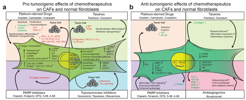
Figure
Figure 2.
2. Effects
Effects of therapeutic agents
of therapeutic agentson
oncancer
cancerassociated
associatedfibroblasts
fibroblasts (CAF)s.
(CAF)s. TheThe diverse
diverse therapeutics
therapeutics usedused in the
in the treat-
treatment
ment of ovarian cancer, including platinum agents, taxanes, Poly ADP-ribose polymerases (PARP) inhibitors,
of ovarian cancer, including platinum agents, taxanes, Poly ADP-ribose polymerases (PARP) inhibitors, tomoisomerase tomoiso-
merase inhibitors,
inhibitors, and anti-angiogenics,
and anti-angiogenics, may havemay
(a)have (a) pro-tumorigenic
pro-tumorigenic or (b) anti-tumorigenic
or (b) anti-tumorigenic effects
effects on CAFs.on* Experimental
CAFs. * Experi- or
mental or clinical observations in normal fibroblasts or fibroblasts from disease states other than cancer.
clinical observations in normal fibroblasts or fibroblasts from disease states other than cancer.
Additional
Additionalresearch
researchusing
usingmore
more physiologically
physiologically relevant
relevant models
models willwill be
be necessary
necessary ifif
we are to illuminate the complex processes that occur in the treated
we are to illuminate the complex processes that occur in the treated TME. Organotypic TME. Organotypic
model
modelsystems
systemsthatthatincorporate
incorporate both
both tumor
tumorandand
stromal
stromalcell cell
types havehave
types proven to beto
proven par-
be
ticularly valuable
particularly in studies
valuable that that
in studies reveal heterotypic
reveal cross-talk,
heterotypic cancer
cross-talk, progression,
cancer and and
progression, op-
portunities
opportunities forfor
drug
drugdiscovery
discovery[26,63].
[26,63]. For instance,
For instance,using
usinga acomplex
complexmodelmodelsystem,
system, Gao
Gao
et al., found that CAFs can form metastatic units with ascitic tumor cells
et al., found that CAFs can form metastatic units with ascitic tumor cells and drive peri- and drive perito-
neal metastasis
toneal metastasisformation,
formation, which
whichis iscommon
commonininovarian
ovariancancer
cancer[124].
[124].Using
Usingsuch
suchcomplex
complex
models can also identify mechanisms of chemo-resistance and pinpoint drug candidates
more likely to be effective in vivo, since malignant cells often exhibit profound differences
in sensitivity to therapeutic agents depending on the culture system utilized [125–128].
Furthermore, while patients receive well-defined cycles of chemotherapy, most studies,
both in vitro and in vivo, do not mirror the concentrations and durations of treatment
typical of clinical exposures.Cancers 2021, 13, 3136 10 of 17
In addition, preclinical examinations of chemotherapy-induced changes in the TME
have been largely restricted to subcutaneous xenograft models that do not fully reca-
pitulate all components of the TME. Studies using orthotopic or genetically engineered
mouse models are needed to more fully understand disease processes. In HGSOC, most
tumor cells harbor mutations in TP53, and mouse models have been recently developed
with syngeneic murine ovarian cancer cells engineered to express mutant p53 protein via
CRISPR/Cas9 [129]. Researchers must carefully consider candidate cell lines for their
experiments, since unique and characteristic TMEs arise depending on the HGSOC model
employed [130].
CAFs originate from numerous sources and are composed of discrete subpopulations.
Therefore, it is difficult to generalize microenvironmental responses to chemotherapy due
to heterogeneity within single tissues and between anatomic sites. For example, while
cisplatin treatment may downregulate α-SMA in lung fibroblasts, studies in bladder and
foreskin fibroblasts have found upregulation of α-SMA or no changes at all [24,35,44].
Furthermore, few studies have examined differences in CAFs between humans and other
model organisms [131]. Research in the ovarian cancer TME has identified at least four
different CAF subpopulations that express distinct molecular signatures characterized
by variable expression of CD29, CD10, FAP, α-SMA, FSP1, PDGFR-β, podoplanin, and
caveolin-1. This suggests that the use of single markers such as α-SMA to define CAF
identity is imprecise. Embracing multiple CAF markers with complementary functional
studies has the potential to elucidate whether therapy-induced changes reflect reprogram-
ming of stromal fibroblasts or the selection of discrete populations. We anticipate that
single-cell sequencing will serve to spatially and temporally resolve CAF subpopulations,
as well as shed light on the epigenetic and evolutionary dynamics within specific microen-
vironments [132–134]. Advances in mass spectrometry-based proteomics have generated
opportunities to characterize the tumor and stromal proteomes in situ [135], quantify the
phosphorylation levels of critical signaling pathways [136,137], and identify secreted fac-
tors derived from both tumor and stromal cells [138]. In addition, imaging mass cytometry
enables the multiplexed, spatial characterization of proteins in both tumor and stromal
compartments and has already revealed important aspects of cellular communication and
“cellular neighborhoods” in breast cancer [139].
Understanding how chemotherapeutics impact cell types in the TME other than CAFs
may also inform opportunities for therapeutic intervention. Patients can experience vary-
ing degrees of myelosuppression, mucous membrane reactions, and alopecia, all evidence
of unintended cellular targets [19,140]. In addition, taxanes and platinum-based agents can
have significant neurotoxicity [141,142]. Because neurons interact with the TME in myriad
ways, releasing neurotransmitters, peptides, and growth factors [143–145], there are likely
to be unexamined roles for chemotherapeutic agents in the regulation of the communica-
tion between neurons and cancer cells [146]. Despite a growing literature showing that
chemotherapeutics perturb nonmalignant TME cells, in most situations, the mechanistic
basis of these effects remains unclear. As well as cross-linking DNA, cisplatin is also
known to generate reactive oxygen species, lower GSH and NADH levels, disrupt calcium
homeostasis, and activate ERK, JNK, and AKT signaling pathways [147]. PARP inhibitors
may disrupt the roles of PARP in DNA damage repair and influence epigenetic remodeling.
Several of these therapy-induced effects on fibroblasts are therefore unsurprising, whereas
others require further elucidation. For example, the general upregulation of α-SMA may be
due to cisplatin-induced changes in SMAD3 signaling, PAI-1 autocrine signaling, or other
mechanisms [148]. To understand these aspects of treatment, future studies should inves-
tigate the transcriptional and translational changes as well as the epitranscriptomic and
epigenetic remodeling that occurs in response to anticancer agents. How chemotherapeutic
agents may remodel the pre-metastatic niche has not yet been explored.
Beyond their likely prognostic and diagnostic value [149], CAFs have garnered interest
as targets of cancer therapy [150]. This could be of particular relevance to OvCa, since
there are few targetable mutations. Rather than aiming to ablate whole CAF populations, itCancers 2021, 13, 3136 11 of 17
may be possible to reprogram CAFs towards an anti-tumorigenic phenotype [151]. Indeed,
emerging research provides a rationale for targeting the TME with all-trans retinoic acid to
induce CAF quiescence [152] or using CXCL12 receptor inhibitors to disrupt CAF-mediated
immune evasion, as well as targeting many other CAF behaviors [153]. Fully realizing the
clinical efficacy of these emerging chemotherapies will require a robust understanding of
how common treatments influence the tumor microenvironment.
10. Conclusions
In summary, chemotherapy and radiation does not only affect the cancer cells, but has
a profound imapct on the TME. Understanding the effect of various treatments on cancer
cells is necessary to completely comprehend their action and develop novel therapeutics
that interrupt undesired tumor–stroma interactions.
Author Contributions: Conceptualization, M.A.E.; writing—original draft preparation, M.A.E., C.O.,
M.J., and J.X.; writing—review and editing, M.A.E., C.O., J.X., M.J., and E.L.; funding acquisition, E.L.
and M.A.E. All authors have read and agreed to the published version of the manuscript.
Funding: E.L. was supported by the National Cancer Institute (NCI) grants R01CA211916 and
R01CA169604. M.A.E. was supported by the Ovarian Cancer Research Alliance (OCRA) Liz Tilberis
Early Career Award 650339.
Institutional Review Board Statement: Not applicable.
Informed Consent Statement: Not applicable.
Data Availability Statement: Not applicable.
Conflicts of Interest: E.L. receives funding from Arsenal Bio and Abbvie to support preclinical
research unrelated to the manuscript. All other authors declare no conflict of interest. The funders
had no role in the design of the study; in the collection, analyses, or interpretation of data; in the
writing of the manuscript, or in the decision to publish the results.
References
1. Foley, O.W.; Rauh-Hain, J.A.; Del Carmen, M.G. Recurrent epithelial ovarian cancer: An update on treatment. Oncology 2013,
27, 288.
2. Kayl, A.E.; Meyers, C.A. Side-effects of chemotherapy and quality of life in ovarian and breast cancer patients. Curr. Opin. Obstet.
Gynecol. 2006, 18, 24–28. [CrossRef] [PubMed]
3. Mikuła-Pietrasik, J.; Witucka, A.; Pakuła, M.; Uruski, P.; Begier-Krasińska, B.; Niklas, A.; Tykarski, A.; Ksia˛żek, K. Comprehensive
review on how platinum- and taxane-based chemotherapy of ovarian cancer affects biology of normal cells. Cell. Mol. Life Sci.
2019, 76, 681–697. [CrossRef] [PubMed]
4. Kalluri, R. The biology and function of fibroblasts in cancer. Nat. Rev. Cancer 2016, 16, 582–598. [CrossRef] [PubMed]
5. Sahai, E.; Astsaturov, I.; Cukierman, E.; De Nardo, D.G.; Egeblad, M.; Evans, R.M.; Fearon, D.; Greten, F.R.; Hingorani, S.R.;
Hunter, T.; et al. A framework for advancing our understanding of cancer-associated fibroblasts. Nat. Rev. Cancer 2020, 20,
174–186. [CrossRef]
6. Santi, A.; Kugeratski, F.; Zanivan, S. Cancer Associated Fibroblasts: The Architects of Stroma Remodeling. Proteomics 2018, 18,
e1700167. [CrossRef]
7. Özdemir, B.C.; Pentcheva-Hoang, T.; Carstens, J.; Zheng, X.; Wu, C.-C.; Simpson, T.R.; Laklai, H.; Sugimoto, H.; Kahlert, C.;
Novitskiy, S.V.; et al. Depletion of Carcinoma-Associated Fibroblasts and Fibrosis Induces Immunosuppression and Accelerates
Pancreas Cancer with Reduced Survival. Cancer Cell 2014, 25, 719–734. [CrossRef]
8. Gascard, P.; Tlsty, T.D. Carcinoma-associated fibroblasts: Orchestrating the composition of malignancy. Genes Dev. 2016, 30,
1002–1019. [CrossRef]
9. Gerling, M.; Büller, N.V.J.A.; Kirn, L.M.; Joost, S.; Frings, O.; Englert, B.; Bergström, Å.; Kuiper, R.V.; Blaas, L.; Wielenga, M.C.B.;
et al. Stromal Hedgehog signalling is downregulated in colon cancer and its restoration restrains tumour growth. Nat. Commun.
2016, 7, 12321. [CrossRef]
10. Vergote, I.; Coens, C.; Nankivell, M.; Kristensen, G.B.; Parmar, M.K.B.; Ehlen, T.; Jayson, G.C.; Johnson, N.; Swart, A.M.; Verheijen,
R.; et al. Neoadjuvant chemotherapy versus debulking surgery in advanced tubo-ovarian cancers: Pooled analysis of individual
patient data from the EORTC 55971 and CHORUS trials. Lancet Oncol. 2018, 19, 1680–1687. [CrossRef]
11. Kurnit, K.C.; Fleming, G.F.; Lengyel, E. Updates and New Options in Advanced Epithelial Ovarian Cancer Treatment. Obstet.
Gynecol. 2021, 137, 108–121. [CrossRef] [PubMed]Cancers 2021, 13, 3136 12 of 17
12. Javellana, M.; Hoppenot, C.; Lengyel, E. The road to long-term survival: Surgical approach and longitudinal treatments of
long-term survivors of advanced-stage serous ovarian cancer. Gynecol. Oncol. 2019, 152, 228–234. [CrossRef] [PubMed]
13. Davis, A.; Tinker, A.V.; Friedlander, M. “Platinum resistant” ovarian cancer: What is it, who to treat and how to measure benefit?
Gynecol. Oncol. 2014, 133, 624–631. [CrossRef] [PubMed]
14. Luvero, D.; Milani, A.; Ledermann, J.A. Treatment options in recurrent ovarian cancer: Latest evidence and clinical potential.
Ther. Adv. Med. Oncol. 2014, 6, 229–239. [CrossRef]
15. Moore, K.; Colombo, N.; Scambia, G.; Kim, B.-G.; Oaknin, A.; Friedlander, M.; Lisyanskaya, A.; Floquet, A.; Leary, A.; Sonke, G.S.;
et al. Maintenance Olaparib in Patients with Newly Diagnosed Advanced Ovarian Cancer. N. Engl. J. Med. 2018, 379, 2495–2505.
[CrossRef]
16. González-Martín, A.; Sánchez-Lorenzo, L. Immunotherapy with checkpoint inhibitors in patients with ovarian cancer: Still
promising? Cancer 2019, 125, 4616–4622. [CrossRef]
17. Doo, D.W.; Norian, L.A.; Arend, R.C. Checkpoint inhibitors in ovarian cancer: A review of preclinical data. Gynecol. Oncol. Rep.
2019, 29, 48–54. [CrossRef] [PubMed]
18. Fields, E.C.; McGuire, W.P.; Lin, L.; Temkin, S.M. Radiation Treatment in Women with Ovarian Cancer: Past, Present, and Future.
Front. Oncol. 2017, 7, 177. [CrossRef]
19. Chi, D.; Berchuck, A.; Dizon, D.; Yasher, C. Principles and Practice of Gynecologic Oncology, 7th ed.; Wolters Kluwer: Baltimore, MD,
USA, 2017.
20. NCCN. Ovarian Cancer Including Fallopian Tube Cancer and Primary Peritoneal Cancer. Version 2. 2013. Available online:
https://www2.tri-kobe.org/nccn/guideline/archive/gynecological2012/english/ovarian.pdf (accessed on 3 May 2021).
21. Neijt, J.P.; Engelholm, S.A.; Tuxen, M.K.; Sørensen, P.G.; Hansen, M.; Sessa, C.; De Swart, C.A.M.; Hirsch, F.R.; Lund, B.; Van
Houwelingen, H.C. Exploratory Phase III Study of Paclitaxel and Cisplatin Versus Paclitaxel and Carboplatin in Advanced
Ovarian Cancer. J. Clin. Oncol. 2000, 18, 3084–3092. [CrossRef]
22. Holzer, A.K.; Manorek, G.H.; Howell, S.B. Contribution of the Major Copper Influx Transporter CTR1 to the Cellular Accumulation
of Cisplatin, Carboplatin, and Oxaliplatin. Mol. Pharmacol. 2006, 70, 1390–1394. [CrossRef]
23. Blair, B.G.; Larson, C.A.; Safaei, R.; Howell, S.B. Copper Transporter 2 Regulates the Cellular Accumulation and Cytotoxicity of
Cisplatin and Carboplatin. Clin. Cancer Res. 2009, 15, 4312–4321. [CrossRef]
24. Long, X.; Xiong, W.; Zeng, X.; Qi, L.; Cai, Y.; Mo, M.; Jiang, H.; Zhu, B.; Chen, Z.; Li, Y. Cancer-associated fibroblasts promote
cisplatin resistance in bladder cancer cells by increasing IGF-1/ERβ/Bcl-2 signalling. Cell Death Dis. 2019, 10, 375. [CrossRef]
25. Che, Y.; Wang, J.; Li, Y.; Lu, Z.; Huang, J.; Sun, S.; Mao, S.; Lei, Y.; Zang, R.; Sun, N.; et al. Cisplatin-activated PAI-1 secretion in the
cancer-associated fibroblasts with paracrine effects promoting esophageal squamous cell carcinoma progression and causing
chemoresistance. Cell Death Dis. 2018, 9, 1–13. [CrossRef] [PubMed]
26. Masuda, T.; Nakashima, T.; Namba, M.; Yamaguchi, K.; Sakamoto, S.; Horimasu, Y.; Miyamoto, S.; Iwamoto, H.; Fujitaka,
K.; Miyata, Y.; et al. Inhibition of PAI-1 limits chemotherapy resistance in lung cancer through suppressing myofibroblast
characteristics of cancer-associated fibroblasts. J. Cell. Mol. Med. 2019, 23, 2984–2994. [CrossRef] [PubMed]
27. Tao, L.; Huang, G.; Wang, R.; Pan, Y.; He, Z.; Chu, X.; Song, H.; Chen, L. Cancer-associated fibroblasts treated with cisplatin
facilitates chemoresistance of lung adenocarcinoma through IL-11/IL-11R/STAT3 signaling pathway. Sci. Rep. 2016, 6, 38408.
[CrossRef]
28. Yan, H.; Guo, B.-Y.; Zhang, S. Cancer-associated fibroblasts attenuate Cisplatin-induced apoptosis in ovarian cancer cells by
promoting STAT3 signaling. Biochem. Biophys. Res. Commun. 2016, 470, 947–954. [CrossRef]
29. Xu, S.; Yang, Z.-Y.; Jin, P.; Yang, X.; Li, X.; Wei, X.; Wang, Y.; Long, S.; Zhang, T.; Chen, G.; et al. Metformin Suppresses Tumor
Progression by Inactivating Stromal Fibroblasts in Ovarian Cancer. Mol. Cancer Ther. 2018, 17, 1291–1302. [CrossRef]
30. Zhou, B.; Sun, C.; Li, N.; Shan, W.; Lu, H.; Guo, L.; Guo, E.; Xia, M.; Weng, D.; Meng, L.; et al. Cisplatin-induced CCL5 secretion
from CAFs promotes cisplatin-resistance in ovarian cancer via regulation of the STAT3 and PI3K/Akt signaling pathways. Int. J.
Oncol. 2016, 48, 2087–2097. [CrossRef] [PubMed]
31. Li, M.; Li, M.; Yin, T.; Shi, H.; Wen, Y.; Zhang, B.; Chen, M.; Xu, G.; Ren, K.; Wei, Y. Targeting of cancer-associated fibroblasts
enhances the efficacy of cancer chemotherapy by regulating the tumor microenvironment. Mol. Med. Rep. 2016, 13, 2476–2484.
[CrossRef] [PubMed]
32. Yang, X.; Li, Y.; Zou, L.; Zhu, Z. Role of Exosomes in Crosstalk Between Cancer-Associated Fibroblasts and Cancer Cells. Front.
Oncol. 2019, 9, 356. [CrossRef]
33. Zhang, H.; Deng, T.; Liu, R.; Ning, T.; Yang, H.; Liu, D.; Zhang, Q.; Lin, D.; Ge, S.; Bai, M.; et al. CAF secreted miR-522 suppresses
ferroptosis and promotes acquired chemo-resistance in gastric cancer. Mol. Cancer 2020, 19, 43. [CrossRef] [PubMed]
34. Qin, X.; Guo, H.; Wang, X.; Zhu, X.; Yan, M.; Wang, X.; Xu, Q.; Shi, J.; Lu, E.; Chen, W.; et al. Exosomal miR-196a derived from
cancer-associated fibroblasts confers cisplatin resistance in head and neck cancer through targeting CDKN1B and ING5. Genome
Biol. 2019, 20, 1–21. [CrossRef]
35. Peiris-Pagès, M.; Sotgia, F.; Lisanti, M. Chemotherapy induces the cancer-associated fibroblast phenotype, activating paracrine
Hedgehog-GLI signalling in breast cancer cells. Oncotarget 2015, 6, 10728–10745. [CrossRef]
36. Nallanthighal, S.; Rada, M.; Heiserman, J.P.; Cha, J.; Sage, J.; Zhou, B.; Yang, W.; Hu, Y.; Korgaonkar, C.; Hanos, C.T.; et al.
Inhibition of collagen XI alpha 1-induced fatty acid oxidation triggers apoptotic cell death in cisplatin-resistant ovarian cancer.
Cell Death Dis. 2020, 11, 1–12. [CrossRef]You can also read