The role of the Golgi apparatus in disease (Review) - Spandidos ...
←
→
Page content transcription
If your browser does not render page correctly, please read the page content below
INTERNATIONAL JOURNAL OF MOlecular medicine 47: 38, 2021

                The role of the Golgi apparatus in disease (Review)
             JIANYANG LIU, YAN HUANG, TING LI, ZHENG JIANG, LIUWANG ZENG and ZHIPING HU

     Department of Neurology, Second Xiangya Hospital, Central South University, Changsha, Hunan 410011, P.R. China

                                       Received August 12, 2020; Accepted January 15, 2021

                                                   DOI: 10.3892/ijmm.2021.4871

Abstract. The Golgi apparatus is known to underpin many              1. Introduction
important cellular homeostatic functions, including trafficking,
sorting and modifications of proteins or lipids. These functions     The Golgi apparatus is a processing and sorting hub in the
are dysregulated in neurodegenerative diseases, cancer, infec‑       transport and targeting of soluble cargo proteins and lipids to
tious diseases and cardiovascular diseases, and the number of        different destinations in the cell (1). Considering its central
disease‑related genes associated with Golgi apparatus is on the      role in the secretory pathway, alterations in the structure
increase. Recently, many studies have suggested that the muta‑       and function of the Golgi apparatus are expected to affect
tions in the genes encoding Golgi resident proteins can trigger      the homeostasis of cellular proteins and lipids. Increasing
the occurrence of diseases. By summarizing the pathogenesis of       evidence suggests that structural changes and functional
these genetic diseases, it was found that most of these diseases     disorder of the Golgi apparatus are involved in many human
have defects in membrane trafficking. Such defects typically         diseases such as neurodegenerative diseases (2‑4), ischemic
result in mislocalization of proteins, impaired glycosylation of     stroke (5,6), cardiovascular diseases (7,8), pulmonary arterial
proteins, and the accumulation of undegraded proteins. In the        hypertension (9,10), infectious diseases (11‑13), and cancer (14).
present review, we aim to understand the patterns of mutations       However, much work is still needed to elucidate how the Golgi
in the genes encoding Golgi resident proteins and decipher the       apparatus affects the progression of these diseases.
interplay between Golgi resident proteins and membrane traf‑             In this review, we describe the central roles of the Golgi
ficking pathway in cells. Furthermore, the detection of Golgi        apparatus in cells, and discuss diseases associated with struc‑
resident protein in human serum samples has the potential to         tural changes and functional disorder of the Golgi apparatus.
be used as a diagnostic tool for diseases, and its central role in   We highlight some of the studies that explore links between
membrane trafficking pathways provides possible targets for          mutation in genes encoding Golgi resident proteins and human
disease therapy. Thus, we also introduced the clinical value of      diseases. By analyzing their pathophysiology, we found that
Golgi apparatus in the present review.                               the majority of genes leading to human diseases are involved
                                                                     in membrane trafficking. Considering the mechanistic links
                                                                     between Golgi resident proteins, membrane trafficking, and
Contents                                                             the development of genetic diseases, we suggest a term for
                                                                     these disorders based on their similar pathophysiology: Golgi
1. Introduction                                                      apparatus membrane trafficking disorders.
2. Golgi apparatus structure and function
3.	Structural and functional changes of the Golgi apparatus in      2. Golgi apparatus structure and function
    diseases
4. Mutant Golgi resident proteins involved in disease                In 1898, the Italian anatomist Camillio Golgi initially
5. Golgi apparatus membrane trafficking disorders                    described the cell organelle that bears his name, the Golgi
6. Clinical value of Golgi apparatus                                 apparatus (15). The Golgi apparatus is characterized by a
7. Conclusion                                                        series of flattened, cisternal membrane structures forming the
                                                                     so‑called Golgi stack, which is surrounded by vesicles. Based
                                                                     on the distribution of resident proteins, the Golgi stack can be
                                                                     divided into three regions: The cis‑, medial‑, and trans‑Golgi
                                                                     cisternae (16). The Golgi stacks in vertebrate cells are later‑
                                                                     ally interconnected by tubular membranes and exhibit a
Correspondence to: Professor Zhiping Hu, Department of
                                                                     twisted ribbon‑like network known as the Golgi ribbon (17).
Neurology, Second Xiangya Hospital, Central South University,
139 Renming Road, Changsha, Hunan 410011, P.R. China                 The structure of the Golgi ribbon is supported by the Golgi
E‑mail: zhipinghu@csu.edu.cn                                         matrix (18). The Golgi matrix is believed to comprise highly
                                                                     dynamic structural proteins, which is important for structural
Key words: Golgi apparatus, Golgi dysfunction, Golgi resident        integrity and vesicular trafficking.
protein, disease, diagnosis, therapy                                     The Golgi apparatus has two main functions. The first
                                                                     is the post‑translational protein modification. Similar to
                                                                     glycosylation, it is a common post‑translational modification
2 LIU et al: GOLGI APPARATUS IN DISEASE
occurring in the endoplasmic reticulum (ER) and Golgi and of apoptosis, but a very early event in the pathological
the glycan processing occurs throughout the Golgi stacks. The cascade in neurodegenerative disorders and precedes other
second is the sorting, packing, routing and recycling of these pathological changes in the neuron (33). Golgi fragmentation
modified cargos to the appropriate cellular destinations (1). may alter neuronal physiology, and induce failures in trans‑
The main secretory pathway can be divided into the following port to axons, dendrites, and synapses (34). Finally, Golgi
steps (19): First, newly synthesized proteins or lipids enter the alteration may trigger a stress response and, as consequence,
exit sites of the ER and are sorted into budding vesicles that result in neuronal death. Furthermore, Golgi fragmentation
are dependent on the COPII. Second, vesicles move to the in neurodegenerative disease alters protein trafficking and
ER‑Golgi intermediate compartment (ERGIC) and forward production, such as amyloid precursor protein in Alzheimer's
to the cis‑Golgi networks (CGN). Third, proteins or lipids disease (35), and sodium‑dependent vitamin C transporter 2 in
enter cis‑Golgi cisternae and move towards the trans‑Golgi Huntington's disease (36). The causes of Golgi fragmentation
cisternae. Vesicular transport and cisternal maturation are the in neurodegenerative diseases may be diverse. First, alteration
two classical models of intra‑Golgi transport (20). The vesic‑ of the microtubule and microfilament stabilization may also
ular transport model proposes that Golgi cisternae are static, be the cause (37). In Alzheimer's disease and other tauopa‑
and the cargos are transported through them by COPI vesicles. thies, tau‑induced microtubule‑bundling may result in Golgi
The cisternal maturation model suggests that cisternae are fragmentation (38). Furthermore, perturbations in Golgi pH
dynamic structures, while Golgi enzymes are recycled via are also responsible for Golgi fragmentation. The Purkinje
retrograde transport of COPI vesicles. Fourth, vesicles reach cells from the Golgi pH regulator conditional knockout mice
the trans‑Golgi networks (TGN), which are involved in the exhibited Golgi fragmentation, followed by axonal degenera‑
sorting of products to their final destinations such as lyso‑ tion and neuronal loss (39).
somes, endosomes, or the plasma membrane.
Infectious disease. Golgi fragmentation has been identified in
3. Structural and functional changes of the Golgi apparatus diseases such as infection by Orf virus (12), Chlamydia tracho‑
in diseases matis (40,41), Hepatitis C virus (HCV) (42), Human Rhinovirus
(HRV) (13), and Rickettsia rickettsii (43). Golgi fragmentation
The structural integrity of the Golgi apparatus is vital for its in these infectious diseases is mainly reflected in two aspects:
normal function, and Golgi fragmentation could result in a i) Escaping from the immune response. In infected cells,
wide range of diseases and disorders. Functional changes of Golgi fragmentation reduces MHC class I complex surface
the Golgi Apparatus include perturbations in Golgi pH, aber‑ expression by defective membrane trafficking (43,44), which
rant Golgi glycosylation, and membrane trafficking. Golgi may aid in escaping host cellular immune recognition (12);
fragmentation has been found to often be an early causative ii) Enhancing viral replication. In human rhinovirus‑1A infec‑
event in the process of cell apoptosis (21,22). With pharma‑ tion, the Golgi in host cells is fragmented and rearranged into
cological or oxidative stress, a series of changes occur in the vesicles that appear to be used as the membrane source for the
Golgi apparatus, such as cargo overloading, ionic imbalance, assembly of viruses (45). Similarly, in Oropouche virus repli‑
and abnormal luminal acidity. These changes can lead to cation, proteins in the endosomal sorting complex required
defects in membrane trafficking. We previously presented for transport in the host cell are hijacked in Golgi cisternae
‘Golgi stress’ as a new concept to explain the Golgi‑specific to mediate remodeling of Golgi membranes, resulting in
stress response (23). The Golgi stress response constitutes enlargement of the Golgi stacks, where the endosomal sorting
autoregulation to repair the Golgi apparatus and may initiate complex required for transport participates in the assembly
signaling pathways to alleviate stress. The nucleus signaling of viral factories (46). Thus, structural changes in the Golgi
pathways of the Golgi stress response was identified in a apparatus may enhance viral replication in infectious diseases
previous study: The procaspase‑2/golgin‑160, TFE3, HSP47, by providing membranes.
and the CREB3‑ARF4 pathways (24). If these pathways fail to
repair overstimulation, the Golgi is completely disassembled, Cancer. Aberrant Golgi glycosylation is reported to regulate
inducing cell apoptosis. invasion of cancer cells, such as in prostate (47), breast (48),
Apoptosis triggered by structural changes and functional and gastric cancer (49). Golgi glycosylation is involved in basic
disorder of the Golgi contributes to the pathogenesis of many molecular and cellular biology processes occurring in cancer,
diseases, such as neurodegenerative diseases (25), ischemic such as cell signaling transduction and communication,
stroke (5,6), cardiovascular diseases (26), pulmonary arte‑ cancer cell dissociation and invasion, cell‑matrix adhesion,
rial hypertension (9,10), infectious diseases (12,13), and cancer angiogenesis, immune regulation and metastasis (50).
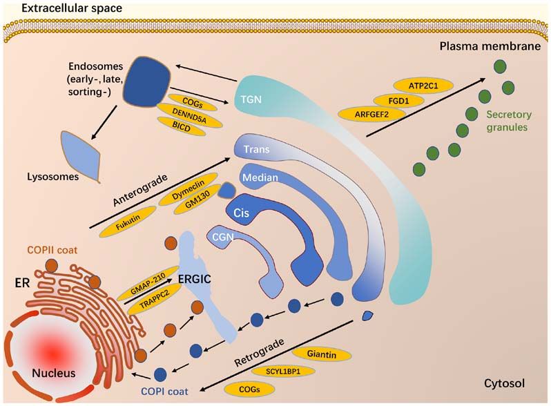
cancer (27). A summary of diseases relating to the Golgi Similar to epithelial cadherin, a transmembrane glycoprotein,
apparatus, classified on the basis of the main organ affected is involved in epithelial cell‑cell adhesion in tumors (51). The
is shown in Fig. 1. Golgi glycosylation of N‑linked glycans on epithelial cadherin
can affect the epithelial‑mesenchymal transition, which
Neurodegenerative disease. Structural and functional changes is related to the formation of metastatic lesions (49). This
of the Golgi apparatus are associated with several neurode‑ process is suggested to help cancer cells leave their original
generative diseases, such as Amyotrophic lateral sclerosis (28), position during wound healing and other normal physiological
Alzheimer's disease (29), Parkinson's disease (3), Huntington's processes, which is an essential mechanism for metastasis and
disease (30), Creutzfeldt‑Jacob disease (31) and multiple diffusion of cancer cells (52,53). The GOLPH3 complex is an
system atrophy (32). Golgi fragmentation is not a consequence important molecular component in the process of Golgi‑drivenINTERNATIONAL JOURNAL OF MOlecular medicine 47: 38, 2021 3
Figure 1. Disorders relating to Golgi dysfunction. Disorders relating to Golgi apparatus dysfunction are grouped according to the main tissues/organs affected.
tumor progression. The role of the GOLPH3 complex in cancer 4. Mutant Golgi resident proteins involved in disease
includes: i) Regulating Golgi glycosylation, which is important
in driving the cancer phenotype (54); ii) promoting the cellular In addition to being an intermediate site in pathogenic cascades
DNA damage response that enhances cellular survival under in diseases, the Golgi apparatus can be the primary target for
DNA damage (55); iii) interacting with components of the diseases caused by genetic mutations in Golgi resident proteins.
retromer complex that enhances growth‑factor‑induced mTOR Mutations in proteins localized to the Golgi apparatus can be
signaling (56); and iv) regulating cell migration by promoting deleterious for the structure and function of this organelle,
reorientation of the Golgi apparatus towards the leading impeding membrane trafficking pathways through it (Fig. 2)
edge (57). In addition to GOLPH3, the Golgi protein GM130 and resulting in disease. We highlight some of the studies that
is important in Golgi glycosylation and protein membrane explore links between Golgi resident proteins and disease.
trafficking in cancer cells. Downregulation of GM130 induces
autophagy, inhibits glycosylation, decreases angiogenesis, Golgi matrix protein and diseases. Adjacent Golgi stacks
and suppresses tumorigenesis (58). In general, aberrant Golgi are linked by tubules forming a membrane network termed
glycosylation causes carcinogenesis, but may also be a conse‑ the Golgi ribbon (66). This structure is a highly ordered
quence of cancer progression. and continuous structure that is adjacent to the nucleus.
The Golgi ribbon comprises proteins that mediate cisternal
Other diseases. Golgi dysfunction was also observed in stacking and the material supporting the Golgi ribbon is
pulmonary arterial hypertension, and cardiovascular diseases. the Golgi matrix (67). The concept of the Golgi matrix was
In an in vivo model of pulmonary arterial hypertension, introduced by Slusarewicz and colleagues, who isolated a
Golgi dysfunction and intracellular trafficking with trap‑ detergent‑insoluble, salt‑resistant Golgi fraction in 1994 (18).
ping of diverse vesicle tethers, giantin, p115, and soluble The main function of the Golgi matrix is maintaining normal
N‑ethylmaleimide‑sensitive factor attachment protein recep‑ structure and mediating protein trafficking through the Golgi
tors (SNAREs) were observed in the Golgi membranes of cisternae. During cisternal progression, the Golgi matrix must
enlarged pulmonary arterial endothelial cells and smooth be dynamic to adapt to Golgi structural changes.
muscle cells (9,10,59). Golgi‑mediated membrane trafficking Golgi matrix proteins include golgins and Golgi reas‑
dysfunctions play important roles in the pathogenesis of sembly stacking proteins (GRASPs) (67), both of which are
pulmonary arterial hypertension (60). important for maintaining Golgi structure and regulating
Structural changes and functional disorder of the Golgi protein and lipid trafficking through the stacks. Golgins are a
apparatus have been identified in many cardiovascular diseases, family of conserved coiled‑coil proteins that were originally
such as heart failure, dilated cardiomyopathy, arrhythmia, and identified as a group of Golgi‑localized antigens (68,69). The
chronic arial fibrillation (61‑64). A previous review clarified golgins not only capture incoming vesicles, but also clearly
the relationship between the Golgi apparatus and various distinguish vesicles from different origins (70). GRASPs
cardiovascular diseases (26). For example, in dilated cardio‑ include GRASP65 (71) and GRASP55 (72). The former local‑
myopathy patients, morphological changes in Golgi vesicle are izes to the cis‑Golgi cisternae while the latter localizes to
consistent with the secretion of natriuretic peptide as the rate the medial/trans‑Golgi cisternae. The functions of GRASPs
of protein secretion affects the morphology and size of Golgi include Golgi structure formation, specific cargo transport,
vesicles (7). In addition, the Golgi vesicle area is inversely apoptosis, and cell migration (73).
proportional to the left ventricular end‑diastolic diameter Given the important multiple functions of Golgi matrix
and the end‑systolic diameter, and is proportional to the left proteins, mutation of Golgi matrix proteins has serious
ventricular ejection fraction (65). consequences on health. Increasing studies support that4 LIU et al: GOLGI APPARATUS IN DISEASE
Figure 2. Golgi resident proteins and membrane trafficking pathway. The main membrane trafficking pathways are included. Newly synthesized proteins
enter the ER and are sorted into budding vesicles that are dependent on the COPII. Vesicles move to the ERGIC and forward to the CGN and the trans‑Golgi
cisternae. Finally, vesicles reach the TGN and cargos sort to their final destinations such as lysosomes, endosomes or the plasma membrane. Different mutation
in Golgi resident proteins affect different membrane trafficking pathway: i) GM130, Giantin, Fukutin, Dymeclin and SCYL1BP1 (involving anterograde traf‑
ficking); ii) COGs (involving retrograde trafficking); iii) TRAPPC2 and GMAP‑210 (involving ER to ERGIC); iv) FGD1, ATP2C1 and ARFGEF2 (involving
TGN to plasma membrane); and v) COGs, DENND5A and BICD (involving endosome to TGN).
the mutation of Golgi matrix proteins including GM130, findings showed that, missense mutations in BICD resulted
Bicaudal‑D (BICD), GMAP‑210, giantin (74), and SCYL1BP1 in spinal muscular atrophy (84,85) and hereditary spastic
(also known as GORAB) (75), leads to diseases. The present paraplegia (86) by changing the normal morphological struc‑
review included some proteins as examples to elaborate on the ture of the golgi. The core pathogenetic mechanism may be
pathogenic mechanism of Golgi matrix proteins. a BICD2 mutation resulting in abnormal cargo trafficking in
The first example is GM130 (also known as GOLGA2), the motor neurons. This trafficking results in neuronal growth
first identified Golgi matrix protein (76). GM130 is a peripheral disorders and eventually neuronal dysfunction.
membrane protein attached to the Golgi membrane that is impor‑ The third example is giantin, encoded by the Golgb1 gene.
tant in maintaining the adaxial Golgi reticular structure (77). In Giantin is a member of the golgin family and is a tethering
neurodegenerative diseases, GM130 knockout in hippocampal factor for COPI vesicles and functions in the CGN (87).
neurons is reported to cause damage to dendritic structures (78). Mutations in the Golgb1 gene lead to lack of expression of
In mouse neuron experiments, specific knockout of GM130 giantin protein and a pleiotropic phenotype including osteo‑
resulted in disruption of the Golgi architecture and positioning chondrodysplasia in a rat model (88) and a ciliopathy‑like
in cerebellar Purkinje cells and to deficient secretory cargo phenotype in a zebrafish model (74). Both pathogenetic mecha‑
trafficking. As a consequence, progressive cerebellar atrophy nisms involve disturbance of extracellular matrix components,
of Purkinje cells resulted in delayed movement and ataxia in which are transported by intracellular membrane trafficking
mice (79). This animal experimental study indicates that GM130 systems. Giantin knockout leads to changes in expression of
mutations are causative in neurodegenerative disease. Golgi‑resident glycosyltransferases, which could affect extra‑
A second example is BICD, a golgin that interacts with cellular matrix deposition (89).
Rab6 on the TGN (80). Of two homologous sequences, BICD1 The fourth example is GORAB (also known as SCYL1BP1).
and BICD2, the latter binds to a subgroup of motility protein GORAB, localized to the trans‑side of the Golgi, is a member
activator proteins and is a connecting molecule between the of the golgin family and interacts with Rab6. Mutation in
motility protein and cargo (81). High expression of BICD GORAB results in gerodermia osteodysplastica (GO) char‑
in normal nervous systems is important for maintaining the acterized by wrinkly skin and osteoporosis (75). GORAB
normal lamellar structure of the cerebral cortex, hippocampus, functions in COPI trafficking, and acts as a scaffolding factor
and cerebellar cortex (82). The brain cortex, hippocampus for COPI assembly at the TGN by interacting with Scyl1.
and cerebellar cortex neurons of BICD2‑knockout mice have GORAB mutations perturb COPI assembly at the TGN, and
impaired migration function (82,83) and eventually, damage result in reduced recycling of COPI‑mediated retrieval of
the brain and cerebellar cortex layer structure. Previous trans‑Golgi enzymes and improper glycosylation (90).INTERNATIONAL JOURNAL OF MOlecular medicine 47: 38, 2021 5
A final example of the effects of loss of expression of a cutis laxa (106). Autosomal recessive cutis laxa type II is a
Golgi matrix protein is GMAP‑210 (also known as TRIP11). heterogeneous condition characterized by sagging, inelastic,
This CGN golgin acts in asymmetric membrane tethering (91). and wrinkled skin (107,108). The mechanism may involve
In animal experiments, a nonsense mutation in Trip11 led to impaired intracellular acidification of the Golgi and damaged
a loss of GMAP‑210, which led to abnormal Golgi‑mediated retrograde trafficking from the Golgi to the ER (100,108).
glycosylation and cellular transport of proteins in chondrocytes ATP7A and ATP7B are the key regulators of cellular
and osteoblasts of mice (92). Similarly, GMAP‑210 mutations Cu 2+ metabolism. Under basal conditions (normal copper
were found in patients with human chondrodysplasia achon‑ levels), ATP7A is located in the TGN and travels to the plasma
drogenesis 1A (92), and odontochondrodysplasia (93). membrane at high copper levels. Mutations in the ATP7A
result in mislocalization of ATP7A protein and impaired
Other Golgi resident proteins and diseases. In addition copper‑responsive trafficking between the TGN and plasma
to matrix proteins, several proteins that localize to Golgi membrane, which contributes to the development of Menkes
membranes are also important for normal Golgi structure disease (109). Menkes disease is a lethal multisystemic
and function such as the tethering factors Rab GTPases and disorder characterized by neurodegeneration and connective
SNAREs, which regulate the specific targeting and fusion of tissue abnormalities as well as typical sparse and steely hair.
transport carriers with Golgi membranes. The maintenance Similarly, mutations in the ATP7B contributes to the develop‑
of Golgi luminal ion concentrations depends on the secre‑ ment of Wilson's disease (110). Wilson's disease, also known
tory pathway Ca 2+/Mn 2+ ATPases and vacuolar H+ ATPase as hepatolenticular degeneration, results in hepatic and/or
(V‑ATPase). Therefore, the impaired performance of mutated neurological deficits, including dystonia and parkinsonism.
Golgi resident proteins creates serious and highly diverse
pathologies in the Golgi. Emerging studies on patient genetics Golgi resident glycosyltransferase. The Golgi apparatus is
have identified mutations in Golgi resident protein‑coding an important organelle for the post‑translational modification
genes that are related to diseases. We focus on some of these of cargos. The post‑translational modification of secreted
proteins, and discuss the activities of mutated Golgi resident and membrane proteins is mediated by the Golgi resident
proteins that result in disease. enzymes such as glycosyltransferases, glycosidases, and
kinases. Glycosylation is an enzymatic reaction that chemi‑
Golgi ion pump. The release and uptake of Ca 2+ by Golgi cally links monosaccharides or polysaccharides (glycans) to
membranes is mainly mediated by secretory pathway other saccharides, proteins, or lipids (111). Golgi glycosylation
Ca2+/Mn2+ ATPases (SPCA1 and SPCA2), which are encoded is a modification by Golgi‑resident glycosylation enzymes
by the ATP2C1/ATP2C2 genes. The proteins transfer Ca 2+ including glycosidases and glycosyltransferases (112). The
from the cytoplasm to the Golgi and maintain the stability normal function of Golgi glycosylation depends on the
of intracellular free Ca 2+ (94). The maintenance of Golgi precise Golgi localization and normal activities of Golgi
luminal Ca2+ and Mn 2+ directly affects the optimal activity resident enzymes. The proper localization of Golgi resident
of Golgi glycosyltransferase and the trafficking of cell adhe‑ enzymes is controlled by finely regulated vesicular traf‑
sion proteins to the cell plasma membrane (95). Knockdown ficking in the Golgi. If the balance between anterograde and
of SPCA1 affects the morphology and structure of the Golgi retrograde trafficking is defective, Golgi glycosylation is
and causes mis‑localization of proteins. Clinically, muta‑ affected, resulting in Golgi glycosylation abnormalities (113).
tions in the ATP2C1 gene on chromosome 3q21 can lead to Mutations in Golgi resident putative glycosyltransferases are
Hailey‑Hailey disease, an autosomal dominant skin disorder directly linked to human congenital muscular dystrophies:
in humans (96,97). The possible pathogenetic mechanism may Like‑acetylglucosaminyl‑transferase (LARGE) in congenital
be dysfunction in Ca2+ signaling at the Golgi membrane and muscular dystrophy syndrome (114), fukutin in Fukuyama‑type
dysfunction of processing, modification and trafficking of congenital muscular dystrophy (115), and fukutin‑related
desmosomal proteins (98). protein in band muscular dystrophy syndrome (116). These
Golgi acidity is an important role for maintaining the mutations appear to affect cell migration in the developing
morphological integrity of the Golgi and transporting various brain, resulting in combined clinical manifestations in muscle
kinds of cargo (99,100). Under normal conditions, the Golgi and brain development. In an animal model, mutations in
cavity is weakly acidic and the pH of the Golgi reticular Golgi resident glycosyltransferases are also associated with
structure decreases gradually from the CGN to the TGN (101). the neurodegenerative disease, such as ST3GAL5, β1,4‑gala
The Golgi luminal pH is regulated by V‑ATPase (102), AE2a ctosyltransferase 4 (B4GalT4) (117), and glycosyltransferase
HCO3‑/Cl‑ exchanger, and Golgi pH regulator (103). Luminal 8 domain containing 1 (GLT8D1). GLT8D1 is a glycosyl‑
pH is closely tied to Golgi function. Partial V‑ATPase transferase enzyme located in the Golgi apparatus. A recent
dysfunction is related to multiple disease states (104). study reported that mutated GLT8D1 induces motor deficits
ATP6V1E1, ATP6V1A, and ATP6V0A2 encode different in zebrafish embryos consistent with amyotrophic lateral scle‑
subunits of the V‑ATPase pump. A study showed that Golgi rosis (118). However, another study suggested that GLT8D1 is
subunit‑isoform of the V‑ATPase (ATP6V0A2) mutations not likely the causative gene for ALS in mainland China (119).
lead to structural changes in the extracellular matrix that is
responsible for skin elasticity (105). Clinically, the dysfunction Rab GTPase. Rab proteins are members of the small Ras‑like
of the Golgi‑localized V‑ATPase caused by mutations in the GTPase family that regulate the four steps of membrane
ATP6VOA2 gene is directly related to cutis laxa. Mutations transport by recruiting effector molecules. Golgi‑associated
in ATP6V1E1 or ATP6V1A also cause autosomal‑recessive Rab proteins including Rab1, Rab2, Rab6, Rab18, Rab33B, and6 LIU et al: GOLGI APPARATUS IN DISEASE
Rab43 have a central role in Golgi organization and membrane Considering the mechanistic links between Golgi resi‑
trafficking (120). Rab33B is localized to medial‑Golgi dent proteins, membrane trafficking, and the development of
cisternae and is important in Golgi‑to‑ER retrograde traf‑ genetic diseases, we suggest a term for these disorders based
ficking. Rab39B, a neuronal‑specific protein, is a novel Rab on their similar pathophysiology: Golgi apparatus membrane
GTPase that localizes to the Golgi and is related to synapse trafficking disorders. It is a group of genetic diseases in which
formation. Mutations in the Rab33B coding gene cause the mutation of the gene encoding Golgi resident protein
Smith‑McCort dysplasia (121) and mutations in the Rab39B results in membrane trafficking defects within the cells. Golgi
gene cause X‑linked mental retardation (122). apparatus membrane trafficking defects typically result in
the accumulation of undegraded proteins, mislocalization of
SNAREs. SNAREs are proteins involved in docking and proteins, and impaired glycosylation of proteins. However, the
fusion of transport to intermediate membranes. Golgi SNAP cascade events following the Golgi apparatus and defective
receptor complex member 2 (GOSR2) is a member of the membrane trafficking, ultimately leading to human diseases,
SNAREs family that localizes to the CGN and is involved remain to be clarified in further research.
in ER‑to‑Golgi trafficking (123). Homozygous mutations in Although the Golgi apparatus‑mediated membrane traf‑
GOSR2 lead to progressive myoclonus epilepsy (124). Clinical ficking pathway exists in all kinds of tissues and organs in
manifestations include early ataxia, myoclonus, and convulsive human, the trafficking defects on tissues is often selective. The
seizures. A possible mechanism involves GOSR2 mutations most sensitive to membrane trafficking defects is the nervous
leading to GOSR2 protein that cannot be localized to the CGN system, skin, bone, cartilage, and skeletal muscle and the
and blocks SNAREs complex formation. SNAREs complex reasons for mutations occurring in these genes mostly affecting
dysfunction could lead to the impaired fusion of vesicles with these tissues remain to be elucidated. Firstly, neurons are
cis‑Golgi cisternae, hindering ER‑to‑Golgi membrane traf‑ extraordinarily polarized cells, the extension of dendrites and
ficking. The perturbation of early ER‑to‑Golgi transport may axons requires a significant expansion of the cell surface area,
result in changes in the regulated release of neurotransmitters and new plasma membrane proteins must be delivered through
and proper sorting of neurotransmitter receptors at synapses in the membrane trafficking. For the nervous system, intracel‑
neurons, potentially leading to epilepsy (125,126). lular trafficking functionally impacts neuronal development,
homeostasis, as well as neurodegeneration (133). Secondly, it is
5. Golgi apparatus membrane trafficking disorders generally known that skin, bone, cartilage, and skeletal muscle
fiber comprise large amounts of the extracellular matrix
In the above section, we introduced the pathophysiology of which define the structure and physical properties. Almost
some diseases related to Golgi resident proteins. A summary all extracellular matrix components are transported by intra‑
of genetic diseases caused by mutations in genes encoding cellular trafficking systems. Alterations in Golgi apparatus
Golgi resident proteins is presented in Table I. By analyzing the membrane trafficking can lead to glycosylation abnormalities.
pathophysiology of these diseases, we found that the majority The assembly and maintenance of the extracellular matrix are
of genes leading to human diseases are involved in defects susceptible to impairment of matrix protein glycosylation.
in membrane trafficking (Fig. 2). For example, TRAPPC2 Thus, the skin, bone, cartilage, and skeletal muscle are most
mutation, involving the membrane trafficking pathway sensitive to impaired glycosylation of cargo proteins, and
between ER‑to‑Golgi in bone cells and chondrocytes, results membrane trafficking defects. Therefore, the loss of some
in X‑linked spondyloepiphyseal dysplasia tarda (127). The Golgi resident proteins, such as ATP6V1A, ATP6V1E1 (106),
conserved oligomeric Golgi (COG) complex is a conserved, ATP6VOA2 (108), TMEM165 (134), GOLGB1 (88),
hetero‑octameric protein complex localized in the Golgi SCYL1BP1 (75), TRAPPC11 (135), TRAPPC2 (136), and
cis/medial cisternae (128). In addition to the COG3 subunit, TRIP11 (92), manifest primarily in these matrix‑rich tissues.
mutations in seven other COG subunits result in human
congenital disorders of glycosylation (CDG) type II, which is 6. Clinical value of Golgi apparatus
mainly marked by misregulation of protein glycosylation, and
defects in retrograde trafficking through the Golgi (129,130). The Golgi apparatus participates in the occurrence and devel‑
The mutation in FGD1 resulting in Aarskog‑Scott syndrome opment of disease and could be the key to finding new targets
may lead to the obstruction of post‑Golgi trafficking, such for disease diagnosis and therapy.
as the Golgi‑to‑plasma membrane trafficking pathway (131).
Mutation in TRIP11 mainly involves ER to ERGIC and Biomarker discovery. Golgi glycoprotein 73 (GP73, also
anterograde trafficking (132). Therefore, membrane trafficking referred to as GOLPH2), a resident Golgi membrane protein,
defects play a major role in the pathogenic process of muta‑ is predominantly expressed in biliary epithelial cells in the
tion in genes encoding Golgi resident protein. Intracellular normal human liver (137). GP73 expression is upregulated
membrane trafficking is a fundamental process responsible in chronic Hepatitis B virus (HBV) infection (138), chronic
for compartmentalization of the biosynthesis pathway HCV infection (139), non‑alcoholic fatty liver disease (140),
and secretion cargos, including hormones, growth factors, and hepatocellular carcinoma (HCC) (141,142). Serum GP73, a
antibodies, matrix and serum proteins, digestive enzymes, new marker for HCC, is reported to appear earlier than serum
and many more. Defective membrane trafficking results in α‑fetoprotein. The combined detection of serum α‑fetoprotein
protein sorting defects, undegraded proteins due to defective and GP73 can improve sensitivity and specificity for HCC
Golgi‑to‑lysosome trafficking, downregulation of protein diagnosis (143,144). However, several studies showed GP73
secretion, and mislocalization of proteins. levels were not higher in HCC patients than in patients withTable I. Human diseases caused by mutations in genes encoding Golgi resident proteins.
Gene Function Disease Main clinical manifestation Cellular effect (Refs.)
AP1S2 Coat adapter X‑linked mental retardation Mental retardation Brain‑specific defect of AP‑1‑dependent (165)
syndrome intracellular protein trafficking
AP3D1 Coat adapter Hermansky‑Pudlak syndrome Immunodeficiency; Impaired lysosomal trafficking (166)
Neurodevelopmental delay; Seizure
ARFGEF2 GTPase activator Periventricular nodular heterotopia Malformation of cortical development Defective TGN‑cell membrane trafficking (167)
ATP2C1 Ion pump Hailey‑Hailey disease Skin disorder Defective trafficking of desmosomal proteins (96)
to cell membrane
ATP6V1A Ion pump Cutis laxa type II Wrinkled skin Defective retrograde transport; Abnormal (106,168)
glycosylation
ATP6V1E1 Ion pump Cutis laxa type II Wrinkled skin Defective retrograde transport; Abnormal (106)
glycosylation
ATP6VOA2 Ion pump Cutis laxa type II Wrinkled skin Defective Golgi trafficking; Abnormal (108)
glycosylation of CDG‑II
ATP7A Ion pump Menkes disease; Occipital horn Neurodegeneration; Connective tissue Defective Golgi trafficking of copper; (109)
disease disorder
ATP7B Ion pump Wilson's disease Hepatic and/or neurological disorder Defective Golgi trafficking of copper (110)
ATXN2 Signaling Spinocerebellar ataxia type 2 Progressive ataxia; slow saccades Disrupted calcium homeostasis (169)
Bicaudal‑D Golgin SMA; HSP Neurodegeneration defective targeting and transport of Golgi (84,86)
resident proteins.
COG Tethering CDG‑type II Neurodegenerative disorder Defective retrograde and endosome‑to‑TGN (170)
trafficking; Abnormal glycosylation
COPA Coat COPA syndrome Interstitial lung, joint and kidney Defective membrane trafficking (171)
disorder
DENND5A GTPase activator Epileptic Encephalopathy Refractory seizures and cognitive Defective endosome‑TGN trafficking (172)
arrest
INTERNATIONAL JOURNAL OF MOlecular medicine 47: 38, 2021
DYM Unknown Dyggve‑Melchior‑Clausen syndrome Spondyloepimetaphyseal dysplasia; Defective ER‑Golgi trafficking (173)
intellectual disability
FGD1 GTPase activator Aarskog‑Scott syndrome Faciogenital dysplasia Reduction in FGD1 trafficking from Golgi (131)
FKRP Glycosyltransferases Limb girdle muscular dystrophy Muscular dystrophy Abnormal glycosylation (116)
Fukutin Glycosyltransferases FCMD Muscular dystrophy Abnormal glycosylation; Impaired ER‑to‑Golgi (115)
trafficking of mutant protein
GOSR2 SNARE Progressive myoclonus epilepsy Seizure Mislocalization of mutant protein to cis‑Golgi; (124)
Defective cis to trans Golgi compartment trafficking
HERC1 GTPase activator Idiopathic intellectual disability Intellectual disability Misregulation of mTOR pathway (174)
78
Table I. Continued.
Gene Function Disease Main clinical manifestation Cellular effect (Refs.)
LARGE Glycosyltransferases Congenital muscular dystrophy Muscular dystrophy Abnormal glycosylation (114)
Type 1D
OSBPL2 Lipid transport Autosomal dominant nonsyndromic Hearing loss Abnormal lipid metabolism (175)
hearing loss
RAB33B Rab GTPase Smith‑McCort dysplasia Skeletal dysplasia Golgi fragmentation; Defective Golgi membrane (176)
trafficking
RAB39B Rab GTPase X‑linked Mental retardation Mental Retardation; Autism; Defective Golgi membrane trafficking (122)
Epilepsy; Macrocephaly
S1P Serine protease Spondyloepimetaphyseal dysplasia Skeletal dysplasia Defective Golgi‑to‑lysosome transport (177,178)
SCYL1BP1 Golgin Gerodermia osteodysplastica Osteoporosis; Wrinkly skin Reduced recycling of trans‑Golgi enzymes; (75,90)
Defective COPI traffic and glycosylation
SLC35A1 CMP Synal CDG‑II Neurodegenerative disorder Abnormal glycosylation (179)
Transporter
SLC35A2 UDP Gal CDG Developmental delay; Seizures; Abnormal glycosylation (180)
Transporter Ataxia
TMEM165 Ion pump CDG‑II Neurodegenerative disorder Mislocalization of mutant protein resulting in (134)
LIU et al: GOLGI APPARATUS IN DISEASE
abnormal Golgi glycosylation
TRAPPC11 Tethering Congenital muscular dystrophy Muscular dystrophy Defective trafficking and Hypoglycosylation of (135)
mutant protein
TRAPPC2 Tethering Spondyloepiphyseal dysplasia tarda Skeletal dysplasia Abnormal trafficking between ER and Golgi (136)
TRIP11 Golgin Achondrogenesis type 1A; Skeletal dysplasia Golgi fragmentation; Abnormal Golgi‑mediated (92,93)
Odontochondrodysplasia glycosylation
VPS53 Unknown Progressive cerebello‑cerebral Mental retardation; Microcephaly; Impaired NPC2 protein sorting to lysosome and (181)
atrophy type 2 Epilepsy cholesterol accumulationINTERNATIONAL JOURNAL OF MOlecular medicine 47: 38, 2021 9
other liver diseases such as cirrhosis (145,146). In addition to photothermal therapy. In this method, a photothermal ablation
being a marker, the expression of GP73 is critical for chemo‑ agent converts light energy into heat and kills cancer cells
therapeutic resistance in HCC cell lines (147). with high specificity and minimal invasiveness by hyper‑
Transmembrane protein 165 (TMEM165) functions in ion pyrexia (162). Another research team developed a prodrug
homeostasis, membrane trafficking, and glycosylation in the nanoparticle system, which appeared to target the Golgi
Golgi apparatus (148). Findings of a study showed that muta‑ apparatus and realized retinoic acid release under an acidic
tions in TMEM165 cause CDG type II in humans (134). Other environment. The retinoic acid‑conjugated chondroitin sulfate
research has found that expression of TMEM165 mRNA could reduce the expression of metastasis‑associated proteins
and protein is apparently increased in HCC patient tissues by inducing Golgi fragmentation (163). Those findings suggest
and contributes to the invasive activity of cancer cells (149). that the Golgi apparatus is a promising target for the develop‑
This result indicates that TMEM165 is a possible biomarker ment of novel drugs. A review summarized small molecules
for HCC. GS28 is a member of the SNAREs protein family. as drugs targeting the Golgi apparatus for the treatment of
GS28 protein immunoreactivity was observed in both nuclear diseases (164), such as LTX‑401, inhibitors of Golgi‑associated
and cytoplasmic compartments of cancer cells. High nuclear lipid transfer proteins, glucosylceramide synthase inhibitors,
expression of GS28 is associated with poor prognosis for O‑glycosylation inhibitors, PI4KIIIb inhibitors and inhibi‑
colorectal (150) and cervical cancer patients (151). tors of ARF activation. Whether these drugs that target the
Anti‑Golgi antibodies (AGAs) were first found in 1982 in Golgi apparatus can be applied in clinical practice needs to be
the serum of patients with Sjogren's syndrome complicated determined.
with lymphoma (152). AGAs have also been found in other
immunological diseases (153‑155). Currently, at least 20 7. Conclusion
Golgi autoantigens are known, including golgin‑97, golgin‑67,
golgin‑245, golgin‑95, golgin‑160, and giantin. AGA positivity The central role of the Golgi apparatus in critical cell processes
is commonly found in connective tissue diseases such as such as the transport, processing, and sorting of proteins and
Sjogren's syndrome, rheumatoid arthritis, and systemic lupus lipids has placed it at the forefront of cell science. Several
erythematosus (154,156); cerebellar malignant disease such as previous studies have suggested that the Golgi apparatus
idiopathic late‑onset cerebellar ataxia (157); infectious diseases plays a critical role in diseases, particularly in neurode‑
such as HBV/HCV infection, Epstein‑Barr virus infection generative diseases. However, few studies focus on human
and HIV infection (155,158,159); and tumors, such as HCC diseases caused by mutations in genes encoding Golgi resident
and lung cancer (160). Although AGAs are not specific to any proteins and summarize the common features of these genetic
disease, their clinical detection may be helpful for classifying diseases. In the present review, we summed up the genetic
and following the progress of some connective tissue diseases. diseases caused by mutations in genes encoding Golgi resident
For example, compared to anti‑BICD2‑negative patients, single proteins. By analyzing their pathophysiology, we identified
specificity anti‑BICD2 patients may be more associated with that the majority of genes are involved in membrane traf‑
inflammatory myopathy and interstitial lung disease (161). ficking. The nervous system, skin, bone, cartilage, and skeletal
Biomarkers are crucial for early diagnosis, assessing response muscle are the most sensitive tissues to defective membrane
to treatment, and classifying diseases into subtypes. Biomarker trafficking. It is reasonable to hope that our basic knowledge of
discovery involves many critical steps such as clinical study Golgi‑mediated membrane trafficking will continue to provide
design, sample collection, data integration, and protein/peptide insights into the pathogenesis of genetic diseases and that
identification and preservation. These steps should be carefully studies of these diseases will continue to enhance our under‑
controlled before confirmation and verification. Therefore, in standing of the critical role of the Golgi apparatus in diseases.
clinical applications, these biomarkers are potential diagnostic In addition, the finding of Golgi‑related biomarker and
markers. Large‑scale investigations are needed and more sensi‑ Golgi‑based therapeutics further emphasize the importance of
tive and specific detection methods need to be researched. Golgi apparatus in human pathology. Taken together, advances
in Golgi apparatus biology provide opportunities to translate
Golgi‑based therapeutics. In addition to biomarker discovery, discoveries into clinical medicine. Thus, we highlighted the
the functions of the Golgi apparatus and its associated importance of underlying clinical insights and provided a new
molecules in maintaining cell structural integrity and its direction for future research.
central role in membrane trafficking pathways provide
possible targets for disease therapy. These targets may be Acknowledgements
direct, due to genetic disease (Table I), or indirect, as in
cancer. Compared to non‑transformed and normal cells, Not applicable.
cancer cells have morphological and functional changes in
the Golgi apparatus that drive invasion and migration in a Funding
unique microenvironment. These changes provide therapeutic
targets for interventions. A research team developed a bovine The present study was supported by grants from the National
serum albumin pH‑responsive photothermal ablation agent Natural Science Foundation of China (grant no. 81974213).
that preferentially accumulates in the Golgi of cancer cells
compared to normal cells due to morphological changes in the Availability of data and materials
Golgi apparatus (162). The agent is activated by the weakly
acidic microenvironment of the Golgi in cancer cells for Not applicable.10 LIU et al: GOLGI APPARATUS IN DISEASE
Authors' contributions 14. Tan X, Banerjee P, Guo HF, Ireland S, Pankova D, Ahn YH,
Nikolaidis IM, Liu X, Zhao Y, Xue Y, et al: Epithelial‑to-
mesenchymal transition drives a pro‑metastatic Golgi compaction
JL and YH were mainly responsible for collecting relevant process through scaffolding protein PAQR11. J Clin Invest 127:
information and completing this review. ZJ, LZ and TL were 117‑131, 2017.
15. Golgi C: On the structure of nerve cells. 1898. J Microsc 155:
mainly responsible for consulting literature materials and 3‑7, 1989.
revising the manuscript. ZH was responsible for the concep‑ 16. Mollenhauer HH and Morré DJ: Perspectives on Golgi appa‑
tion of this review and the assignment of tasks. There was no ratus form and function. J Electron Microsc Tech 17: 2‑14,
additional assistance with manuscript preparation. All authors 1991.
17. Storrie B, White J, Röttger S, Stelzer EH, Suganuma T and
read and approved the final manuscript. Nilsson T: Recycling of golgi‑resident glycosyltransferases
through the ER reveals a novel pathway and provides an explana‑
Ethics approval and consent to participate tion for nocodazole‑induced Golgi scattering. J Cell Biol 143:
1505‑1521, 1998.
18. Slusarewicz P, Nilsson T, Hui N, Watson R and Warren G:
Not applicable. Isolation of a matrix that binds medial Golgi enzymes. J Cell
Biol 124: 405‑413, 1994.
19. Papanikou E and Glick BS: Golgi compartmentation and identity.
Patient consent for publication Curr Opin Cell Biol 29: 74‑81, 2014.
20. Glick BS and Luini A: Models for Golgi traffic: A critical assess‑
Not applicable. ment. Cold Spring Harb Perspect Biol 3: a005215, 2011.
21. Sundaramoorthy V, Sultana JM and Atkin JD: Golgi fragmen‑
tation in amyotrophic lateral sclerosis, an overview of possible
Competing interests triggers and consequences. Front Neurosci 9: 400, 2015.
22. Gonatas NK, Stieber A and Gonatas JO: Fragmentation of the
Golgi apparatus in neurodegenerative diseases and cell death.
The authors declare that they have no competing interests. J Neurol Sci 246: 21‑30, 2006.
23. Jiang Z, Hu Z, Zeng L, Lu W, Zhang H, Li T and Xiao H: The
References role of the Golgi apparatus in oxidative stress: Is this organelle
less significant than mitochondria? Free Radic Biol Med 50:
907‑917, 2011.
1. Rios RM and Bornens M: The Golgi apparatus at the cell centre. 24. Liu JY, He JL, Huang Y, Xiao H, Jiang Z and Hu ZP: The Golgi
Curr Opin Cell Biol 15: 60‑66, 2003. apparatus in neurorestoration. J Neuroresstoratology 7: 116‑128,
2. Gautam M, Jara JH, Sekerkova G, Yasvoina MV, Martina M and 2019.
Özdinler PH: Absence of alsin function leads to corticospinal 25. Fan J, Hu Z, Zeng L, Lu W, Tang X, Zhang J and Li T: Golgi
motor neuron vulnerability via novel disease mechanisms. Hum apparatus and neurodegenerative diseases. Int J Dev Neurosci 26:
Mol Genet 25: 1074‑1087, 2016. 523‑534, 2008.
3. Rendón WO, Martínez‑Alonso E, Tomás M, Martínez‑Martínez N 26. Lu L, Zhou Q, Chen Z and Chen L: The significant role of the
and Martínez‑Menárguez JA: Golgi fragmentation is Rab and Golgi apparatus in cardiovascular diseases. J Cell Physiol 233:
SNARE dependent in cellular models of Parkinson's disease. 2911‑2919, 2018.
Histochem Cell Biol 139: 671‑684, 2013. 27. Millarte V and Farhan H: The Golgi in cell migration: Regulation
4. Brandstaetter H, Kruppa AJ and Buss F: Huntingtin is required by signal transduction and its implications for cancer cell metas‑
for ER‑to‑Golgi transport and for secretory vesicle fusion at the tasis. ScientificWorldJournal 2012: 498278, 2012.
plasma membrane. Dis Model Mech 7: 1335‑1340, 2014. 28. Mourelatos Z, Gonatas NK, Stieber A, Gurney ME and
5. Yuan D, Liu C and Hu B: Dysfunction of membrane trafficking Dal Canto MC: The Golgi apparatus of spinal cord motor
leads to ischemia‑reperfusion injury after transient cerebral neurons in transgenic mice expressing mutant Cu,Zn superoxide
ischemia. Transl Stroke Res 9: 215‑222, 2018. dismutase becomes fragmented in early, preclinical stages of the
6. Li T, You H, Mo X, He W, Tang X, Jiang Z, Chen S, Chen Y, disease. Proc Natl Acad Sci USA 93: 5472‑5477, 1996.
Zhang J and Hu Z: GOLPH3 mediated Golgi stress response in 29. Joshi G, Bekier ME II and Wang Y: Golgi fragmentation in
modulating N2A cell death upon oxygen‑glucose deprivation and Alzheimer's disease. Front Neurosci 9: 340, 2015.
reoxygenation injury. Mol Neurobiol 53: 1377‑1385, 2016. 30. Strehlow AN, Li JZ and Myers RM: Wild‑type huntingtin
7. Tarazón E, Roselló‑Lletí E, Ortega A, Gil‑Cayuela C, participates in protein trafficking between the Golgi and the
González‑Juanatey JR, Lago F, Martínez‑Dolz L, Portolés M and extracellular space. Hum Mol Genet 16: 391‑409, 2007.
Rivera M: Changes in human Golgi apparatus reflect new left 31. Sakurai A, Okamoto K, Fujita Y, Nakazato Y, Wakabayashi K,
ventricular dimensions and function in dilated cardiomyopathy Takahashi H and Gonatas NK: Fragmentation of the Golgi
patients. Eur J Heart Fail 19: 280‑282, 2017. apparatus of the ballooned neurons in patients with corti‑
8. Stancu CS, Toma L and Sima AV: Dual role of lipoproteins cobasal degeneration and Creutzfeldt‑Jakob disease. Acta
in endothelial cell dysfunction in atherosclerosis. Cell Tissue Neuropathol 100: 270‑274, 2000.
Res 349: 433‑446, 2012. 32. Sakurai A, Okamoto K, Yaguchi M, Fujita Y, Mizuno Y,
9. Lee J, Reich R, Xu F and Sehgal PB: Golgi, trafficking, and Nakazato Y and Gonatas NK: Pathology of the inferior
mitosis dysfunctions in pulmonary arterial endothelial cells olivary nucleus in patients with multiple system atrophy. Acta
exposed to monocrotaline pyrrole and NO scavenging. Am J Neuropathol 103: 550‑554, 2002.
Physiol Lung Cell Mol Physiol 297: L715‑L728, 2009. 33. van Dis V, Kuijpers M, Haasdijk ED, Teuling E, Oakes SA,
10. Sehgal PB, Mukhopadhyay S, Xu F, Patel K and Shah M: Hoogenraad CC and Jaarsma D: Golgi fragmentation precedes
Dysfunction of Golgi tethers, SNAREs, and SNAPs in monocro‑ neuromuscular denervation and is associated with endosome
taline‑induced pulmonary hypertension. Am J Physiol Lung Cell abnormalities in SOD1‑ALS mouse motor neurons. Acta
Mol Physiol 292: L1526‑L1542, 2007. Neuropathol Commun 2: 38, 2014.
11. Zhu H, Li H, Wang P, Chen M, Huang Z, Li K, Li Y, He J, Han J 34. Pottorf T, Mann A, Fross S, Mansel C and Vohra BPS:
and Zhang Q: Persistent and acute chlamydial infections induce Nicotinamide mononucleotide adenylyltransferase 2 maintains
different structural changes in the Golgi apparatus. Int J Med neuronal structural integrity through the maintenance of golgi
Microbiol 304: 577‑585, 2014. structure. Neurochem Int 121: 86‑97, 2018.
12. Rohde J, Emschermann F, Knittler MR and Rziha HJ: Orf virus 35. Joshi G and Wang Y: Golgi defects enhance APP amyloidogenic
interferes with MHC class I surface expression by targeting processing in Alzheimer's disease. Bioessays 37: 240‑247, 2015.
vesicular transport and Golgi. BMC Vet Res 8: 114, 2012. 36. Covarrubias‑Pinto A, Parra AV, Mayorga‑Weber G, Papic E,
13. Mousnier A, Swieboda D, Pinto A, Guedán A, Rogers AV, Vicencio I, Ehrenfeld P, Rivera FJ and Castro MA: Impaired
Walton R, Johnston SL and Solari R: Human rhinovirus 16 intracellular trafficking of sodium‑dependent vitamin C trans‑
causes Golgi apparatus fragmentation without blocking protein porter 2 contributes to the redox imbalance in Huntington's
secretion. J Virol 88: 11671‑11685, 2014. disease. J Neurosci Res 99: 223‑235, 2021.INTERNATIONAL JOURNAL OF MOlecular medicine 47: 38, 2021 11
37. Mani M, Thao DT, Kim BC, Lee UH, Kim DJ, Jang SH, 57. Xing M, Peterman MC, Davis RL, Oegema K, Shiau AK and
Back SH, Lee BJ, Cho WJ, Han IS and Park JW: DRG2 knock‑ Field SJ: GOLPH3 drives cell migration by promoting Golgi
down induces Golgi fragmentation via GSK3β phosphorylation reorientation and directional trafficking to the leading edge. Mol
and microtubule stabilization. Biochim Biophys Acta Mol Cell Biol Cell 27: 3828‑3840, 2016.
Res 1866: 1463‑1474, 2019. 58. Chang SH, Hong SH, Jiang HL, Minai‑Tehrani A, Yu KN, Lee JH,
38. Rodríguez‑Cruz F, Torres‑Cruz FM, Monroy‑Ramírez HC, Kim JE, Shin JY, Kang B, Park S, et al: GOLGA2/GM130,
Escobar‑Herrera J, Basurto‑Islas G, Avila J and García‑Sierra F: cis‑Golgi matrix protein, is a novel target of anticancer gene
Fragmentation of the Golgi apparatus in neuroblastoma cells is therapy. Mol Ther 20: 2052‑2063, 2012.
associated with tau‑induced ring‑shaped microtubule bundles. 59. Sehgal PB, Mukhopadhyay S, Patel K, Xu F, Almodóvar S,
J Alzheimers Dis 65: 1185‑1207, 2018. Tuder RM and Flores SC: Golgi dysfunction is a common feature
39. Sou YS, Kakuta S, Kamikubo Y, Niisato K, Sakurai T, Parajuli LK, in idiopathic human pulmonary hypertension and vascular
Tanida I, Saito H, Suzuki N, Sakimura K, et al: Cerebellar lesions in SHIV‑nef‑infected macaques. Am J Physiol Lung Cell
neurodegeneration and neuronal circuit remodeling in Golgi pH Mol Physiol 297: L729‑L737, 2009.
regulator‑deficient mice. eNeuro 6: ENEURO.0427‑18.2019, 2019. 60. Sehgal PB and Lee JE: Protein trafficking dysfunctions: Role
40. Heuer D, Rejman Lipinski A, Machuy N, Karlas A, Wehrens A, in the pathogenesis of pulmonary arterial hypertension. Pulm
Siedler F, Brinkmann V and Meyer TF: Chlamydia causes Circ 1: 17‑32, 2011.
fragmentation of the Golgi compartment to ensure reproduction. 61. Muhammad E, Levitas A, Singh SR, Braiman A, Ofir R, Etzion S,
Nature 457: 731‑735, 2009. Sheffield VC, Etzion Y, Carrier L and Parvari R: PLEKHM2
41. Pruneda JN, Bastidas RJ, Bertsoulaki E, Swatek KN, mutation leads to abnormal localization of lysosomes, impaired
Santhanam B, Clague MJ, Valdivia RH, Urbé S and Komander D: autophagy flux and associates with recessive dilated cardiomy‑
A chlamydia effector combining deubiquitination and acetyla‑ opathy and left ventricular noncompaction. Hum Mol Genet 24:
tion activities induces Golgi fragmentation. Nat Microbiol 3: 7227‑7240, 2015.
1377‑1384, 2018. 62. Hatt PY: Cellular changes and damage in mechanically over‑
42. Hansen MD, Johnsen IB, Stiberg KA, Sherstova T, Wakita T, loaded hearts. Recent Adv Stud Cardiac Struct Metab 6: 325‑333,
Richard GM, Kandasamy RK, Meurs EF and Anthonsen MW: 1975.
Hepatitis C virus triggers Golgi fragmentation and autophagy 63. Satoh H: Sino‑atrial nodal cells of mammalian hearts: Ionic
through the immunity‑related GTPase M. Proc Natl Acad Sci currents and gene expression of pacemaker ionic channels.
USA 114: E3462‑E3471, 2017. J Smooth Muscle Res 39: 175‑193, 2003.
43. Aistleitner K, Clark T, Dooley C and Hackstadt T: Selective frag‑ 64. Jungk L, Franke H, Salameh A and Dhein S: Golgi fragmentation
mentation of the trans‑Golgi apparatus by Rickettsia rickettsii. in human patients with chronic atrial fibrillation: A new aspect of
PLoS Pathog 16: e1008582, 2020. remodeling. Thorac Cardiovasc Surg 67: 98‑106, 2019.
44. Ga nesa n M, Mathews S, Ma ka rov E, Pet rosya n A, 65. Prasad K and Singal PK: Ultrastructure of failing myocardium
Kharbanda KK, Kidambi S, Poluektova LY, Casey CA and due to induced chronic mitral insufficiency in dogs. Br J Exp
Osna NA: Acetaldehyde suppresses HBV‑MHC class I complex Pathol 58: 289‑300, 1977.
presentation on hepatocytes via induction of ER stress and Golgi 66. Rambourg A, Clermont Y and Hermo L: Three‑dimensional
fragmentation. Am J Physiol Gastrointest Liver Physiol 319: architecture of the golgi apparatus in sertoli cells of the rat. Am
G432‑G442, 2020. J Anat 154: 455‑476, 1979.
45. Quiner CA and Jackson WT: Fragmentation of the Golgi appa‑ 67. Xiang Y and Wang Y: New components of the Golgi matrix. Cell
ratus provides replication membranes for human rhinovirus 1A. Tissue Res 344: 365‑379, 2011.
Virology 407: 185‑195, 2010. 68. Kooy J, Toh BH, Pettitt JM, Erlich R and Gleeson PA: Human
46. Barbosa NS, Mendonça LR, Dias MVS, Pontelli MC, autoantibodies as reagents to conserved Golgi components.
da Silva EZM, Criado MF, da Silva‑Januário ME, Schindler M, Characterization of a peripheral, 230‑kDa compartment‑specific
Jamur MC, Oliver C, et al: ESCRT machinery components Golgi protein. J Biol Chem 267: 20255‑20263, 1992.
are required for orthobunyavirus particle production in Golgi 69. Fritzler MJ, Hamel JC, Ochs RL and Chan EK: Molecular
compartments. PLoS Pathog 14: e1007047, 2018. characterization of two human autoantigens: Unique cDNAs
47. Petrosyan A, Holzapfel MS, Muirhead DE and Cheng PW: encoding 95‑ and 160‑kD proteins of a putative family in the
Restoration of compact Golgi morphology in advanced prostate Golgi complex. J Exp Med 178: 49‑62, 1993.
cancer enhances susceptibility to galectin‑1‑induced apoptosis 70. Wong M and Munro S: Membrane trafficking. The specificity of
by modifying mucin O‑glycan synthesis. Mol Cancer Res 12: vesicle traffic to the Golgi is encoded in the golgin coiled‑coil
1704‑1716, 2014. proteins. Science 346: 1256898, 2014.
48. Tokuda E, Itoh T, Hasegawa J, Ijuin T, Takeuchi Y, Irino Y, 71. Barr FA, Puype M, Vandekerckhove J and Warren G: GRASP65,
Fukumoto M and Takenawa T: Phosphatidylinositol 4‑phosphate a protein involved in the stacking of Golgi cisternae. Cell 91:
in the Golgi apparatus regulates cell‑cell adhesion and invasive cell 253‑262, 1997.
migration in human breast cancer. Cancer Res 74: 3054‑3066, 2014. 72. Shorter J and Warren G: A role for the vesicle tethering protein,
49. Zhao J, Yang C, Guo S and Wu Y: GM130 regulates epithe‑ p115, in the post‑mitotic stacking of reassembling Golgi cisternae
lial‑to‑mesenchymal transition and invasion of gastric cancer in a cell‑free system. J Cell Biol 146: 57‑70, 1999.
cells via snail. Int J Clin Exp Pathol 8: 10784‑10791, 2015. 73. Vinke FP, Grieve AG and Rabouille C: The multiple facets of
50. Pinho SS and Reis CA: Glycosylation in cancer: Mechanisms the Golgi reassembly stacking proteins. Biochem J 433: 423‑433,
and clinical implications. Nat Rev Cancer 15: 540‑555, 2015. 2011.
51. Pinho SS, Seruca R, Gärtner F, Yamaguchi Y, Gu J, Taniguchi N 74. Bergen DJM, Stevenson NL, Skinner REH, Stephens DJ and
and Reis CA: Modulation of E‑cadherin function and dysfunc‑ Hammond CL: The Golgi matrix protein giantin is required for
tion by N‑glycosylation. Cell Mol Life Sci 68: 1011‑1020, 2011. normal cilia function in zebrafish. Biol Open 6: 1180‑1189, 2017.
52. Baschieri F, Confalonieri S, Bertalot G, Di Fiore PP, Dietmaier W, 75. Hennies HC, Kornak U, Zhang H, Egerer J, Zhang X, Seifert W,
Leist M, Crespo P, Macara IG and Farhan H: Spatial control Kühnisch J, Budde B, Nätebus M, Brancati F, et al: Gerodermia
of Cdc42 signalling by a GM130‑RasGRF complex regulates osteodysplastica is caused by mutations in SCYL1BP1, a Rab‑6
polarity and tumorigenesis. Nat Commun 5: 4839, 2014. interacting golgin. Nat Genet 40: 1410‑1412, 2008.
53. Taniguchi N and Kizuka Y: Glycans and cancer: Role of 76. Nakamura N, Rabouille C, Watson R, Nilsson T, Hui N,
N‑glycans in cancer biomarker, progression and metastasis, and Slusarewicz P, Kreis TE and Warren G: Characterization of a
therapeutics. Adv Cancer Res 126: 11‑51, 2015. cis‑Golgi matrix protein, GM130. J Cell Biol 131: 1715‑1726, 1995.
54. Rizzo R, Parashuraman S, D'Angelo G and Luini A: GOLPH3 77. Alvarez C, Garcia‑Mata R, Hauri HP and Sztul E: The p115‑inter‑
and oncogenesis: What is the molecular link? Tissue Cell 49: active proteins GM130 and giantin participate in endoplasmic
170‑174, 2017. reticulum‑Golgi traffic. J Biol Chem 276: 2693‑2700, 2001.
55. Farber‑Katz SE, Dippold HC, Buschman MD, Peterman MC, 78. Huang W, She L, Chang XY, Yang RR, Wang L, Ji HB, Jiao JW
Xing M, Noakes CJ, Tat J, Ng MM, Rahajeng J, Cowan DM, et al: and Poo MM: Protein kinase LKB1 regulates polarized dendrite
DNA damage triggers Golgi dispersal via DNA‑PK and formation of adult hippocampal newborn neurons. Proc Natl
GOLPH3. Cell 156: 413‑427, 2014. Acad Sci USA 111: 469‑474, 2014.
56. Scott KL, Kabbarah O, Liang MC, Ivanova E, Anagnostou V, 79. Liu C, Mei M, Li Q, Roboti P, Pang Q, Ying Z, Gao F, Lowe M
Wu J, Dhakal S, Wu M, Chen S, Feinberg T, et al: GOLPH3 and Bao S: Loss of the golgin GM130 causes Golgi disruption,
modulates mTOR signalling and rapamycin sensitivity in cancer. Purkinje neuron loss, and ataxia in mice. Proc Natl Acad Sci
Nature 459: 1085‑1090, 2009. USA 114: 346‑351, 2017.You can also read