THE MECHANICS AND THERMODYNAMICS OF TUBULE FORMATION IN BIOLOGICAL MEMBRANES
←
→
Page content transcription
If your browser does not render page correctly, please read the page content below
T HE MECHANICS AND THERMODYNAMICS OF TUBULE
                                                                      FORMATION IN BIOLOGICAL MEMBRANES

                                                                                                      A P REPRINT

                                                                          Arijit Mahapatra*, Can Uysalel*, and Padmini Rangamani**
arXiv:2007.07510v1 [cond-mat.soft] 15 Jul 2020

                                                                                      * both these authors contributed equally
                                                                                  ** to whom correspondence must be addressed
                                                              Department of Mechanical and Aerospace Engineering, University of California San Diego,
                                                                                  9500 Gilman Drive, La Jolla, CA 92093, U.S.A.
                                                                           Email address of corresponding author: prangamani@ucsd.edu

                                                                                                      July 16, 2020

                                                                                                     A BSTRACT
                                                          Membrane tubulation is an ubiquitous process that occurs at the plasma membrane and at the in-
                                                          tracellular organelle membranes. These tubulation events are known to be mediated by the forces
                                                          applied on the membrane by motor proteins and due to the interactions between membrane proteins
                                                          binding onto the membrane. These experimental observations have been amply supported by math-
                                                          ematical modeling of membrane mechanics and have provided insights into the force-displacement
                                                          relationships for tubulation. The advances in quantitative biophysical measurements of membrane-
                                                          protein interactions and tubule formation have necessitated the need for advances in modeling that
                                                          will account for the interplay of multiple physics that occur simultaneously. Here, we present a
                                                          comprehensive review of experimental observations of tubule formation and provide context from
                                                          the framework of continuum modeling. Finally, we explore the scope for future research in this area
                                                          with an emphasis on iterative modeling and experimental measurements to enable us to expand our
                                                          understanding of the tubulation processes in cells.
                                                          Keywords membrane tubule formation · membrane-protein interactions · membrane mechanics ·
                                                          thermodynamics

                                                          Abbreviations AC - anterograde carriers; BAR - Bin/Amphiphysin/Rvs; BDP - BAR domain pro-
                                                          tein; BFA - brefeldin A; BIN1 - Bridging Integrator 1; CICR - calcium-induced calcium release;
                                                          ER - endoplasmic reticulum; ERES - ER exit site; ERGIC - ER-Golgi intermediate compartment;
                                                          FBP17- formin-binding protein 17; GFP - Green Fluorescent Proteins; GTPase - guanosine triphos-
                                                          phatase; GUV - Giant Unilamellar Vesicle; iPALM - interferometric photoactivated localization
                                                          microscopy; LTCC - L-type Calcium Channel; PEC - Protrusion, Engorgement, and Consolidation;
                                                          PSGL-1 - P-selectin glycoprotein ligand-1; RBC - red blood cell; RyRs - Ryanodine receptors; SR -
                                                          sarcoplasmic reticulum; TC - transport carrier; T-tubules - Transverse tubules; wtENTH - wild-type
                                                          epsin1 ENTH.

                                                 1   Introduction
                                                 The curvature generation capacity of biological membranes is critical for many cellular functions. In the past few
                                                 decades, the study of curvature generation in cellular and synthetic systems has given us physical insights into the un-
                                                 derpinnings of curvature generation in membranes (Figure 3a) [1]. Many of these studies have revealed the quantitative
                                                 relationships between protein density, applied force, and curvature generated [2, 3, 4].
                                                 The membrane deformations can broadly classified as buds, pearled structures, and tubes [5]. In this review, we
                                                 focus on the formation of membrane tubes exclusively because of the broad application to membrane physiology. In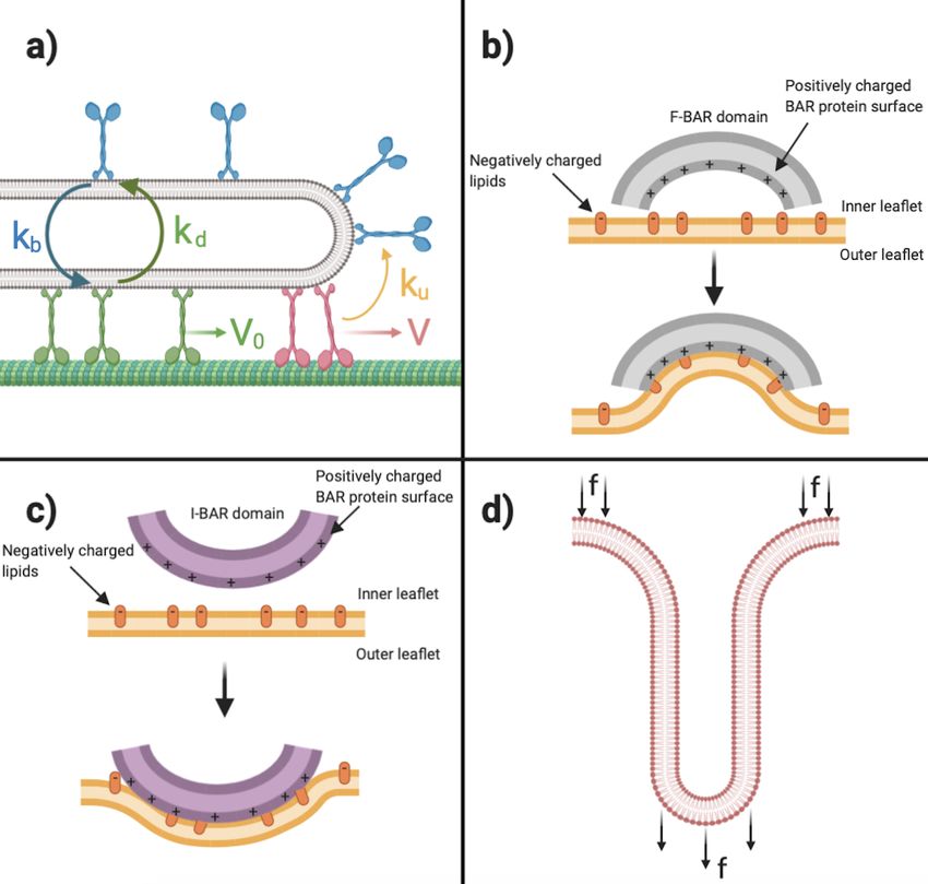
A PREPRINT - J ULY 16, 2020

eukaryotic cells, we find numerous applications of tubular protrusions at the plasma membrane. A motile cell uses the
actin-dense tubular structure, filopodia, to probe the environment during migration [6]. Filopodia also play a crucial
role in neurite growth, the formation of dendritic spines, wound healing, cellular trafficking [7]. They also lead to
cellularization in Drosophila embryo [8] and adhesion of epithelial cells during embryo development [9]. Tubular
protrusions from the plasma membrane also aid in the trafficking of cargoes (through transport carriers) [10] and
transport of ions through tubular cores (t-tubules) [11].
Furthermore, beyond the plasma membrane, organelle membranes, such as ER and the Golgi apparatus can generate
complex and dynamic tubular protrusions [12, 13]. The generality of tube formation in vesicle based systems, protein
crowding [14], liquid-liquid phase separation [15, 16], osmotic pressure [17], polymer binding [18] and even triblock
copolymers [19] indicate that there are multiple ways to induce the compressive stresses associated with membrane
tube formation.
A critical aspect of research in the area of membrane mechanics is the close interactions between theoretical develop-
ments and experimental observations. Indeed, for nearly five decades, the iterative development of theory, simulation,
and experiment has resulted in a rich field. In that spirit, we review some key highlights of tube formation in different
experimental systems (Section 2), the associated mechanical models to explain these observations (Section 3), and
the thermodynamic underpinnings of tube formation (Section 4). We conclude with some critical open questions for
future studies and suggest new interdisciplinary efforts in Section 5.

2     Experimental observations of membrane tubes

2.1     Tubular protrusion in cells and their myriad functions

Tubular membrane are ubiquitously found on the plasma membrane and intracellular organelles and these tubules are
implicated in a variety of cellular functions including membrane trafficking, cell migration, signaling, and probing the
extracellular environment. These tubular structures are found in all eukaryotic cells. We present a few examples of
these tubules to elaborate on their detailed structure and function relationships (Figure 1).

2.1.1    Tubular formation at the plasma membrane

Filopodia: Filopodia are finger-like cellular protrusions that play a crucial role in many cellular processes such as
cell migration, axon, and dendrite formation in neural growth, wound healing, and adhesion to the extracellular ma-
trix. This structure is mainly supported by a branch of actin filaments that also controls the length and elongation of
the filopodia with the help of regulated polymerization and depolarization of actin monomers [20]. Filaments from
the lamellipodial actin network can elongate and therefore filopodial protrusion happens with the help of ’convergent
elongation’ [21] process. The plasma membrane plays an important role in the formation of filopodia; the actin fil-
ament nucleating proteins (formins, Arp2/3, spire, etc.) bind to the membrane and induce polymerization of actin
filaments, which eventually causes tubular protrusion [22]. Additionally, there are instances of membrane deformation
in filopodial-like precursors in the dendritic spine where filopodial structure forms without an actin array [23]. Com-
mon to the spectrum of different scenarios that induce filopodial formation is the interaction of the plasma membrane
with a variety of regulating proteins that play an important role in the initiation and the regulation of the filopodial
geometry.

Tubule formation during membrane trafficking: Eukaryotic cells have multiple internal organelles, each of which
has specific function. Proteins and lipids are transported from one compartment to another through the membrane-
bound organelles, called transport carrier (TC) [24, 25]. TCs can be made of small vesicles, single tubes, or complex
tubular membrane structures. In particular, the tubule-shaped TC can transport large cargo over longer distances when
compared to vesicular TCs [10]. The mechanism of the tubular TC formation includes three basic steps– budding of
the membrane loaded with the cargo from the donor membrane, tube elongation and tubular fission, and finally, fusion
to the acceptor membrane [26]. Membrane scaffolding proteins help this tubular protrusion at the beginning from the
donor site and subsequently support the elongation of the tube [27]. Similar tubular elements are responsible for the
transport of cargoes through the endocytic pathway [28]. According to Sens et al. [27], membrane nanotubes in vitro
can also be used to observe the role of membrane curvature in several processes such as membrane trafficking. Kwok
and colleagues demonstrated the formation of tubular membrane protrusions in ternary mixtures of sphingomyelin,
phosphatidylcholine and cholesterol. Kwok et al. [29] noted nearly four decades ago that there exists a possibility that
membrane curvature and phase separation depend on lipid sorting.

                                                           2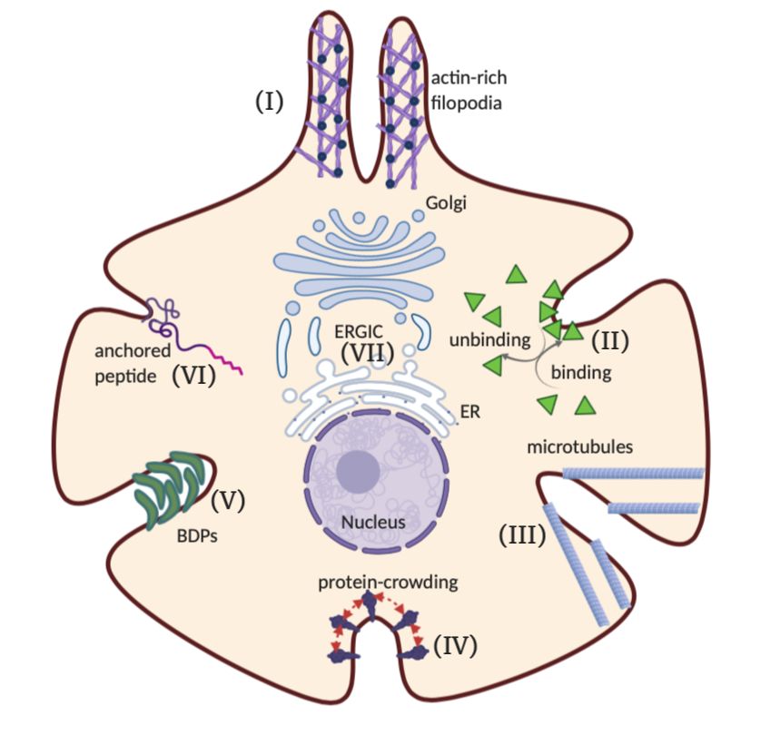
A PREPRINT - J ULY 16, 2020

Figure 1: Mechanisms of membrane tubulation in cell and intracellular organelles. (I) Actin-driven filopodial protru-
sion, (II) Tubular protrusion due to force generation caused by binding/unbinding of proteins to the membrane, (III)
Tubular structure supported by microtubules in the cytoskeleton, (IV) Tubular shape transformation of the membrane
due to steric effect of crowded proteins, (V) Spontaneous tubulation of membrane due to anisotropic intrinsic curvature
induced by BDPs, (VI) Tubulation due to anchored motor protein or peptides, and (VII) Tubular TC during membrane
trafficking in ERGIC.

2.1.2   Tubule formation in intracellular organelles

In Golgi-ER complexes ERGIC: In mammalian cells, protein cargo is transported from the ER to Golgi through a
tubulovesicular cluster of the membrane, which is often called as ER-Golgi intermediate compartment (ERGIC) [30].
This tubular structure is extremely dynamic in nature and works as a mobile transport complex that delivers cargoes
from the ER to Golgi [31]. The complexity of transport in ERGIC ranges from transport through a vesicle with a
coat protein complex (COPI and COPII) [32] to the movement of the large carrier along microtubule with the help
of TCs [24] and AC [33] that contains fusion protein from ERES. Microtubules in the cytoskeleton interact with the
tubular membrane and regulate these dynamics with the help of motor proteins in the early secretory pathway [34, 35].
However, forces from the motor proteins alone are unable to overcome the initial energy barrier of tubular protrusion
[36]; tubulation happens in the presence of GTPase and other curvature generating agents [37]. The ERGIC transport
machinery also contains the SNARE-complex [38] and other tethering proteins [39] that help with transporting the
multiprotein complex.

2.1.3   Select functions of tubules in whole cells

We focus on some select functions of tubular structures in whole cells based on some of our emerging research
interests. While not exhaustive, these functions give us some context on how the shape of the membrane tubule is
closely tied to cellular function.

                                                          3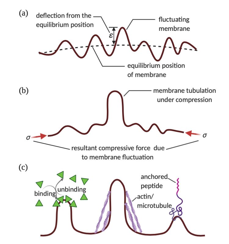
A PREPRINT - J ULY 16, 2020

Cardiac T-tubules: T-tubules are the tubular structures that present in the skeletal muscle cells and cardiac my-
ocytes and play a major role in muscle contraction. In cardiac myocytes, these tubular structures invaginate from the
sarcolemma and are organized along the z-disc surrounding the myofilaments [11]. The t-tubules are organized in the
close proximity to the SR and assist in the rapid entry of Ca2+ from there to the z-discs through its tubular core [40].
The LTCC on the membrane of the t-tubule stays in contact with RyRs of the SR membrane and forms dyads [41] that
help to stabilize the tubular structure. This spatial organization of the calcium handling units in the cardiac myocytes
is thought to be important for the spatiotemporal dynamics of calcium in these cells [42, 43].
The tubular morphology of t-tubules is found to be dynamic in nature and loss of tubules can occur in many disease
state [44] and can result in delayed kinetics of CICR. Even though the t-tubule structure dedifferentiates completely
in vitro, the studies have confirmed that the tubular structure does not protrude as a result of forces applied to the
membrane [45]. Furthermore, these tubules are found absent in the stem cell regeneration for cardiac myocyte [46],
which suggests that the mechanism of t-tubule formation is yet to be completely understood. Many studies suggest that
the BDP BIN1 that attaches to the dyad [47, 48, 49, 50] is crucial to the formation of the tubular structure, indicating
that curvature generating proteins play an important role in the formation of t-tubules.

In Neurons: Another excitable cell type, where the formation of tubules plays an important role is neurons. Neu-
ronal precursors undergo a series of morphological changes through tubular protrusions in multiple stages before they
develop into a mature neuron [51]. Early stages in these steps include the formation and elongation of smaller length
scale filopodial and lamellipodial structures [52]. Many of the filopodial protrusions further elongate in a longer length
scale with the help of actin-rich growth cone and form neurites [52]. In subsequent stages, one of the neurites under-
goes further rapid elongation and develops into the axon, whereas the remaining neurites become dendrites. The final
stage consists of forming early dendritic spine (locations of synaptic contact) and axonal branches, which are protru-
sions at a smaller length scale. Neuritogenesis, the process of neurite formation, is largely an actin-driven membrane
deformation and the process happens in coordination with the actin cytoskeleton and membrane scaffolding proteins.
The tubular geometry along with their electric property efficiently transmits the signals received from synaptic input
to other cells [53].
Membrane tubule formation is also important at the small length scale for neuronal function. Dendritic spines are
small scale (Length∼1–5 µm) protrusions along a dendrite that are sites of signal input from a neighboring neuron.
Similar to t-tubule, the tubular structure in spines is also very dynamic in nature and changes both with age and
excitatory stage [54]. The early spines are made of long and highly motile filopodial structures that seek a synaptic
partner[55]. Eventually, the long filopodia develop into a dendritic spine if the synaptic pathway strengthens and
firings of neurons occur [56]. These spines undergo structural changes with afferent input and in many cases disappear
from the old location [57]. This remodeling of spine morphology, known as structural plasticity, causes strengthening
and weakening of the structure of dendritic spines, which contributes to memory and learning [58].
The growth cone, as mentioned earlier, is the actin-rich filopodial structure that elongates from the early filopodial
structure to mature neurite and often produces a neural circuit in the brain [59]. The growth cone is very motile in
nature and constitutes of three major structural regions– actin enriched peripheral domain often known as P-domain,
central domain consisting of organelles and microtubule, and a transition domain where actin interacts with micro-
tubules [60]. The entire structure flows and elongates with the same rate of axon elongation with the help of PEC
mechanism [61]. Thus, the plasma membrane plays pivotal roles in the structure and motility of the growth cone by
assisting actin polymerization, receptor trafficking, recycling and turnover of membrane surface area, and adhesion to
the extracellular environment [62].

Development and Cellularization: Cellularization is the process that produces cell membranes for each nucleus
in Drosophila embryo after they undergo mitotic division. During this nuclear division, the plasma membrane is
covered with many finger-like small protrusions, known as microvilli [63]. At the same time, many cleavages and
furrows occur in the plasma membrane, which eventually propagate and form compartments [8]. Microvilli contain
much of the membrane that is required for furrow ingression in early cellularization [63]. Additionally, the furrow
canals contain proteins such as Myosin 2, Anillin, F-actin which actively control the compartmentalization process
[64]. Figard et al. [63] stated that since pulling forces of furrow ingression induce high plasma membrane tension;
this tension can be sufficient to limit and/or stall actin polymerization at microvillar tips. The interaction between the
plasma membrane, trafficking machinery, and force generating machinery is thought to be critical for the process of
cellularization. Taking these arguments into account, we note that microvilli unfolding depends on (a) interaction of
the plasma membrane with BDPs; (b) interaction of the plasma membrane with actin filaments; and (c) membrane
tension through regulation of furrow invagination and membrane trafficking.

                                                            4
A PREPRINT - J ULY 16, 2020
2.2 Tubule formation using forces and membrane-protein interaction
In this section, we focus on how the observations of tubule formation in cells can be studied in experiments with
reconstituted systems to identify the biophysical mechanisms involved. Synthetic and reconstituted systems such as
GUVs are useful systems to study the biophysical interactions of membranes and curvature inducing components
in a systematic manner. These systems also help to build iterative feedback between mathematical modeling and
experimental observations [65].
2.2.1 Membrane forces and tubes
In a synthetic system using GUVs and optical tweezers, tubular protrusions can be generated by the forces exerted
on membranes by motor proteins [66]. By using constant suction pressure, Shao and colleagues held an anti-CD162
or anti-CD45-coated bead at the micropipette tip. After the cell was moved toward the bead, a constant pulling force
was applied on experimental system. Shao et al. [67] revealed that tubular protrusions can be generated when point
force is applied on their tips. Similarly, Xu et al. [68] modeled the tube pulling force by examining the cell motion by
piezoelectric stage holding the micropipette and reported that localized forces applied to the membrane are sufficient
to generate tubules. Evans et al. [69] and Heinrich et al. [70] studied the detachment dynamics of P-selectin from
PSGL-1 and observed an elastic-like deformation in the initial stage of tube formation.
Koster et al. [71] hypothesized a mechanism in which the individual motor proteins can dynamically form clusters of
motor proteins and these can apply force to generate tubular protrusion. Due to the liquid nature of the bilayer, motor
proteins can diffuse laterally on the vesicle and Klopfenstein et al. [72] argued that certain kinesin motor proteins
can bind to lipids directly and they can induce dynamic preclustering mechanism. Koster et al. [71] measured these
forces with optical tweezers and revealed that these forces depend on membrane parameters such as membrane tension
and membrane bending ridigity. By varying the number of motors of the vesicle and by varying the properties of the
membrane such as tension, Koster et al. [71] observed that multiple motors have to cooperate to generate tubular
protrusion. These studies highlighted that the dynamic association of motor proteins with the cytoskeleton plays a
significant role in the cellular context of formation of tubular protrusions and in control of the structure of intracellular
membrane structures.
Another important environmental stressor known to form tubular protrusions is osmotic pressure. Sanborn and col-
leagues generated osmotic gradients by exposing giant vesicles to sucrose and concentration gradients. The induced
osmotic gradient was either positive or negative; the negative osmotic gradient can be induced by entrapping pre-
designated concentrations of sugar within the vesicular compartments during electroformation [17]. This gradient
was utilized to drive hydrodynamic water influx in the vesicular interior. This behavior gave rise to an osmotically
generated membrane tension resulting into cylindrical protrusions [17]. On the other hand, positive osmotic gradient
generated giant cylindrical vesicles that show pearling-like behavior and transient pearling state leads to stable net-
work of interconnected, spherical daughter vesicles [17]. In both cases, Sanborn et al. [17] observed that water influx
causes dynamic behavior of osmotic pressure which characterizes the dynamics of the tubular formations.
There are several examples that illustrate the role of membrane tension and force in tubular protrusion formation (Fig-
ure 2d). As cells store membrane in surface reservoirs of pits and protrusions, the formation of membrane reservoirs at
the cell surface depends on membrane physical properties such as bending elasticity, membrane tension and membrane
proteins such as BDPs [73].
2.2.2 Tubule formation from membrane-protein interaction
In this section, we focus on observations in reconstituted systems for curvature generation by proteins’ interaction
with the bilayer. Several protein classes, such as motor proteins of the dynein and kinesin families can mediate the
interactions of membranes with microtubules (Figure 2a) [74]. According to [75, 76], in vivo and in vitro microtubule-
based motor activity are both required in BFA-induced tubulation of Golgi membranes. Endophilin, amphiphysin, and
the GTPase dynamin have a role in membrane fission events, and these proteins can directly bind to membranes with
lipid binding domains. Such proteins can also generate tubular protrusions from liposomes in vitro [77, 78, 79]. More
recently, Busch et al. [80] studied the curvature generating role of endocytic adaptor proteins Epsin1 and AP180 in
the membrane. Stachowiak and colleagues studied tubular protrusion formations with protein densities on membrane
surfaces by exposing GUVs to wtENTH. Stachowiak et al. [81] showed that tubular protrusions are generated by
lateral pressure generated by collisions between bound proteins and steric congestion on cellular membranes.
Stachowiak and colleagues revealed that protein crowding on lipid domain surfaces forms a protein layer that buckles
outward. This buckling bends the domain into stable buds and tubules spontaneously. Lipid domains can confine
protein binding on vesicle surfaces and protein binding can generate buds and tubular protrusions by using two global
parameters: domain size and membrane tension. Stachowiak et al. [14] demonstrated how steric interactions between
5A PREPRINT - J ULY 16, 2020
proteins and lipids can induce membrane bending. Coat proteins such as clathrin can generate bud protrusions and
BDPs can generate tubular protrusions [82]. More recently, Stachowiak and colleagues showed that liquid-liquid phase
separation on the surface of the membrane can lead to the formation of tubules [16].
Protein-induced membrane bending generates the curvature of clathrin-coated pits and caveolae. During clathrin-
mediated endocytosis, epsin family proteins can insert amphipathic helices in the cytoplasmic membrane leaflet [14].
The structured clathrin coat concentrates epsins leading to membrane budding [3]. It was also hypothesized that
caveolins deform the bilayer through application of steric pressure [83]. To explore the interaction between protein-
lipid domain interactions in membrane protrusions, Stachowiak and colleagues generated a model system using GUVs
and revealed that domains can concentrate protein binding interactions, which can lead to the formation of buds and
tubular protrusions. Stachowiak et al. [14] observed that tubular protrusion formation depends on the presence of fluid-
phase lipids in the domain and requires a high density of protein attachment. These experiments led to a quantitative
observation that tubule length has a linear relationship with vesicle diameter and a specific protein structure is not a
requisite for tubular protrusion formation.
As curvatures that are required to generate tubular protrusions also emerge through mechanical forces from localized
activities of motor proteins, and cytoskeleton [19], and reconstitution, localization, and crowding of membrane proteins
affect the tubular protrusion formation [84]. Kamioka et al. [85] established the FBP17-dynamin interaction and the
role of FBP17 in tubular protrusion formation during dynamin-mediated endocytosis. Snead et al. [86] demonstrated
membrane fission by dynamin and revealed the tubular protrusion formation upon exposure to curvature-inducing
proteins. Roux et al. [87] revealed that tubular protrusions and complex tubular networks can be generated from
GUVs by microtubules and ATP. Moreover, Roux and colleagues demonstrated that bundles of tubes can be generated
if tubes connect at different points to the same microtubule and demonstrated that motor proteins are prerequisite
elements for generating tubular protrusions [87]. Girard et al. [88] investigated the role of protein content in tubular
protrusion formation during the reconstitution of membrane proteins into GUVs. As lipid domains are transformed
into tubular protrusions by protein binding, Stachowiak and colleagues reported the synthesis and membrane behavior
of a lipid-like molecule, DPIDA, and studied the role of lipid membrane composition in tubular protrusion formation.
Stachowiak et al. [89] used lipid membrane composition with thermal treatment to control the structure of tubular
protrusions and observed that steric interactions of surface-bound proteins can transform the lipid domains into tubular
protrusions. Also, Leduc et al. [90] conducted experiments on dynamics of motors and tube growth, and observed
tubular protrusions in vitro by kinesins that are in contact with GUVs and microtubules, establishing the role of
membrane tension and motor density in tubular protrusion formation.
6A PREPRINT - J ULY 16, 2020
Figure 2: (a) Schematic of a growing tubular protrusion (brown) along a microtubule (green). The motors are attached
to the membrane and they can be either bound (green and purple) or unbound (blue) from the microtubule. When
bound motors far from the tip (green), they move with velocity V0 and detach at a rate kd . Unbound motors reattach
to the tube at a rate kb . The bound motors at the tip (red) detach at a rate ku . The tube growth velocity is V . (b)
Illustration of binding mechanism of F-BAR domain protein (grey) to a lipid bilayer (yellow) that generate membrane
invagination. (c) Illustration of binding mechanism of I-BAR domain protein (purple) to a lipid bilayer (yellow) that
generate membrane exvagination. (d) Tubular protrusion formation by forces that are exerted by cytoskeleton.
7A PREPRINT - J ULY 16, 2020
3 Mechanics of tube formation
The formation of tubular protrusions on membranes can be understood by considering the balance of forces on the
membrane. We note that the mechanics approach is valuable for both equilibrium and dynamic configurations. The
fundamental feature underlying many of these models is the elastic nature of the lipid bilayer. The lipid bilayer is a
thin elastic sheet, fluid in plane but solid in bending. As a result, there have been significant advances in theoretical
developments in the field of membrane mechanics [4, 5, 91, 92, 93, 94, 95, 96, 97]. We summarize them here in the
context of membrane tube formation.
3.1 Helfrich energy for membrane mechanics
The Canham-Helfrich energy [98, 99] is commonly utilized for modeling the elastic bending energy of lipid bilayers
in membrane mechanics. This model proposes that the strain energy of a lipid bilayer can be written as a function
of the surface curvatures and the minimization of this energy will give us the equilibrium shapes of the membrane
[99, 100, 101] Subsequently, this model has been adapted for modeling the behavior of proteins that form spherical
coats by inducing an isotropic spontaneous curvature [5]. The strain energy per unit area is given by
w = κ(H − C)2 + κG K [99] (1)
The total energy of the membrane is then given by
Z
W = (κ(H − C)2 + κG K)dA [99] (2)
A
where κ is the bending modulus, H is the mean curvature of the membrane (average of the two principal curvatures),
C is the spontaneous curvature, κG is the Gaussian modulus, K is the Gaussian curvature (product of the two principal
curvatures) and A is the total membrane surface area. We should point out that for Equation 1 and Equation 2, strain
energy per unit area (w) and total energy of the membrane (W ) differ from the classical Helfrich model [99] by a
factor of 2, which was compensated by using the value of bending modulus to be twice that of the standard bending
modulus typically measured in the literature.
3.2 Tubule formation using forces and tension
A classic result using the Helfrich energy for membrane tube dimensions and how they are related to the applied forces
were presented in [4]. We briefly summarize it here to demonstrate the utility of mechanical models in predicting
quantitative relationships between the applied force and the tubule radius. Derenyi et al. [4] studied the membrane
pulling with the point force f and showed that the total membrane energy can be expressed as
E = πκL/r + 2πσrL − f L [4] (3)
where σ is the membrane tension, p is the transmembrane pressure, r is the radius of tubular protrusion and L is the
length of tubular protrusion. Minimizing the energy of a tubular protrusion with respect to r and L yields
∂E
= −πκL/r2 + 2πσL = 0 (4)
∂r
∂E
= πκ/r + 2πσr − f = 0 (5)
∂L
pκ √
Therefore, the equilibrium tube radius is given by 2σ and the static force to hold the tube is 2π 2σκ.
Separately, the role of membrane tension and lipid flow was explored in substantial detail by Hochmuth and colleagues
in a series of papers inspired by their experimental measurements. Considering that a majority of cellular membranes
consist of two monolayers, Hochmuth et al. [102] formed a tubular protrusion from a cell body by using micropipette
aspiration. Additionally, Hochmuth et al. [102] reported that tube pulling force is affected by membrane surface
viscosity, the slip viscosity between the two monolayers, and the viscosity between membrane and cytoskeleton.
Leduc and colleagues studied a biomimetic system which involves GUVs, kinesin-1 motors and microtubules in the
presence of ATP. Leduc et al. [103] presented both theoretical and experimental results based on fluorescence mi-
croscopy that elucidate the dynamics of membrane tube formation, growth and stalling. The results demonstrate that
8A PREPRINT - J ULY 16, 2020
molecular motors are able to pull membrane tubes without the aid of other proteins, and that tube formation should de-
pend on both motor protein density and membrane tension. Evans [104] modeled a micropipette aspiration experiment
for flaccid red cell and reported that minimum energy cell membrane contours is directly related to pipette suction
force. Also, Hochmuth and colleagues formed tubular protrusion formations by pulling tubes from RBCs by using mi-
cropipette aspiration. They modeled the tube as a thin shell and demonstrated that there is an inverse relation between
radius of tubular protrusion and axial tension [105, 106]. Furthermore, Waugh [107] conducted a micropipette aspira-
tion experiment in which the lipid membrane bilayer was subjected to fluid drag and noted that osmotic properties of
the vesicle interior do not have a contribution to tube formation.
3.3 Modeling the interaction of membranes with BDPs in tubule formation
Anisotropic components play a significant role in membrane morphology. Spherical buds require a fixed contact angle
on the lipid membrane along a circular contact line [108]. On the other hand, for tubular protrusion forming proteins
such as BDPs, the contact line with the planar membrane is highly anisotropic [108]. Anisotropic components generate
a spontaneous deviatoric curvature, which aids in the formation of a cylinder [109].
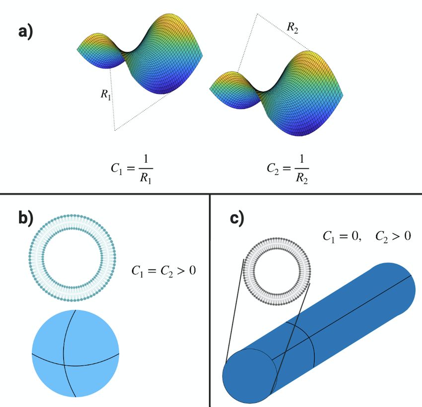
To model a tube formation, we note that for a cylindrical shape (Figure 3c), unlike a spherical shape (Figure 3b),
normal curvature along longitudinal axis is different from the normal curvature along the circumferential direction
[110]. Spontaneous curvatures generated by tubule forming proteins, such as BDPs, are inherently anisotropic in
nature.
It follows that the use of the isotropic spontaneous curvature model is insufficient for capturing the shapes of tubules
and the relationship between tubule dimensions and protein densities on the membrane surface. To address this issue,
a membrane strain energy density that captures the anisotropic curvature was proposed by many groups [5, 91, 97,
109, 111]. This modified Helfrich Model was used for modeling the behavior of proteins that form tubular protrusions
and induce an anisotropic curvature. The energy per unit area in this case is written as
w= κ(H − C)2 + κ(D − D0 )2 (6)
| {z } | {z }
Elastic effects Deviatoric effects
Here, H is the mean curvature as before and D captures the difference between the two principal curvatures. D0 is
the spontaneous deviatoric curvature. The total energy of the membrane is calculated as
Z
2 2
W = κ (H − C) + (D − D0 ) dA. (7)
A
There are several applications that use deviatoric curvature model to enhance our understanding of tube formations.
Bobrovska et al. [92] and Alimohamadi et al. [91] modeled tube formation by using deviatoric curvature model to
implement the effects of membrane elements and attached proteins with anisotropic properties. By using deviatoric
curvature, Iglič and colleagues generated anisotropy bending energy model for anisotropic membranes. During their
analysis of the stability of tubular protrusion formations, Iglič et al. [112, 113] used deviatoric curvature model and
observed that anisotropic membrane components play an important role in stability of tubular protrusion formations.
Also, Iglič et al. [114] and Kabaso et al. [115] studied deviatoric curvature model to demonstrate that BDPs generate
and stabilize tubular protrusion formations.
BDPs have an intrinsic curvature that provides these proteins the ability to bind to the membrane surface and bend the
membrane in what is known as the scaffold mechanism [97]. An additional mechanism that has been proposed for
BDP induced tubulation is the amphipathic wedge mechanism, which proposes that curvature is induced as a buckling
response to the insertion of amphipathic sequences into the leaflet of the bilayer [82]. The adhesion of F-BAR domain
protein to the lipid bilayer induces positive curvature (Figure 2b), while the adhesion of the I-BAR domain protein to
the lipid bilayer induces negative curvature (Figure 2c) [116]. These features can be captured by the curvature deviator
model.
3.4 Current state of the art and future needs in dynamic measurements of tube formation in lipid membranes
Thus far, we have focused on the equilibrium aspects of membrane tubule formation. We now turn our attention
to the dynamic measurements of tubule formation. Dynamic measurements of tube formation in lipid membranes
9A PREPRINT - J ULY 16, 2020
Figure 3: (a) R1 and R2 are principle radii of a hyperbolic paraboloid surface and C1 and C2 are principal curvatures
of a hyperbolic paraboloid surface. (b) Principal curvatures of a sphere. (c) Principal curvatures of a tube.
can be achieved using optical tweezers; such optical tweezers are used to characterize the mechanical properties
of plasma membrane in terms of tether formation [117]. According to [117], compared to other tether formation
techniques, optical tweezers provide noninvasive manipulation of cells with comparably great force resolution (∼ 0.1
pN) and provide continuous monitoring of instantaneous tether force. Indeed, there is no dearth of data for dynamic
measurements of tubule formation [93, 94, 95, 96, 102, 118, 119].
There are also have been several models of the dynamics of tubule protrusion. Simunovic and colleagues modeled
the dynamics of tube formation by mimicking the tubular protrusion formation. This is done by pulling membrane
nanotubes from GUVs using optical tweezers [96]. They constructed their model based on certain balance laws
involving parameters such as external force, tube area, change in tube area, tube length, change in tube length, and
membrane tension. Simunovic et al. [96] combined their model with in vivo and in vitro experiments and demonstrated
that motors provide tube pulling force as well as frictional force for both tube formation and BAR domain scaffold.
Hochmuth and colleagues developed a thermodynamic analysis of tether formation process and they developed exper-
iments which can be used to analyse neuronal growth cones. Hochmuth et al. [102] demonstrated that during dynamic
measurements, membrane viscosity is one of the important considerations since it determines the rate of membrane
deformation and it influences diffusion rate of particles in the surface plane [107]. Tian et al. [119] and Sorre et al.
[118] examined the role of membrane curvature in lipid and protein sorting. Their models revealed that spontaneous
10A PREPRINT - J ULY 16, 2020
curvature in tube formations contributes to curvature sorting and this curvature sorting can be achieved by cooperative
operation of lipids. Also, Sorre et al. [118] generated a model which demonstrates that curvature-induced lipid sorting
is generated by the lipid cooperativity and can be influenced by lipid-clustering proteins.
Separately, based on experiments conducted in a multilamellar lipid system with osmotic pressure as a driver, Ranga-
mani and colleagues developed a model including fluid drag, transmembrane pressure, and membrane tension along a
tubular protrusion. The model predicted that the three stages during tubular protrusion are initiation, elongation, and
termination. Based on experimental data Rangamani et al. [95] constructed a mathematical model that can predict the
tubular protrusion growth. They reported that their force balance approach can explain the elongation phase of tubular
protrusion and that confinement-based tubule growth system is regulated by osmotic pressure and drag.
Their simple force balance approach has also been used to explain the dynamics of elongation of acrosomes [93] and
neurite retraction [94]. The applications of this model to different processes have revealed that the viscoelasticity of
the membrane and membrane tension are significant factors in governing the dynamic behavior of membranes. In
certain cases, model predictions were verified experimentally [94].
A comprehensive class of models incorporating surface lipid viscosity has also been developed [95, 120, 121, 122,
123]. Specifically, Rahimi et al. [121] built a continuum model that can explain the behavior of a bilayer configuration
and its internal dissipative mechanisms. Using lipid hydrodynamics with shape dynamics and the Helfrich-Canham
energy approach, they developed a dynamical model of tubular protrusion and generated dynamics by modeling the
system as a constrained optimization problem. Their continuum model shows that the viscoelastic behavior of tube
formation depends on membrane bending elasticity and inter-monolayer friction.
4 Thermodynamic considerations of tube formation
Thus far, we have discussed the mechanical considerations of tube formation in lipid bilayers. The applied forces and
membrane-protein interactions are also influenced by thermodynamic considerations and will be briefly discussed in
what follows.
4.1 Role of thermal fluctuations in tubule formation
Lipid bilayers are moderately soft compared to Boltzmann energy (kB T ) at physiological temperatures. As a result of
this, they undergo shape undulations due to the thermal movement of the fluid molecules in the surrounding domain
(Figure 4a). Experiments have reported the observations of membrane fluctuations in vesicles [124, 125, 126]. These
undulations cause mechanical softening of the membrane [127] and can influence shape instabilities in the bilayer
[128].
There are a series of theoretical studies [129, 130, 131] and Monte-Carlo simulations [132] that have reported that
thermal fluctuations soften the membrane to a significant amount and also reduce local tension of the membrane. It
was proposed that the effective bending rigidity in the presence of thermal fluctuations can be written as [130]
3 qmax
κ(T, λ, a) = κ0 − kB T ln , (8)
4π qmin
and the effective tension is given by
3kB T 2 2
σ(T, a) ' − (qmax − qmin ), (9)
8
where qmin and qmax are the magnitude of maximum and minimum wave numbers of the undulations.
It is worth mentioning that the equipartition of energy limits the energy of each undulation mode. Thus, the magni-
tude of the deflection correlates inversely with the square of the wavenumber of that particular mode of undulation.
Further, the ratio of these wavenumbers correlated with the maximum and minimum size of the wavelengths (λ) of the
undulations as
qmax λmax
= . (10)
qmin λmin
The highest value of the wavelength (λmax ) is of the order of the size of the membrane (L), whereas the least value of
it scales with the diameter of the lipid molecules (a).
Considering Equation 8 and the fact that thermal softening is directly correlated with the size of the domain, the role of
fluctuations can become prominent on a larger length scale. In contrast, for a lower length scale, the effect of thermal
fluctuation will be negligible. Thus, the persistent length ξ below which the membrane behaves as a rigid surface is
given by [131] [133]
11A PREPRINT - J ULY 16, 2020
ξ = ξ0 exp (4πκ0 /3T ) . (11)
The changes in physical properties of the membrane resulting from the effect of thermal fluctuations can facilitate
shape instabilities, many of which lead to the formation of tubular protrusions [128]. For low surface tension mem-
branes, the shape undulation generates a negative tension and thus inserts a compression in the plane of the membrane.
As a result of this compression, the membrane undergoes a buckling instability resulting in the formation of a tubule
out of the plane (Figure 4b). Such tubular structures have been observed in many experiments [134, 135]. Further-
more, the shape undulations alter the binding probability of the molecules from the surrounding fluid [136], which
confer additional surface area on the membrane and impose compressive stresses that support tubulation.
The coupling between shape fluctuations and membrane-protein interactions can result in the clustering of proteins on
the membrane surface due to in-plane attraction [137]. These protein clusters can lead to tubulation of the membrane
by means of a steric effect [14] or by spontaneous tubulation [138].
4.2 Thermodynamics of protein binding, aggregation, and phase separation
The coupling between membrane mechanics and the thermodynamics of the membrane protein interactions results
in thermophysical phenomena such as aggregation of proteins, separation of protein and lipid phase, binding and
unbinding of proteins to the membrane, etc. Proteins that do not interact with one another prefer a homogeneous
distribution in the lipid bilayer to maximize the entropy of the system [139]. However, proteins that interact with
each other can experience a net attraction force among themselves and form a cluster [15]. Additionally, due to the
difference in chemical composition from the lipid, the protein-coated region forms a separate phase on the lipid bilayer.
The unbalanced force in the transition region induces line tension that drives formation of a cluster of the same phase
energetically favorable [140, 141, 142, 143].
Theoretically, the effect of aggregation can be modeled by incorporating an aggregation potential in addition to the
membrane bending energy [144, 145, 146]. Furthermore, binding and unbinding of proteins to the membrane and
adhesion of the proteins on the membrane can decrease the free energy of the system and are energetically favor-
able [147]. Each of these thermophysical phenomena influences membrane bending and is conversely dependent on
membrane curvature created by bending (Figure 4c). Veksler and Gov [148] presented a detailed theoretical model of
filopodial protrusion where they considered the effect of protein adhesion, the force due to actin polymerization, and
membrane tension separately on aggregation. They observed that force due to polymerization increases the critical
temperature, whereas, adhesion strength and tension decrease the critical temperature for transition.
Curvature plays a significant role in aggregation and phase segregation. We discussed in Section 3.3 how BDPs insert
anisotropic curvature to the membrane and induce spontaneous tubulation. Further, BDPs are the flexible rod-like
proteins that undergo elastic deformation in addition to inducing membrane curvature. The energy for elastic bending
of the BDPs is thus dependent on the membrane curvature and hence minimizes the total energy with a preferable
distribution [113]. Moreover, the anisotropic curvatures of proteins also induce an orientation entropy in the system.
A series of studies [92, 112, 113, 149, 150] modeled the thermodynamics of BDP interaction with the membrane
by considering the energy of bending of both the membrane and the BDPs along with the entropy for configuration
and orientation of BDPs. These studies suggest that BDP undergoes curvature induced aggregation that eventually
results in a tubular protrusion of the membrane, corresponding to the minimum energy state of the system and the
orientational entropy favors this process [150].
Another thermodynamic effect that influences tubulation of the membrane is protein crowding. Protein crowding is the
phenomenon that is associated with a high concentration of proteins in the lipid bilayers. When such macromolecules
adsorb onto the membrane, they tend to interact among themselves with steric effects, and often result in tubulation
of membranes [14, 151]. Stachowiak et. al. [14] demonstrated this kind of tubular protrusion as membrane tension
dominated, and proposes a physical model for the estimation of membrane tension (λ) as the function of protein-lipid
binding energy (∆G)
3∆GAD
λ≈ , (12)
AP
where AD is the fractional area of protein domain and AP is the fractional binding area of protein. They further
assumed that membranes with crowded proteins undergo area dilation under this high tension. Such crowded proteins
can collide with each other and generate a lateral pressure [152] that can lead to tubulation. This crowding pres-
sure, when sufficiently large, can also facilitate membrane fission [153, 154]. Darganc and Copic [155] theoretically
modeled the curvature generation due to crowding pressure and estimated a spontaneous curvature as a function of
12A PREPRINT - J ULY 16, 2020
Figure 4: (a) A fluctuating membrane with deflection ε from the equilibrium position, (b) Membrane tubulation
under compressive force caused by thermal fluctuation, (c) Tubulation due to active forces – binding and unbinding
of proteins, pulling of actin, microtubules and motor proteins in cytoskeleton, and force induced by anchored and
tethered proteins.
difference of crowding pressure between two monolayers, given by
δpC
C0 = , (13)
`
where δpC is the difference between crowding pressure between two leaflets of lipid bilayer and ` is the modified length
scale evaluated as the ratio of the projected area proteins to their height. Further, the crowding pressure is modeled
in the same fashion as thermodynamic gas pressure, which encounters the effect of collision between the proteins.
This process of curvature generation with a crowding pressure is also considered as one of the basic thermodynamic
mechanisms of curvature generation in protein-lipid interface [156, 157].
13A PREPRINT - J ULY 16, 2020
5 Future perspectives and open questions
In the previous sections, we have elaborated on how cell-based experiments, model systems, and mechanical models
have focused on the problem of membrane tubulation. Here, we discuss certain new avenues for this area of research
and how we might be able to bridge some of the gaps between mechanics and cell biology.
From a modeling standpoint, we need to develop models that take multiple physics into account. We are seeing
an increase in extensions of models of membrane bending that go beyond the classical descriptors of spontaneous
curvature, and include other features such as lipid viscosity and protein diffusion [95, 120, 123, 158]. However, these
models need to be brought closer to the experimental observations. A challenge that lies ahead is the development
of numerical methods that are robust [159, 160]. An additional opportunity lies in bridging molecular dynamics
simulations to continuum mechanics simulations to build a truly multiscale model [161].
From an experimental standpoint, increasing the resolution of quantitative measurements in time and space in GUV
based systems (e.g. protein density, tubule radius, surface coverage) would provide invaluable data to constrain the
free parameters in model development process. Of course, as discussed earlier, dynamic measurements of the tubule
formation process are critical for informing the relevant timescales in the models.
The next opportunity, in our view, lies in the gaps between models built for synthetic or purified systems and models
for cellular processes. For instance, Shtengel et al. [162] built an iPALM, a simultaneous multiphase interferometry
that provides both molecular specification and resolution of cellular nanoarchitecture. Thus, there is an opportunity for
the modeling community to interact more closely to work with large experimental data sets to identify the key physics
underlying these processes. Finally, we would like to iterate that there are many opportunities that call for truly
interdisciplinary collaborations with open science approaches that can help us gain more insight into the fundamental
process of tube formation in cellular membranes.
Acknowledgments
The authors would like to thank their many collaborators in the field of membrane mechanics for discussing ideas
and the organizers of the International Symposium on Cell Surface Macromolecules 2020 for engaging discussions.
They would also like to acknowledge Haleh Alimohamadi, Elizabeth Heyde, Matt Akamatsu, and Ali Behzadan for
providing their critical comments and feedback for the manuscript. This work was supported by NIH R01GM132106
to P.R.
References
[1] Haleh Alimohamadi and Padmini Rangamani. Modeling membrane curvature generation due to membrane–
protein interactions. Biomolecules, 8(4):120, 2018.
[2] Michael C Heinrich, Benjamin R Capraro, Aiwei Tian, Jose M Isas, Ralf Langen, and Tobias Baumgart. Quan-
tifying membrane curvature generation of drosophila amphiphysin n-bar domains. J. Phys. Chem. letters,
1(23):3401–3406, 2010.
[3] Marijn GJ Ford, Ian G Mills, Brian J Peter, Yvonne Vallis, Gerrit JK Praefcke, Philip R Evans, and Harvey T
McMahon. Curvature of clathrin-coated pits driven by epsin. Nature, 419(6905):361–366, 2002.
[4] Imre Derényi, Frank Jülicher, and Jacques Prost. Formation and interaction of membrane tubes. Phys. Rev.
Lett., 88(23):238101, 2002.
[5] Nikhil Walani, Jennifer Torres, and Ashutosh Agrawal. Anisotropic spontaneous curvatures in lipid membranes.
Phys. Rev. E, 89(6):062715, 2014.
[6] Catherine G Galbraith, Kenneth M Yamada, and James A Galbraith. Polymerizing actin fibers position integrins
primed to probe for adhesion sites. Science, 315(5814):992–995, 2007.
[7] Pieta K Mattila and Pekka Lappalainen. Filopodia: molecular architecture and cellular functions. Nat. rev. Mol.
Cell Biol., 9(6):446–454, 2008.
[8] Anna Marie Sokac and Eric Wieschaus. Local actin-dependent endocytosis is zygotically controlled to initiate
drosophila cellularization. Dev. Cell, 14(5):775–786, 2008.
[9] Valeri Vasioukhin, Christoph Bauer, Mei Yin, and Elaine Fuchs. Directed actin polymerization is the driving
force for epithelial cell–cell adhesion. Cell, 100(2):209–219, 2000.
14A PREPRINT - J ULY 16, 2020
[10] Frédéric Bard and Vivek Malhotra. The formation of tgn-to-plasma-membrane transport carriers. Annu. Rev.
Cell Dev. Biol., 22:439–455, 2006.
[11] TingTing Hong and Robin M Shaw. Cardiac t-tubule microanatomy and function. Physiol. Rev., 97(1):227–252,
2017.
[12] Christopher Lee and Lan Bo Chen. Dynamic behavior of endoplasmic reticulum in living cells. Cell, 54(1):37–
46, 1988.
[13] HH Mollenhauer and D James Morré. The tubular network of the golgi apparatus. Histochemistry and cell
biology, 109(5-6):533–543, 1998.
[14] J. C. Stachowiak, C. C. Hayden, and D. Y. Sasaki. Steric confinement of proteins on lipid membranes can drive
curvature and tubulation. Proc. Natl. Acad. Sci. U.S.A. U.S.A., 107(17):7781–7786, 2010.
[15] Benjamin S Schuster, Ellen H Reed, Ranganath Parthasarathy, Craig N Jahnke, Reese M Caldwell, Jessica G
Bermudez, Holly Ramage, Matthew C Good, and Daniel A Hammer. Controllable protein phase separation and
modular recruitment to form responsive membraneless organelles. Nat. Commun., 9(1):1–12, 2018.
[16] Feng Yuan, Haleh Alimohamadi, Brandon Bakka, Andrea N. Trementozzi, Nicolas L. Fawzi, Padmini Ranga-
mani, and Jeanne C. Stachowiak. Membrane bending by protein phase separation. 2020.
[17] Jeremy Sanborn, Kamila Oglecka, Rachel S Kraut, and Atul N Parikh. Transient pearling and vesiculation of
membrane tubes under osmotic gradients. Faraday Discuss., 161:167–176, 2013.
[18] Fèlix Campelo and Aurora Hernández-Machado. Polymer-induced tubulation in lipid vesicles. Phys. Rev. Lett.,
100(15):158103, 2008.
[19] Seng Koon Lim, Andrew SW Wong, Hans-Peter M de Hoog, Padmini Rangamani, Atul N Parikh, Madhavan
Nallani, Sara Sandin, and Bo Liedberg. Spontaneous formation of nanometer scale tubular vesicles in aqueous
mixtures of lipid and block copolymer amphiphiles. Soft Matter, 13(6):1107–1115, 2017.
[20] Matthew D Welch and R Dyche Mullins. Cellular control of actin nucleation. Annu. Rev. Cell Dev. Biol.,
18(1):247–288, 2002.
[21] Tatyana M Svitkina, Elena A Bulanova, Oleg Y Chaga, Danijela M Vignjevic, Shin-ichiro Kojima, Jury M
Vasiliev, and Gary G Borisy. Mechanism of filopodia initiation by reorganization of a dendritic network. J. Cell
Biol., 160(3):409–421, 2003.
[22] Elif Nur Firat-Karalar and Matthew D Welch. New mechanisms and functions of actin nucleation. Curr. Opin.
Cell Biol., 23(1):4–13, 2011.
[23] Yuko Sekino, Nobuhiko Kojima, and Tomoaki Shirao. Role of actin cytoskeleton in dendritic spine morpho-
genesis. Neurochem. Int., 51(2-4):92–104, 2007.
[24] David J Stephens and Rainer Pepperkok. Illuminating the secretory pathway: when do we need vesicles? J.
Cell. Sci., 114(6):1053–1059, 2001.
[25] JENNIFER LIPPINCOTT-SCHWARTZ. Dynamics of secretory membrane trafficking. Annals of the New York
Academy of Sciences, 1038(1):115–124, 2004.
[26] Roman S Polishchuk, Mariagrazia Capestrano, and Elena V Polishchuk. Shaping tubular carriers for intracel-
lular membrane transport. FEBS lett., 583(23):3847–3856, 2009.
[27] Pierre Sens, Ludger Johannes, and Patricia Bassereau. Biophysical approaches to protein-induced membrane
deformations in trafficking. Curr. Opin. Cell Biol., 20(4):476–482, 2008.
[28] P J Cullen. Endosomal sorting and signalling: an emerging role for sorting nexins. Nat. rev. Mol. cell biol.,
9(7):574–582, 2008.
[29] R Kwok and Evan Evans. Thermoelasticity of large lecithin bilayer vesicles. Biophys. J., 35(3):637–652, 1981.
[30] Hans-Peter Hauri and Anja Schweizer. The endoplasmic reticulumgolgi intermediate compartment. Curr. Opin.
Cell Biol., 4(4):600–608, 1992.
[31] Heinrich Horstmann, Chee Peng Ng, Bor Luen Tang, and Wanjin Hong. Ultrastructural characterization of
endoplasmic reticulumgolgi transport containers (egtc). J. Cell. Sci., 115(22):4263–4273, 2002.
[32] Meir Aridor, Sergei I Bannykh, Tony Rowe, and William E Balch. Sequential coupling between copii and copi
vesicle coats in endoplasmic reticulum to golgi transport. J. Cell Biol., 131(4):875–893, 1995.
[33] Houchaima Ben-Tekaya, Kota Miura, Rainer Pepperkok, and Hans-Peter Hauri. Live imaging of bidirectional
traffic from the ergic. J. Cell. Sci., 118(2):357–367, 2005.
15A PREPRINT - J ULY 16, 2020
[34] D J Stephens. Functional coupling of microtubules to membranes–implications for membrane structure and
dynamics. J. Cell. Sci., 125(12):2795–2804, 2012.
[35] Gerbrand Koster, Martijn VanDuijn, Bas Hofs, and Marileen Dogterom. Membrane tube formation from giant
vesicles by dynamic association of motor proteins. Proc. Natl. Acad. Sci. U.S.A., 100(26):15583–15588, 2003.
[36] Gerbrand Koster, Angelo Cacciuto, Imre Derényi, Daan Frenkel, and Marileen Dogterom. Force barriers for
membrane tube formation. Phys. Rev. Lett., 94(6):068101, 2005.
[37] Anna Bielli, Charles J Haney, Gavin Gabreski, Simon C Watkins, Sergei I Bannykh, and Meir Aridor. Reg-
ulation of sar1 nh2 terminus by gtp binding and hydrolysis promotes membrane deformation to control copii
vesicle fission. J. Cell Biol., 171(6):919–924, 2005.
[38] Yu A Chen and Richard H Scheller. Snare-mediated membrane fusion. Nat. Rev. Mol. Cell Biol., 2(2):98–106,
2001.
[39] Alison K Gillingham and Sean Munro. Long coiled-coil proteins and membrane traffic. Biochim. Biophys.
Acta, Mol. Cell. Res., 1641(2-3):71–85, 2003.
[40] Alexandre Fabiato. Calcium-induced release of calcium from the cardiac sarcoplasmic reticulum. American
Journal of Physiology-Cell Physiology, 245(1):C1–C14, 1983.
[41] Heping Cheng, WJ Lederer, and Mark B Cannell. Calcium sparks: elementary events underlying excitation-
contraction coupling in heart muscle. Science, 262(5134):740–744, 1993.
[42] Alexandre Fabiato. Calcium-induced release of calcium from the cardiac sarcoplasmic reticulum. Am. J.
Physiol. Cell Physiol., 245(1):C1–C14, 1983.
[43] Heping Cheng, W Jonathan Lederer, and Mark B Cannell. Calcium sparks: elementary events underlying
excitation-contraction coupling in heart muscle. Science, 262(5134):740–744, 1993.
[44] William E Louch, Ole M Sejersted, and Fredrik Swift. There goes the neighborhood: Pathological alterations in
t-tubule morphology and consequences for cardiomyocyte ca++. BioMed Research International, 2010, 2010.
[45] A. Di Maio, K. Karko, Rose M. Snopko, Rafael Mejı́a-Alvarez, and Clara Franzini-Armstrong. T-tubule for-
mation in cardiacmyocytes: two possible mechanisms? J. Muscle Res. Cell. Motil., 28(4-5):231–241, 2007.
[46] Deborah K Lieu, Jing Liu, Chung-Wah Siu, Gregory P McNerney, Hung-Fat Tse, Amir Abu-Khalil, Thomas
Huser, and Ronald A Li. Absence of transverse tubules contributes to non-uniform ca2+ wavefronts in mouse
and human embryonic stem cell–derived cardiomyocytes. Stem cells and development, 18(10):1493–1500,
2009.
[47] TingTing Hong, Huanghe Yang, Shan-Shan Zhang, Hee Cheol Cho, Mariya Kalashnikova, Baiming Sun, Hao
Zhang, Anamika Bhargava, Michael Grabe, Jeffrey Olgin, et al. Cardiac bin1 folds t-tubule membrane, con-
trolling ion flux and limiting arrhythmia. Nat. Med., 20(6):624, 2014.
[48] Avery D Posey Jr, Kaitlin E Swanson, Manuel G Alvarez, Swathi Krishnan, Judy U Earley, Hamid Band, Peter
Pytel, Elizabeth M McNally, and Alexis R Demonbreun. Ehd1 mediates vesicle trafficking required for normal
muscle growth and transverse tubule development. Dev. Biol., 387(2):179–190, 2014.
[49] Min Wu, Bo Huang, Morven Graham, Andrea Raimondi, John E Heuser, Xiaowei Zhuang, and Pietro
De Camilli. Coupling between clathrin-dependent endocytic budding and f-bar-dependent tubulation in a cell-
free system. Nat. Cell Biol., 12(9):902–908, 2010.
[50] Margaret Husta Butler, Carol David, Gian-Carlo Ochoa, Zachary Freyberg, Laurie Daniell, Detlev Grabs, Ot-
tavio Cremona, and Pietro De Camilli. Amphiphysin ii (sh3p9; bin1), a member of the amphiphysin/rvs family,
is concentrated in the cortical cytomatrix of axon initial segments and nodes of ranvier in brain and around t
tubules in skeletal muscle. J. Cell Biol., 137(6):1355–1367, 1997.
[51] Stefanie Kaech and Gary Banker. Culturing hippocampal neurons. Nat. Protoc., 1(5):2406, 2006.
[52] Carlos G Dotti, Christopher A Sullivan, and Gary A Banker. The establishment of polarity by hippocampal
neurons in culture. J. Neurosci., 8(4):1454–1468, 1988.
[53] JOHN P Miller and GWEN A Jacobs. Relationships between neuronal structure and function. J. Exp. Biol.,
112(1):129–145, 1984.
[54] Michael E Dailey and Stephen J Smith. The dynamics of dendritic structure in developing hippocampal slices.
J. Neurosci., 16(9):2983–2994, 1996.
[55] John C Fiala, Marcia Feinberg, Viktor Popov, and Kristen M Harris. Synaptogenesis via dendritic filopodia in
developing hippocampal area ca1. J. Neurosci., 18(21):8900–8911, 1998.
16You can also read