What's a Biofilm?-How the Choice of the Biofilm Model Impacts the Protein Inventory of Clostridioides difficile - Frontiers
←
→
Page content transcription
If your browser does not render page correctly, please read the page content below
ORIGINAL RESEARCH
                                                                                                                                                  published: 10 June 2021
                                                                                                                                         doi: 10.3389/fmicb.2021.682111

                                             What’s a Biofilm?—How the Choice
                                             of the Biofilm Model Impacts the
                                             Protein Inventory of Clostridioides
                                             difficile
                                             Madita Brauer 1 , Christian Lassek 1 , Christian Hinze 1 , Juliane Hoyer 2 , Dörte Becher 2 ,
                                             Dieter Jahn 3 , Susanne Sievers 1 and Katharina Riedel 1*
                                             1
                                               Department for Microbial Physiology and Molecular Biology, Institute of Microbiology, University of Greifswald, Greifswald,
                                             Germany, 2 Department for Microbial Proteomics, Institute of Microbiology, University of Greifswald, Greifswald, Germany,
                                             3
                                               Braunschweig Integrated Centre of Systems Biology (BRICS), Institute of Microbiology, Technische Universität
                                             Braunschweig, Braunschweig, Germany

                                             The anaerobic pathogen Clostridioides difficile is perfectly equipped to survive and
                                             persist inside the mammalian intestine. When facing unfavorable conditions C. difficile
                                             is able to form highly resistant endospores. Likewise, biofilms are currently discussed
                                             as form of persistence. Here a comprehensive proteomics approach was applied to
                                             investigate the molecular processes of C. difficile strain 6301erm underlying biofilm
                            Edited by:       formation. The comparison of the proteome from two different forms of biofilm-
                        George Grant,        like growth, namely aggregate biofilms and colonies on agar plates, revealed major
               University of Aberdeen,
                                             differences in the formation of cell surface proteins, as well as enzymes of its energy and
                      United Kingdom
                                             stress metabolism. For instance, while the obtained data suggest that aggregate biofilm
                       Reviewed by:
                    Thomas Candela,          cells express both flagella, type IV pili and enzymes required for biosynthesis of cell-
      Université Paris-Saclay, France        surface polysaccharides, the S-layer protein SlpA and most cell wall proteins (CWPs)
                     Anthony Buckley,
 University of Leeds, United Kingdom         encoded adjacent to SlpA were detected in significantly lower amounts in aggregate
                   *Correspondence:          biofilm cells than in colony biofilms. Moreover, the obtained data suggested that
                      Katharina Riedel       aggregate biofilm cells are rather actively growing cells while colony biofilm cells most
             riedela@uni-greifswald.de
                                             likely severely suffer from a lack of reductive equivalents what requires induction of the
                    Specialty section:       Wood-Ljungdahl pathway and C. difficile’s V-type ATPase to maintain cell homeostasis.
         This article was submitted to       In agreement with this, aggregate biofilm cells, in contrast to colony biofilm cells, neither
                   Infectious Diseases,
               a section of the journal
                                             induced toxin nor spore production. Finally, the data revealed that the sigma factor
             Frontiers in Microbiology       SigL/RpoN and its dependent regulators are noticeably induced in aggregate biofilms
           Received: 17 March 2021           suggesting an important role of SigL/RpoN in aggregate biofilm formation.
            Accepted: 12 May 2021
           Published: 10 June 2021           Keywords: biofilm, colony biofilm, aggregate biofilm, Clostridioides difficile, proteomics, cell surface antigens,
                                             RpoN signaling
                             Citation:
        Brauer M, Lassek C, Hinze C,
Hoyer J, Becher D, Jahn D, Sievers S
          and Riedel K (2021) What’s
                                            INTRODUCTION
  a Biofilm?—How the Choice of the
   Biofilm Model Impacts the Protein
                                            In recent years, the anaerobic gastrointestinal pathogen Clostridioides difficile has established itself
   Inventory of Clostridioides difficile.   as one of the major causative agents of pseudomembranous colitis and toxic megacolon (Wiegand
         Front. Microbiol. 12:682111.       et al., 2012; Nasiri et al., 2018; Martínez-Meléndez et al., 2020; Doll et al., 2021). C. difficile does not
    doi: 10.3389/fmicb.2021.682111          only produce enterotoxins that damage the gastrointestinal epithelium (Fletcher et al., 2021) but

Frontiers in Microbiology | www.frontiersin.org                                      1                                             June 2021 | Volume 12 | Article 682111
Brauer et al.                                                                                                      Protein Inventory of C. difficile Biofilms

also forms easily transmittable spores that significantly contribute         and persistence behavior in rodent models (Barketi-Klai et al.,
to C. difficile’s efficient spreading in the environment, hospitals          2011; Dawson et al., 2012; Ðapa et al., 2013; Batah et al., 2017;
and elderly homes (McLure et al., 2019; Hernandez et al.,                    McKee et al., 2018a; Slater et al., 2019). Corresponding cell-
2020; Werner et al., 2020; Khader et al., 2021). If conditions               cell aggregates were observed in a CDI murine model and
are favorable, e.g., after antibiotic-induced dysbiosis when the             multispecies biofilms have been shown to be a reservoir for
reduced microbiome fails to convert primary bile acids into                  C. difficile spores in triple-stage chemostat human gut model
secondary bile acids, C. difficile spores are able to germinate              (Lawley et al., 2012; Soavelomandroso et al., 2017; Normington
to successively colonize the large intestine (Theriot et al., 2014;          et al., 2021). While these data clearly indicate that biofilm
Pike and Theriot, 2020). Even though antibiosis mostly stops                 formation is indeed an important virulence factor our knowledge
the acute infection, C. difficile spores as well as some vegetative          on the physiology of C. difficile biofilms is still scarce. Moreover, it
cells survive in the intestine and can subsequently cause a                  remains to be determined whether in vitro biofilm model systems
relapse as soon as antibiotic concentrations are sufficiently low            sufficiently resemble in vivo biofilm formation. Currently, in vitro
(Goulding et al., 2009; Chilton et al., 2018; Castro-Córdova et al.,         C. difficile biofilms are often studied in plastic microtiter plates,
2020; Feuerstadt et al., 2021; Normington et al., 2021). Initially,          where cells initially attach to the surface, followed by a maturation
sporulation was assumed to be one of the major prerequisites for             of the biofilm and a final detachment of cell-cell-aggregates
C. difficile’s persistence under clinical circumstances. However,            (Dawson et al., 2012; Ðapa et al., 2013; Dubois et al., 2019).
recent publications failed to correlate sporulation efficiency of a          Similar biofilms have been observed in continuous-flow micro-
certain strain with the corresponding virulence and persistence              fermenters (Poquet et al., 2018). Alternatively, colony biofilms on
potential, i.e., highly infectious strains do not necessarily produce        agar plates and related biofilm models have been used (Crowther
more spores than other strains (Sirard et al., 2011; Oka et al., 2012;       et al., 2014; Semenyuk et al., 2014; James et al., 2018). Inconsistent
Plaza-Garrido et al., 2015; Gómez et al., 2017). In conclusion,              results from in vitro experiments suggest that biofilm physiology
persistence does obviously not solely rely on sporulation but                strongly depends on the chosen strain, growth conditions and
also on additional features of C. difficile (Smits, 2013). In this           phase as well as the experimental setup (Ðapa et al., 2013;
context, biofilm formation was proposed to be a major additional             Maldarelli et al., 2016; Mathur et al., 2016; Pantaléon et al., 2018).
factor (Dawson et al., 2012; Ðapa et al., 2013). Surface-associated          For example, differences in flagella production and contribution
biofilms, consisting of multiple microorganisms embedded in                  to biofilm formation has been reported for different strains
a slimy extracellular matrix, represent a form of community                  and phases of biofilm formation (Ðapa et al., 2013; Maldarelli
lifestyle for many bacteria (Hansen et al., 2007; Oliveira et al.,           et al., 2016). Analogously, a comprehensive RNA-seq approach
2015; Ren et al., 2015). Many nosocomial infections caused                   revealed that the expression levels of some genes which had
by Staphylococcus aureus, Streptococci and numerous other                    previously been linked to biofilm formation were significantly
pathogens rely on biofilms (Jamal et al., 2018). In this context,            different between C. difficile colony biofilms grown on agar plates
biofilm formation has been frequently linked to pro-longed                   and biofilms formed on glass beads (Maldarelli et al., 2016).
infection and persistence (Burmølle et al., 2010; Jamal et al.,                  Considering the strong evidence that C. difficile colonizes
2018). An important feature of biofilms is their extracellular               the gut in a biofilm-like manner a profound molecular
matrix, which mostly consists of extracellular DNA (eDNA),                   characterization of C. difficile biofilms is crucial, e.g., for the
lipids, proteins and polysaccharides. The extracellular matrix               targeted development of therapeutics and vaccines. Since most
provides protection against chemical and mechanical stress and               vaccines are directed against cell-surface exposed structures,
confers resistance to therapeutics and the host immune system                the comprehensive characterization of C. difficile’s cell-surface
(Karygianni et al., 2020). Anaerobic bacteria, including C. difficile        proteins and polysaccharides in biofilms is of particular
have also been found to produce biofilms in vitro, and biofilm-like          importance (Bruxelle et al., 2017; Kirk et al., 2017a; Péchiné et al.,
structures have been observed on the intestinal mucosal surface              2018; Bradshaw et al., 2019). Here, we applied a comparative
of C. difficile infected mice (Donelli et al., 2012; Lawley et al.,          proteomics approach to investigate the proteome repertoire of
2012; Soavelomandroso et al., 2017).                                         C. difficile strain 6301erm grown either as aggregate or colony
    Initial biofilm studies using C. difficile revealed mechanisms           biofilm to (i) identify proteins contributing to biofilm formation
of formation and involved components comparable to those                     of one or the other growth condition and (ii) elucidate the
determined for other bacteria (Dawson et al., 2012; Vuotto et al.,           underlying regulatory networks.
2018). For instance, the involvement of adhesion-mediating cell
surface structures, such as flagella, pili and various adhesion
molecules, has been reported (Reynolds et al., 2011; Ðapa et al.,            MATERIALS AND METHODS
2013; Pantaléon et al., 2015). Similarly, c-di-GMP signaling,
quorum sensing, and regulators, such as Spo0A and CodY, are                  Bacterial Strains and Media
involved in coordination of the process of biofilm formation                 The reference strain C. difficile 6301erm (DSM28645) (Hussain
(Ðapa et al., 2013; Purcell et al., 2016; McKee et al., 2018b;               et al., 2005; van Eijk et al., 2015) was grown at 37◦ C in
Dubois et al., 2019). Knockout mutants of some genes encoding                an anaerobic workstation (Toepffer Lab Systems, Germany) in
certain proteins commonly linked to biofilm formation, such                  Brain Heart Infusion (BHIS; Oxoid (Thermo Fisher Scientific),
as adhesion and cell signaling proteins, were found impaired                 Waltham, MA) supplemented with L-cysteine (0.1%(wt/vol),
in biofilm formation in vitro and revealed a reduced infection               Sigma-Aldrich, St. Louis, MO), and yeast extract (5 mg/ml; Carl

Frontiers in Microbiology | www.frontiersin.org                          2                                        June 2021 | Volume 12 | Article 682111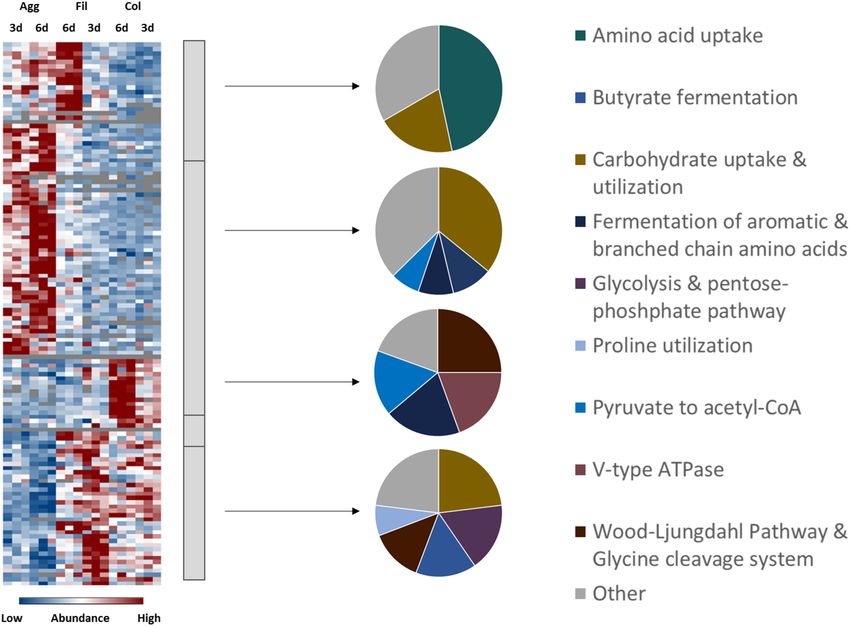
Brauer et al.                                                                                                 Protein Inventory of C. difficile Biofilms

Roth, Germany) as described earlier (Ðapa et al., 2013). Prior            Cell debris was removed by centrifugation at 6,000 × g for
to cultivation, spores were allowed to germinate for 72 h in              20 min at 4◦ C. 200 µl of the resulting lysates were precipitated
BHIS medium. Subsequently, the germinated cells were used                 by ice-cold acetone (in a 1:7 ratio v/v) for 20 h at −20◦ C.
to inoculate pre-cultures which were grown for 18 h in BHIS               Subsequently, samples were allowed to warm up at RT and were
medium. For planktonic growth main cultures were inoculated               centrifuged at 22,000 × g for 45 min at RT. The supernatant was
to an optical density of 0.05 at 600 nm.                                  discarded and the pellets were dried at RT. The protein pellets
                                                                          were solubilized in 100 µl of an SDS-containing urea-buffer (7 M
Growth of Colony and Aggregate                                            urea, 2 M thiourea, 1% SDS v/v). In order to estimate the relative
                                                                          protein concentration, 10 µl of each sample was mixed with
Biofilms                                                                  SDS-loading buffer and separated by SDS-PAGE (Criterion TGX
The growth of colony biofilms was performed as previously                 Precast Gels 12%, Biorad, Hercules, CA, Laemmli, 1970). Protein
described with some modifications (Semenyuk et al., 2014).                gels were fixed for 1 h at RT (40% EtOH, 10% glacial acidic acid
Briefly, BHI medium was supplemented with yeast extract and               and 50% H2 O), washed in H2 O and stained by the Flamingo
agarose (1.5% v/v) and autoclaved for 15 min. Subsequently,               fluorescent dye (Biorad, Hercules, CA) for 1 h at RT. Remaining
sterile-filtered cysteine in BHI medium was added to the medium,          dye was removed by a washing step in H2 O and fluorescence
mixed and transferred to petri dishes. Plates were allowed to dry         signals of the samples were obtained by Typhoon scanner (GE
for 24 h in the anaerobic chamber. Prior to cultivation, a filter         Healthcare, Little Chalfont, United Kingdom). The fluorescence
membrane with a pore size of 0.45 µm (cellulose ester, Sigma-             signals of the gel image were quantified by ImageQuant (Biorad,
Aldrich, St. Louis, MO) was placed on top of the BHIS agar plate.         Hercules, CA). These fluorescent signal intensities were used
Per plate 100 µl of C. difficile-containing BHIS medium were              for normalization to load comparable protein amounts (30 µg
placed on top of the filter membrane. The BHIS medium was                 of protein per sample) on the final SDS-Gel. For each sample,
inoculated by an exponentially grown C. difficile main culture            the entire gel lane was cut into 10 gel blocks and proteins were
(OD600nm : ∼0.5) in a 100-fold dilution. Plates were wrapped              digested in-gel with trypsin as follows: the excised gel pieces were
with parafilm in order to protect them from drying. The colony            destained using 50% (v/v) methanol in 100 mM NH4 HCO3 .
biofilms were grown for three and six days at 37◦ C in the                Subsequently, gel pieces were dehydrated using 100% ACN and
anaerobic environment.                                                    allowed to dry. Modified trypsin (sequencing grade, Promega,
   Aggregated biofilms were cultivated as previously reported             Germany) was added to a final ratio of 1:10 (trypsin/sample)
with some modifications (Dawson et al., 2012). Briefly, sterile           in 50 mM Tris/HCl, pH 7.5 and the sample incubated at 37◦ C
6-well plates (polystyrene, Corning, NY) were filled with 2 ml            overnight. Peptides were iteratively extracted from the gel by a
C. difficile-containing BHIS medium. The BHIS medium was                  four-step procedure, using ACN, 5% (v/v) formic acid in ddH2 O
inoculated by an exponentially grown C. difficile main culture            and further two steps of ACN. Peptide-containing supernatants
(OD600nm : ∼0.5) in a 100-fold dilution. The 6-well plates were           were pooled and completely dried using a Speedvac concentrator
placed in a plastic bag to avoid evaporation of the medium.               (Eppendorf AG). Samples were subsequently resolved in buffer
Cells were grown for three and six days at 37◦ C in the                   A (5% (v/v) ACN, 0.1% (v/v) formic acid) and desalted using
anaerobic environment.                                                    ZipTips (C18, Millipore). Desalted peptides were again vacuum-
                                                                          dried and stored at −20◦ C. Finally, the samples were solubilised
Cell Harvest and Protein Extraction                                       in 10 µL 0,1% acetic acid solution and transferred into vials
Planktonically-grown cells were harvested after 6 and 12 h of             for MS-analysis.
cultivation at 6.000 × g for 20 min and pellets were stored
at −80◦ C. Filter membranes with colony biofilms were placed
into 15 ml reaction tubes and stored at −80◦ C. In order to               Mass Spectrometric Measurement and
separate aggregated cells from free-living planktonic cells the           Data Analysis
culture-medium was carefully removed from the 6-well plates               Samples were subjected to LC-MS/MS measurements using an
and pooled (one 6-well plate = one biological sample). The pooled         EASYnLC 1,000 (Thermo Fisher Scientific, Odense, Denmark)
culture-medium was filtered through a 10 µm filter (IsoporeTM             with self-packed columns [(Luna 3 µC18(2) 100A, Phenomenex,
PC Membrane, Merck Millipore, Tullagreen, Ireland), washed                Germany)] in a one-column setup on-line coupled to an Orbitrap
with 5 ml of 0.9% NaCl (w/v) and subsequently the filter was              Elite (Thermo Fisher Scientific, Bremen, Germany) setting
stored at −80◦ C. The filtrate containing the planktonic cells was        parameters as previously described (Berges et al., 2018).
centrifuged at 6.000 × g and the cell pellet was stored at −80◦ C.            Database search and intensity based absolute quantification
    The samples (cell pellets of filtrate samples and membrane            (iBAQ) was achieved using the MaxQuant proteomics software
filters of both biofilm types) were kept in 0.6–1 ml of an                package (Cox and Mann, 2008; Tyanova et al., 2016a; version:
urea-containing buffer (7 M urea, 2 M thiourea, 50 mM                     1.6.10.43). A protein sequence database of C. difficile strain
dichlorodiphenyltrichloroethane (DDT), 4% (w/v) 3-[(3-                    6301erm containing 3781 entries was obtained from UniProt.
cholamidopropyl)          dimethylammonio]-1-propanesulfonate             Common laboratory contaminants and reverse sequences were
(CHAPS), 50 mM Tris-HCl). Cell lysis was performed by                     added by the MaxQuant software. Parameters were set as
sonication in six cycles on ice as done previously (Sonoplus,             follows: Trypsin cleavage with a maximum of two missed
Bandelin, Berlin, Germany, Otto et al., 2016; Berges et al., 2018).       cleavages was assumed and oxidation of methionine was set as

Frontiers in Microbiology | www.frontiersin.org                       3                                      June 2021 | Volume 12 | Article 682111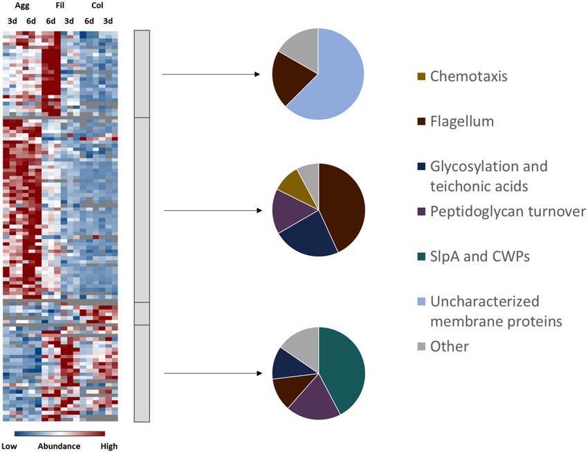
Brauer et al.                                                                                                               Protein Inventory of C. difficile Biofilms

variable modification. Default parameters were used for protein                excluded that the growth conditions that bacteria face during this
identification. For label-free protein quantification unique and               form of multicellular living are relevant during infection. Colony
razor peptides were considered with a minimum ratio count                      biofilms were directly analyzed after scraping them off the plates.
of 2. Match between runs was enabled with default settings                     Aggregate biofilms from 6-well plates were harvested by filtration
within each sample group. C difficile proteins were considered                 and cells that passed the filter were analyzed in parallel as non-
as identified if they were identified with at least two unique                 biofilm fraction. Previous studies investigating C. difficile biofilm
peptides in at least two out of three biological replicates. riBAQ             gene expression by RNA-seq used planktonically-grown cells as
values were calculated as published previously (Shin et al., 2013).            reference (Maldarelli et al., 2016; Poquet et al., 2018). Thus,
Averaged riBAQs were used to calculate log2 fold changes.                      planktonically-grown cells from exponential (6 h) and stationary
Proteins significantly altered in their abundance between two                  (12 h) growth phase were also included in this study (Figure 1).
conditions were identified by two-way analysis of variance                         Principal component analyses revealed that the various
(ANOVA) followed by a Tukey post hoc test using the Perseus                    proteome parameters obtained for every tested condition
software package (Tyanova et al., 2016b; version: 1.6.2.2). Cellular           clustered together within the biological replicates but revealed
localization of identified proteins was predicted by PSORTb (Yu                a clear cut separated distribution in dependence of the growth
et al., 2010; version: 3.0).                                                   condition and time (Supplementary Figure 1). This was also
                                                                               true for the data obtained for cells from the filtrate (sample C
Voronoi-Regulon Treemaps                                                       of Supplementary Figure 1) of the aggregate biofilms which
Global protein expression patterns were analyzed and visualized                clearly separated from the corresponding biofilm cells (sample
using Voronoi treemaps (Bernhardt et al., 2013) which were                     B of Supplementary Figure 1), although both types of cells
adapted to illustrate regulons of several well defined global                  experienced the same cultivation time and growth medium
regulators of C. difficile as described in the literature. For this            along with nutrient limitation and accumulated waste products.
purpose, data extracted from eight transcriptomic studies was                  We propose physiological differences between these cells to
used to generate a scaffold for the Voronoi-regulon-treemaps.                  be mainly attributed to cell aggregation/biofilm formation and
In these previous studies, regulons of fourteen global regulators              therefore used the filtrate samples as the reference for non-
were characterized, i.e., SigB (Kint et al., 2017), SigH (Saujet et al.,       biofilm conditions rather than the planktonic cells (sample A of
2011), SigD (El Meouche et al., 2013), SigL/RpoN (Soutourina                   Supplementary Figure 1) which were not further considered.
et al., 2020), CodY (Dineen et al., 2010), CcpA (Antunes et al.,                   Global hierarchical cluster analyses on all proteins that were
2012), Fur (Ho and Ellermeier, 2015), Hfq (Boudry et al., 2014),               found differentially expressed between the six remaining data
c-di-GMP (McKee et al., 2018b), Spo0A (Pettit et al., 2014), SigF,             sets using ANOVA (analysis of variance) yielded four major
SigE, SigG, and SigK (Fimlaid et al., 2013). The corresponding                 clusters A–D (Figure 2 and Supplementary Table 2). Cluster
regulons, comprising negatively as well as positively regulated                A contained proteins with higher abundance in 6-day old
genes, differ in size in the range of 30 to more than 400 genes                filtrate cells and consisted of a high proportion of membrane
(Table 1). In total, these transcriptional regulators modulate the
expression of 1252 different C. difficile genes. For the illustration
of the defined regulons each regulated gene was assigned to its                TABLE 1 | Selected regulators of C. difficile gene expression.
corresponding regulator(s). Additionally, the regulatory effect
                                                                               Regulator      Regulatory event               Regulon size References
(positive or negative) of a regulator on the expression of a specific
                                                                                                                               (genes)
gene is indicated by a plus or minus symbol. Finally, C. difficile
log2 fold changes of biofilm models compared to filtrate samples               SigB           General stress response              663          Kint et al., 2017
were mapped onto the regulon maps.                                             SigH           Transition phase                     490          Saujet et al., 2011
                                                                               SigD           Motility, toxin production           146          El Meouche et al.,
                                                                                                                                                2013
                                                                               SigL/RpoN      Amino acid catabolism                114          Soutourina et al.,
RESULTS AND DISCUSSION                                                                                                                          2020
                                                                               CodY           Regulation of metabolism             160          Dineen et al., 2010
A Comparative Proteomics Approach to                                           CcpA           Carbohydrate catabolism              313          Antunes et al.,
Investigate the Protein Repertoire of                                                                                                           2012
                                                                               Fur            Iron acquisition                     125          Ho and Ellermeier,
C. difficile Biofilms                                                                                                                           2015
In order to identify proteins required for and/or characteristic               Hfq            Posttranscriptional                  203          Boudry et al., 2014
of C. difficile biofilm formation the protein inventory of biofilm-                           (pleiotropic) regulation
and planktonically-grown bacteria was comparatively analyzed.                  c-di-GMP       Motility                             160          McKee et al.,
Two different types of biofilms were investigated: 1. colony                                                                                    2018b
biofilms on agar plates and 2. aggregate biofilms formed in 6-                 Spo0A          Sporulation                          297          Pettit et al., 2014
well plates. Although colonies on agar plates do not meet the                  SigF           Sporulation (forespore)              181          Fimlaid et al., 2013
traditional criteria of a biofilm, the densely packed cells likewise           SigE           Sporulation (mother cell)            164          Fimlaid et al., 2013
present a form of multicellular growth attached to a biotic surface            SigG           Sporulation (forespore)               34          Fimlaid et al., 2013
(Gingichashvili et al., 2017; Kesel et al., 2017) and it cannot be             SigK           Sporulation (mother cell)             30          Fimlaid et al., 2013

Frontiers in Microbiology | www.frontiersin.org                            4                                               June 2021 | Volume 12 | Article 682111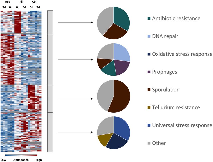
Brauer et al.                                                                                                                         Protein Inventory of C. difficile Biofilms

  FIGURE 1 | Experimental setup for a comparative proteomics approach to investigate biofilm formation in C. difficile. C. difficile strain 6301erm was grown as
  planktonic culture for 6 and 12 h (A), or as aggregate biofilm in six-well plates for 3 or 6 days (B) or as colony biofilm on solid growth medium for 3 or 6 days (D). In
  addition, cells from the filtrate of aggregate biofilms were analyzed (C). Cells were harvested and their protein repertoire was subsequently analyzed using a
  LC-MS/MS approach.

proteins (54%) while all other clusters contained approximately                           type IV pili proteins (i.e., PilT, PilB2, and PilM2) were detected
75% cytosolic proteins. In line with this, cluster A contained                            in higher concentrations in aggregate biofilms than in colony
transporters for cations and amino acids. The largest cluster B                           biofilms and filtrates. Similar behavior was observed for proteins
contained 607 proteins of high abundance in aggregate biofilms                            involved in cell wall biogenesis like enzymes of the mur operon
which were assigned to the functional categories “cell wall                               (i.e., MurB, MurE, MurG), enzymes involved in lipoteichoic acid
biosynthesis”, “cell surface polysaccharide biosynthesis”, “flagella                      synthesis (GtaB, GtaB1) and modification (DltA), enzymes of
biosynthesis”, “regulation and cell signaling”, and “amino acid                           the Cell Wall Glycopolymer (CWG) locus (CD2783 to CD2769),
and carbohydrate metabolism”. Cluster C was composed of 99                                and proteins of membrane-lipid biosynthesis encoded by the
proteins highly abundant in colony biofilms, and the 267 proteins                         fab operon (i.e., FabD, FabH, FabK). In contrast, the S-layer
of cluster D were found in higher abundance in colony biofilms                            protein SlpA and most CWPs (i.e., Cwp12, Cwp16, Cwp19,
and filtrate cells. Both clusters share a high proportion of cell-                        Cwp22, Cwp84) were of significantly lower abundance in
wall proteins including the S-layer protein SlpA and 12 of the                            aggregate biofilms.
28 cell wall proteins (CWPs) encoded in one operon with SlpA.
Additionally, “fermentation and energy generation” proteins of                            Flagella and Type IV Pili
cluster C were assigned to the Wood-Ljungdahl pathway and the                             While comparable RNA-seq based expression profiles were
corresponding glycine cleavage system, and to subunits of the                             observed for cell wall and S-layer biogenesis genes when
V-type ATPase, but also “sporulation”-related proteins such as                            aggregate biofilms and planktonic cells were compared (Poquet
CotA, CotB, and SipL were observed.                                                       et al., 2018), transcription of flagella genes has been found to
   In agreement with transcriptome data comparing biofilm and                             be reduced in aggregate and colony biofilms in comparison
non-biofilm cells (Maldarelli et al., 2016; Poquet et al., 2018),                         to planktonic cells of C. difficile (Maldarelli et al., 2016;
we identified genes encoding cell-surface exposed proteins, but                           Poquet et al., 2018). However, both transcriptomic studies
also genes from the categories “energy metabolism”, “stress                               were performed with biofilms grown in different setups and
response”, “virulence” and “regulation and cell signaling” as                             to different timepoints. Hence, flagella might be required for
biofilm signature.                                                                        aggregate biofilm growth in our rather static set up, but might be
                                                                                          obsolete or even obstructive for biofilm formation in continuous
Production of Cell Surface-Associated                                                     flow systems (Poquet et al., 2018) or for biofilms grown on glass
Proteins in C. difficile Biofilms                                                         beads (Maldarelli et al., 2016). As discussed above, somewhat
Adhesion to epithelial cells as well as cell-cell-aggregation                             contradictory observations have been obtained regarding the
is mainly mediated by cell-surface exposed proteins,                                      role of flagella during C. difficile biofilm formation (Ðapa et al.,
polysaccharides and cell appendices such as pili and flagella                             2013; Jain et al., 2017) that may either reflect the complexity
(Ðapa et al., 2013; McKee et al., 2018a; Arato et al., 2019).                             of the regulatory system underlying flagella expression in
Notably, proteins from the respective categories were found                               C. difficile (El Meouche et al., 2013; Soutourina et al., 2013;
differently expressed between colony and aggregate biofilms                               Anjuwon-Foster and Tamayo, 2017, 2018) and/or the different
(Figure 3 and Supplementary Table 3). Most flagella proteins                              functions of flagella that depend on their posttranslational
(i.e., FliE, FliF, FliG, FliH, FliM, FlgE, FlgG, FhlA, FhlF) and                          modification state (Twine et al., 2009; Faulds-Pain et al., 2014;

Frontiers in Microbiology | www.frontiersin.org                                       5                                              June 2021 | Volume 12 | Article 682111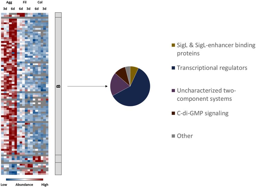
Brauer et al.                                                                                                                            Protein Inventory of C. difficile Biofilms

  FIGURE 2 | Hierarchical cluster analyses of C. difficile proteome data sets. The heatmap on the left side shows differentially abundant proteins between aggregate
  biofilms (Agg), filtrate cells (Fil), and colony biofilms (Col) from day three (3d) and day six (6d) for three biological replicates (Bio1-3). Z-transformed abundance values
  are displayed as a color gradient where blue colors indicate weakest abundance levels and red colors the strongest abundance levels among the growth conditions
  with maximum z-scores of +/–1.5. Hierarchical clustering revealed four major cluster (A–D) representing 6-day old filtrate cells cluster (A), aggregate biofilms (B),
  colony biofilms (C) or both, colony biofilms and filtrate samples (D). In the pie charts on the right the various clusters shown on left were further analyzed with regard
  to the subcellular location (middle: C for cytoplasmic; CM for cytoplasmic membrane; CW for cell wall; E for extracellular; U for unassigned). In addition, proteins
  within a cluster were grouped according to their function and the most prominent functional groups of each cluster are presented in the pie charts on the right (WLP
  for Wood-Ljungdahl pathway). A detailed list of all significantly differentially abundant proteins including fold changes can be found in Supplementary Table 2
  (Supplementary Material). Agg: Aggregate biofilms; Col: Colony biofilms; Fil: Filtrate samples; 3d: Samples from day three; 6d: Samples from day six; C,
  cytoplasmic; CM, cytoplasmic membrane; CW, cell wall; E, extracellular; U, unassigned; WLP, Wood-Ljungdahl pathway; CWPs, cell wall proteins.

Bouché et al., 2016; Valiente et al., 2016). Additionally, biofilms                         2016; McKee et al., 2018a). We found type IV pili-associated
are often composed of subpopulations (Vlamakis et al., 2008;                                proteins like PilT, PilB2 and PilM2 in aggregate biofilms and in
Besharova et al., 2016) and flagella in one or the other                                    lower amounts in filtrates but not in colony biofilms. Therefore,
modification state might be relevant for one subpopulation                                  our data suggest that aggregate biofilms in contrast to colony
and obstructive for the other. Alterations of flagella post-                                biofilms obviously utilized glycosylated flagella and certain pili
translational modifications were shown to impact motility                                   components during their formation.
and cell-cell-aggregation properties of C. difficile (Faulds-Pain
et al., 2014; Valiente et al., 2016). Significant upregulation of                           Cell Surface Glyco-Polymers and Teichoic Acids
C. difficile’s conserved glycosyltransferase CDIF630erm_00362 in                            Proteins from the PSII locus for the synthesis of the teichoic-
conjunction with the flagella proteins in aggregate biofilms in                             acid-like cell-surface polysaccharide II (e.g., CDIF630erm_03033,
our study might suggest that flagella in aggregate biofilms were                            CDIF630erm_03035, CDIF630erm_03041), GtaB and GtaB1
glycosylated. The CDIF630erm_00362 gene is encoded directly                                 providing UDP-glucose for teichoic acid synthesis and DltA
downstream of fliC which is essential for flagella formation                                involved in D-alanylation of teichoic acids were detected in
and the bacterium’s virulence in vivo (Valiente et al., 2016).                              aggregate biofilms and in lower amounts in the filtrates, and even
Obviously, flagella are not always required for biofilm formation                           lower or not in colony biofilms. Polysaccharide II is one of three
in C. difficile but might be beneficial depending on their state of                         of C. difficile’s cell surface polysaccharides (Ganeshapillai et al.,
modification in some cases.                                                                 2008; Reid et al., 2012; Willing et al., 2015). The ubiquitously
   In contrast, there is growing consensus that type IV pili                                found polysaccharide, attached to the cell surface, represents
are dispensable for early biofilm development, but required in                              either an important antigen or is masking surface antigens with
mature biofilms of C. difficile (Maldarelli et al., 2016; Purcell et al.,                   higher inflammatory potential. Its presence in C. difficile biofilms

Frontiers in Microbiology | www.frontiersin.org                                         6                                              June 2021 | Volume 12 | Article 682111
Brauer et al.                                                                                                                        Protein Inventory of C. difficile Biofilms

  FIGURE 3 | Differential abundance of cell surface-associated proteins. 108 surface-associated proteins were found differentially abundant (blue is down, red is up)
  between aggregate biofilms (Agg), filtrate samples (Fil) and colony (Col) from day three (3d) and day six (6d). Z-transformed abundance values are displayed as a
  color gradient where blue colors indicate weakest abundance levels and red colors the strongest abundance levels among the growth conditions with maximum
  z-scores of +/–1.5. The heatmap on the left site shows that they were found in all 4 clusters, representing the 6-day old filtrate cells cluster (A), aggregate biofilms
  (B), colony biofilms (C) or both, colony biofilms and filtrate samples (D). Pie charts on the right reveal that each individual cluster comprised mostly proteins from a
  specific functional group of cell surface-associated proteins. A list of the underlying proteins for this heatmap including their fold changes in abundance can be found
  in Supplementary Table 3. Agg, Aggregate biofilms; Col, Colony biofilms; Fil, Filtrate samples; 3d, Samples from day three; 6d, Samples from day six; CWPs, cell
  wall proteins.

was reported before (Ðapa et al., 2013; Chu et al., 2016). Likewise,                     the S-layer, was found to play a role in cell-cell-aggregation and
lipoteichoic acids revealed a significant immunogenic potential.                         attachment to epithelial cells (Waligora et al., 2001; Merrigan
They have been found to fulfill numerous functions in antibiotic                         et al., 2013; Bradshaw et al., 2017; Kirk et al., 2017b; Richards
resistance, cell wall homeostasis, cell division and metabolism                          et al., 2018). Interestingly, the proteome data revealed that SlpA
(McBride and Sonenshein, 2011; Schade and Weidenmaier,                                   and 23 of its adjacent CWPs could be identified in colony
2016).                                                                                   biofilms and filtrate samples but were almost not detected in
                                                                                         aggregate biofilms. This is in line with observations made by
The S-Layer                                                                              Janoir et al. (2013) who reported the downregulation of SlpA
The S-layer covers the cell surface of C. difficile and consists of the                  in the late stage of a mouse colonization model. The cysteine
main S-layer protein SlpA which is encoded in one operon with                            protease Cwp084, which was among the lower abundant CWPs,
28 CWPs. C. difficile’s S-layer protein SlpA and the 28 CWPs have                        is responsible for the proteolytic processing of S-layer proteins
been shown to be immunogenic (Péchiné et al., 2011; Bruxelle                             (Kirby et al., 2009; Gooyit and Janda, 2016). Interestingly, a
et al., 2016; Mizrahi et al., 2018). Deletion of the S-layer encoding                    cwp84 mutant produces more biofilm mass compared to the
genes renders a pathogenic C. difficile strain apathogenic (La Riva                      wildtype (Pantaléon et al., 2015). Since the mutant was unable to
et al., 2011; Kirk et al., 2017b; Vaz et al., 2019). Furthermore,                        cleave the SlpA precursor protein, a different more hydrophobic

Frontiers in Microbiology | www.frontiersin.org                                      7                                             June 2021 | Volume 12 | Article 682111
Brauer et al. Protein Inventory of C. difficile Biofilms
surface and different matrix proteome composition was observed, Amino Acid Utilization
which might lead to an enhanced surface attachment of the Several systems of the oxidative and reductive branches of the
cells, potentially explaining the increased biofilm production Stickland amino acid fermentation were found differentially
(La Riva et al., 2011; Pantaléon et al., 2015). Pantaléon et al. produced under the different biofilm conditions investigated in
(2015) further detected most CWPs in the biofilm matrix (Cwp5, comparison to the filtrate samples. To start with, proteins from
Cwp6, Cwp 9, Cwp14, Cwp21) and supernatant (CwpV, Cwp2, the CDIF630erm_00522-. . .-etfA1 operon (AcdB, EtfA1, EtfB1)
Cwp11, Cwp12, Cwp13, Cwp16, Cwp18, Cwp19, Cwp22, Cwp25, for the reductive fermentation of leucine and phenylalanine
Cwp66) rather than in the surface proteome by comparative to 3-phenylproprionate/isocoproate and the activator protein
MALDI TOF analyses. of the 2-hydroxyisocaproyl-CoA dehydratase, HadI, were
higher abundant in aggregate biofilms compared to other
The Potential Role of PrkC Kinase During Biofilm culture conditions. The reductive fermentation of leucine and
Formation of C. difficile phenylalanine is indirectly coupled to the Rnf complex via
Cwp7 was one of the few CWPs that revealed a differential ferredoxin (Kim et al., 2006; Schiffels and Selmer, 2019). In
expression pattern between the analyzed growth conditions and agreement, proteins from the Rnf complex (RnfB, RnfC, RnfD,
was significantly lower abundant in colony biofilms than in RnfG) were higher abundant in filtrate and aggregate biofilms
filtrates although not as low in aggregate biofilms as most than in colony biofilms. In contrast, the proline reductase (PrdA,
other CWPs. Interestingly, a previous investigation showed that PrdB) for reductive degradation of proline to 5-aminovalerate,
deletion of the membrane-associated serine/threonine kinase which is directly coupled to the Rnf complex (Jackson et al.,
PrkC which is involved in cell wall homeostasis and antimicrobial 2006), showed the highest abundance levels in filtrate samples.
resistance led to the downregulation of almost all CWPs but to an Similarly, the subunits of the glycine reductase (GrdB, GrdC,
upregulation of Cwp7. Additionally, the 1prkC mutant showed GrdD) and associated proteins TrxA2 and TrxB3 for degradation
an increased release of polysaccharide II into the supernatant, of glycine to acetate were higher abundant in filtrate cells than in
was less motile and produced more biofilm (Cuenot et al., 2019). biofilm cells. Enzymes of the oxidative branch for fermentation
Although PrkC was found to be significantly higher abundant in of branched chain (VorA1, VorB1, VorC1, Ptb1) and aromatic
the 3-day old aggregate biofilms and significantly lower abundant amino acids (CDIF630erm_02622, IorA, IorB, Ptb1) were higher
in 6-day old colony biofilms, the similar expression pattern of abundant in colony biofilms. To react to the reduced availability
aggregate biofilms and the 1prkC mutant suggest a possible role of nutrients, both filtrate samples and aggregate biofilms were
of PrkC in the regulation of biofilm formation in C. difficile. found to have transporters for the uptake of amino acids such
In summary, aggregate biofilms analyzed here were as those encoded by the app operon for oligo-peptide transport
characterized by higher abundance of potentially glycosylated and MetNQ required for methionine uptake. In contrast,
flagella, type IV pili proteins, proteins required for cell surface colony biofilms showed an overall lower abundance of any kind
polysaccharide synthesis and proteins involved in cell wall and of transporters which might reflect the even more impaired
membrane turnover compared to colony biofilms while SlpA and diffusion of nutrients inside the dense biofilm matrix. It cannot
CWPs were lower abundant in aggregate biofilms compared to be excluded that membrane protein extraction was less efficient
colony biofilms. for colony biofilms and reinforced this observation. However,
overall only a very minor bias toward cytoplasmic and against
Energy Metabolism membrane proteins was observed and significant changes in
The unique energy metabolism of C. difficile preferentially utilizes protein formation between the different analyzed conditions
amino acids through a process called Stickland fermentation caused by regulatory processes were clearly visible.
which firstly produces ATP via substrate level phosphorylation
(Stickland, 1934; Neumann-Schaal et al., 2015). Secondly,
various carbohydrates are the basis for a complex mixed acid Carbohydrate Utilization
fermentation (Riedel et al., 2017; Hofmann et al., 2021). Some of C. difficile degrades carbohydrates via glycolysis and the pentose-
the involved processes are coupled via the membrane associated phosphate pathway yielding pyruvate (Hofmann et al., 2018).
Rnf complex to the formation of an ion gradient which in turn Proteome analysis showed that enzymes of both pathways,
drives ATP formation via a classical FO F1 -ATPase (Müller et al., such as PfkA, Pgi, Tpi, and Eno from the glycolysis and
2008). Alternative carbon sources like ethanolamine are used Tal, Tal1, RpiB1, RpiB2, Rpe1, and Tkt’ from the pentose-
additionally (Nawrocki et al., 2018; Hofmann et al., 2021). When phosphate pathway, were higher abundant in filtrate samples.
preferred amino acids and glucose are depleted from the medium, This is in contradiction to results of Poquet et al. (2018) who
C. difficile is able to generate energy from lactate fermentation reported induction of glycolysis and pentose-phosphate pathway
and to fix CO2 via the Wood-Ljungdahl pathway (Köpke et al., in aggregate biofilms compared to planktonically-grown cells.
2013; Hofmann et al., 2021). However, both data sets agree on active carbohydrate utilization
Although all biofilm and filtrate cells were obviously subject to within aggregate biofilms. As observed for amino acid uptake
nutrient limitations due to extended incubation time, proteomic systems, both filtrate samples and aggregate biofilms revealed
profiles for proteins involved in various pathways of energy the presence of PTS systems for the uptake of carbohydrates.
generation indicated an aggregate- and colony biofilm specific Moreover, aggregate biofilms revealed an extensive amount of
energy metabolism (Figure 4 and Supplementary Table 4). enzymes for the utilization of carbohydrates which underlines
Frontiers in Microbiology | www.frontiersin.org 8 June 2021 | Volume 12 | Article 682111Brauer et al. Protein Inventory of C. difficile Biofilms
the importance of carbohydrates for C. difficile under infection- This suggests that the V-type ATPase is of importance for the
relevant conditions (Theriot et al., 2014; Jenior et al., 2017, 2018; colony biofilms.
Fletcher et al., 2018). Also, Dubois et al. (2019) reported that
induction of biofilm formation by deoxycholate was enhanced in Role of the Wood-Ljungdahl Pathway
the presence of fermentable carbohydrates. In line with this, several proteins from the Wood-Ljungdahl
pathway (WLP) were higher abundant in colony biofilms
Ethanolamine Utilization while repressed in aggregate biofilms in comparison to filtrate
cells. With the exception of AcsABE that were found higher
An abundant nutrient source in the gut is the membrane
abundant in aggregate biofilms, almost all enzymes of the
lipid phosphatidylethanolamine. The derived amino alcohol
WLP, such as the carbon monoxide dehydrogenase (CooS,
ethanolamine can serve as carbon and nitrogen source.
CDIFerm_00297, CDIFerm_00298), Fhs, FchA, FoldD, MetV
Availability of ethanolamine has been shown to reduce virulence
and MetF, and the glycine cleavage system proteins (GcvTPA,
in C. difficile and to delay the onset of CDI in a hamster
GcvPB), which provide 5,10-methylene-tetrahydrofolate for the
model (Nawrocki et al., 2018). Interestingly, proteins involved
WLP, were significantly higher abundant in colony biofilms but
in ethanolamine utilization were more or less exclusively
lower abundant in aggregate biofilms compared to filtrate cells.
identified in filtrate cells. However, e.g., in Entercoccus faecalis
The WLP, also known as the reductive Acetyl-CoA pathway,
ethanolamine utilization is dependent on the presence of
is able to generate acetyl-CoA from CO2 (Stupperich et al.,
cobalamin (Del Papa and Perego, 2008). Assuming similar
1983; Köpke et al., 2013). Moreover, the WLP has recently been
regulation in C. difficile, induction of cobalamin biosynthesis
suggested as an electron sink to maintain cell homeostasis in
pathways in the aggregate biofilms (Supplementary Table 1)
the absence of Stickland acceptors (Gencic and Grahame, 2020).
suggests that these biofilms were limited in cobalamin what
Taken together, the higher levels of proteins from the oxidative
might explain the inhibition of ethanolamine utilization gene
branch of Stickland fermentation, of butyrate fermentation
expression under these growth conditions.
and of the V-type ATPase discussed above as well of WLP
proteins in colony biofilms strongly support the hypothesis
Utilization of the Intermediate Products Pyruvate and that the WLP plays an important role in maintaining cell
Acetyl-CoA growth in environments depleted in reductive equivalents and
The intermediate product of the glycolysis as well as alanine potentially maintains the membrane potential in concert with the
oxidation, pyruvate, is further metabolized to either acetyl-CoA coupled V-type ATPase (Gencic and Grahame, 2020). Of note,
and formate via the pyruvate formate-lyase PflDE or to lactate by Poquet et al. (2018) reported a downregulation of WLP, glycine
the lactate dehydrogenase Ldh (Dannheim et al., 2017; Hofmann cleavage system and V-type ATPase genes in aggregate biofilms
et al., 2018). Acetyl-CoA is in turn degraded to acetate by Ptb1 compared to planktonic cells which is in line with results of this
and AckA or to butyrate via the butyrate fermentation pathway proteome analysis.
encoded in the bcd2-. . .-thlA and 4hbD-. . .-CDIFerm_02583 In conclusion, higher production of proteins for less favorable
operons (Hofmann et al., 2018). Formate can be further energy pathways such as the Wood-Ljungdahl pathway and
metabolized to hydrogen by the formate hydrogenases FdhD and the lactate dehydrogenase and lower production of proteins
FhdF and the hydrogenases HydN1, HydN2, and HydA (Berges for more favorable energy pathways and enzymes such as the
et al., 2018). According to our proteome data, several proteins proline and glycine reductase as well as glycolysis in biofilm
for butyrate fermentation (ThlA1, Hbd, Crt2, EtfB3, Ptb, AbfD) cells compared to filtrate cells suggest that biofilms were more
and the hydrogenase HydN2 were found in higher amounts in affected by the nutrient limitation than filtrate cells. However,
colony biofilms and in lower amounts in aggregate biofilms. In both biofilms obviously responded differently. Presence of several
contrast, the electron bifurcating lactate dehydrogenase encoded PTS and enzymes for carbohydrate uptake and degradation
by CDIF630erm_01319-01321 was detected in higher amounts and the induction of cofactor biosynthesis proteins in aggregate
in aggregate biofilms. Pyruvate formate-lyase PflDE was found biofilms suggest that aggregate biofilms are (1) rather active
induced in biofilms compared to filtrate cells. cells that are able to invest ATP to take up nutrients from the
environments and (2) permeable enough to allow nutrients to
Expression of FO F1 -Type and V-Type ATPase reach cells inside the biofilm. In contrast, the observed uniform
Rarely observed among bacteria, C. difficile encodes for two induction of WLP, V-type ATPase, butyrate fermentation and
ATPases. In addition to its FO F1 -ATPase, which uses the oxidative branch of the Stickland fermentation in colony biofilms
membrane potential to generate ATP, C. difficile encodes a Na + – compared to aggregate biofilm and filtrate cells likely points at
or H + -transporting V-type ATPase which uses ATP to maintain severe limitation of reductive equivalents in colony biofilms that
the ion gradient across the membrane and which has been rather supports survival than reproduction.
linked to the Wood-Ljungdahl pathway and glycine cleavage
system before (Saujet et al., 2011; Poquet et al., 2018). While Stress Response and Virulence
the subunits of the FO F1 -ATPase were overall lower abundant in In general, biofilm cells are embedded inside an extracellular
both biofilm types compared to filtrate cells, the V-type ATPase matrix that protects cells from antibiotics and disinfectants
subunits were significantly higher abundant in colony biofilms but at the same time also impairs diffusion of nutrients and
but less abundant in aggregate biofilms compared to filtrate cells. waste products (Anwar et al., 1992; Karygianni et al., 2020).
Frontiers in Microbiology | www.frontiersin.org 9 June 2021 | Volume 12 | Article 682111Brauer et al. Protein Inventory of C. difficile Biofilms
Consequently, cells from the inner biofilm have to cope with matrix and differential regulation of antibiotic resistance markers
nutrient limitation and accumulation of waste products. To (Høiby et al., 2010) increased mutation rates as a result of
survive such stressful conditions C. difficile’s genome encodes for accumulating metabolic waste products as well as increased
various stress response systems as a first line of defense (Sebaihia horizontal gene transfer that is often observed in biofilms
et al., 2006). If conditions remain unfavorable, C. difficile initiates where cell densities are particularly high are assumed to boost
toxin synthesis or, as a last resort, sporulation (Onderdonk et al., antibiotic resistance of biofilms (Molin and Tolker-Nielsen, 2003;
1979; Lawley et al., 2009; Underwood et al., 2009). Analysis of Boles and Singh, 2008; Levin and Cornejo, 2009; Ryder et al.,
C. difficile’s stress reponse systems revealed that both biofilm types 2012). Accordingly, levels of proteins required for homologous
and filtrate cells revealed different expression of stress response recombination and DNA repair (RecN, RuvB, RadA, UvrABC,
and virulence-associated pathways similar to the results for the MutLS, SbcCD, LexA) were significantly higher in aggregate
energy metabolism (Figure 5 and Supplementary Table 5). biofilms than in filtrate samples. In line with this, it was shown
before, that the induction of the SOS response (RecA, UvrABC)
Stress Response in a lexA deletion mutant resulted in an increased biofilm
As a strictly anaerobic pathogen, C. difficile requires an effective mass further demonstrating the importance of gene transfer
oxidative stress response to be able to react to oxygen and reactive and genetic evolution for efficient biofilm formation (Walter
oxygen species (Neumann-Schaal et al., 2018). Interestingly, our et al., 2015). Again, colony biofilms revealed even lower protein
proteome data set revealed that some of C. difficile’s oxidative amounts of mentioned proteins than filtrate cells.
stress response proteins, such as the rubrerythrin Rbr and the
reverse rubrerythrins Rbr2 and Rbr3, were drastically lower Toxin Synthesis
abundant in aggregate biofilms compared to filtrate samples Both forms of biofilms had in common a decreased production
but slightly higher abundant in colony biofilms than in filtrate of toxin A and B compared to filtrate samples although the
samples. Since PerR, a transcriptional regulator which represses effect was more pronounced in aggregate biofilms. In agreement,
oxidative stress response proteins, is inactive in strain 1erm toxin B mRNA levels were previously determined to be lower
due to a single nucleotide polymorphism in the perR gene in aggregate biofilms than in colony biofilms. Toxin A mRNA
(Troitzsch et al., 2021), these effects are possibly a result of post- levels were found to be decreased in aggregate biofilms vs.
transcriptional regulation. On the other hand, 6-day old colony planktonic cells (Maldarelli et al., 2016; Poquet et al., 2018).
biofilms revealed an induction of some oxidative stress response Worth mentioning, toxin expression in C. difficile underlies
proteins such as NorR and SodA. Since no molecular oxygen a sophisticated regulatory network that is tightly coupled to
was present in any of the tested conditions, the oxidative stress the energy metabolism (Dineen et al., 2007; Antunes et al.,
response proteins identified here were either expressed to react to 2011; Dubois et al., 2016; Hofmann et al., 2018). In summary,
other oxidative species such as reactive nitrogen species or other although both biofilms revealed significantly different expression
yet unknown signals not present in aggregate biofilms. Similarly, profiles with regard to energy metabolism, toxins were found
other stress response-associated proteins such as DnaK, GrpE, downregulated in both biofilm models indicating that the biofilm
GroL, and ClpC, which were previously shown to be induced lifestyle rather facilitates persistence than infection.
in response to heat stress, bile acids and antibiotics (Jain et al.,
2011; Ternan et al., 2012; Chong et al., 2014; Sievers et al., Sporulation
2019), were significantly lower abundant in aggregate biofilms Although biofilms were initially assumed to be hot spots
compared to filtrate samples while the transcriptional regulators of sporulation and a potential reservoir for spores during
CtsR and HrcA, which repress the above mentioned proteins in persistence, recent data suggest that this may not be the case
other firmicutes species (Schulz and Schumann, 1996; Derré et al., and only a few spores can be found in C. difficile biofilms
1999), were higher abundant in aggregate biofilms compared to which additionally were determined to be different from other
the filtrate samples. In general, aggregate biofilms seemed to face spores with regard to germination efficiency and heat resistance
less stress than colony biofilms and filtrate samples. (Ðapa et al., 2013; Semenyuk et al., 2014; Pizarro-Guajardo
et al., 2016; Dubois et al., 2019). Moreover, it was reported
Antibiotic Resistance that sporulation rates in biofilms vary between strains and do
In contrast, antibiotic resistance-associated proteins such as ClnA not correlate with severity of disease (Semenyuk et al., 2014).
and ClnR involved in cationic antimicrobial peptide resistance In agreement with the previous observations, we determined
(Woods et al., 2018), the tetracycline resistance protein TetM that sporulation and spore proteins such as spore coat proteins
(Mullany et al., 1990) and putative multidrug ATP-type transport CotA, CotB, SipL, and SpoIVA were less abundant in aggregate
proteins such as CDIF630erm_00291, CDIF630erm_00940, and biofilms than in filtrate samples which matches the concomitant
CDIF630erm_02245 revealed highest protein levels in aggregate higher abundance of the negative regulators of sporulation, KipI
biofilms, but were rarely detected in colony biofilms. Indeed, and Soj, in aggregate biofilms (Ðapa et al., 2013; Poquet et al.,
most studies addressing antibiotic resistance of C. difficile 2018; Dubois et al., 2019). In contrast, we found spore proteins
biofilms consistently showed that biofilms are more resistant to significantly higher abundant in colony biofilms. This, however,
various antibiotics such as metronidazole (Semenyuk et al., 2014), matches the observation that the carbohydrate utilization-
vancomycin and linezolide (Tijerina-Rodríguez et al., 2019). In and the amino acid uptake systems App and Opp, whose
addition to impaired diffusion through the dense extracellular expression was found negatively correlated with sporulation
Frontiers in Microbiology | www.frontiersin.org 10 June 2021 | Volume 12 | Article 682111Brauer et al. Protein Inventory of C. difficile Biofilms
FIGURE 4 | Differential abundance of energy metabolism-associated proteins. 185 metabolism-related proteins were found differentially abundant between
aggregate biofilms (Agg), filtrate samples (Fil) and colony (Col) from day three (3d) and day six (6d). Z-transformed abundance values are displayed as a color
gradient where blue colors indicate weakest abundance levels and red colors the strongest abundance levels among the growth conditions with maximum z-scores
of +/–1.5. The heatmap on the left site shows that they were found in all 4 clusters, representing the 6-day old filtrate cells cluster (A), aggregate biofilms (B), colony
biofilms (C) or both, colony biofilms and filtrate samples (D). Pie charts on the right reveal that each individual cluster comprised mostly proteins from a specific
functional metabolic pathway. A list of proteins from this heatmap including their fold changes in abundance can be found in Supplementary Table 4. Agg,
Aggregate biofilms; Col, Colony biofilms; Fil, Filtrate samples; 3d, Samples from day three; 6d, Samples from day six.
before (Antunes et al., 2012; Edwards et al., 2014), as well as underlines the urgent need to answer the question of which type
proteins involved in translation, ribosome maturation and cell of biofilm is produced in the host.
division such as RumA, MiaB, BipA, Obg, and InfB showed lower
abundance in our colony biofilms than in aggregate biofilms and
filtrate samples. Again, these data indicate a higher metabolic Global Regulatory Circuits and Cell
activity of C. difficile in aggregate biofilms and in filtrate samples Signaling
compared to colony biofilms. Finally, proteins involved in regulation and cell signaling were
Taken together, the lower response levels of aggregate biofilms analyzed to uncover which regulatory circuits possibly underlie
to unknown metabolic stresses and lower sporulation rates but the observations discussed in the previous sections. In view of
higher production of antibiotic resistance markers suggest that the extensive remodeling of the cell envelope and of metabolic
the extracellular matrix of aggregate biofilms is possibly less pathways and the tight control of toxin synthesis and sporulation,
dense than in colony biofilms what insufficiently protects cells it was not surprising that multiple central regulatory networks
from antibiotics but prevents accumulation of waste products have been affected during biofilm formation in C. difficile
and allows diffusion of nutrients. While Poquet et al. (2018) (Ðapa et al., 2013; Purcell et al., 2017; Dubois et al., 2019).
who cultivated aggregate biofilms in continuous flow systems For example, c-di-GMP signaling and quorum sensing were
argued that the constant renewal of medium is responsible for shown to be required for biofilm formation before (Ðapa
the observed metabolic activity and low sporulation rates, the et al., 2013; Purcell et al., 2017; Slater et al., 2019). Moreover,
data presented here indicate that aggregate biofilms comprise several publications have reported that deletion of various
active growing cells regardless of the nutrient supply. Overall, the regulatory proteins including Spo0A (Dawson et al., 2012; Ðapa
differential expression of sporulation proteins depending on the et al., 2013), Hfq (Boudry et al., 2014), CcpA (Dubois et al.,
choice of biofilm model is an interesting observation and further 2019), CodY (Dubois et al., 2019), and as mentioned above
Frontiers in Microbiology | www.frontiersin.org 11 June 2021 | Volume 12 | Article 682111You can also read