Curcumin and its Potential for Systemic Targeting of Inflamm-Aging and Metabolic Reprogramming in Cancer - MDPI
←
→
Page content transcription
If your browser does not render page correctly, please read the page content below
International Journal of
           Molecular Sciences

Review
Curcumin and its Potential for Systemic Targeting
of Inflamm-Aging and Metabolic Reprogramming
in Cancer
Renata Novak Kujundžić 1 , Višnja Stepanić 1 , Lidija Milković 2 , Ana Čipak Gašparović 2 ,
Marko Tomljanović 1 and Koraljka Gall Trošelj 1, *
 1    Laboratory for Epigenomics, Ruđer Bošković Institute, Division of Molecular Medicine, 10 000 Zagreb,
      Croatia; rnovak@irb.hr (R.N.K.); stepanic@irb.hr (V.S.); Marko.Tomljanovic@irb.hr (M.T.)
 2    Laboratory for Oxidative Stress (LabOS), Ruđer Bošković Institute, Division of Molecular Medicine, 10
      000 Zagreb, Croatia; Lidija.Milkovic@irb.hr (L.M.); Ana.Cipak.Gasparovic@irb.hr (A.Č.G.)
 *    Correspondence: troselj@irb.hr; Tel.: +385-1-4560-972

 Received: 25 January 2019; Accepted: 5 March 2019; Published: 8 March 2019

 Abstract: Pleiotropic effects of curcumin have been the subject of intensive research. The interest
 in this molecule for preventive medicine may further increase because of its potential to modulate
 inflamm-aging. Although direct data related to its effect on inflamm-aging does not exist, there
 is a strong possibility that its well-known anti-inflammatory properties may be relevant to this
 phenomenon. Curcumin’s binding to various proteins, which was shown to be dependent on cellular
 oxidative status, is yet another feature for exploration in depth. Finally, the binding of curcumin to
 various metabolic enzymes is crucial to curcumin’s interference with powerful metabolic machinery,
 and can also be crucial for metabolic reprogramming of cancer cells. This review offers a synthesis
 and functional links that may better explain older data, some observational, in light of the most recent
 findings on curcumin. Our focus is on its modes of action that have the potential to alleviate specific
 morbidities of the 21st century.

 Keywords: curcumin; oxidative metabolites; inflamm-aging; cancer; metabolic reprogramming;
 direct protein binding; IL-17; STAT3; SHMT2

1. Introduction
      During the past one and a half decades, we have been witnessing increased interest in natural
compounds and their applications to everyday life. This fact should not be surprising, especially in the
field of preventive medicine. Thanks to various interventions including, but not limited to, vaccination,
high hygienic standards, avoidance of smoking and alcohol abuse, healthy diet, maintenance of body
weight and exercising, human life span is extended. According to the World Health Organization
(WHO), the average global life expectancy for those born in 2016 is 72.0 years (males 69.8 years; females
74.2 years), with a significant differential depending on the geographical region: African region (only)
61.2 years, versus European region 77.5 years [1]. The United Nations Department of Economic and
Social Affairs (DESA) has forecasted that global life expectancy at birth, for both sexes combined,
is going to rise to 76.9 years by years 2045–2050 [2].
      However, an inevitable trade-off of extended lifespan is the increased incidence of various
age-related diseases, of which cancer is certainly a very important one.
      Chronic inflammation is common to aging and age-related diseases. The concept of a causal
relationship between inflammation and cancer dates back to Rudolf Virchow who suggested, in his
lectures in 1858, that the “lymphoreticular infiltrate” reflects the origin of cancer at sites of chronic
inflammation. He pointed out that there are striking similarities between ulcers, wound healing,

Int. J. Mol. Sci. 2019, 20, 1180; doi:10.3390/ijms20051180                           www.mdpi.com/journal/ijms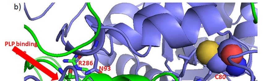
Int. J. Mol. Sci. 2019, 20, 1180                                                                    2 of 24

and cancer [3,4]. More than a century later, Harold F. Dvorak published an essay in the New England
Journal of Medicine entitled “Tumors: Wounds that do not heal. Similarities between tumor stroma
generation and wound-healing.” He proposed that solid tumors act as parasites that promote a
wound-healing response to acquire the stroma needed for their survival and growth [5]. Aging is
often accompanied by an impaired healing response [6], accumulation of senescent cells, and chronic
inflammation [7].
     The link between aging and inflammation is very accurately coined in the term “inflamm-aging”,
which denotes the up-regulation of certain pro-inflammatory cytokines at older ages, and is associated
with chronic diseases. This term was originally introduced in 2006 [8], to describe the imbalance
between inflammatory and anti-inflammatory networks, which results in a low grade chronic,
age-associated, pro-inflammatory status. Currently, deregulated cytokine production is appreciated
as a very important consequence of the remodeling of the immune system in old age (recently
reviewed) [9]. It has been recognized that, in older subjects, high levels of interleukins IL-6 and IL-1,
tumor necrosis factor-alpha (TNF-α), and C-reactive protein (CRP) are associated with an increased
risk of morbidity and mortality [10].
     The rate of cancer incidence is increasing, especially in the Western world. According to
GLOBOCAN 2018, there have been 18.1 million new reported cases of cancer in 2018 (9.5 million males
and 8.6 million females). The same database predicted 9.6 million cancer deaths in 2018 (5.4 million
males and 4.2 million females) [11]. For 2040, the International Agency for Research on Cancer
predicted 29.5 million newly diagnosed cancer patients, and 16.4 million cancer deaths [12].
     Early detection screenings represent a valid effort in fighting cancer. In the same setting, the era
of biotechnology implemented in molecular medicine, enables an insightful understanding of the
molecular mechanisms of action of natural compounds which have been used in traditional medicine
for centuries. One such compound is curcumin, which possesses properties relevant to successful
cancer chemoprevention. This is particularly important for the elder population.

2. Curcumin and Inflamm-aging
     The natural source of curcumin is the rhizome of the medicinal plant, Curcuma longa, a perennial
herb in the family Zingiberaceae [13]. The curcuminoid complex, found in the rhizome of turmeric
(2.5–6%) contains: curcumin (CUR - diferuloylmethane, ~85%); demethoxycurcumin (DEM, ~15%);
bis-demethoxycurcumin (bis-DEM, ~5%) and cyclocurcumin [14]. In the commercially available
formulations, the major curcuminoid complexes are reported to be in a similar range: 77% of CUR;
17% of DEM; and 3% of bis-DEM) [15]. By 2022, the U.S. curcumin market is expected to approach
40 million dollars [16]. It is also recognized that over 52% of curcumin production will be used for
pharmaceutical applications, in the U.S.
     Well-known for its healing properties, curcumin has been extensively used in traditional Ayurveda,
Unani and Siddha medicine for treating various diseases. An early mention of curcumin in modern
medical literature was in 1937, appearing in the Lancet, one of the most prestigious clinical medical
journals [17]. The article, describing curcumin applications to humans, was written by Albert
Oppenheimer—then an assistant professor at the American University of Beirut, Lebanon, who
applied curcumin orally (up to 800 mg daily) for the treatment of 67 patients suffering from various
forms of subacute, recurrent, or chronic cholecystitis. The positive therapeutic response recorded then,
was the basis for future interest in curcumin and its healing properties, especially its anti-inflammatory
properties, which were among the first studied [18].
     Is it reasonable to conclude that the anti-inflammatory property of curcumin may be used for
alleviating inflamm-aging? If that is so, can it explain the beneficial effects of curcumin in various
experimental models of diseases of elderly humans? We will present some older and some very recent
data which we consider central in the context of this puzzle.
     In2009, Smith et al. published data showing that the total projected cancer incidence will increase
in the U.S. by approximately 45%, in only 20 years, from 1.6 million (2010) to 2.3 million, in 2030 [19].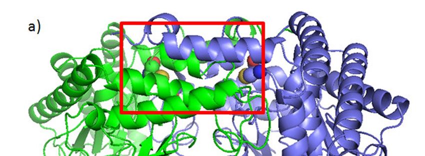
Int. J. Mol. Sci. 2019, 20, 1180                                                                      3 of 24

This prediction seems to be very accurate since, for 2019, 1,762,450 new cases and 606,880 cancer deaths
were estimated to occur in the U.S. [20]. The Smith’s study predicted something that was way ahead
of the time when the article was published. A 67% increase in cancer incidence was anticipated for
older adults [19]. Eight years later, in 2017, Nolen et al. have shown that cancer prevalence and cancer
incidence increases until ages 85-89, after which the rates decrease until 100+ [21].
      Inflammation has been recognized as a strong contributor to the acquisition of core hallmark
cancer capabilities [22]. The most recent data convincingly show that the high level of pro-inflammatory
cytokines (such as IL-6 and IL-8) may predict some crucial prognostic parameters in cancer patients [23,
24]. In addition to an increased level of IL-6 and IL-8, colorectal cancer patients also have alterations in
their serum amino-acid profile. Low levels of serum glutamine, histidine, alanine and high glycine
levels were shown to be associated with advanced stage of cancer and with poor cancer-specific
survival (N = 336; univariate analysis) [25].
      There are also quite convincing data on neuroinflammation as a crucial process in the pathogenesis
of the two the most common neurodegenerative diseases in the elder population: Alzheimer’s disease
(AD) and Parkinson’s disease (PD) [26]. Based on numerous data, it has been proposed that both
disorders are strongly related to inflamm-aging [27,28]. As recently reviewed, three shared strong
mediators of inflammatory reaction are increased in patients suffering from these two disorders: IL-6,
IL-1β and TNF-α [29].
      These inflammatory molecules are integrative parts of the canonical activation of the NF-kappa B
(nuclear factor kappa-light-chain-enhancer of activated B cells; NF-κB) signaling pathway. Generally,
this pathway has been considered as protumorigenic, although there are several models showing
its opposite action [30]. During the last few years, the interleukin 17 (IL-17) has become the focus
of various types of research, including the research related to cancer, Alzheimer’s and Parkinson’s
disease. There are several models in which the communication between NF-κB and IL-17 takes the
place. The most recent data indicate that IL-17 can promote the proliferation and migration of glioma
cells via PI3K/AKT1/NF-κB-p65 activation [31].

2.1. Inflamm-Aging, Interleukin-17 and Curcumin
     Originally, in 2002, human colonic subepithelial myofibroblasts (SEMFs) were used as a model
system for showing that SEMFs secrete IL-6, IL-8, and MCP-1 (Monocyte Chemoattractant Protein-1),
in response to IL-17 [32]. The most recent data point out the critical role for IL-17-producing T
lymphocytes in sporadic PD, in humans. In vitro, the midbrain neurons (MBNs) were shown
to increasingly die consequentially to the upregulation of IL-17 receptor (IL-17R) and NF-κB
activation [33]. The involvement of IL-17-producing T lymphocytes in the onset of Alzheimer’s
disease was also shown, in the animal model [34].
     Although majority studies performed so far indicate the presence/an increase of IL-17 as a
negative prognostic factor for cancer patients, the results are not entirely conclusive and depend on
numerous factors. These include the type of the tumor, the types of genetic aberrations, which may
significantly vary, and the host’s immune response. Of importance, different cell types expressing
IL-17 can play different roles, not only in the tumor microenvironment, but, it seems, may provide
insight into the overall picture of the malignant tumor. In a cohort of 573 gastric cancer patients [35],
high levels of IL-17+ neutrophils were shown in the tumorous tissue, where the high level of IL-17
induced the migration of neutrophils into gastric cancer, via cancer cell–derived CXC chemokines.
These neutrophils were further shown to stimulate the proangiogenic activity of tumor cells, both
in vitro and in vivo. The increase in IL-17-expressing cells in peripheral blood, particularly Th17,
was associated with tumor progression in HNC (Head and Neck Carcinoma) patients (N = 120) [36].
     While there are many data on curcumin and NF-κB, there are only a few of them in relation
to curcumin and its effect on IL-17producing cells. In relation to HNC, it is quite interesting that
the testing in 2015 of a novel microgranular curcumin formulation (15 patients and eight healthy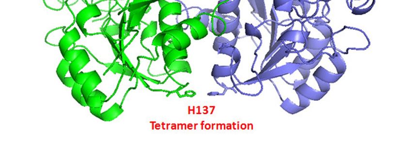
Int. J. Mol. Sci. 2019, 20, 1180                                                                      4 of 24

volunteers) showed that curcumin strongly decreases the serum level of IL-17 at one hour post-
ingestion; p = 0.0342 [37].

2.2. Breast Tissue Inflamm-Aging and Curcumin
      There are many research papers discussing curcumin’s action on sex-hormone related cancer
(breast, ovary, prostate), in vitro. In breast cancer cells, the effects obtained depend not only on the
status of estrogen and progesterone receptors (estrogen receptor; ER and progesterone receptor; PR),
but also on the specific genetic/epigenetic cellular background. A 20 year old paper presented the
strong inhibitory action of curcumin alone and in combination with isoflavonoids in both ER-positive
human breast cancer cells (MCF-7 and T47D) and ER-negative MDA-MB-231 cells exposed to
environmental estrogens (pesticide o,p’-DDT and the pollutants 4-nonylphenol and 4-octylphenol) [38].
The mechanism of curcumin’s inhibitory effect on MCF-7 and MDA-MB-231 was explained four years
later [39]. Curcumin exerts antiproliferative effects on MCF-7 cells through: (a) inhibiting the exogenous
17-β estradiol’s stimulatory growth effect on these cells; (b) blocking the expression of downstream
ER-responsive genes, pS2 and TGF-α, when applied in high concentrations. In MDA-MB-231 cells,
this mechanism was absent. Instead, curcumin strongly decreased the level of MMP-2 (matrix
metalloproteinase 2).
      In this paper, we reinforce the fact that breast cancer has been recognized as a systemic disease [40].
There is strong experimental data showing that postmenopausal breast cancer in obese women also
can be considered as „breast inflamm-aging“ disease. At the outset of the pathophysiological process,
subclinical, local breast inflammation, characterized by crown-like structures (CLS) consisting of
dead adipocytes surrounded by macrophages, occurs in breast white adipose tissue [41]. This type
of inflammation is paralleled by increased NF-κB binding activity joined with elevated levels of
proinflammatory mediators and a key enzyme in the biosynthesis of estrogen in menopause, aromatase
(estrogen synthase) [42]. Through aromatization of androgen precursors in adipose tissue (through
tissue specific reactions), this enzyme makes a strong link between inflammation, obesity, and an
increased risk for hormone receptor-positive breast cancer [43].
      On the other hand, lipolysis, which is also increased in obesity, results in increased concentrations
of free fatty acids [44]. Saturated fatty acids trigger the activation of NF-κB in macrophages resulting
in increased production of proinflammatory mediators which additionally induce aromatase in
preadipocytes [45]. Thus, there is a complete circulus vitiosus, associated with increased activity
of NF-κB, which can be modified by curcumin.
      As shown in vitro, curcumin can significantly suppress the inductive effects of stearic acid–treated
macrophages, on aromatase mRNA and aromatase activity in preadipocytes, through suppression of
NF-κB and Akt signaling pathways [46]. In the cited paper [46], a mixture of multiple polyphenols
(Zyflamend; curcumin included), was given to animals kept on a high-fat-diet. As a result, the increased
levels of aromatase mRNA and its activity in the mammary glands were partially suppressed. Although
this data constitutes a solid basis for future research on chemoprevention in humans, there is one
obvious problem. Measurement of the effect which occurs locally requires a biopsy. This significant
obstacle is one, but not the only one, of the major reasons for serious gaps in this area of research [47].
      There is no data, to the best of our knowledge showing the organ-specific, local effects and/or
systemic effects of curcumin in humans, which would be gender-specific.

2.3. Curcumin and the Concept of Network Medicine
     The NF-κB signaling pathway is a master regulator of inflammation-associated signaling
pathways. Age-related, tissue-specific, brain inflammation mediated by NF-κB, when joined with
Nitric Oxide and Reactive Oxygen Species (NOROS), in macrophages has been recognized as a most
significant risk factor for developing Alzheimer’s disease [48]. Activation of this pathway has been also
recognized in pathogenesis of PD [49]. Thus, if we focus on the pathophysiology of these three diseases
(cancer, AD and PD) and their increased incidence in older subjects, we need to take note of some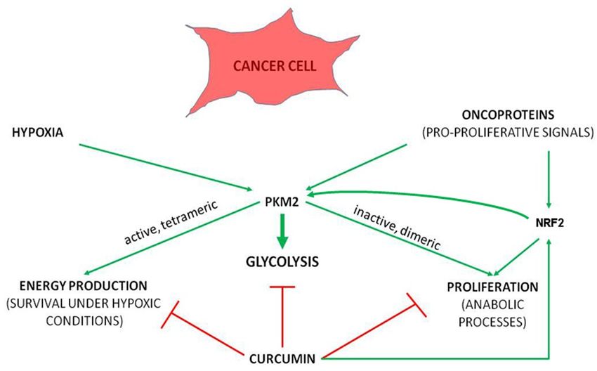
Int. J. Mol. Sci. 2019, 20, 1180                                                                    5 of 24

shared general molecular features, of which the “inflamm-age status” represents a very important
factor. It may, indeed, be the strongest indicator of perturbations which affect complex intracellular and
intercellular networks which communicate at different levels, as suggested by postulates of Network
Medicine. This discipline proposes a highly interconnected nature of the interactome and considers
that a gene, protein or metabolite can be implicated in several disease modules. Accordingly, at the
molecular level, it leads to the conclusion that diseases are not independent of one another [50].
     Can this interconnective dependency explain the beneficial effects of multilevel acting pleiotropic
curcumin [51] as a molecule that is considered useful for the prevention of these diseases, as recorded
in various models/diseases?
     First of all, surprisingly, the combined terms “curcumin” and “inflamm-age” appear in only a few
research papers in the PubMed database. Secondly, there are only a few studies based on measuring
cytokines in cancer-free human subjects who were taking curcumin. Different types of curcumin
formulations given, in addition to various doses applied and duration of administration, makes the
picture confusing. In a cohort of 72 migraine patients, only the combined daily administration of
2500 mg of ω-3 fatty acids (2 × 1250 mg) and 80 mg nano-curcumin (1 × 80 mg), led to a significant
decrease of TNF-α in the serum (p = 0.001). Administration of curcumin alone had no significant
effects [52]. However, a decrease of TNF-α was recorded (from 103.90 ± 13.29 ng/dL to 92.56 ±
8.70 ng/dL). The question is whether these subtle changes, although not statistically significant when
taking into consideration the whole group of subjects, may be significant for the physiology of a
particular patient. This would be in accord with the postulates of personalized medicine.
     In a study which included 117 subjects suffering from metabolic syndrome, 1 g of curcumin daily
and placebo were given to 59 and 58 subjects, respectively, for a period of eight weeks. In this research
study, between-group comparison suggested significantly greater reductions in serum concentrations
of TNF-α, IL-6, TGF-β (transforming growth factor beta) and MCP-1 in the curcumin versus the
placebo group (p < 0.001). When adjusted for potential confounders, changes in all parameters (serum
glucose and lipids, baseline serum concentration of the cytokines), except IL-6, remained statistically
significant [53].
     These two examples show that we are far away from firm conclusions when considering the
potential beneficial effects of curcumin. The problem relating to the lack of comprehensive data
associated with inflammation markers and curcumin application to healthy subjects, was very
accurately addressed by Hewlings and Kalman [54], who concluded that measuring potential benefits
of curcumin in healthy populations may be challenging because the benefits may not be as immediate
and measurable if (bio)markers are normal at the baseline. Thus, for reaching meaningful conclusions,
subjects will need to be followed over an extended time period, which would require a high level of
participants’ cooperation. But even if the cooperation and stringent follow-up is provided, caution in
interpretation is needed.
     The necessity of careful interpretation of all this data and the effects obtained must be put in the
context of the most recent findings.

3. Oxidative Metabolites of Curcumin
      The newest data show that the anti-inflammatory effects of curcumin, those mediated through
inhibition of NF-κB [55,56], do not depend on the parent molecule, but rather on its oxidized
products [57]. It was very convincingly shown that oxidative metabolites of curcumin adduct to and
inhibit IKK β (inhibitor of nuclear factor kappa-B kinase subunit beta; known also as IKK2/NFKBIKB).
If the cells were pretreated with N-acetylcysteine, a biosynthetic precursor of glutathione (GSH), the
potency of curcumin was decreased, probably due to GSH-mediated scavenge and inactivation of
curcumin-derived electrophiles. Finally, as concluded in the cited article [57], oxidative metabolites of
curcumin, which occur in vitro, adduct to cellular proteins. As the authors stated, this may explain
the wide range of cellular targets of curcumin identified in vitro. On the other hand, insufficient
bioactivation in vivo may underlie the inconclusive data in human studies. This may mean that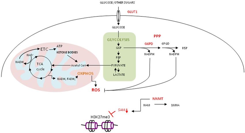
Int. J. Mol. Sci. 2019, 20, 1180                                                                   6 of 24

healthy humans who exhibit a lower level of oxidative stress, may be less likely to experience benefits
from the oxidative activation of curcumin. However, the oxidative stress theory of aging, which
is based on the hypothesis that age-associated functional losses are due to the accumulation of
NOROS-induced damages [58], joined with recent data on age-related inflammation (inflamm-age),
may strongly favor the protective effects of curcumin in older age. Thus, before coming to any final
conclusion, some molecular events related to curcumin’s pro-oxidative capability, obtained in vitro,
need to be addressed.
     Recently published data on the anti-tumorigenic effects of curcumin on CML-derived human
leukemic cells, in a xenograft model and in vitro culture system [59], confirmed, mostly indirectly, data
obtained in experiments with oxidized curcumin metabolites [57]. In the leukemic cells treated with
curcumin, the strong cytotoxic effect was joined with the strongest increase of ROS. On the other hand,
when GSH was added into the system, the level of ROS was, expectedly, lower, as was the cytotoxic
effect. The most probable explanation for these phenomena may be that GSH associated decrease of
ROS consequentially leads to a decreased level of oxidized curcumin metabolites. As a consequence,
the binding of curcumin to its potential protein-partners decreases. In the leukemic cell model, these
protein-partners were shown to be metabolic enzymes, a few of which, like NQO1 (NAD(P)H Quinone
Dehydrogenase 1), are well-known targets of the NRF2 (Nuclear Factor (Erythroid-Derived 2)-Like 2)
transcription factor.
     In addition to NQO-1, some other enzymes discovered as curcumin-binding partners, such as
CBR1 (carbonyl reductase 1), GSTP1 (glutathione-S-transferase phi 1), AKR1C1 (aldo-keto reductase
family 1 member 1), GLO1 (Glyoxalase I), exert their detoxifying function in the ROS-related metabolic
pathway. It is not surprising then that ectopic overexpression of these enzymes led to the same
effect as was gained with GSH: decreased level of ROS accompanied by a decrease in the curcumin’s
cytotoxic effect.

Curcumin and ROS-Producing Compounds
      When contemplating these in vitro systems, one may ask whether oxidized curcumin metabolites
lay in the background of commonly-recorded synergistic effects when applied with cytotoxic drugs,
for example – with cisplatin [60]. It has been shown that in cancer cells cisplatin exposure induces
a mitochondria-dependent increase in ROS levels [61]. This ROS may be the fuel for occurrence
of oxidized curcumin metabolites, which may block the NF-κB signaling pathway (as already
described) [57] and, additionally, decrease the activity of detoxifying enzymes through direct binding.
This action probably includes balancing many factors, as curcumin (or its oxidative metabolites?) can
restore NRF2 transcription through demethylation of the NRF2 promoter (first five CpGs positioned
between -1086 and -1226) [62]. Then, on the other hand, curcumin uses its oxidized metabolites for
binding to at least some protein targets of NRF2 (as is the case of NQO-1, which is considered as a bona
fide NRF2 target) [63]. This is a good example of the complexity in the field and, as already mentioned,
the necessity for multilevel research approach which needs to include as many as possible explorations
of various interactions.
      In considering the meaning of these recent discoveries, one should remember that, traditionally,
various effective combinations of curcumin are, indeed, those which are based on its combination with
the ROS-producing compounds. For example, oligomeric proanthocyanidins, which are known
to exhibit anticancer properties are, in some studies, shown as ROS generation activators [64].
The effect seems to be cell-type-specific [65]. The combination of proanthocyanidins and curcumin
was investigated in several models of colorectal cancer (cell lines, mice xenografts, colorectal cancer
patient-derived organoids), where the combined application showed superior anticancer properties.
It has been again shown that the expression of some very important genes/proteins remained intact
when compounds were applied separately, while combined application induced a strong change in the
activity of some genes. For example, a strong decrease of glucose-6-phosphate dehydrogenase (G6PD),
a key enzyme in the Pentose Phosphate Pathway (PPP) was discovered [66]. Deficiency of G6PD
Int. J. Mol. Sci. 2019, 20, 1180 7 of 24
was recognized to be associated with a lower cancer risk already in 1965 [67]. However, metabolic
reprogramming was (officially) recognized as a hallmark of cancer many years later [22]. With respect
to curcumin activity, metabolic reprogramming is becoming a hot topic of scientific interest.
4. Curcumin, Cancer and Metabolic Reprogramming
“Diffuse, evolutionary and developmental processes that we diagnose as cancer” [68], comprise,
through initiation and progression of the disease, dynamic, stepwise changes in fundamental
physiological characteristics of cells and tissues. A variety of stresses, either extrinsic (environmental)
or intrinsic (genetic, epigenetic, metabolic), induce damage to cellular components that can lead
to malignant transformation. Survival of damaged, potentially dangerous cells strongly depends
on stress response pathways. One of the main barriers to propagation of cells that are at risk for
malignant transformation is the onset of cellular senescence, a phenomenon that closely relates to
cellular metabolism.
4.1. Cellular Senescence (CS), Hypoxia and Cancer Metabolic Plasticity
Cellular senescence represents stress response which is needed for permanent arrest of division
of damaged cells. It is evolutionary well conserved, highly sophisticated and extremely complex.
Although CS is of utmost importance in preventing proliferation of cells that are at risk of malignant
transformation, the accumulation of senescent cells in aging tissues can compromise normal tissue
microenvironment and facilitate cancer progression [69,70]. We are aware that the role of CS in
molecular pathophysiology of many age-associated diseases, not only cancer, is multifaceted and can
be detrimental.
Senescent cells are not inert. To the contrary, they are metabolically very active, and capable to
secrete a plethora of bioactive molecules - pro-inflammatory cytokines and other factors which are
able to change tissue structure. In the setting of cancer, they contribute in creating a cancer permissive
microenvironment. Most anti-cancer therapeutic modalities promote senescence, which is beneficial for
inhibiting tumor cell proliferation. But, at the same time, it is not selective and can induce senescence
of adjacent non-tumor cells with consequential local inflammation, occurrence of secondary tumors
and cancer relapse.
There are two hypotheses on the protumorigenic role of cellular senescence. According to a
cell non-autonomous model, senescent cells secrete a variety of cytokines and factors which modify
surrounding cells that have not fully entered senescence [71]. According to cell autonomous model,
some senescent cells can bypass senescence and develop stem cell properties [72]. Malignant tumors are
composed of a heterogeneous population of cells. This heterogeneity is not only based on genetically
distinct subpopulations of tumor cells, but also on their functional heterogeneity.
Cancer is anything but a static system in terms of metabolism. A prominent characteristic of
cancer cells is uncontrolled proliferation, which requires nutrients and energy to accommodate their
augmented biosynthetic activity. The difference in glucose metabolism between normal and cancer cells
was first noted by Otto Warburg [73]. He observed that cancer cells from ascites “ferment” glucose into
lactate, even when enough oxygen, needed for supporting mitochondrial oxidative phosphorylation
(OXPHOS), is available. Proliferative cancer cells exhibit a high rate of glycolysis, for meeting their
high energetic and biosynthetic requirements. Although glycolysis is less efficient in producing ATP
per molecule of glucose, high glycolytic flux may provide more cellular ATP from glycolysis than from
OXPHOS. This is important for supporting cellular anabolic reactions [74–76]. Otto Warburg originally
postulated that cancer cells acquire a defect in the mitochondria that disturbs aerobic respiration.
We now know that, although mitochondrial function is often disturbed in cancer, cancer cells retain a
substantial capacity to produce energy in the mitochondria.
Considering that rapid proliferation of tumor cells is not always adequately accompanied with
vascularization of tumor mass, cancer cells are forced to adapt to nutrient- and oxygen-deprived
environment. To do so, cancers select metabolically plastic cells, those with the highest capacity forInt. J. Mol. Sci. 2019, 20, 1180 8 of 24
metabolism reprogramming. This metabolic adaptivity supports cellular survival under different
types of stress.
Cellular metabolic phenotype changes considerably as malignant tumors progress. In
H-RasV12/E1A transformed fibroblasts, an increase in OXPHOS activity precedes the increase
of glycolytic rate [77]. The strong mediator of this switch is transcription factor HIF-1α
(Hypoxia-Inducible Factor 1α).
Permanent increase of ROS, a by-product of OXPHOS, has a major role in stabilizing HIF-1α and
promoting aerobic glycolysis [78–80]. The stability of cellular HIF-1α is post-translationally regulated
by O2 −, Fe2+ - and α-ketoglutarate - dependent hydroxylation of proline residues. Hydroxylation of
HIF-1α promotes its ubiquitination and degradation, thereby preventing transcription of its target
genes. An elevated level of cellular ROS oxidizes Fe2+ to Fe3+ , abolishes HIF-1α hydroxylation/
ubiquitination/degradation. As a consequence, HIF-1α accumulates in the cell [79] where it promotes
pseudohypoxic state, permissive of mitochondrial dysfunction.
In Hep G2 hepatocellular carcinoma cells, application of curcumin led to a significant decrease
of HIF-1α protein. It also suppressed its transcriptional activity under hypoxia, which resulted
in decreased expression of vascular endothelial growth factor (VEGF), known as a major HIF-1 α
target [81]. The most recent data point out the strong simultaneous inhibitory effect of polymeric
nano-encapsulated curcumin on HIF-1α and REL A (P65; NFKB3), in lung and breast cancer cell
lines [82]. Curcumin’s inhibitory effects on gastric cancer in experimental animals were also shown
to be dependent not only on HIF-1α/VEGF decrease (shown immunohistochemically), but also on
decrease of STAT3 (Signal Transducer and Activator of Transcription 3) transcription factor [83].
What is the role of STAT3 in cellular metabolism, how it relates to inflammation and senescence,
and, finally, what kind of influence curcumin may have in relation to this potent transcription factor?
4.2. Curcumin, STAT3, and Modes of Survival
Constitutively activated in various malignancies, STAT3 has an important role in inflammation-
associated tumorigenesis [84]. It has recently been reported that curcumin inhibits highly active
STAT3 in H-Ras transformed breast epithelial cells (H-Ras MCF10A), through direct binding to
its cysteine residue 259. This cysteine (Cys) is critical for STAT3 phosphorylation, dimerization,
nuclear translocation and DNA binding [85], upon activation by various cytokines, hormones and
growth factors.
In addition to inflammation-associated tumorigenesis [86], STAT3 is mandatory for the survival
of cells, which, upon bypassing oncogene-induced senescence in premalignant lesions, acquire stem
cell properties. This was recently shown in a model of pancreatic cancer [87]. The mechanisms by
which cells circumvent senescence in tumors that spontaneously develop from premalignant lesions
are still not fully elucidated.
A prominent feature of senescent cells is STAT3 decrease, due to its proteasomal degradation [88].
Considering STAT3 involvement in promoting mitochondrial functions which are needed for cancer
cell stemness and metabolic reprogramming, cancer cells that bypass senescence and acquire stem cell
properties are sensitive to depletion and/or inactivation of STAT3. Napabucasin, a small molecule
compound which was developed to inhibit several pathways in cancer stem cell-like cells, exerts at
least some of its therapeutic activity through binding to the SH2 domain of STAT3 and, consequentially,
suppresses STAT3 activity [89]. Accordingly, curcumin’s binding to STAT3 Cys259 will result in
multiple consequences, in addition to those which were recently discovered [85].
High level of STAT3 expression and its activity was recorded in metabolically plastic, glucose
deprivation-resistant, ovarian cancer cells, accompanied by increased expression of metabolic
genes G6PD, GLUT1 (Glucose transporter 1) and NNMT (nicotinamide N-methyltransferase) [90].
In cancer stem cells (CSC) obtained from patients with epithelial ovarian cancer, glucose deprivation
leads to enrichment of cells with a high rate of OXPHOS and PPP, joined with a high level of
ROS [91]. These features represent cancer stem cell properties. High expression of GLUT1 in glucoseInt. J. Mol. Sci. 2019, 20, 1180 9 of 24
deprivation-resistant cells allows them to utilize other sugars (D-fructose, D-arabinose, mannan,
maltotriose and dextrin) and ketone bodies for energy production. Ketone bodies and lactic acid enter
tricarboxylic acid (TCA) cycle to produce NADH (Nicotinamide Adenine Dinucleotide) and FADH2
(Flavin Adenine Dinucleotide) that feed OXPHOS to generate ATP. Elevated OXPHOS activity leads to
increased
Int. levels
J. Mol. Sci. of xROS which needs to be counterbalanced by the activity of PPP. The rate-limiting
2019, 20, 9 of 25
enzyme in PPP is G6PD. It generates NADPH (Nicotinamide Adenine Dinucleotide Phosphate) needed
Dinucleotide
to reduce oxidized Phosphate) needed
glutathione toto reduceglutathione,
reduced oxidized glutathione to reduced
required for glutathione,
reduction required
of ROS (Figure 1). for
reduction of ROS (Figure 1).
Figure 1.
Figure 1. Derailed
Derailedmetabolism
metabolism of of
cancer
cancerstem cell cell
stem resistant to glucose
resistant deprivation.
to glucose Metabolically
deprivation. plastic
Metabolically
cancer cell,
plastic resistant
cancer to glucose
cell, resistant todeprivation, with highwith
glucose deprivation, expression of GLUT1, of
high expression G6PDH,
GLUT1, G6PD and NNMT,
G6PDH, G6PD
reprograms
and NNMT,metabolism
reprograms to metabolism
efficiently produce energy produce
to efficiently through utilization of sugars
energy through other than
utilization of glucose
sugars
(other
D -fructose, D -arabinose,
than glucose mannan,
(D-fructose, maltotriose
D-arabinose, and dextrin),
mannan, ketoneand
maltotriose bodies and lactate
dextrin), ketoneinbodies
OXPHOS.and
To counteract high level of ROS due to high OXPHOS, PPP is highly active. The
lactate in OXPHOS. To counteract high level of ROS due to high OXPHOS, PPP is highly active. upregulation of NNMTThe
consumes methyl
upregulation of groups
NNMTfrom S-adenosyl
consumes methionine
methyl groups(SAM)fromforS-adenosyl
methylationmethionine
of nicotinamide
(SAM)(NAM).
for
The level of SAM
methylation drops and hinders
of nicotinamide (NAM)cellular
. The methylation
level of SAM potential.
drops and hinders cellular methylation
potential.
The role of NNMT in metabolic plasticity and cancer cell stemness is emerging. Contrary to
the long-standing
The role of NNMT belief in
that the sole plasticity
metabolic function of andthis enzyme
cancer cellisstemness
methylation of nicotinamide
is emerging. Contrary(NAM)to the
long-standing belief that the sole function of this enzyme is methylation of nicotinamideof(NAM)
and its excretion from the body, when in excess, NNMT is implicated in a plethora important
and
cellular processes [92]. This enzyme is overexpressed in various malignancies.
its excretion from the body, when in excess, NNMT is implicated in a plethora of important cellular In Hep G2 cells
was shown
processes [92]. NNMT
thatThis enzyme promoter activity depends
is overexpressed in variouson the activation of
malignancies. In STAT3
Hep G2[93]. cells The
was NNMT
shown
methylates NAM, creating a stable metabolic product 1-methylnicotinamide
that NNMT promoter activity depends on the activation of STAT3 [93]. The NNMT methylates (1MNA). This reaction
consumes
NAM, methyl
creating units from
a stable SAM.product
metabolic As a consequence, the methylation
1-methylnicotinamide (1MNA).index (thereaction
This ratio SAM/SAH;
consumes
SAH - S-adenosylhomocysteine) of the cell changes. This way
methyl units from SAM. As a consequence, the methylation index (the ratio SAM/SAH; of acting puts NNMT in the central node-
SAH
involved in metabolic regulation of cellular methylation potential.
S-adenosylhomocysteine) of the cell changes. This way of acting puts NNMT in the central node Ulanovskaya et al. showed that
1MNA,
involved rather than being
in metabolic an active,of
regulation pro-tumorigenic metabolite,
cellular methylation servesUlanovskaya
potential. as “a stable sink”et al.ofshowed
methylation
that
groups in cancer cells [94]. The NNMT activity has a strong, methionine
1MNA, rather than being an active, pro-tumorigenic metabolite, serves as “a stable sink” of concentration-dependent,
impact on protein
methylation groupsmethylation.
in cancer Thecells
effect[94].
on protein
The methylation
NNMT activity has beenhasobserved
a strong, onlymethionine
in the cells
concentration-dependent, impact on protein methylation. The effect on protein methylationgrown
cultured with low methionine concentration (10-20 µM), while there was no effect in cells has beenin
high methionine concentration (100 µM) [94].
observed only in the cells cultured with low methionine concentration (10-20 µM), while there was
Likewise,
no effect in cellsthe concentration
grown of NAM varies
in high methionine in mammalian
concentration (100 µM) tissues
[94].and can impact the biological
outcome of NNMT activity. In NAM-limited conditions,
Likewise, the concentration of NAM varies in mammalian tissues NNMT has
and a potential,
can impactin addition
the to
biological
negatively influencing methylation processes, to change the NAD + /NADH ratio. This is a consequence
outcome of NNMT activity. In NAM-limited conditions, NNMT has a potential, in addition to
of the permanent loss of methylated NAM from theto NAD + recycling process. Another very significant
negatively influencing methylation processes, change the NAD+/NADH ratio. This is a
consequence of the permanent loss of methylated NAM from the impact
observation of this study [94] is that NNMT does not equally methylation
NAD+ recycling processes
process. Another of
very significant observation of this study [94] is that NNMT does not equally impact methylation
processes of all biomolecules. Instead, the specificity of its targeting specific methylation pathways
depends most probably on the relative Km values of individual methyltransferases for SAM and
SAH. Methyltransferases with higher Km for SAM and SAH are more sensitive to the activity of
NNMT [94]. Sperber et al. [95] reported high level of NNMT in naive human pluripotent stem cellsInt. J. Mol. Sci. 2019, 20, 1180 10 of 24
all biomolecules. Instead, the specificity of its targeting specific methylation pathways depends
most probably on the relative Km values of individual methyltransferases for SAM and SAH.
Methyltransferases with higher Km for SAM and SAH are more sensitive to the activity of NNMT [94].
Sperber et al. [95] reported high level of NNMT in naive human pluripotent stem cells (hPSC), in which
it contributed to low values of SAM and H3K27me3. Down-regulation of NNMT caused naive to
primed state transition in hPSC.
Considering that curcumin also has a negative effect on STAT3 phosphorylation [96], it is not
surprising that it down-regulates expression of NNMT (MDA-MB-468 breast cancer cells and HT29
colon cancer cells) [93].
4.3. Oxidative Stress, Curcumin and Gerometabolite NAD+
The oxidative stress-induced pseudohypoxic state leads to depletion of “gerometabolites”,
small-molecule components of normal metabolism [97], and consequential decline in mitochondrial
function [98,99]. One of the well-recognized gerometabolites is nicotinamide adenine dinucleotide,
NAD+ [97].
NAD+ is an important co-factor in many metabolic reactions and is co-substrate for PARP-1 (Poly
(ADP-Ribose) Polymerase 1) and SIRT1 (NAD-Dependent Deacetylase; Sirtuin-1), very important
enzymes involved in cellular stress response [100]. We consider them as crucial nodes at the
intersections of cellular metabolic and stress response pathways. For the cancer cell, adjustment
of metabolic requirements must be precisely balanced with adequate stress response, as an imperative
for survival. The pleiotropic activity of curcumin should be considered as a multilevel attack on
this redundant cellular communication network in which “metabolic” meeting “inflammatory” and
“oncogenic” serves as the basis for a unique cellular interactome, in accord with the postulates of
Network Medicine [50].
4.4. Curcumin in Regulation of Metabolic and Stress Response Pathways
Curcumin supports cellular antioxidant defense through stimulating NRF2, a master regulator
of ROS-scavenging enzymes. It has been shown that NRF2 may induce metabolic reprogramming
by directing glucose and glutamine into the anabolic pathway. The activity of the anabolic pathway
correlates with the presence of pyruvate kinase isozyme M2 (PKM2) [101]. The primary transcript
of the PKM gene may be spliced in several different ways, and the mode of splicing is regulated by
heterogeneous nuclear ribonucleoproteins (hnRNPs) [102]. The PKM1 transcript includes exon 10,
while the PKM2 transcript contains part of intron 9 instead of exon 10. Of note, these two fragments are
of the same length [103]. Functionally, two major protein isoforms, PKM1 (Isoform M1-PK; SwissProt
Identifier P14618-2); and PKM2 (Isoform M2-PK; SwissProt Identifier P14618-1), may occur.
In contrast to the PKM1 isozyme which is normally expressed in differentiated cells, embryonic-
and cancer-specific PKM2 confers a proliferative advantage to tumor cells. PKM2 can form dimers, with
low activity, or active tetramers. Dimeric PKM2 diverts glucose metabolism towards aerobic glycolysis,
thereby supporting biosynthetic processes, while tetrameric PKM2 promotes ATP production via
OXPHOS. Since biosynthetic and energetic requirements of highly proliferative cancer cells should
be simultaneously met and well adjusted, the ratio of PKM2 dimers and tetramers is critical for
tumorigenesis [104]. This metabolic switch, dependent on the active axis PI3K-AKT, a part of the
receptor tyrosine kinase/PI3K/AKT/mammalian target of rapamycin (RTK/PI3K/AKT/mTOR)
signaling pathway, accelerates tumor proliferation and contributes to its aggressiveness [105].
Tumor cells with constitutively activated K-ras oncogenic signaling have upregulated PKM2,
which is mandatory for accumulating phosphoenolpyruvate (PEP), its shunting to an alternative
glycolytic pathway and utilization for anabolic processes [106]. Despite the stimulatory effect on
NRF2, curcumin antagonizes metabolic reprogramming towards glycolysis (Figure 2). One of the
mechanisms involved in this process is curcumin-mediated negative regulation of TNF-α, with
consequent prevention of inflammatory environment-induced onset of aerobic glycolysis. Thus,Int. J. Mol. Sci. 2019, 20, 1180 11 of 24
curcumin makes yet another link between inflammation and metabolism. This mechanism was shown
in breast epithelial cells [107]. The same negative impact of curcumin toward glycolysis was shown in
Int. J. Mol. Sci.
Dalton’s 2019, 20, x in mice [108].
lymphoma, 11 of 25
Figure 2. The principle underlying antitumorigenic effect of curcumin. Curcumin targets cellular
Figure 2. The principle underlying antitumorigenic effect of curcumin. Curcumin targets cellular
processes central to the ability of cancer cell to survive by coordinately hindering aerobic glycolysis-
processes central to the ability of cancer cell to survive by coordinately hindering aerobic
dependent anabolic processes and energy production, despite of stimulating NRF2.
glycolysis-dependent anabolic processes and energy production, despite of stimulating NRF2.
The unrestricted proliferative potential of cancer cells is closely dependent on PI3K/AKT/mTOR
The unrestricted proliferative potential of cancer cells is closely dependent on PI3K/AKT/mTOR
signaling pathway [109]. For example, mTOR activation in cancer cells upregulates the expression
signaling pathway [109]. For example, mTOR activation in cancer cells upregulates the expression of
of PKM2 through HIF-1α mediated transcriptional activation of PKM, and Myc-hnRNPs-mediated
PKM2 through HIF-1α mediated transcriptional activation of PKM, and Myc-hnRNPs-mediated
splicing of PKM pre-mRNA, in favor of PKM2. In a nude mouse xenograft tumor model, stable
splicing of PKM pre-mRNA, in favor of PKM2. In a nude mouse xenograft tumor model, stable
knock down of PKM2 in PC3 cells (a human prostate cancer cell line with PTEN deficiency and
knock down of PKM2 in PC3 cells (a human prostate cancer cell line with PTEN deficiency and
mTOR hyperactivation) significantly extended the survival of tumor-bearing mice [110]. Silencing of
mTOR hyperactivation) significantly extended the survival of tumor-bearing mice [110]. Silencing of
PKM2 (transduction with shPKM2) suppresses mTOR-mediated tumorigenesis [110]. Due to the (a)
PKM2 (transduction with shPKM2) suppresses mTOR-mediated tumorigenesis [110]. Due to the a)
dependence of PKM2 on mTOR activation and (b) mTOR-mediated cancer cell proliferation associated
dependence of PKM2 on mTOR activation and b) mTOR-mediated cancer cell proliferation
with PKM2 shunting PEP into alternative glycolytic pathway supporting anabolic processes, cancer
associated with PKM2 shunting PEP into alternative glycolytic pathway supporting anabolic
cells with hyperactive mTOR are particularly sensitive to dual inhibition of mTOR and glycolysis [111].
processes, cancer cells with hyperactive mTOR are particularly sensitive to dual inhibition of mTOR
One of the prominent features of the multifaceted antitumorigenic effects of curcumin is its
and glycolysis [111].
ability to simultaneously inhibit glycolysis and mTOR [112]. Recently, it has been shown that 20 µM
One of the prominent features of the multifaceted antitumorigenic effects of curcumin is its
curcumin inhibits glucose uptake and down-regulates PKM2 and lactate production in various cancer
ability to simultaneously inhibit glycolysis and mTOR [112]. Recently, it has been shown that 20 µM
cell lines (H1299, MCF-7, HeLa and PC3) [113]. In curcumin-treated cells, decreased phosphorylation
curcumin inhibits glucose uptake and down-regulates PKM2 and lactate production in various
of p70S6 kinase (T389) was associated with the concomitant lowering of HIF-1α protein expression.
cancer cell lines (H1299, MCF-7, HeLa and PC3) [113]. In curcumin-treated cells, decreased
The involvement of mTOR/HIF-1α signaling inhibition, in curcumin-mediated down-regulation of
phosphorylation of p70S6 kinase (T389) was associated with the concomitant lowering of HIF-1α
PKM2 expression, was further validated by the observation that rapamycin, a well-known mTOR
protein expression. The involvement of mTOR/HIF-1α signaling inhibition, in curcumin-mediated
inhibitor, likewise down-regulates PKM2 [113]. In addition to a negative effect on PKM2 expression,
down-regulation of PKM2 expression, was further validated by the observation that rapamycin, a
curcumin treatment, in the stated cellular models, caused decreased expression of glucose transporter
well-known mTOR inhibitor, likewise down-regulates PKM2 [113]. In addition to a negative effect
GLUT1 and hexokinase II (HKII) transcripts. In HCT116 and HT29 colon cancer cells, curcumin
on PKM2 expression, curcumin treatment, in the stated cellular models, caused decreased
has previously been reported to down-regulate HKII, the enzyme which catalyzes the first step in
expression of glucose transporter GLUT1 and hexokinase II (HKII) transcripts. In HCT116 and HT29
glycolysis—phosphorylation of glucose to form glucose-6-phosphate [114]. Finally, in esophageal
colon cancer cells, curcumin has previously been reported to down-regulate HKII, the enzyme
squamous cell carcinoma EC109 cells, curcumin down-regulates the expression of glycolytic enzymes,
which catalyzes the first step in glycolysis - phosphorylation of glucose to form glucose-6-phosphate
in dose- and AMPK (AMP-Activated Kinase) -dependent manner [115].
[114]. Finally, in esophageal squamous cell carcinoma EC109 cells, curcumin down-regulates the
These data indicate that the actions of curcumin are highly dependent on the context of intrinsic
expression of glycolytic enzymes, in dose- and AMPK (AMP-Activated Kinase) -dependent manner
cellular makeup, encompassing genetic, epigenetic, metabolic and cell proliferation status, together
[115].
with external influences (microenvironmental cues). All these factors may also modulate its ability for
These data indicate that the actions of curcumin are highly dependent on the context of intrinsic
binding to various cellular proteins.
cellular makeup, encompassing genetic, epigenetic, metabolic and cell proliferation status, together
with external influences (microenvironmental cues). All these factors may also modulate its ability
for binding to various cellular proteins.
4.5. Metabolic Enzymes as Targets for Curcumin Binding
Approximately 60 proteins - curcumin’s binding partners, were discovered after incubating the
HEI-193 human schwannoma cells with a very high concentration of biotinylated curcumin (1
mg/mL – 2.71 mM) for 30 min. The most abundant binding partners were heat shock proteins (HSPs)Int. J. Mol. Sci. 2019, 20, 1180 12 of 24
4.5. Metabolic Enzymes as Targets for Curcumin Binding
Approximately 60 proteins—curcumin’s binding partners, were discovered after incubating the
HEI-193 human schwannoma cells with a very high concentration of biotinylated curcumin (1 mg/mL
– 2.71 mM) for 30 min. The most abundant binding partners were heat shock proteins (HSPs) -70
and -90, molecular chaperones involved in the proper folding of client proteins, 3-phosphoglycerate
dehydrogenase (PHDGH) and β-actin [116]. Abegg and collaborators profiled 42 proteins, covalently
bound to curcumin through their cysteine residues, in HeLa cervical cancer cells [117]. A relatively
small subset of Cys residues has been considered to be involved in cell signaling (contrary to
”sensing“) [118]. However, curcumin’s binding to cysteine residues of metabolic enzymes may be very
important for cellular reprogramming mechanisms.
A recently published paper [119] on in situ proteomic profiling of curcumin targets in HCT116
colon cancer cell line, reports a list of 197 curcumin-binding proteins, among which are many
cancer-related metabolic enzymes: GAPDH (Glyceraldehyde 3-Phosphate Dehydrogenase), PKM
isozymes M1/M2, LDHA (L-Lactate Dehydrogenase A), MDH1/2 (cytoplasmic/mitochondrial Malate
Dehydrogenase), SHMT2 (Serine Hydroxymethyltransferase; mitochondrial), ADP/ATP translocase 1.
Additionally, curcumin targets PARP-1, NAD+ -consuming enzyme involved in numerous cellular
processes including DNA-damage detection and repair, transcription and intracellular localization and
activity of many proteins. Binding to numerous hnRNPs, including the alternative splicing repressors
hnRNP A1/A2 which influence PKM splicing [103], point to the possible role of curcumin in regulating
alternative splicing. So far, its influence on splicing was shown in fibroblasts from patients with SMA
type II (Survival of Motor Neuron 2) in which curcumin increased the proportion of SMN2_exon
7-containing transcript [120].
How curcumin binding to its protein targets influences their activity is mostly unknown. The
SiteMap calculation performed by Angelo et al. [116] predicted the binding of curcumin to NAD+
binding site on PHGDH, which is crucial for PHGDH enzymatic activity. The curcumin binding
enzymes, PHGDH and SHMT2, belong to the serine-glycine metabolic pathway. Both amino acids,
serine and glycine, serve as intermediates for the biosynthesis of other amino acids, nucleic acids and
lipids. What is the possible scenario of curcumin’s binding to these enzymes?
Phosphoglycerate Dehydrogenase and Serine Hydroxymethyltransferase 2
PHGDH, the first, rate-limiting enzyme in the pathway of serine biosynthesis, catalyzes the
transition of 3-phosphoglycerate (3PG) into 3-phosphohydroxypyruvate, using NAD+ /NADH as
a cofactor. Shunting of the glycolytic intermediate 3PG to serine synthesis is permitted by PKM2.
Low activity of PKM2 in tumor cells leads to an accumulation of its substrate PEP, which participates
in phosphorylation and the catalytic activation of phosphoglycerate mutase (PGAM1) [106]. As a
consequence, there is an increase of its product, 2-phosphoglycerate, which activates PHGDH and
serine biosynthesis [121].
PHGDH protein level is elevated in 70% of estrogen receptor (ER)-negative breast cancers and
suppression of PHGDH in breast cancer cell lines decreases cell proliferation and reduces serine
synthesis [122]. In addition to increasing PHGDH, an increase of nicotinamide phosphoribosyltransferase
(NAMPT), a rate-limiting enzyme of NAD+ salvage pathway, was also shown in ER-negative breast
cancer lines and patient-derived breast tumors [123]. Their interconnection was shown experimentally
on PHGDHhigh MDA-MB-468 and PHGDHlow MDA-MB-231 breast cancer cells by treating them with
well-established NAMPT inhibitor (FK866). This treatment caused a drop in NAD+ level and almost
completely abrogated serine synthesis in PHGDHhigh MDA-MB-468 cells, while the drop in NAD+
level and serine synthesis was insignificant in PHGDHlow MDA-MB-231 cells [123].
Although pursued in cancer treatment, NAD+ salvage pathway inhibitors exert various
efficacy. Thus, it was suggested that NAMPT inhibitors may be effective for treating a subset of
PHGDH-dependent cancers [123]. Therefore, it is possible that curcumin, in addition to inhibiting
PHGDH directly by binding to its NAD+ pocket [116], hinders its activity indirectly by down-regulatingInt. J. Mol. Sci. 2019, 20, 1180 13 of 24
expression of NAMPT (also known as visfatin) as has been demonstrated in breast cancer cell lines
(MDA-MB-231, MDA-MB-468, and MCF-7) [124]. Knocking down PHGDH in HeLa cells significantly
inhibited cell proliferation and increased cisplatin chemosensitivity [125].
Serine can be directly converted to glycine by two serine hydroxymethyltransferases: cytoplasmic
Int. J. Mol. Sci. 2019, 20, x 13 of 25
(SHMT1) and mitochondrial (SHMT2). SHMT2 is the main source of glycine in proliferating cells [126]
and its importance
proliferating for tumor
cells [126] and itscells survival in
importance forhypoxia was demonstrated
tumor cells in glioblastoma
survival in hypoxia multiforme
was demonstrated in
(GM) [127].
glioblastoma multiforme (GM) [127].
Competent eukaryotic
Competent eukaryotic SHMT2
SHMT2 is is aa tetrameric
tetrameric protein
protein built
built as
as dimers
dimers of of “tight”
“tight” dimers
dimers which
which
correspond to its minimal catalytic active units. A dimer-to-tetramer transition
correspond to its minimal catalytic active units. A dimer-to-tetramer transition is triggered by the is triggered by the
binding of the isozyme SHMT2 cofactor, pyridoxal 5’-phosphate (PLP)
binding of the isozyme SHMT2 cofactor, pyridoxal 5’-phosphate (PLP) [128]. Curcumin-binding [128]. Curcumin-binding
Cys80 [116]
Cys80 [116] indirectly
indirectly influences
influences the
the formation
formation of of the
the binding
binding site
site for
for PLP,
PLP, through
through interacting with
interacting with
residues Arg283 and Gly284 to Arg286 (Figure 3). Cys80 also participates
residues Arg283 and Gly284 to Arg286 (Figure 3). Cys80 also participates in protein-proteinin protein-protein interaction
of the tightof
interaction dimer, through
the tight interacting
dimer, through with Asn93with
interacting of the otherofchain.
Asn93 Thus,chain.
the other covalent binding
Thus, covalentof
curcuminoftocurcumin
binding Cys80 maytobeCys80
expectedmayto be
impact both, the
expected structureboth,
to impact and thethe catalytic
structureactivity of the
and the active
catalytic
form of SHMT2.
activity of the active form of SHMT2.
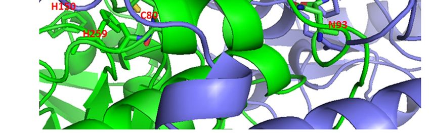
Figure 3.
Figure 3. Possible
Possibleeffects
effectsof of
curcumin’s
curcumin’sbinding to SHMT2
binding Cys80.
to SHMT2 (a) The
Cys80. (a)symmetric dimer ofdimer
The symmetric SHMT2 of
(PDB: 4PVF) showing amino acid residues Cys80 (spheres) and His137 (sticks)
SHMT2 (PDB: 4PVF) showing amino acid residues Cys80 (spheres) and His137 (sticks) participated participated in the
tetramer
in formation
the tetramer [128].[128].
formation Monomers
Monomers are shown in green
are shown andand
in green purple.
purple.(b) (b)The
Thepart
partof
of the 4PVF
the 4PVF
structure within red rectangular in (a), with marked amino acid residues making interactions
structure within red rectangular in (a), with marked amino acid residues making interactions with with the
nonmodified
the nonmodifiedCys80. The pyridoxal
Cys80. 5’-phosphate
The pyridoxal (PLP)(PLP)
5’-phosphate binding site is site
binding encompassed by marked
is encompassed His150
by marked
and His259.
His150 and His259.
As recently described in GM, SHMT2 activity limits the activity of PKM2 and reduces oxygen
As recently described in GM, SHMT2 activity limits the activity of PKM2 and reduces oxygen
consumption, thereby eliciting a metabolic state permissive of tumor cells survival in poorly
consumption, thereby eliciting a metabolic state permissive of tumor cells survival in poorly
vascularized tumor regions. It is plausible to expect that curcumin’s binding to Cys80 may hinder
formation of SHMT2 tetramers, inhibit SHMT2 activity and prevent PKM2-mediated metabolic
reprogramming, needed for facilitating cell survival in hypoxia [127]. The negative effect of elevated
SHMT2 activity on PKM2 may stem from the increased catabolism of serine, a known PKM2
activator [129]. Transformation of serine in the mitochondria by SHMT2 increases the cellularYou can also read