A multifaceted analysis reveals two distinct phases of chloroplast biogenesis during de-etiolation in Arabidopsis - eLife
←
→
Page content transcription
If your browser does not render page correctly, please read the page content below
RESEARCH ARTICLE

                                    A multifaceted analysis reveals two
                                    distinct phases of chloroplast biogenesis
                                    during de-etiolation in Arabidopsis
                                    Rosa Pipitone1, Simona Eicke2, Barbara Pfister2, Gaetan Glauser3,
                                    Denis Falconet4, Clarisse Uwizeye4, Thibaut Pralon1, Samuel C Zeeman2,
                                    Felix Kessler1*, Emilie Demarsy1,5*
                                    1
                                     Plant Physiology Laboratory, University of Neuchâtel, Neuchâtel, Switzerland;
                                    2
                                     Institute of Molecular Plant Biology, Department of Biology, ETH Zurich, Zurich,
                                    Switzerland; 3Neuchâtel Platform of Analytical Chemistry, University of Neuchâtel,
                                    Neuchâtel, Switzerland; 4Université Grenoble Alpes, CNRS, CEA, INRAE, IRIG-
                                    DBSCI-LPCV, Grenoble, France; 5Department of Botany and Plant Biology,
                                    University of Geneva, Geneva, Switzerland

                                    Abstract Light triggers chloroplast differentiation whereby the etioplast transforms into a
                                    photosynthesizing chloroplast and the thylakoid rapidly emerges. However, the sequence of events
                                    during chloroplast differentiation remains poorly understood. Using Serial Block Face Scanning
                                    Electron Microscopy (SBF-SEM), we generated a series of chloroplast 3D reconstructions during
                                    differentiation, revealing chloroplast number and volume and the extent of envelope and thylakoid
                                    membrane surfaces. Furthermore, we used quantitative lipid and whole proteome data to
                                    complement the (ultra)structural data, providing a time-resolved, multi-dimensional description of
                                    chloroplast differentiation. This showed two distinct phases of chloroplast biogenesis: an initial
                                    photosynthesis-enabling ‘Structure Establishment Phase’ followed by a ‘Chloroplast Proliferation
                                    Phase’ during cell expansion. Moreover, these data detail thylakoid membrane expansion during
*For correspondence:                de-etiolation at the seedling level and the relative contribution and differential regulation of
felix.kessler@unine.ch (FK);        proteins and lipids at each developmental stage. Altogether, we establish a roadmap for
emilie.demarsy@unige.ch (ED)        chloroplast differentiation, a critical process for plant photoautotrophic growth and survival.
Competing interests: The
authors declare that no
competing interests exist.
                                    Introduction
Funding: See page 27                Seedling development relies on successful chloroplast biogenesis, ensuring the transition from het-
Received: 02 September 2020         erotrophic to autotrophic growth. Light is a crucial factor for chloroplast differentiation. For seeds
Accepted: 04 February 2021          that germinate in the light, chloroplasts may differentiate directly from proplastids present in cotyle-
Published: 25 February 2021         dons. However, as seeds most often germinate underneath soil, seedling development typically
                                    begins in darkness and follows a skotomorphogenic program called etiolation, characterized by
Reviewing editor: Caroline
Gutjahr, Technical University of
                                    rapid hypocotyl elongation and etioplast development. Light promotes seedling de-etiolation, which
Munich, Germany                     involves a series of morphological changes, such as cotyledon expansion, hypocotyl growth inhibi-
                                    tion, and greening, that accompanies the onset of photosynthesis in chloroplasts. During de-etiola-
   Copyright Pipitone et al. This
                                    tion, etioplast–chloroplast transition is thereby rapidly triggered by light following seedling
article is distributed under the
                                    emergence at the soil surface (Jarvis and López-Juez, 2013; Solymosi and Schoefs, 2010;
terms of the Creative Commons
Attribution License, which          Weier and Brown, 1970). A hallmark of chloroplast differentiation is the biogenesis of thylakoids, a
permits unrestricted use and        network of internal membranes where the components of the photosynthetic electron transport
redistribution provided that the    chain assemble. Thylakoid biogenesis and the onset of photosynthesis rely on the concerted synthe-
original author and source are      sis and coordinated assembly of chlorophylls, lipids, and proteins in both space and time (Jarvis and
credited.                           López-Juez, 2013).

Pipitone et al. eLife 2021;10:e62709. DOI: https://doi.org/10.7554/eLife.62709                                                       1 of 32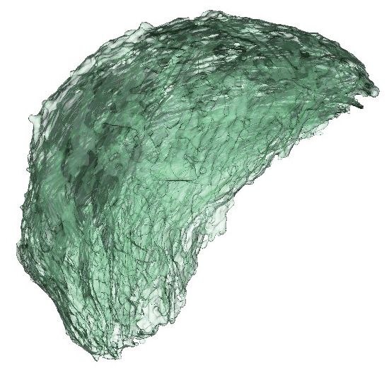
Research article                                                                                                   Plant Biology

                                        The thylakoids harbor the photosynthetic electron transport chain, which is composed of three
                                    complexes: photosystem II (PSII), the cytochrome b6f complex (Cyt b6f), and photosystem I (PSI).
                                    Electron transfer between these complexes is facilitated by mobile electron carriers, specifically the
                                    low-molecular-weight, membrane-soluble plastoquinone (electron transfer from PSII to Cyt b6f) and
                                    the lumenal protein plastocyanin (electron transfer from Cyt b6f to PSI) (Eberhard et al., 2008). Elec-
                                    tron transfer leads to successive reduction and oxidation of electron transport chain components.
                                    The final reduction step catalyzed by ferredoxin-NADP(+) reductase (FNR) leads to NADPH produc-
                                    tion. Oxidation of water by PSII and of plastoquinone by Cyt b6f releases protons into the lumen,
                                    generating a proton gradient across the thylakoid membrane that drives the activity of the thyla-
                                    koid-localized chloroplast ATP synthase complex. Each of the photosynthetic complexes consists of
                                    multiple subunits encoded by the plastid or nuclear genome (Allen et al., 2011; Jarvis and López-
                                    Juez, 2013) PSII and PSI have core complexes comprising 25–30 and 15 proteins, respectively
                                    (Amunts and Nelson, 2009; Caffarri et al., 2014). The antenna proteins from the Light Harvesting
                                    Complexes (LHC) surround the PSI and PSII core complexes contributing to the formation of super-
                                    complexes. Cyt b6f is an eight-subunit dimeric complex (Schöttler et al., 2015). Each complex of
                                    the electron transport chain has a specific dimension, orientation, and location within the thylakoid
                                    membrane, occupying a defined surface, and their dimensions have been reported in several studies
                                    giving congruent results (Caffarri et al., 2014; Kurisu et al., 2003; van Bezouwen et al., 2017).
                                    During de-etiolation, massive protein synthesis is required for assembly of the highly abundant pho-
                                    tosynthetic complexes embedded in thylakoids. The photomorphogenic program is controlled by
                                    regulation of gene expression at different levels (Wu, 2014). Transcriptome analyses have revealed
                                    that upon light exposure, up to one-third of Arabidopsis genes are differentially expressed, with 3/5
                                    being upregulated and 2/5 downregulated (Ma et al., 2001). Chloroplast proteins encoded by the
                                    nuclear genome must be imported from the cytoplasm (Jarvis and López-Juez, 2013). The general
                                    chloroplast protein import machinery is composed of the multimeric complexes Translocon at the
                                    Outer membrane of the Chloroplast (TOC) and Translocon at the Inner membrane
                                    of the Chloroplast (TIC), and selective import is based on specific recognition of transit peptide
                                    sequences by TOC receptors (Agne and Kessler, 2010; Richardson and Schnell, 2020).
                                        Reminiscent of their cyanobacterial origin, chloroplast membranes are composed mostly of glyco-
                                    lipids (mono- and di-galactosyldiacylglycerol; MGDG and DGDG) and are poor in phospholipids
                                    compared to other membranes in the cell (Bastien et al., 2016; Block et al., 1983; Kobaya-
                                    shi, 2016). Galactolipids comprise a glycerol backbone esterified to contain a single (MGDG) or dou-
                                    ble (DGDG) galactose units at the sn1 position and two fatty acid chains at the sn2 and sn3
                                    positions. In addition to the number of galactose units at sn1, galactolipids also differ by the length
                                    and degrees of saturation of the fatty acid chains. In some species, including Arabidopsis, galactoli-
                                    pid synthesis relies on two different pathways, defined as the eukaryotic and prokaryotic pathways
                                    depending on the organellar origin of the diacylglycerol precursor. The eukaryotic pathway requires
                                    the import of diacyl-glycerol (DAG) synthesized in the endoplasmic reticulum (ER) into the plastids
                                    and is referred to as the ER pathway, whereas the prokaryotic pathway is entirely restricted to the
                                    plastid (PL) and is referred to as the PL pathway (Ohlrogge and Browse, 1995). As signatures, ER
                                    pathway-derived galactolipids harbor an 18-carbon chain, whereas PL pathway–derived galactolipids
                                    harbor a 16-carbon chain at the sn2 position. In addition to constituting the lipid bilayer, galactoli-
                                    pids are integral components of photosystems and thereby contribute to photochemistry and photo-
                                    protection (Aronsson et al., 2008; Kobayashi, 2016). Thylakoids also contain neutral lipids such as
                                    chlorophyll, carotenoids, tocopherols, and plastoquinone. These may exist freely or be associated
                                    with the photosynthetic complexes, having either a direct role in photosynthesis (chlorophyll, carote-
                                    noids, plastoquinone) or participating indirectly in the optimization of light usage and/or mitigation
                                    of potentially damaging effects (tocopherols in addition to carotenoids and plastoquinone)
                                    (Hashimoto et al., 2003; van Wijk and Kessler, 2017).
                                        Past studies used conventional electron microscopy to first describe the architecture of the thyla-
                                    koid membrane network. Based on these 2D observations, researchers proposed that plant thylakoid
                                    membranes are organized as single lamellae connected to appressed multi-lamellar regions called
                                    grana. How these lamellae are interconnected was revealed only later following the development of
                                    3D electron microscopic techniques (Staehelin and Paolillo, 2020). Tremendous technological prog-
                                    ress in the field of electron microscopy has been made recently, leading to improved descriptions of
                                    chloroplast ultrastructure (Daum et al., 2010; Daum and Kühlbrandt, 2011). Electron tomography

Pipitone et al. eLife 2021;10:e62709. DOI: https://doi.org/10.7554/eLife.62709                                                      2 of 32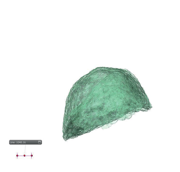
Research article                                                                                                    Plant Biology

                                    substantially improved our comprehension of the 3D organization of the thylakoid network in chloro-
                                    plasts at different developmental stages and in different photosynthetic organisms, including Arabi-
                                    dopsis (Austin and Staehelin, 2011; Liang et al., 2018), Chlamydomonas (Engel et al., 2015),
                                    runner bean (Kowalewska et al., 2016), and Phaeodactylum tricornutum (Flori et al., 2017). Elec-
                                    tron tomography also provided quantitative information on thylakoid structure such as the thylakoid
                                    layer number within the grana stack and the thickness of the stacking repeat distance of grana mem-
                                    brane (Daum et al., 2010; Kirchhoff et al., 2011). These quantitative data allowed a greater under-
                                    standing of the spatial organization of the thylakoid membrane in relation to the embedded
                                    photosynthetic complexes (Wietrzynski et al., 2020). Although electron tomography offers extraor-
                                    dinary resolution at the nanometer level, its main drawback is a limit to the volume of the observa-
                                    tion, enabling only a partial 3D reconstruction of a chloroplast. Serial Block Face-Scanning Electron
                                    Microscopy (SBF-SEM) is a technique where the embedded specimen is imaged by scanning the
                                    face of the block with an electron beam. After imaging, the face of the block is shaved automatically
                                    (e.g. 60-nm-thick slices) by an ultramicrotome mounted in the vacuum chamber. The section is dis-
                                    carded and the newly revealed block face is imaged again. Repeated imaging and cutting allows the
                                    collection of a tomographic sequence of hundreds of images of the same area. Thereby, a much
                                    larger volume can be reconstructed in 3D to show cellular organization (Peddie and Collinson,
                                    2014; Pinali and Kitmitto, 2014).
                                       In combination with electron microscopy, biochemical fractionation of thylakoids has revealed dif-
                                    ferential lipid and protein compositions of the grana and the stroma lamellae. The grana are
                                    enriched in DGDG and PSII, whereas the stroma lamellae are enriched in MGDG, Cyt b6/f, and PSI
                                    (Demé et al., 2014; Koochak et al., 2019; Tomizioli et al., 2014; Wietrzynski et al., 2020).
                                    Changes in lipid and protein compositions during etioplast–chloroplast transition are tightly linked
                                    to the thylakoid architecture. In particular, changes in MGDG to DGDG ratio are correlated with the
                                    transition from prolamellar body (PLB) and prothylakoid (PT) structures (tubular membrane) to thyla-
                                    koid membranes (lamellar structure) (Bottier et al., 2007; Demé et al., 2014; Mazur et al., 2019).
                                       Individual studies have provided much insight regarding specific dynamics of the soluble chloro-
                                    plast proteome, the chloroplast transcriptome, photosynthesis-related protein accumulation and
                                    photosynthetic activity, chloroplast lipids, and changes in thylakoid architecture (Armarego-
                                    Marriott et al., 2019; Dubreuil et al., 2018; Kleffmann et al., 2007; Kowalewska et al., 2016;
                                    Liang et al., 2018; Rudowska et al., 2012). However, these studies were mostly qualitative, focused
                                    on one or two aspects, and were performed in different model organisms. Therefore, chemical data
                                    related to thylakoid biogenesis remain sparse and quantitative information is rare. Here, we present
                                    a systems-level study that integrates quantitative information on ultrastructural changes of the thyla-
                                    koids with lipid and protein composition during de-etiolation of Arabidopsis seedlings.

                                    Results
                                    The photosynthetic machinery is functional after 14 hr of de-etiolation
                                    We analyzed etioplast–chloroplast transition in Arabidopsis seedlings grown in the absence of exog-
                                    enous sucrose for 3 days in darkness and then exposed to constant white light (Figure 1A). These
                                    experimental conditions were chosen to avoid effects of exogenous sucrose on seedling develop-
                                    ment and variations due to circadian rhythm. Upon illumination, the etiolated seedlings switched
                                    from the skotomorphogenic to the photomorphogenic developmental program, evidenced by open-
                                    ing of the apical hook and cotyledon greening and expansion (Figure 1B; Kami et al., 2010). We
                                    stopped the analysis following 96 hr of illumination (T96), before the emergence of the primary
                                    leaves. Samples were collected at different selected time points during de-etiolation (Figure 1A).
                                        In angiosperms, chlorophyll synthesis arrests in the dark but starts immediately upon seedling
                                    irradiation (Von Wettstein et al., 1995). Chlorophyll levels in whole seedlings increased within the
                                    first 4 hr of illumination (T4) and continued to increase linearly during subsequent illumination as the
                                    seedlings grew (Figure 1C). To evaluate photosynthetic efficiency during de-etiolation, we measured
                                    chlorophyll fluorescence and calculated the maximum quantum yield of PSII (Fv/Fm, Figure 1D and
                                    Figure 1—figure supplement 1). PSII maximum quantum yield increased during the initial period of
                                    illumination and was near the maximal value of 0.8 at 14 hr of light exposure (T14), independent of
                                    light intensity (Figure 1D and Figure 1—figure supplement 1A). Other photosynthetic parameters

Pipitone et al. eLife 2021;10:e62709. DOI: https://doi.org/10.7554/eLife.62709                                                       3 of 32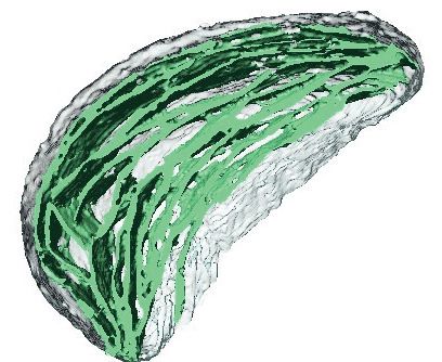
Research article                                                                                                   Plant Biology

                   A
                                                   STRATIFICATION        ETIOLATION
                                                     3 days 4°C           3 days 22°C                        DE-ETIOLATION

                                                                                              0 4 8 12       24              48          72         96     Hours
                                                                                 TEM

                                                                               SBF-SEM
                                                                               Proteomics
                                                                               Lipidomics

                   B
                                                         T0                        T4                         T24                             T96

                   C                                                                                     D
   Chlorophyll concentration (ng/seedling)

                                                                                                                    1
                                             300
                                                                                                                   0.7

                                             200
                                                                                                           Fv/Fm

                                                                                                                   0.5

                                             100
                                                                                                                   0.2

                                              0                                                                     0
                                                   0          25          50             75          100                 0    10       20     30      40       50
                                                               Time of light exposure (h)                                         Time of light exposure (h)

Figure 1. Photosynthesis onset during de-etiolation. (A) Scheme of the experimental design. Seeds of Arabidopsis thaliana (Columbia) sown on agar
plates were stratified for three days at 4˚C and then transferred to 22˚C in the dark. After 3 days, etiolated seedlings were exposed to continuous white
light (40 mmol/m2/s) and harvested at different time points during de-etiolation. Selected time points used for different analyses are indicated. (B)
Cotyledon phenotype of etiolated seedlings (T0) after 4 hr (T4), 24 hr (T24), and 96 (T96) hr in continuous white light. Scale bars: 0.5 mm. (C) Chlorophyll
quantification at different time points upon illumination. Error bars indicate ± SD (n = 3). (D) Maximum quantum yield of photosystem II (Fv/Fm). Error
bars indicate ± SD (n = 4–10). For some data points, the error bars are inferior to the size of the symbol. Measurements of further photosynthetic
parameters are presented in Figure 1—figure supplement 1.
The online version of this article includes the following figure supplement(s) for figure 1:
Figure supplement 1. Photosynthesis parameters during de-etiolation.

                                                                            (photochemical quenching, qP and PSII quantum yield in the light, FPSII, Figure 1—figure supple-
                                                                            ment 1B and C) reached maximum values at T14 and remained stable thereafter, indicating that the
                                                                            assembly of a fully functional photosynthetic machinery occurs within the first 14 hr of de-etiolation,
                                                                            and that further biosynthesis of photosynthesis related compounds is efficiently coordinated.

                                                                            Major thylakoid structural changes occur within 24 hr of de-etiolation
                                                                            We determined the dynamics of thylakoid biogenesis during the etioplast–chloroplast transition by
                                                                            observing chloroplast ultrastructure in cotyledons using transmission electron microscopy (TEM)

Pipitone et al. eLife 2021;10:e62709. DOI: https://doi.org/10.7554/eLife.62709                                                                                              4 of 32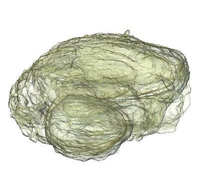
Research article                                                                                                                    Plant Biology

                                     (Figure 2). Plastids present in cotyledons of etiolated seedlings displayed the typical etioplast ultra-
                                     structure with a paracrystalline PLB and tubular PTs (Figure 2A). The observed PLBs were consti-
                                     tuted of hexagonal units with diameters of 0.8–1 mm (Figure 2E). By T4, the highly structured PLBs
                                     progressively disappeared and thylakoid lamellae were formed (Figure 2B). The lamellae were blurry
                                     and their thickness varied between 15 and 70 nm (Figure 2F). After 24 hr of illumination (T24), the
                                     density of lamellae per chloroplast was higher than that at T4 due to an increase in lamellar length
                                     and number. Appressed regions corresponding to developing grana stacks also appeared by T24
                                     (Figure 2C and G). These early grana stacks consisted of 2–6 lamellae with a thickness of 13 nm
                                     each (Figure 2—figure supplement 1). In addition, starch granules were present at T24, supporting
                                     the notion that these chloroplasts are photosynthetically functional and able to assimilate carbon
                                     dioxide (CO2). At T96, thylakoid membrane organization was visually similar to that at T24, but with
                                     more layers per grana (up to 10 lamellae per grana; Figure 2D and H). In addition, singular lamella
                                     thickness at T96 increased by 2–3 nm compared to that at T24 (Figure 2—figure supplement 1).
                                     The major differences observed between T24 and T96 were increases in starch granule size and
                                     number and overall chloroplast size (Figure 2C and D and Table 1). Etioplast average length (esti-
                                     mated by measuring the maximum distance on individual slices) was 2 mm (±0.9, n = 10) in the dark

                    T0                              T4                                 T24                                      T96
     A                              B                                   C                    SG              D                         SG
                    PLB

                              PE
                                                        PE

     E                 PT                 F                               G                                   H

                      PLB

                                                                                                                                               GS
                                                  SL                                      GS
                                                                                               SL
                                    PE

                                                                                                                                          SL

Figure 2. Qualitative analysis of chloroplast ultrastructure during de-etiolation. Transmission electron microscopy (TEM) images of cotyledon cells of 3-
day-old, dark-grown Arabidopsis thaliana (Columbia) seedlings illuminated for 0 hr (T0, A and E), 4 hr (T4, B and F), 24 hr (T24, C and G), and 96 hr (T96,
D and H) in continuous white light (40 mmol/m2/s). (A–D) Scale bars: 500 nm, (E–H) higher magnification of A–D images; Scale bars: 200 nm. PLB:
prolamellar body; PT: prothylakoid; PE: plastid envelope; SG: starch grain; GS: grana stack; SL: single lamella. Specific details for measurements of
lamella thickness are provided in Figure 2—figure supplement 1.
The online version of this article includes the following figure supplement(s) for figure 2:
Figure supplement 1. Measurement of lamella thickness.

Pipitone et al. eLife 2021;10:e62709. DOI: https://doi.org/10.7554/eLife.62709                                                                       5 of 32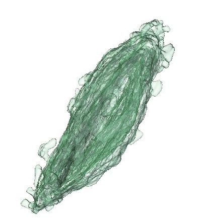
Research article                                                                                                                          Plant Biology

Table 1. Collection of quantitative data.
Morphometric data corresponding to thylakoid surfaces and volumes, thylakoid/envelope surface ratio, and chloroplast and cell vol-
umes were collected after SBF-SEM and 3D reconstruction. Chloroplast and cell volumes were also quantified by subsequent confocal
microscopy analysis, whereas plastid length was measured using TEM images. Molecular data for galactolipids (GLs) were analyzed by
lipidomics, whereas PsbA, PsaC, and PetC were quantified by quantitative immunodetection.
                              Method         T0             T4             T8            T12            T24            T48            T72             T96
Chloroplast volume            SBF-SEM        12.27 (±2.3)   9.4 (±4.8)     -             -              62 (±2.04)     -              -               112.14 (±4.3)
(mm3)
Thylakoid surface             SBF-SEM        -              67 (±29.5)     -             -              1476 (±146)    -              -               2086 (±393)
(mm2)
Grana lamellae/total                         -              -              -             -              2.55 (±0.11)   -              -               2.08 (±0.57)
thylakoid surface
Thylakoid/envelope                           -              1.02 (±0.15)   -             -              7.37 (±0.51)   -              -               6.83 (±1.40)
surface
Length of plastid             TEM            2 (±0.90)      2.8 (±0.90)    -             -              5.1 (±1.47)    -              -               6
(mm)                                                                                                                                                  (±1.62)
Stroma lamellae volume        SBF-SEM                       2.43 (±0.95)   -             -              17.87 (±1.04) -               -               29.17 (±1.94)
(mm3)
Chloroplast volume            Confocal       -              -              -             -              61.5 (±11.2)   70.1 (±10.2)   85              -
(mm3)                                                                                                                                 (±22)
Cell volume                   SBF-SEM        1173 (±284)    1891 (±362)    -             -              6103 (±1309) -                -               52597
(mm3)                                                                                                                                                 (±12671)
Cell perimeter                TEM                                                                       55.3 (±14.1)   46.4 (±6.1)    71.7 (±19.1)    92.8 (±22.1)
(mm)
Number of chloroplast per SBF-SEM            22 (±6)        25 (±8)        -             -              26             -              -               112
cell                                                                                                    (±6)                                          (±29)
Number of cells per                          -              -              -             -              ~3000          -              -               ~3000
seedling
Protein / GLs surface                        0.19           0.23 (±0.04)   0.34          0.52 (±0.07)   0.80 (±0.14)   0.80 (±0.17)   0.78 (0.07)     0.87 (±0.25)
                                             (±0.05)                       (±0.03)
GLs (nmol/seedling)           Lipidomics     0.31           0.31 (±0.02)   0.32          0.54 (±0.02)   0.67 (±0.04)   1.28 (±0.12)   1.84 (±0.01)    2.20 (±0.09)
                                             (±0.03)                       (±0.02)
PsbA (nmol/seedling)          Immuno-        6.9E-06        9.2E-06        1.5E-05       3.2E-05        9.3E-05        2.0E-04        3.9E-04         6.2E-04
                              detection      (±1.8E-06)     (±1.7E-06)     (±0.07E-05)   (±0.4E-05)     (±2E-05)       (±0.6E-04)     (±0.4E-04)      (±1.7E-04)
PsaC (nmol/seedling)          Immuno-                                                    1.6E-05 (±0.2 7.3E-05         1.1E-04        1.7E-04         2.3E-04
                              detection                                                  E-05)         (±2E-05)        (±0.7E-04)     (±0.4E-04)      (±1E-04)
PetC                          Immuno-        2.7E-05        2.8E-05        2.5E-05       5.3E-05        1.2E-04        1.8E-04 (±0. 5.7E-04           7.9E-04
(nmol /seedling)              detection      (±0.8E-05)     (±1E-05)       (±0.4E-05)    (±2.2E-05)     (±0.4E-04)     E-04)        (±1.8E-04)        (±3.7E-04)

                                         (T0), whereas chloroplast average length was 6 mm (±1.62, n = 10) at T96 (Table 1). Collectively,
                                         these data show that photosynthetically functional thylakoid membranes form rapidly during the first
                                         24 hr of de-etiolation. This implies that there are efficient mechanisms for thylakoid assembly and
                                         structural organization. Subsequent changes seem to involve the expansion of pre-existing structures
                                         (i.e. lamellae length and grana size) and the initiation of photosynthetic carbon fixation (reflected by
                                         starch content).

                                         Quantitative analysis of thylakoid surface area per chloroplast during
                                         de-etiolation
                                         To visualize entire chloroplasts and thylakoid networks in 3D, and to obtain a quantitative view of
                                         the total thylakoid surface area during chloroplast development, we prepared and imaged cotyle-
                                         dons at different developmental stages by SBF-SEM (Figure 3A–D). PLBs, thylakoids, and envelope
                                         membranes were selected, and segmented images were used for 3D reconstruction (Figure 3E–L,
                                         and Videos 1–4; see also Figure 2—figure supplement 1 and Figure 3—figure supplement 1 for
                                         grana segmentation). Similar to that observed by TEM (Figure 2), a drastic switch from PLB to

Pipitone et al. eLife 2021;10:e62709. DOI: https://doi.org/10.7554/eLife.62709                                                                                6 of 32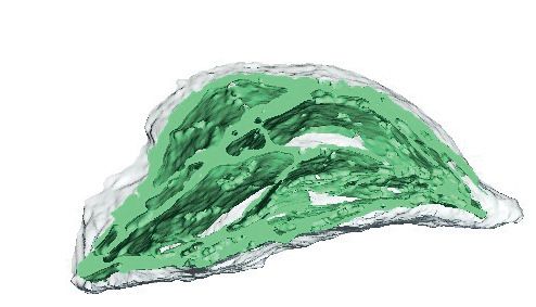
Research article                                                                                                                    Plant Biology

    A                                      B                                C                                  D

    E                                      F                                G                                  H
                                                                                                                         PE
                          PE
                                                                                 PE                                                SL

                                                                                                          SL
                                                                                                        GS

                                                                                                                                        GS
                                                                 PE
                       PT                              SL
           PLB

    I                                      J                                 K                                 L

Figure 3. 3D reconstructions of chloroplast thylakoid networks during de-etiolation. (A–D) Scanning electron microscopy (SEM) micrographs of
representative etioplasts and chloroplasts from 3-day-old, dark-grown Arabidopsis thaliana seedlings illuminated for 0 hr (T0; A), 4 hr (T4; B), 24 hr (T24;
C), and 96 hr (T96; D) in continuous white light (40 mmol/m2/s). (E–H) Partial 3D reconstruction of thylakoid membranes (green) and envelope (blue) at
T0 (E), T4 (F), T24 (G), and T96 (H). Z-depth of thylakoid membrane reconstruction corresponds to 0.06 mm (E), 0.10 mm (F), 0.13 mm (G), and 0.15 mm
(H). (I–L) 3D reconstruction of a thylakoid membrane of an etioplast at T0 (I) or a chloroplast at T4 (J), T24 (K), and T96 (L). Scale bars = 1 mm. Details of
grana segmentation at T24 are provided in Figure 3—figure supplement 1. PLB: prolamellar body; PT: prothylakoid; PE: plastid envelope; SG: starch
grain; GS: grana stack; SL: single lamella.
The online version of this article includes the following figure supplement(s) for figure 3:
Figure supplement 1. Grana segmentation (T24).

                                      thylakoid membrane occurred by T4: the typical structure of the PLB connected to PTs disappeared
                                      leaving only elongated lamellar structures (Figure 3E–F and Videos 1 and 2). At T24 and T96, thyla-
                                      koid membranes were organized in appressed and non-appressed regions and large spaces occu-
                                      pied by starch granules were observed (Figure 3G–H and Videos 3 and 4). 3D reconstruction
                                      revealed a change in plastid shape from ovoid at T0 and T4 to hemispheric at T24 and T96
                                      (Figure 3I–L).
                                          Using 3D reconstruction of the thylakoid network for three or four chloroplasts for each develop-
                                      mental stage, quantitative data such as chloroplast volume and membrane surface area were
                                      extracted and calculated (Figure 4A and B, Figure 3—figure supplement 1 and Table 1). The total
                                      chloroplast volume increased about 11-fold from T4 (9.4 mm3) to T96 (112.14 mm3) (Table 1). In par-
                                      allel, the thylakoid surface area (stroma side) increased about 30-fold reaching 2086 (±393) mm2 per
                                      chloroplast at T96 (Figure 4A and Table 1). The surface area increased drastically between T4 and

Pipitone et al. eLife 2021;10:e62709. DOI: https://doi.org/10.7554/eLife.62709                                                                         7 of 32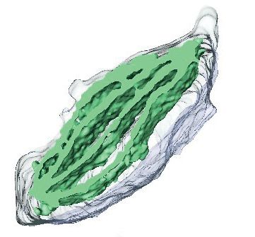
Research article                                                                                                               Plant Biology

                                                                                                T24 (about 22-fold) and much less (about 1.4-
                                                                                                fold) between T24 and T96. Accordingly, quantifi-
                                                                                                cation of the envelope surface area indicated that
                                                                                                the ratio of the thylakoid to envelope surface
                                                                                                area increased drastically from T4 to T24, but
                                                                                                decreased slightly between T24 and T96
                                                                                                (Table 1).
                                                                                                   Our quantitative observations confirmed that
                                                                                                during chloroplast development the major ultra-
                                                                                                structural changes (disappearance of prolamellar
                                                                                                body, build-up of the thylakoids and their organi-
                                    Video 1. Representative sequential sections showing
                                                                                                zation into grana) occurs within the first 24 hr of
                                    etioplasts (T0) followed by segmentation and 3D
                                    reconstruction of envelope (blue), and prothylakoids        de-etiolation, and no drastic changes occur there-
                                    and prolamellar body (yellow) of a single etioplast. The    after. We further analyzed these temporal pro-
                                    tour of the etioplast reveals its ovoid shape. The          cesses at the molecular level focusing on proteins
                                    sequential view of the 3D reconstruction and final          and lipids that constitute the thylakoid
                                    partial 3D visualization reveals a single prolamellar       membrane.
                                    body and interconnected prothylakoids.
                                    https://elifesciences.org/articles/62709#video1             Dynamics of plastid proteins
                                                                                                related to thylakoid biogenesis
                                                                                       We analyzed the full proteome to reveal the
                                                                                       dynamics of protein accumulation during de-etio-
                                    lation. Total proteins were prepared from 3-day-old etiolated seedlings exposed to light for 0–96 hr
                                    (eight time points; Figure 1A) and quantified by label-free shot-gun mass spectrometry. For relative
                                    quantification of protein abundances between different samples, peptide ion abundances were nor-
                                    malized to total protein (see Materials and methods). We considered further only those proteins that
                                    were identified with a minimum of two different peptides (with at least one being unique; see
                                    Materials and methods for information on protein grouping), resulting in the robust identification
                                    and quantification of more than 5000 proteins.
                                        Based on this proteomic approach, the first 12 hr of illumination (T12) saw very few statistically
                                    significant changes in protein abundance (Figure 5—source data 1). Considering a q-value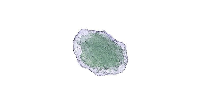
Research article                                                                                                     Plant Biology

                                    which belonged to the chlorophyll a/b binding
                                    proteins category involved in photoprotection
                                    (AT1G44575 = PsbS; AT4G10340 = Lhcb5;
                                    AT1G15820 = Lhcb6; Chen et al., 2018;
                                    Li et al., 2000). Relaxing the statistical threshold
                                    value to 0.05, cryptochrome 2 and Lhcb6 levels
                                    were respectively decreased and increased
                                    already after 4 hr of illumination and the abun-
                                    dance of two other proteins (ATCG00790 =
                                    Ribosomal protein L16; AT4G15630 = Uncharac-
                                    terized protein family UPF0497) increased
                                                                                           Video 4. Representative sequential sections of a
                                    slightly (fold changes of 1.9 and 1.7, respec-
                                                                                           chloroplast (T96) followed by segmentation and 3D
                                    tively). At 8 hr, a total of 36 proteins displayed a
                                                                                           reconstruction of envelope (blue), and thylakoids
                                    change in abundance with a q-value 2; q-value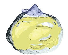
Research article                                                                                                                   Plant Biology

   A                                                                                           B
    Thylakoid surface / chloroplast (ȝP2)

                                                                                               &KORURSODVWYROXPHFKORURSODVW ȝP3)
                                            3500                                                                                         140

                                            3000                                                                                         120

                                            2500                                                                                         100

                                            2000                                                                                         80

                                            1500                                                                                         60

                                            1000                                                                                         40

                                            500                                                                                          20

                                              0                                                                                           0
                                                        4             24           96                                                          0      4               24         96
                                                         7LPHRIOLJKWH[SRVXUH K                                                               7LPHRIOLJKWH[SRVXUH K

Figure 4. Quantitative analysis of chloroplast volume and thylakoid surface during de-etiolation. Quantification of thylakoid surface per chloroplast (A)
and chloroplast volume (B) using 3-day-old, dark-grown Arabidopsis thaliana (Columbia) seedlings illuminated for 0 hr, 4 hr, 24 hr, and 96 hr in
continuous white light (40 mmol/m2/s). Morphometric data were quantified by Labels analysis module of Amira software. Error bars indicate ± SD
(n = 3). The total thylakoid surface indicated in A corresponds to the thylakoid surface exposed to the stroma, calculated in Amira software, in addition
to the percentage of the grana surface (%Gs) calculated as described in Figure 3—figure supplement 1.
The online version of this article includes the following source data for figure 4:
Source data 1. Quantitative chloroplast morphomotric data.

                                                                     (Blomqvist et al., 2008; Runge et al., 1996; Von Wettstein et al., 1995). In agreement, illumination
                                                                     resulted in increased amounts of most of all detected proteins of the chlorophyll biosynthesis path-
                                                                     way, except PORA and to a lesser extent PORB, which clearly decreased and were separated from
                                                                     other chlorophyll-related proteins (Figure 5C and Figure 5—source data 1). We also selected pro-
                                                                     teins involved in protein import in chloroplasts, focusing on the TOC-TIC machinery (Figure 5D) that
                                                                     is the major route for plastid protein import and essential for chloroplast biogenesis (Kessler and
                                                                     Schnell, 2006). Past studies identified several TOC preprotein receptors that are proposed to dis-
                                                                     play differential specificities for preprotein classes (Bauer et al., 2000; Bischof et al., 2011). The
                                                                     composition of plastid import complexes varies with developmental stages and in different tissues,
                                                                     thereby adjusting the selectivity of the import apparatus to the demands of the plastid and influenc-
                                                                     ing its proteome composition (Demarsy et al., 2014; Kubis et al., 2003). Accordingly, the TOC
                                                                     receptors TOC120 and TOC132, which are important for the import of proteins in non-photosyn-
                                                                     thetic tissues, were more abundant in etioplasts compared to fully-developed chloroplasts (compare
                                                                     T0 and T96). TOC120 and TOC132 were part of a cluster separated from other components of the
                                                                     plastid machinery, such as the TOC159 receptor associated with large-scale import of proteins in
                                                                     chloroplasts. The general import channel TOC75 (TOC75 III) maintained stable expression levels
                                                                     throughout de-etiolation, reflecting its general role in protein import. All other components clus-
                                                                     tered with TOC159 and displayed gradual increases in accumulation during de-etiolation. Most of
                                                                     these components have not been reported to confer selectivity to the import machinery, which sug-
                                                                     gests an overall increase of chloroplast protein import capacity.
                                                                         To validate and complement our proteomic data, we used immunoblot analysis to detect and
                                                                     quantify representative proteins linked to photomorphogenesis and etioplast-to-chloroplast
                                                                     transition.
                                                                         Our proteomic data indicated a significant decrease of the abundance of the photoreceptor
                                                                     phyA between 48 and 72 hr of illumination (Figure 5—source data 1). However, immunoblots
                                                                     revealed that the abundance of phyA dropped already during the first 4 hr of light exposure (Fig-
                                                                     ure 6), as previously reported (e.g. Debrieux and Fankhauser, 2010). The transcription factor

Pipitone et al. eLife 2021;10:e62709. DOI: https://doi.org/10.7554/eLife.62709                                                                                                        10 of 32
Research article Plant Biology
A
0 0.5 1 1.5 2 2.5 3
AT1G45474 Lhca5
AT4G15510 PPD1
AT1G76450 PSBP
AT1G30380 PsaK
AT2G40100 Lhcb4.3
ATCG00540 PetA
AT1G03600 Psb27
ATCG00720 PetB
AT2G39470 PPL2
AT1G55670 PsaG
AT3G47470 Lhca4
AT5G54270 Lhcb3
AT4G03280 PetC
AT1G44575 PsbS
AT4G09650 AtpD
ATCG00470 AtpE
AT4G21280 PsbQA
AT1G31330 PsaF
ATCG00020 PsbA
AT1G29930 Lhcb1.3
AT4G10340 Lhcb5
AT1G15820 Lhcb6
AT4G05180 3VE4í
ATCG00710 PsbH
AT4G02770 3VD'í
AT3G61470 Lhca2
AT2G05070 Lhcb2.2
ATCG00580 PsbE
ATCG00680 PsbB
ATCG00560 PsbL
AT4G28750 3VD(í
AT4G12800 PsaL
AT3G54890 Lhca1
AT1G61520 Lhca3
AT5G01530 Lhcb4.1
AT3G08940 Lhcb4.2
AT1G52230 PsaH2
AT2G20260 3VD(í
AT3G50820 PsbO2
ATCG00270 PsbD
ATCG00350 PsaA
ATCG00480 AtpB
ATCG00130 AtpF
AT5G66570 PsbO1
ATCG00280 PsbC
AT1G06680 3VE3í
AT1G79040 PsbR
AT4G04640 AtpC1
ATCG00120 AtpA
AT2G34430 Lhcb1.4
ATCG00340 PsaB
0 4 8 12 24 48 72 96
0.6 0.3 0
Time of light exposure (h)
B
0 0.2 0.4 0.6 0.8 1 1.2
AT4G33030 SQD1
AT2G43710 FAB2
AT1G32200 ACT1
AT4G30950 FAD6
AT3G15850 FAD5
AT3G06510 SFR2
AT1G74960 FAB1
AT3G11170 FAD7
0.4 0.2 0 0 4 8 12 24 48 72 96
Time of light exposure (h)
C
0 0.5 1 1.5 2 2.5 3
AT3G56940 CRD1
AT2G26670 TED4
AT1G04620 HCAR
AT4G37000 ACD2
AT5G08280 HEMC
AT5G63570 GSA1
AT3G48730 GSA2
AT1G44446 CH1
AT3G14930 HEME1
AT1G69740 HEMB1
AT3G44880 ACD1
AT1G03630 PORC
AT2G26540 HEMD
AT4G01690 PPOX
AT5G04900 NOL
AT5G64050 ERS
AT5G13630 GUN5
AT2G30390 FC2
AT2G40490 HEME2
AT5G45930 CHLI2
AT1G08520 ALB1
AT4G18480 CHLI1
AT1G58290 HEMA1
AT5G18660 PCB2
AT4G25080 CHLM
AT5G26030 FC1
AT1G03475 LIN2
AT4G27440 PORB
AT5G54190 PORA
0 4 8 12 24 48 72 96
3.0 1.5 0
Time of light exposure (h)
D
0.5 1 1.5 2
AT2G24820 TIC55
AT4G33350 TIC22
AT3G18890 TIC62
AT5G19620 T2&í9
AT4G25650 7,&í/,.(
AT2G439502(3í
AT2G28900 OEP16
AT5G05000 TOC34
AT1G02280 TOC33
AT1G06950 TIC110
AT4G02510 TOC159
AT3G46740 TOC75 III
AT5G16620 TIC40
AT2G16640 TOC 132
AT5G42960 2(3í
AT3G17970 TOC 64 II
AT3G16620 TOC120
0 4 8 12 24 48 72 96
1.8 0.9 0
Time of light exposure (h)
Figure 5. Accumulation dynamics of plastid proteins during de-etiolation. Three-day-old etiolated seedlings of
Arabidopsis thaliana were illuminated for 0 hr (T0), 4 hr (T4), 8 hr (T8), 12 hr (T12), 24 hr (T24), 48 hr (T48), 72 hr
(T72), and 96 hr (T96) under white light (40 mmol/m2/s). Hierarchical clustering (Euclidean, average linkage) of
normalized protein abundance for photosynthesis-(A), galactolipid metabolism- (B), chlorophyll metabolism- (C),
Figure 5 continued on next page
Pipitone et al. eLife 2021;10:e62709. DOI: https://doi.org/10.7554/eLife.62709 11 of 32Research article Plant Biology
Figure 5 continued
and protein import-related proteins during de-etiolation (D). Protein abundance was quantified by shot-gun
proteomics and heatmap colors indicate the fold change (average of 3–4 replicates) of each selected protein at
each time point of de-etiolation (T0 to T96), relative to the last time point (T96). Note that some PORA values in
panel D were higher than 3.5 and outside of the color range limits. Further hierarchical clustering based on the
accumulation dynamics of all plastid-localized proteins is provided in Figure 5—figure supplement 1.
The online version of this article includes the following source data and figure supplement(s) for figure 5:
Source data 1. Chloroplast localized proteins identified by MS and clusters.
Figure supplement 1. Accumulation dynamics of selected plastid proteins during de-etiolation.
Figure supplement 1—source data 1. List of proteins identified by MS and quantitative data.
ELONGATED HYPOCOTYL 5 (HY5) is a positive regulator of photomorphogenesis, and accumulates
during light exposure (Osterlund and Deng, 1998). The increase of HY5 peptide abundance was
not significant by proteomics but we observed a transient accumulation of the protein between 4
and 72 hr by immunoblot (Figure 5—source data 1; Figure 6) consistent with the previously
reported regulation of abundance during seedling development (Hardtke et al., 2000).
We further compared data obtained by proteomics and immunoblot focusing on chloroplast
localized proteins. Overall, immunoblot and proteomics provided similar results (Figure 6 and Fig-
ure 6—figure supplement 1). PsbA and PsbD (PSII reaction center core), PsbO (Oxygen Evolving
Complex), and Lhcb2 (outer antenna complex) proteins were detectable in seedlings at T4, gradually
increasing thereafter. Accumulation of the PSI proteins PsaC and PsaD and the Cyt b6f complex pro-
tein PetC started later; these proteins were detectable starting at T8 (Figure 6A and Figure 6—fig-
ure supplement 1). Interestingly, AtpC (ATP synthase complex) was detectable in the etioplast, as
described previously (Plöscher et al., 2011). Other proteins were selected as markers of etioplast–
chloroplast transition. As expected, ELIPs (Early Light Induced Protein) transiently accumulated upon
the dark-to-light transition (Figure 6A; Kimura et al., 2003). As in the proteome analysis, PORA
accumulated in etiolated seedlings (T0) and then progressively disappeared upon light exposure.
We performed absolute quantification for PsbA, PsaC, and PetC proteins using recombinant pro-
teins as standards (Figure 6B and C and Figure 6—figure supplement 1). Quantitative data (nmol/
seedling) were obtained and normalized using the last time point (Figure 6C) to compare the
dynamics of protein accumulation. In addition, the comparison of PsbA and PsaC (representative
proteins of PSII and PSI, respectively) showed that PsbA levels were about twice that of PsaC at T96
(Figure 6B and C).
Dynamics of chloroplast membrane lipids
Total lipids were extracted from seedlings collected at different time points during de-etiolation (T0,
T4, T8, T12, T24, T48, T72, and T96), analyzed by ultra-high-pressure liquid chromatography–mass
spectrometry (UHPLC-MS), and quantified against pure standards (Figure 7—source data 1). We
analyzed the quantity and kinetics of accumulation of 12 different species of galactolipids
(Figure 7A and B). MGDG 18:3/16:3, MGDG 18:3/18:3, MGDG 18:3/16:1, DGDG 18:3/18:3, and
DGDG 18:3/16:0 were the most abundant lipids detected at all time points. Accumulation of all gal-
actolipids increased upon de-etiolation; however, clustering analysis identified two distinct kinetic
patterns. One group displayed a leap between T8 and T12, whereas the other group increased later
during the de-etiolation period (Figure 7C). Interestingly, the two clusters separated the lipids
according to the two pathways described for galactolipid synthesis, namely the ER and PL pathways
(Figure 7A and B; Marechal et al., 1997; Ohlrogge and Browse, 1995). During early stages of de-
etiolation (T0–T24), we observed an incremental accumulation of MGDG and DGDG galactolipids
derived from the ER pathway, whereas galactolipids from the PL pathway started to accumulate at
T24 (Figure 7A and B). The MGDG/DGDG ratio decreased between T0 and T8. This was associated
with the transition from PLB (cubic lipid phase) to thylakoid membrane (lamellar structure)
(Bottier et al., 2007). The MGDG/DGDG ratio started to increase gradually at T8 and was constant
by T72 and T96 (Figure 7D).
Pipitone et al. eLife 2021;10:e62709. DOI: https://doi.org/10.7554/eLife.62709 12 of 32Research article Plant Biology
Time of light exposure (h)
A 0 4 8 12 24 48 72 96
38kDa PsbA
38kDa PsbD
38kDa PsbO
21kDa PetC
21kDa PsaD
9kDa PsaC
25kDa Lhcb2
42kDa AtpC
21kDa ELIP
38kDa POR
124kDa phyA
18kDa HY5
48kDa ACTIN
B C
-1 -0.5 0 0.5 1
PsbA
PetC
PsaC
0 4 8 12 24 48 72 96 0 0.2 0.4 0.6 0.8 1 1.2
Time of light exposure (h) nmol/seedling (E-03)
(T96)
Figure 6. Accumulation dynamics of photosynthesis-related proteins during de-etiolation. Three-day-old etiolated seedlings of Arabidopsis thaliana
were illuminated for 0 hr (T0), 4 hr (T4), 8 hr (T8), 12 hr (T12), 24 hr (T24), 48 hr (T48), 72 hr (T72), and 96 hr (T96) under white light (40 mmol/m2/s). (A)
Proteins were separated by SDS-PAGE and transferred onto nitrocellulose membrane and immunodetected with antibodies against PsbA, PsbD, PsbO,
PetC, PsaD, PsaC, Lhcb2, AtpC, ELIP, POR, phyA, HY5, and ACTIN proteins. (B–C) Quantification of PsbA, PetC, and PsaC during de-etiolation.
Figure 6 continued on next page
Pipitone et al. eLife 2021;10:e62709. DOI: https://doi.org/10.7554/eLife.62709 13 of 32Research article Plant Biology
Figure 6 continued
Heatmap (B) was generated after normalization of the amount of each protein relative to the last time point (T96). Graph (C) corresponds to the
absolute quantification of proteins at T96. Error bars indicate ± SD (n = 3). Quantification of photosystem-related proteins during de-etiolation is
detailed in Figure 6—figure supplement 1.
The online version of this article includes the following source data and figure supplement(s) for figure 6:
Source data 1. Quantitative data for immunoblot analysis.
Figure supplement 1. Quantification of photosynthesis-related proteins.
Identification of a chloroplast division phase
We observed a massive increase in the accumulation of photosynthesis-related proteins and galacto-
lipids between T24 and T96, corresponding to FC > 2 in the levels of all major chloroplast proteins
and lipids (Figures 6 and 7). Intriguingly, the total thylakoid surface per chloroplast increased by
only 41% between these two time points (Figure 4A and Table 1). We reasoned that the increase in
chloroplast proteins and lipids between T24 and T96 could be explained by increased chloroplast
number (per cell and thus per seedling) and thus total thylakoid surface per seedling. We therefore
determined chloroplast number per cell and the cell number and volume for each developmental
stage through SBF-SEM analysis (T0, T4, T24, and T96) and confocal microscopy analysis for interme-
diary time points (T24–T96) (Figure 8 and Figure 8—figure supplement 1). The chloroplast number
per cell was constant from T4 (25 ± 8) to T24 (26 ± 6); however, in parallel with cell expansion
(Figure 8A and B), chloroplast number increased sharply (fourfold increase) between T24 (26 ± 6)
and T96 (112 ± 29), indicating that two rounds of chloroplast division occurred during this time.
Immunoblot analysis of FILAMENTOUS TEMPERATURE-SENSITIVE FtsZ1, FtsZ2-1, and FtsZ2-2 pro-
teins showed that these key components of the chloroplast division machinery were already present
during the early time points of de-etiolation. We observed considerably increased accumulation of
these proteins between T24 and T48, consistent with the idea that activation of chloroplast division
takes place at T24, leading the proliferation of chloroplasts (Figure 8C). However, levels of ACCU-
MULATION AND REPLICATION OF CHLOROPLAST 5 (ARC5) protein, another key component of
the chloroplast division machinery, clearly increased during de-etiolation between T8 and T12, pre-
sumably reflecting assembly of the chloroplast division machinery before its activation and the prolif-
eration of chloroplasts (Figure 8D). To test whether there is a correlation between chloroplast
division and either volume or developmental stage, we measured the volume of dividing chloro-
plasts (selected visually based on the presence of a constriction ring, see Figure 8—figure supple-
ment 1) at T24 and T96 using images acquired by SBF-SEM. The average volume of dividing
chloroplasts at T24 and T96 were consistently higher than the average volume of all chloroplasts (96
mm3 and 136 mm3 compared to 62 mm3 and 112 mm3, respectively) (Figure 4B, Figure 8E and Fig-
ure 8—source data 1) indicating that smaller chloroplasts are not dividing. This indicates that devel-
oping chloroplasts only divide once a certain chloroplast volume is reached.
Model of thylakoid surface expansion over time
The quantitative molecular data for the major compounds of thylakoids (galactolipids and proteins)
and estimation of chloroplast number per cell allowed us to mathematically determine the thylakoid
membrane surface area per seedling and its expansion over time (molecular approach hereafter) and
compare it to the surface estimated from the 3D reconstruction (morphometric approach hereafter).
First, we calculated the surface area occupied by the main galactolipids (MGDG and DGDG) and
photosynthesis-related complexes (PSII, Cyt b6f, and PSI) per seedling (Table 2), assuming a 1:1 ratio
between number of PsbA, PetC, and PsaC subunits with their corresponding complexes
(Amunts and Nelson, 2009; Caffarri et al., 2014; Schöttler et al., 2015).
Surface=seedling ¼ nmol=seedling N nm2 per molecule (1)
Quantitative data for MGDG, DGDG, PsbA, PetC, and PsaC (nmol/seedling) obtained from lipi-
domic and immunological analyses (Figures 6 and 7) were converted into number of molecules/
seedling using the Avogadro constant (N). To calculate the surface area of outer membrane of thyla-
koids (i.e. surface exposed to the stroma in lamellae and facing the other thylakoid in appressed
regions) and account for the lipid double layer of the membrane, corresponding values of lipids
Pipitone et al. eLife 2021;10:e62709. DOI: https://doi.org/10.7554/eLife.62709 14 of 32Research article Plant Biology
A B
0.2 0.4 0.6 0.8 1
DGDG 18:3/18:3
DGDG 18:0/18:3
EUKARYOTIC DGDG 18:1/18:3
PATHWAY DGDG 18:3/20:3
MGDG 18:2/18:3
MGDG 18:3/18:3
MGDG 18:3/16:3
DGDG 18:3/16:3
PROKARYOTIC DGDG 18:3/16:0
PATHWAY
MGDG 18:3/16:1
DGDG 18:2/16:0
0 4 8 12 24 48 72 96 0,0 0,1 0,2 0,3 0,4 0,5 0,6 0,7 0,8 0,9
Time of light exposure (h) nmol/seedling
(T96)
C
0.25
0.20
MGDG 18:3/18:3
DGDG 18:3/18:3
(nmol/seedling)
(nmol/seedling)
0.20
0.15
0.15
0.10
0.10
0.05
0.05
0 24 48 72 96 0 24 48 72 96
Time of light exposure (h) Time of light exposure (h)
0.8 0.08 0.06
MGDG 18:3/16:3
MGDG 18:3/16:1
DGDG 18:3/16:0
(nmol/seedling)
(nmol/seedling)
(nmol/seedling)
0.6 0.06
0.04
0.4 0.04
0.02
0.2 0.02
0.0 0.0
0 24 48 72 96 0 24 48 72 96 0 24 48 72 96
Time of light exposure (h) Time of light exposure (h) Time of light exposure (h)
3,50
D
3,00
2,50
MGDG /DGDG
2,00
ratio
1,50
1,00
0,50
0,00
0 4 8 12 24 48 72 96
Time of light exposure (h)
Figure 7. Accumulation dynamics of galactolipids during de-etiolation. Three-day-old etiolated seedlings of Arabidopsis thaliana were illuminated for 0
hr (T0), 4 hr (T4), 8 hr (T8), 12 hr (T12), 24 hr (T24), 48 hr (T48), 72 hr (T72), and 96 hr (T96) under white light (40 mmol/m2/s). (A) Heatmap representation
of galactolipids (MGDG and DGDG) during de-etiolation. Samples were normalized to the last time point (T96). (B) Absolute quantification at T96
expressed in nmol/seedling. Error bars indicate ± SD (n = 4). (C) Absolute quantification (nmol/seedling) of the most abundant chloroplast galactolipids
Figure 7 continued on next page
Pipitone et al. eLife 2021;10:e62709. DOI: https://doi.org/10.7554/eLife.62709 15 of 32Research article Plant Biology
Figure 7 continued
MGDG (MGDG 18:3/18:3, MGDG 18:3/16:3, MGDG 18:3/16:1) and DGDG (DGDG 18:3/18:3, DGDG 18:3/16:0) at different time points during de-
etiolation. Error bars indicate ± SD (n = 4). (D) The MGDG/DGDG ratio was calculated using all 12 species of galactolipids detected during de-
etiolation. Error bars indicate ± SD (n = 4).
The online version of this article includes the following source data for figure 7:
Source data 1. Quantitative data for lipidomics.
(Figure 7—source data 1, Table 2) were divided by 2. In addition, the lipid values were corrected
by subtracting the portion of lipids incorporated into the envelope rather than present in the thyla-
koids (Table 1). The surface area occupied by molecules of MGDG and DGDG, and that of PSII, Cyt
b6f, and PSI photosynthetic complexes (nm2 per molecule, corresponding to stroma-exposed sur-
face) were retrieved from the literature (Table 3). Specifically, we used the minimal molecular area of
MGDG and DGDG (Bottier et al., 2007). To quantify the surface area occupied by the galactolipids
and photosynthetic complexes in thylakoids per seedling, the number of molecules per seedling of
galactolipids was multiplied by the corresponding molecular surface area, whereas the number of
molecules per seedling of PsbA, PetC, and PsaC (subunits of PSII, Cyt b6f, and PSI, respectively)
were multiplied by the surface area of the corresponding complex (see Table 3).
We calculated thylakoid surface (S) per seedling for each time point (t) as the sum of the surface
occupied by MGDG, DGDG, photosynthetic complexes (PS), and e per seedling, the latter of which
corresponds to compounds such as other lipids (e.g. sulfoquinovosyldiacylglycerol, plastoquinone)
or protein complexes (ATP synthase and NDH) that were not quantified.
S thylakoid ðt Þ=seedling ¼ ðS MGDGðtÞ þ S DGDGðt Þ þ S PS ðtÞ þ " Þ=seedling (2)
Omitting the unknown e factor, we plotted the thylakoid surface calculated for each time point
where quantitative molecular data were available (T0, T4, T8, T12, T24, T48, T72, and T96) as a func-
tion of the duration of light exposure (Figure 9—figure supplement 1). The best fitting curve corre-
sponded to a S-shaped logistic function, characterized by a lag phase at early time points (T0–T8),
followed by a phase of near-linear increase, and a final plateau at the final time points (T72–T96). To
model this function, a four-parameter logistic non-linear regression equation was used to describe
the dynamics of the total thylakoid surface over time (Figure 9—figure supplement 1C).
Superimposition of molecular and morphometric data
We compared the values of thylakoid surface, as obtained with the model based on molecular data,
with the values obtained from the morphometric analysis (Figure 9). The total thylakoid surface per
seedling (S_thylakoid_morpho) was calculated by multiplying the thylakoid surface (S_thylakoid) per
chloroplast obtained by morphometrics (Figure 4A) by the number of chloroplasts (nb.cp) per cell
(Figure 8A) and the number of cells (nb.cells) per seedlings for each time point (t).
SthylakoidmorphoðtÞ
¼
seedling (3)
S thylakoidðt Þ=chloroplast nb:cpðt Þ=cell nb:cellsðtÞ=seedling
We estimated cell number per seedling by measuring the total volume occupied by palisade and
spongy cells in cotyledons (that corresponded to 50% of total cotyledon volume; Figure 9—figure
supplement 2) and dividing this by the average cell volume (Table 1). As reported previously
(Pyke and Leech, 1994), cell number was constant during cotyledon development. We estimated
this number as 3000 mesophyll and palisade cells per seedling at T24 and T96 (Figure 9—figure
supplement 2). The thylakoid membrane surface quantified by the morphometric approach was also
estimated at T4, assuming that cell number per cotyledon remained similar between T4 and T24.
We compared the thylakoid surface predicted by our mathematical model to the surface estimated
experimentally with our 3D thylakoid reconstruction and morphometric measurements (Figure 9 and
Table 1). As shown in Figure 9, the two approaches showed very similar total thylakoid surface area
per seedling at T4 and T24 and differences in this parameter by T96. This indicates that the plateau
Pipitone et al. eLife 2021;10:e62709. DOI: https://doi.org/10.7554/eLife.62709 16 of 32Research article Plant Biology
A B
140 70000
120 60000
Cell volume (µm3)
Chloroplasts/ cell
100 50000
80 40000
60 30000
40 20000
20 10000
0 0
0 4 24 96 0 4 24 96
Time of light exposure (h) Time of light exposure (h)
C Time of light exposure (h)
D Time of light exposure (h)
0 4 8 12 24 48 72 96 0 4 8 12 24 48 72 96
FtsZ2-1
42kDa FtsZ2-2 87kDa ARC5
38kDa FtsZ1 42kDa ACTIN
42kDa ACTIN
E
DIVIDING CHLOROPLASTS
180
160
140
Volume (µm3)
120
100
80
60
40
20
0
24 96
Time of light exposure (h)
Figure 8. Relationship between chloroplast proliferation and chloroplast volume. (A–B) Chloroplast number and cell volume in cotyledons of 3-day-old,
dark-grown Arabidopsis thaliana seedlings illuminated for 0 hr (T0), 4 hr (T4), 24 hr (T24), and 96 hr (T96) in continuous white light (40 mmol/m2/s). (A)
Chloroplast number per cell during de-etiolation. Error bars indicate ± SD (n = 6 for T0 and T4; seven for T24; five for T96). (B) Cell volume was
quantified by the Labels analysis module of Amira software. Error bars indicate ± SD (n = 5–6). (C–D) Total proteins were extracted from T0–T96
seedlings, separated on SDS-PAGE, and transferred onto nitrocellulose. Proteins involved in plastid division (C, FtsZ; D, ARC5) and loading control
(actin) were detected using specific antibodies (FtsZ2 antibody recognizes both FtsZ2-1 and FtsZ2-2). (E) Volume of dividing chloroplast at T24 and T96.
Error bars indicate ± SD (n = 3). Further details of chloroplast proliferation in parallel with cell expansion are provided in Figure 8—figure supplement
1.
Figure 8 continued on next page
Pipitone et al. eLife 2021;10:e62709. DOI: https://doi.org/10.7554/eLife.62709 17 of 32Research article Plant Biology
Figure 8 continued
The online version of this article includes the following source data and figure supplement(s) for figure 8:
Source data 1. Quantitative data for chloroplast number, cell and chloroplast volumes.
Figure supplement 1. Chloroplast proliferation in parallel with cell expansion.
phase suggested by the model is not validated and that other components that were not included in
the model probably contributed to the expansion of thylakoids at later time points of de-etiolation.
Discussion
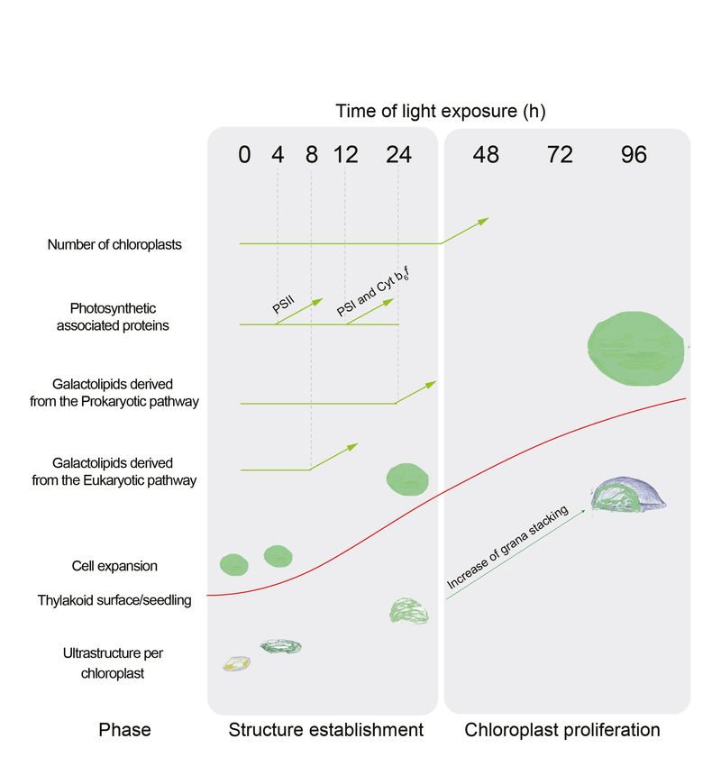
Here, the analysis of 3D structures of entire chloroplasts in Arabidopsis in combination with proteo-
mic and lipidomic analyses provide an overview of thylakoid biogenesis. Figure 10 depicts a sum-
mary of the changes that occur during the de-etiolation process. When considering chloroplast
development, our study shows that de-etiolation is divided into two phases. We documented struc-
tural changes (disassembly of the PLB and the gradual formation of thylakoid lamellae) and initial
increases of ER- and PL-pathway galactolipids and photosynthesis-related proteins (PSII, PSI, and
Cyt b6f) during the ‘Structure Establishment Phase’, which was followed by increased chloroplast
number in parallel with cell expansion in the ‘Chloroplast Proliferation Phase’. Collection of quantita-
tive data allowed us to create a mathematical model of thylakoid membrane expansion and describe
this process during de-etiolation.
A set of 3D reconstructions of whole chloroplasts by SBF-SEM
In contrast to electron tomography, which is limited in the volume of observation, SBF-SEM allows
the acquisition of ultrastructural data from large volumes of mesophyll tissue and the generation of
3D reconstructions of entire cells and chloroplasts (Figure 3 and Figure 8—figure supplement 1,
Videos 1–4). SEM image resolution was sufficient to visualize stromal lamellae and grana contours,
whereas grana segmentation in different lamellae was deduced according to our own TEM analysis
and literature data (Figure 2—figure supplement 1 and Figure 3—figure supplement 1). This
approach allowed us to obtain quantitative data of chloroplast and thylakoid structure at different
developmental stages during de-etiolation at the whole-chloroplast level. By T96, the latest time
point of our analysis, the total surface area of thylakoids present in the seedling cotyledons was
about 700 mm2 (see values in Table 1 for calculation), about 500-fold greater than the surface area
of one cotyledon at this developmental stage. This result is supported by previous estimates made
regarding thylakoid surface area relative to leaf surface area (Bastien et al., 2016; Demé et al.,
2014). Moreover, the extent of thylakoid surface area emphasizes how fast and efficient thylakoid
Table 2. Surface area occupied by the main galactolipids (MGDG and DGDG) and photosynthetic complexes (PSII, cyt b6f, and PSI).
Shown are values at different time points following illumination of 3-day-old etiolated seedlings. Each value indicates the calculated
surface area in mm2 and corresponds to the average of three biological replicates. Errors indicate SD.
T0 T4 T8 T12 T24 T48 T72 T96
MGDG 1.11E+07 1.15E+07 (±0.1E 1.11E+07 (±0.1E 1.75E+07 4.16E+07 (±0.4E 8.65E+07 (±0.6E 1.68E+08 2.35E+08 (±0.2E
(±0.03E+07) +07) +07) (±0.18E+07) +07) +07) (±0.09E+08) +07)
DGDG 3.64E+06 (±0.4E 4.23E+06 (±0.5E 4.10E+06 (±0.1E 6.26E+06 (±0.5E 1.32E+07 (±0.1 2.32E+07 (±0.2 E 3.97E+07 (±0.3E 5.48E+07
+06) +06) +06) +05) 07) +07) +07) (±0.41E+07)
PSII 2.04E+06 (±0.5 E 2.74E+06 (±0.5E 4.40E+06 (±0.2E 9.91E+06 (±1.3E 2.75E+07 (±0.6E 6.06E+07 (±0.2E 1.15E+08 (±0.2E 1.83E+08 (±0.5E
+05) +05) +06) +06) +07) +07) +08) +08)
PSI 0E+00 (±0E+00) 0E+00 (±0E+00) 0E+00 (±0E+00) 8.95E+05 1.33E+07 (±0.4E 2.10E+07 3.04E+07 (±0.8E 4.24E+07
(±4.49E+05) +07) (±1.30E+07) +07) (±1.89E+07)
Cyt b6f 7.99E+05 8.43E+05 7.5E+05 (±1.33E 1.57E+06 (±0.7E 3.44E+06 5.30E+06 1.69E+07 (±0.5E 2.37E+07
(±2.33E+05) (±2.91E+05) +05) +06) (±1.22E+06) (±1.01E+06) +06) (±1.11E+07)
The online version of this article includes the following source data for Table 2:
Source data 1. Quantitative data of surface occupied by galactolipids and proteins.
Pipitone et al. eLife 2021;10:e62709. DOI: https://doi.org/10.7554/eLife.62709 18 of 32You can also read