Cofilin-1 phosphorylation catalyzed by ERK1/2 alters cardiac actin dynamics in dilated cardiomyopathy caused by lamin A/C gene mutation
←
→
Page content transcription
If your browser does not render page correctly, please read the page content below
Human Molecular Genetics, 2018, Vol. 27, No. 17               3060–3078

                                                                                                   doi: 10.1093/hmg/ddy215
                                                                                                   Advance Access Publication Date: 5 June 2018
                                                                                                   Original Article

                ORIGINAL ARTICLE

                Cofilin-1 phosphorylation catalyzed by ERK1/2 alters
                cardiac actin dynamics in dilated cardiomyopathy
                caused by lamin A/C gene mutation
                Maria Chatzifrangkeskou1, David Yadin2, Thibaut Marais1,
                Solenne Chardonnet3, Mathilde Cohen-Tannoudji1, Nathalie Mougenot4,
                Alain Schmitt5, Silvia Crasto6,7, Elisa Di Pasquale6,7, Coline Macquart1,
                Yannick Tanguy1, Imen Jebeniani8, Michel Pucéat8,
                Blanca Morales Rodriguez1, Wolfgang H. Goldmann9, Matteo Dal Ferro10,
                Maria-Grazia Biferi1, Petra Knaus2, Gisèle Bonne1, Howard J. Worman11,12
                and Antoine Muchir1,*
                1
                 Sorbonne Université, UPMC Paris 06, INSERM UMRS974, Center of Research in Myology, F-75013 Paris, France,
                2
                 Institute for Chemistry and Biochemistry, Freie Universität Berlin, 14195 Berlin, Germany, 3Sorbonne Université,
                UPMC Paris 06, INSERM, UMS29 Omique, F-75013 Paris, France, 4Sorbonne Université, UPMC Paris 06, INSERM,
                UMS28 Phénotypage du Petit Animal, Paris F-75013, France, 5Institut Cochin, INSERM U1016-CNRS UMR 8104,
                Université Paris Descartes-Sorbonne Paris Cité, Paris F-75014, France, 6Istituto Clinico Humanitas IRCCS, Milan,
                Italy, 7Istituto Ricerca Genetica e Biomedica, National Research Council of Italy, Milan 20089, Italy, 8Faculté de
                Médecine La Timone, Université Aix-Marseille, INSERM UMR910, Marseille 13005, France, 9Department of Physics,
                Friedrich-Alexander-University of Erlangen-Nuremberg, 91054 Erlangen, Germany, 10Cardiovascular Department,
                Ospedali Riuniti and University of Trieste, Trieste, Italy, 11Department of Medicine and 12Department of
                Pathology and Cell Biology, College of Physicians and Surgeons, Columbia University, New York, NY 10032, USA
                *To whom correspondence should be addressed at: Institut de Myologie, G.H. Pitie-Salpetriere, 47, boulevard de l’Hopital, F-75 651 Paris, Cedex 13 - France.
                Tel: þ33 1 42 16 57 05; Email: a.muchir@institut-myologie.org

                Abstract
                Hyper-activation of extracellular signal-regulated kinase (ERK) 1/2 contributes to heart dysfunction in cardiomyopathy caused
                by mutations in the lamin A/C gene (LMNA cardiomyopathy). The mechanism of how this affects cardiac function is unknown.
                We show that active phosphorylated ERK1/2 directly binds to and catalyzes the phosphorylation of the actin depolymerizing
                factor cofilin-1 on Thr25. Cofilin-1 becomes active and disassembles actin filaments in a large array of cellular and animal
                models of LMNA cardiomyopathy. In vivo expression of cofilin-1, phosphorylated on Thr25 by endogenous ERK1/2 signaling,
                leads to alterations in left ventricular function and cardiac actin. These results demonstrate a novel role for cofilin-1 on actin
                dynamics in cardiac muscle and provide a rationale on how increased ERK1/2 signaling leads to LMNA cardiomyopathy.

                Received: April 13, 2018. Revised: May 30, 2018. Accepted: May 30, 2018
                C The Author(s) 2018. Published by Oxford University Press. All rights reserved.
                V
                For permissions, please email: journals.permissions@oup.com

                                                                                                                                                                   3060

Downloaded from https://academic.oup.com/hmg/article-abstract/27/17/3060/5033381
by Universitaet Erlangen-Nuernberg, Wirtschafts- und Sozialwissenschaftliche Zweigbibliothek user
on 21 August 2018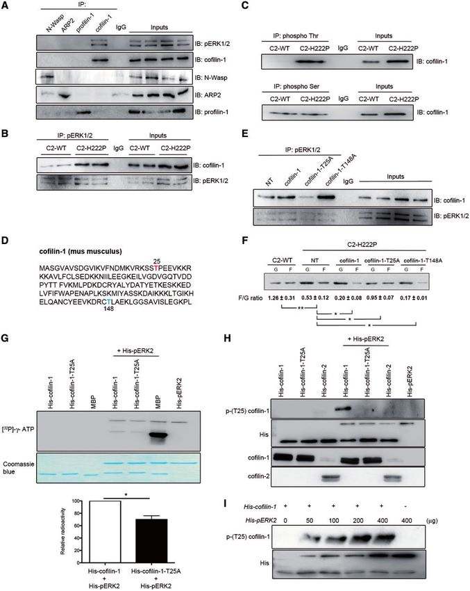
Human Molecular Genetics, 2018, Vol. 27, No. 17 | 3061
Introduction pERK1/2 in C2-H222P cells was further demonstrated by
immunofluorescence microscopy (Fig. 1C).
Mutations in the lamin A/C gene (LMNA) cause an autosomal
Although most ERK1/2 substrates are localized in the
dominant inherited form of dilated cardiomyopathy (hereafter
nucleus, several cytoskeletal proteins are targets (26). We
referred to as LMNA cardiomyopathy), often with concurrent
hypothesized that cytoplasmic pERK1/2 in cells expressing the
muscular dystrophy (1,2). LMNA encodes the A-type nuclear lam-
lamin A H222P variant catalyzes the phosphorylation of cyto-
ins, which arise from alternative RNA splicing (3–5) and along
solic proteins. Given that A-type lamins modulate cytosolic
with B-type lamins are the main constituents of nuclear lamina
actin polymerization (27), we focused on actin dynamics. When
(6). Much of the current research on A-type lamins is focused on
examined by immunoblotting, the ratio of filamentous (F) to
how mutations leading to alterations in these proteins cause
globular (G) actin was significantly lower in C2-H222P cells com-
dilated cardiomyopathy and other inherited diseases. We previ-
pared with C2-WT cells (Fig. 2A). Treating C2-H222P cells with
ously demonstrated that extracellular signal-regulated kinase
cytochalasin D, an inhibitor of actin polymerization, lowered
(ERK) 1/2 is hyper-activated in the heart in LMNA cardiomyopa-
the ratio while treating with jasplakinolide, which promotes
thy (7). However, insights into the molecular mechanisms bridg-
actin polymerization and stabilization, increased the ratio
ing ERK1/2 activation and depressed cardiac function are lacking.
(Fig. 2A). Treating C2-H222P cells with selumetinib, an inhibitor
Alterations in cardiomyocyte (CM) mechanotransduction
of MEK1/2, the kinases that specifically phosphorylate ERK1/2,
likely underlie molecular mechanisms of dilated cardiomyopa-
led to F-actin polymerization (Fig. 2A). These results were con-
thy and progression to heart failure (8,9). Actin is one of the
firmed by immunofluorescence microscopic analysis of F- and
major cytoskeletal proteins in eukaryotic cells that play an
G-actin (Supplementary Material, Fig. S1A). Similar to selumeti-
essential role in several cellular processes, including mechano-
nib, other inhibitors of MEK1/2 gave the same results
resistance and contractile force generation. Actin filaments
(Supplementary Material, Fig. S1B and C). When selumetinib
within sarcomeres, the contractile units of CMs, are uniform in
was washed out from the media in which C2-H222P cells were
length and precisely oriented with their barbed-ends (þ) facing
cultured, the F/G actin ratio analyzed by immunoblotting pro-
the Z-disc, which are capped by CapZ (10) and their pointed-ends
gressively returned to a value similar to that in untreated cells
() directed toward the M-band, which are associated with tropo-
(Supplementary Material, Fig. S1D). This was also observed
modulin. Actin filaments are additionally decorated along their
by immunofluorescence microscopy (Supplementary Material,
length by tropomyosin and a large number of actin-binding pro-
Fig. S1E). Treating C2-WT and C2-H222P cells with cytochalasin
teins, which contribute to maintaining sarcomere structure and
D or latrunculin B induced depolymerization of the actin net-
organization (11–16). A number of actin-binding proteins
work and washing out these drugs led to progressive re-
enhance their turnover, promoting polymerization, depolymeri-
polymerization of F-actin (Supplementary Material, Fig. S1E).
zation or filament severing (17–19). Defective regulation of the
However, actin re-polymerization was delayed in C2-H222P cells
length or the organization of actin filaments in sarcomeres,
compared with C2-WT cells and this delay was diminished
owing to genetic mutations or de-regulated expression of cyto-
when we added selumetinib (Supplementary Material, Fig. S1E).
skeletal proteins, is a hallmark of many heart and skeletal mus-
We next compared the effect of protein extracts from C2-WT
cle disorders (20). Among the regulators of actin, cofilins, which
or C2-H222P cells on the length of F-actin in vitro by microscopic
are actin-depolymerizing factors, play an essential role in the
analysis of fluorescently labeled actin. When actin was poly-
dynamics of filaments. Cofilins enhance actin filament turnover
merized in the presence of extracts from C2-H222P cells, the
by severing and promoting dissociation of actin monomers from
length of F-actin was shorter than in the presence of extracts
the pointed-ends () (21). We now show in a large array of unique
from C2-WT cells (Fig. 2B). This effect on F-actin dynamics was
in vitro and in vivo disease models that phosphorylated ERK1/2
blunted when an extract of C2-H222P cells treated with selume-
(pERK1/2) binds to and activates cofilin-1 in LMNA cardiomyopa-
tinib was used (Fig. 2B). To test if ERK1/2 contributed directly to
thy. The disassembly of actin occurs in CMs from the mouse
F-actin dynamics, we transiently transfected C2-WT cells with
model, leading to left ventricular dysfunction.
wild-type ERK2 or MEK1 constructs. This led to a decrease in the
F/G actin ratio compared with non-transfected cells (Fig. 2C).
Conversely, C2-H222P cells transfected with plasmids encoding
Results ERK2-K52R (kinase dead) or ERK2-T183A/Y185F (dominant nega-
pERK1/2 alters F-actin dynamics in LMNA tive), both of which competitively inhibit activation of endoge-
nous ERK2, had an increased F/G actin ratio compared with
cardiomyopathy
non-transfected C2-H222P cells with the quantity of F-actin in
We set out to unravel the consequences of abnormal ERK1/2 sig- these transfected C2-H222P cells similar to that in C2-WT cells
naling in the heart of LmnaH222P/H222P mice, a model for dilated (Fig. 2C). These data suggest that ERK1/2 triggers depolymeriza-
cardiomyopathy caused by mutation in LMNA (22). As in previous tion of actin in C2-H222P cells expressing a lamin A variant that
studies (7,23), we demonstrated an increase in pERK1/2 in hearts causes dilated cardiomyopathy.
of LmnaH222P/H222P mice compared with wild-type mice (Fig. 1A). To determine whether other lamin A variants have the same
We also previously demonstrated increased pERK1/2 primarily in effect on actin dynamics as lamin A H222P, we transiently
the nucleus of transiently transfected C2C12 cells over- transfected C2C12 cells with plasmids encoding lamin A E358K,
expressing the lamin A H222P variant (7). When we examined L271P and N456I. These mutations have been previously shown
protein extracts from stably transfected C2C12 cells expressing to cause cardiomyopathy (28–30). Similar to lamin A H222P,
H222P lamin A (C2-H222P) at lower levels (24,25), we observed an C2C12 cells expressing these pathogenic lamin A variants
increase in cytoplasmic relative to nuclear pERK1/2 compared showed increased pERK1/2 (Supplementary Material, Fig. S2A)
with cells expressing wild-type lamin A (C2-WT) (Fig. 1B). Total and altered ratios of F-actin to G-actin (Supplementary Material,
cellular pERK1/2 was not changed (data not shown), which is con- Fig. S2B). These findings further suggest that altered F-actin
sistent with previous results showing that it is only increased af- dynamics arises from LMNA mutations as a result of abnormal
ter subjecting these cells to stress (24). The increased cytoplasmic ERK1/2 activation.
Downloaded from https://academic.oup.com/hmg/article-abstract/27/17/3060/5033381
by Universitaet Erlangen-Nuernberg, Wirtschafts- und Sozialwissenschaftliche Zweigbibliothek user
on 21 August 2018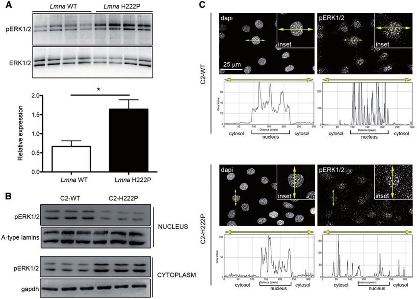
3062     | Human Molecular Genetics, 2018, Vol. 27, No. 17

                Figure 1. Increased ERK1/2 activation in hearts of LmnaH222P/H222P mice and in the cytoplasm of C2-H222P cells. (A) Immunoblots showing pERK1/2 and total ERK1/2 in
                hearts from LmnaH222P/H222P mice (H222P) and wild-type mice (WT). Data in bar graph below are represented as means6SEM (n¼4; *P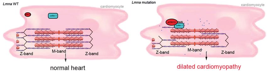
Human Molecular Genetics, 2018, Vol. 27, No. 17                | 3063

                Figure 2. Altered F-actin dynamics in LMNA cardiomyopathy. (A) Immunoblot from one representative experiment showing the effect of lamin A H222P on the
                amounts of G-actin and F-actin and the calculated F/G actin ratios. C2-H222P cells were either untreated (UT), or treated with cytochalasin D, jasplakinolide or selume-
                tinib. Data are represented as means6SEM (n¼3; **P
3064 | Human Molecular Genetics, 2018, Vol. 27, No. 17
Figure 3. Interaction between cofilin-1 and pERK1/2. (A) Immunoblot showing interaction of cofilin-1 and pERK12 in IP experiments. Proteins extracted from C2C12 cells
were subjected to IP using antibodies against cofilin-1, N-Wasp, ARP2 or profilin-1. Proteins in immunoprecipitates were separated by SDS-PAGE and IB using antibodies
against pERK1/2, cofilin-1, N-Wasp, ARP2 or profilin-1. Immunoglobulin G (IgG) was used as a negative control. Representative from three independent repeats.
(B) Immunoblot showing interaction of cofilin-1 and pERK12 in IP experiments from C2-WT and C2-H222P cells. Proteins extracted from C2-WT and C2-H222P cells were
subjected to IP using antibodies against pERK1/2. Proteins in immunoprecipitates were separated by SDS-PAGE and IB using antibodies against pERK1/2 and cofilin-1. IgG
was used as a negative control. Representative from three independent repeats. (C) Proteins extracted from C2-WT and C2-H222P cells were subjected to IP using antibod-
ies specific to phospho Thr or phospho Ser. The immunoprecipitates were separated by SDS-PAGE and IB using antibody against cofilin-1. IgG was used as a negative
control. Representative from three independent repeats. (D) Amino acid sequence of murine cofilin-1 with highlighted threonine 25 (red) and threonine 148 (blue). (E) C2-
H222P cells transfected or not transfected (NT) with plasmids encoding cofilin-1, cofilin-1-T25A or cofilin-1-T148A were subjected to IP using antibodies against pERK1/2.
Proteins in immunoprecipitates were separated by SDS-PAGE and IB using antibodies against cofilin-1 or pERK1/2. IgG was used as a negative control. Representative
from three independent repeats. (F) Immunoblot illustrating the effect of transfection with different cofilin-1 constructs on the amount of G-actin and F-actin and the cal-
culated F/G actin ratio. Data are represented as means6SEM (n¼3; *PHuman Molecular Genetics, 2018, Vol. 27, No. 17 | 3065
depolymerizing factor expressed mainly in striated muscles, does His-cofilin-1 variant with Thr25 replaced by an alanine (Fig. 3H).
not have the Thr-Pro consensus site for phosphorylation cata- Overall, these results demonstrated that pERK1/2 catalyzed the
lyzed by ERK1/2 and therefore did not bind to it (Supplementary phosphorylation of cofilin-1 on Thr25 in cells expressing lamin
Material, Fig. S3D). We further asked whether cofilin-1-T25A could A H222P, which correlated with altered F-actin dynamics.
alter actin dynamics. C2-H222P cells transfected with plasmids To detect endogenous cofilin-1 phosphorylated on Thr25, we
that expressed wild-type cofilin-1 or cofilin-1-T148A had a de- analyzed protein extracts from C2-WT, C2-H222P, and C2-H222P
creased F/G actin ratio compared with non-transfected C2-H222P cells treated with selumetinib using 2-dimensional gel electro-
cells (Fig. 3F). Expression of cofilin-1-T25A partially rescued the F/ phoresis (Supplementary Material, Fig. S5A). Immunoblotting of
G actin ratio in C2-H222P cells (Fig. 3F). Cofilin-1-T25D, a phospho- the separated proteins with antibodies against cofilin-1 identi-
mimetic variant, did not reverse actin depolymerization when fied a specific pattern of expression of cofilin-1 in C2-H222P cells
expressed in C2-H222P cells (Supplementary Material, Fig. S3E). compared with C2-WT cells and C2-H222P cells treated with
Similar results were observed when C2-WT cells were transfected selumetinib (Supplementary Material, Fig. S5B). Given that these
with plasmids that expressed wild-type cofilin-1 or cofilin-1-T25D specific patterns of expression of cofilin-1 could be owing to dif-
(Supplementary Material, Fig. S3F). Expressing cofilin-1-T25A did ference in phosphorylation status, each protein isoform
not alter the F/G actin ratio in C2-WT cells (Supplementary revealed by the antibody against cofilin-1 was excised and sub-
Material, Fig. S3F). jected to in-gel digestion and mass spectrometry analysis
We next generated an antibody recognizing the region (Supplementary Material, Fig. S5C). This analysis identified a
of phosphorylated (Thr25) cofilin-1 (Supplementary Material, total of 12 phosphorylation sites distributed in 8 polypeptides
Fig. S4A). To ensure that the identified phosphorylated (Thr25) corresponding to cofilin-1 (Supplementary Material, Table S1).
cofilin-1 was increased in C2-H222P cells, immunoblots of pro- Phosphopeptide D (Supplementary Material, Fig. S5D) contained
tein lysates were probed with these antibodies. The level of the Thr25 phosphorylation site (SSTPEEVKK) and was observed
phosphorylated (Thr25) cofilin-1 was significantly increased in only in extracted proteins from C2-H222P cells (Table 1;
C2-H222P cells compared with C2-WT cells and reduced when Supplementary Material, Table S2). This phosphopeptide was
treated with selumetinib (Supplementary Material, Fig. S4B). predominantly observed in C2-H222P cells when immunoblot-
Like ERK1/2, phosphorylated (Thr25) cofilin-1 was present in the ted (IB) with a specific antibody against phosphorylated (Thr25)
cytoplasm and nucleus of C2-H222P cells, while it is mostly cofilin-1 (Supplementary Material, Fig. S5D). Phosphopeptides B
in the nucleus of C2-WT cells and essentially absent from and C (Supplementary Material, Fig. S5C) showed increased
C2-H222P cells treated with selumetinib (Supplementary intensity in C2-H222P cells (Supplementary Material, Fig. S5B).
Material, Fig. S4C). To determine whether other lamin A var- Similar to phosphopeptide D, the increase in phosphopeptide B
iants have the same effect on the phosphorylation of cofilin-1 was abrogated by selumetinib, suggesting an ERK1/2 dependent
on Thr25 as lamin A H222P, we transiently transfected C2C12 phosphorylation of cofilin-1. The intensity of phosphopeptide C
cells with plasmids encoding other pathogenic lamin A variant was not affected by selumetinib (Supplementary Material, Fig.
associated with cardiomyopathy. Similar to lamin A H222P, S5B), indicating that cofilin-1 could be phosphorylated by other
C2C12 cells expressing these lamin A variants showed increased kinases.
level of phosphorylated (Thr25) cofilin-1 (Supplementary RhoA and Rho kinase (ROCK) regulate cofilin-1-mediated
Material, Fig. S4D). Expression of the phosphorylated (Thr25) F-actin disassembly through LIMK-catalyzed phosphorylation
cofilin-1 was increased in heart tissue from LmnaH222P/H222P mice of the protein (37). LIMK catalyzes the phosphorylation of
compared with wild-type mice (Supplementary Material, Fig. cofilin-1 on Ser3, and inactivates its actin-severing activity (38).
S4F). The increased expression of phosphorylated cofilin- Phosphorylated (Ser3) cofilin-1 was detected in the protein
1-T25A occurs as early as 2 months of age, when the cardiac extracts analyzed by mass spectrometry (Table 1;
function is not altered (22). We then examined the phosphory- Supplementary Material, Table S2). However, the level of phos-
lated (Thr25) cofilin-1 in human heart samples. We saw an phorylated (Ser3) cofilin-1 was not different between C2-WT
increase of phosphorylated (Thr25) cofilin-1 as well as pERK1/2 and C2-H222P cells (Supplementary Material, Fig. S6).
in heart tissue from humans carrying cardiomyopathy-causing Treatment with Y27632, a selective inhibitor of ROCK, blocks
LMNA mutations compared with controls (Supplementary the phosphorylation of LIMK and in turn the LIMK-catalyzed
Material, Fig. S4F). phosphorylation of cofilin-1, which leads to its activation (39).
We next demonstrated that pERK1/2 catalyzed the phos- Treating C2-WT and C2-H222P cells with Y27632 decreased
phorylation of cofilin-1 on Thr25. In an in vitro kinase assay, phosphorylation of cofilin-1 on Ser3 without affecting the phos-
incubation of histidine-tagged pERK2 (His-pERK2) with phorylation of Thr25 (Supplementary Material, Fig. S6). These
histidine-tagged cofilin-1 (His-cofilin-1) and [32P]-c-ATP resulted results suggested that phosphorylation of Thr25 on cofilin-1
in the phosphorylation of cofilin-1 (Fig. 3G). This in vitro assay was independent of the ROCK pathway.
also showed that incubation of His-pERK2 with [32P]- c-ATP
resulted in 32P incorporation into ERK2, which confirmed the
previously reported autophosphorylation of ERK2 (35,36). The
Phosphorylated (Thr25) cofilin-1 stimulates cardiac
pERK2-catalyzed phosphorylation of cofilin-1 was significantly
dysfunction
reduced in the presence of His-cofilin-1-T25A (Fig. 3G). This sug-
gested that pERK2 catalyzed the phosphorylation of cofilin-1 We next tested the hypothesis that phosphorylated (Thr25)
on Thr25. We further incubated pERK2 with His-cofilin-1, His- cofilin-1 influences left ventricular function in vivo. We injected
cofilin-1-T25A or His-cofilin-2. pERK2 catalyzed the phosphory- adeno-associated virus (AAV) vectors expressing cofilin-1 into
lation of His-cofilin-1 on Thr25, but not His-cofilin-1-T25A 3-month-old Lmnaþ/þ mice. Three months after injection they
or His-cofilin-2 (Fig. 3H). The phosphorylation of cofilin-1 corre- had increased phosphorylated (Thr25) cofilin-1 expression in
lated with amount of His-pERK2 (Fig. 3I), indicating that its the heart (Fig. 4A). Expression of cofilin-2 in the heart was
phosphorylation was catalyzed by ERK2. This phosphorylation unchanged by over-expression of cofilin-1 (Fig. 4A). There was
of cofilin-1 catalyzed by pERK2 was blunted in the presence of a depolymerization of F-actin in Lmnaþ/þ mice expressing viral
Downloaded from https://academic.oup.com/hmg/article-abstract/27/17/3060/5033381
by Universitaet Erlangen-Nuernberg, Wirtschafts- und Sozialwissenschaftliche Zweigbibliothek user
on 21 August 2018on 21 August 2018
3066
Table 1. Cofilin-1 phospho-residues identified by mass spectrometry analysis from C2-WT, C2-H222P and C2-H222P cells treated with selumetinib
Serine S3 S23 S41 S94 S156
Genotype C2-WT C2-H222P C2-H222P C2-WT C2-H222P C2-H222P C2-WT C2-H222P C2-H222P C2-WT C2-H222P C2-H222P C2-WT C2-H222P C2-H222P
(selumetinib) (selumetinib) (selumetinib) (selumetinib) (selumetinib)
A
B
C
D
Threonine T25 T63 T88
| Human Molecular Genetics, 2018, Vol. 27, No. 17
Downloaded from https://academic.oup.com/hmg/article-abstract/27/17/3060/5033381
Genotype C2-WT C2-H222P C2-H222P C2-WT C2-H222P C2-H222P C2-WT C2-H222P C2-H222P
(selumetinib) (selumetinib) (selumetinib)
A
B
C
D x
by Universitaet Erlangen-Nuernberg, Wirtschafts- und Sozialwissenschaftliche Zweigbibliothek user
Tyrosine Y82 Y85 Y89 Y140
Genotype C2-WT C2-H222P C2-H222P C2-WT C2-H222P C2-H222P C2-WT C2-H222P C2-H222P C2-WT C2-H222P C2-H222P
(selumetinib) (selumetinib) (selumetinib) (selumetinib)
A
B
C
D Human Molecular Genetics, 2018, Vol. 27, No. 17 | 3067
Figure 4. Phosphorylated (Thr25) cofilin-1 induces cardiac dysfunction. (A) Immunoblots showing cofilin-1, phosphorylated (Thr25) cofilin-1, cofilin-2 and gapdh in pro-
tein extracts from hearts of Lmnaþ/þ (Lmna WT) transduced with AAV vectors encoding cofilin-1; NT indicates not transduced. Representative from three independent
repeats. (B) Immunoblot illustrating the effect of AAV expressing cofilin-1 construct on the amount of G-actin and F-actin and the calculated F/G actin ratio in hearts
from mice; NT indicates not transduced. Data are represented as means6SEM (n¼3; ***P3068 | Human Molecular Genetics, 2018, Vol. 27, No. 17
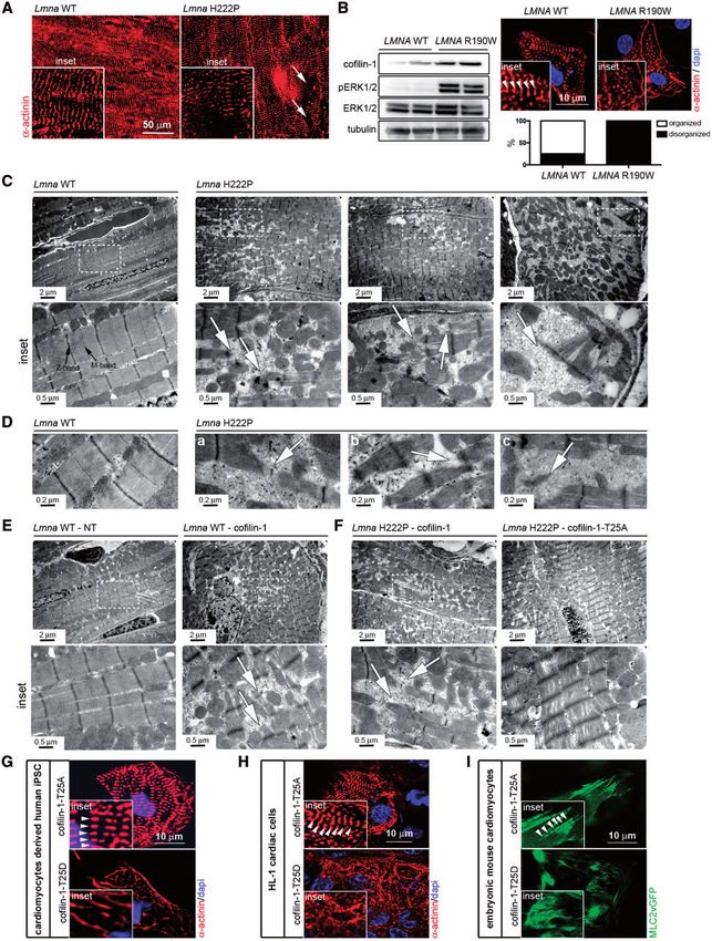
Figure 5. Phosphorylated (Thr25) cofilin-1 alters sarcomeric organization. (A) Micrographs showing a-actinin labeling of cross-sections of hearts from 6-month-old male
Lmnaþ/þ (Lmna WT) mice and LmnaH222P/H222P (Lmna H222P) mice. Arrows indicate areas of disorganized sarcomeres (inset). (B) Immunoblots showing cofilin-1, pERK1/2,
ERK1/2 and tubulin in protein extracts from CMs derived from patient-specific human iPSCs carrying the LMNA p.R190W mutation (LMNA R190W) and control (LMNA WT).
Micrographs showing a-actinin labeling of CMs derived from patient-specific human iPSCs carrying LMNA p.R190W mutation (LMNA R190W) and control (LMNA WT). Dapi
counter-staining (blue) of nuclei is also shown. Inset indicates altered myofibrillar organization. Arrows indicate Z-bands of sarcomeres. Means of organized and (black
bar) and disorganized (while bar) sarcomeres are shown in each bar for each condition. (C) Electron micrographs showing disruption of sarcomeric organization in hearts
from 6-month-old male LmnaH222P/H222P (Lmna H222P) mice compared with Lmnaþ/þ (Lmna WT) mice. Insets show a higher magnification. Arrows indicate disorganized sar-
comeres. (D) Electron micrographs showing disorganized myofibrillar apparatus (a), areas without rods with normal sarcomere appearance (b) and sparse sarcomere
structures (c) indicated by arrows in hearts from 6-month-old male LmnaH222P/H222P (Lmna H222P) mice. (E) Electron micrographs showing sarcomeric organization in
hearts from 6-month-old Lmnaþ/þ (Lmna WT) mice transduced with AAV expressing cofilin-1 construct. NT indicates not transduced. Insets show a higher magnification.
Arrows indicate disorganized sarcomeres. (F) Electron micrographs showing sarcomeric organization in hearts from 6-month-old LmnaH222P/H222P (Lmna H222P) mice trans-
duced with AAV expressing cofilin-1 or cofilin-1-T25A constructs. Insets show a higher magnification. Arrows indicate disorganized sarcomeres. (G) Micrographs showing
a-actinin labeling of CMs derived from human iPSCs transfected with plasmids encoding cofilin-1-T25A and cofilin-1-T25D. Insets highlight altered myofibrillar organiza-
tion. Arrows indicate Z-bands of sarcomeres (inset). Dapi counter-staining (blue) of nuclei is also shown. (H) Micrographs showing a-actinin labeling of mouse HL-1 cardiac
cells transfected with plasmids encoding cofilin-1-T25A and cofilin-1-T25D. Insets highlight altered myofibrillar organization. Arrows indicate Z-bands of sarcomeres
(inset). Dapi counter-staining (blue) of nuclei is also shown. (I) Micrographs showing sarcomeric organization (MLC2vGFP) of embryonic mouse CMs transfected with
plasmids encoding cofilin-1-T25A and cofilin-1-T25D. Insets highlight altered myofibrillar organization. Arrows indicate Z-bands of sarcomeres (inset).
Downloaded from https://academic.oup.com/hmg/article-abstract/27/17/3060/5033381
by Universitaet Erlangen-Nuernberg, Wirtschafts- und Sozialwissenschaftliche Zweigbibliothek user
on 21 August 2018Human Molecular Genetics, 2018, Vol. 27, No. 17 | 3069
Figure 6. A schematic representation of the mechanism of cofilin-1 phosphorylation by ERK1/2 in the heart from Lmna H222P mice with consequences on sarcomeric
actin depolymerization. Blue circle indicates G-actin.
precursor (Fig. 4F). These genes have been previously shown to altered myofibrillar organization compared with transfection
be up-regulated in LMNA cardiomyopathy (23,40–43). with a plasmid encoding the cofilin-1 T25A variant.
We next tested the effect of cofilin-1 variants T25A on left
ventricular function in vivo. Expressing cofilin-1-T25A in
3-month-old LmnaH222P/H222P mice lead to a decrease of the rela-
Discussion
tive expression of phosphorylated (Thr25) to total cofilin-1 in We have shown that ERK1/2-catalyzed phosphorylation of
the heart (Fig. 4C). Expression of cofilin-2 in the heart was cofilin-1 in cells expressing a cardiomyopathy-causing lamin A
unchanged by overexpression of cofilin-1 or cofilin-1-T25A variant leads to depolymerization of F-actin. In a mouse model
(Fig. 4C). Expression of cofilin-1-T25A rescued the cardiac F/G of LMNA cardiomyopathy, this participates in the development
actin ratio of LmnaH222P/H222P mice (Fig. 4D). Compared with mice of cardiac dysfunction. These results suggest a novel model for
that expressed virally encoded cofilin-1, left ventricular frac- A-type lamins in the regulation of actin dynamics and patho-
tional shortening significantly improved in LmnaH222P/H222P mice physiology (Fig. 6). In this model, active pERK1/2 binds to cofilin-
that expressed viral encoded cofilin-1-T25A (Fig. 4E; Table 2). 1, an F-actin binding partner, and catalyzes its phosphorylation
LmnaH222P/H222P mice that received the viral vector encoding on Thr25. This phosphorylation event activates the F-actin
cofilin-1-T25A had decreased expression of Col1a2, Myh7 and depolymerizing function of cofilin-1 in CMs. Inhibition of ERK1/2
Nppa (Fig. 4G), compared with mice that expressed virally suppresses the phosphorylation of cofilin-1 events on Thr25
encoded cofilin-1. and improves left ventricular function. The mechanism by
which LMNA mutation leads to ERK1/2 activation remains to be
elucidated but inference can be made from the literature. ERK1/
2 signaling activation occurred prior to significant cardiomyopa-
Phosphorylated (Thr25) cofilin-1 alters sarcomere
thy in LmnaH222P/H222P mice (7,31) and in LmnaH222P/þ mice, which
organization
do not develop clinical heart disease until 2 years of age (7). This
We hypothesized that phosphorylation of Thr25 on cofilin-1 has is consistent with the hypothesis that ERK1/2 signaling activa-
detrimental effects on cardiac muscle cells. Given that sarco- tion underlies the development of LMNA cardiomyopathy
meres are composed of myosin and actin, we hypothesized that instead of occurring as a consequence of the cardiac disease.
phosphorylated (Thr25) cofilin-1 may affect the dynamics of Accordingly, lamin A/C-deficient cells subjected to cyclic strain
sarcomeric actin. Immunostaining using an a-actinin antibody respond with altered expression of the mechanosensitive
showed disruption of sarcomere organization (Fig. 5A). Similar genes, which are downstream targets of the ERK1/2 signaling
myofibrillars alterations were observed in CMs derived from (44). While it remains unclear how A-type lamins with amino
patient-specific human induced pluripotent stem cells (iPSCs) acid substitutions activate ERK1/2 signaling, our results show
carrying LMNA p.R190W mutation, for which ERK1/2 signaling that they do so when expressed in transfected cells. An intrigu-
was abnormally activated (Fig. 5B). Transmission electron mi- ing question is how certain alterations in A-type lamins, which
croscopy further showed that left ventricular tissue from are expressed in virtually all differentiated somatic cells, acti-
LmnaH222P/H222P mice exhibited severe disruption of myofibrillar vate ERK1/2 signaling specifically in cardiac muscle (7). Given
structure including areas of sarcomere disorganization along- their role in maintaining normal cellular mechanics, a hypothe-
side of normal-looking fibers (Fig. 5C and D). Left ventricular tis- sis is that A-type lamins variants make contractile cells more
sue from Lmnaþ/þ mice that received the vector encoding susceptible to stress-induced damage, which activates ERK1/2
cofilin-1 exhibited severe disruption of myofibrillars, similar to signaling. Our results demonstrate downstream alterations by
the ones observed in LmnaH222P/H222P mice (Fig. 5E). The sarco- which ERK1/2 promotes cardiac dysfunction caused by such
meric organization is improved in left ventricular tissue from mutations. Although cofilin-1 has been known to function in
LmnaH222P/H222P mice that received the vector encoding cofilin- F-actin depolymerization (45), its role in sarcomeric organiza-
1-T25A, compared with LmnaH222P/H222P mice that received the tion and the development of cardiomyopathy was unclear.
vector encoding cofilin-1 (Fig. 5F). We further showed that phos- We previously showed that hearts from LmnaH222P/H222P mice
phorylated (Thr25) cofilin-1 influences sarcomeric organization at 6 months of age have an increase in mostly nuclear pERK1/2
in vitro. Transfection with a plasmid encoding the cofilin-1 T25D (7). Relatively increased cytoplasmic pERK1/2 may therefore
variant in CMs derived from human iPSCs (Fig. 5G), embryonic reflect an early-stage in the development of LMNA cardiomyop-
CMs (Fig. 5H), and HL-1 cardiac muscle cells (Fig. 5I) led to athy. The effect of metabolic stress in C2-H222P cells (24) or age
Downloaded from https://academic.oup.com/hmg/article-abstract/27/17/3060/5033381
by Universitaet Erlangen-Nuernberg, Wirtschafts- und Sozialwissenschaftliche Zweigbibliothek user
on 21 August 20183070 | Human Molecular Genetics, 2018, Vol. 27, No. 17
in mouse hearts (7) might trigger nuclear import of pERK1/2 impedes the emergence of sarcomeric structures from the actin
when total cellular pERK1/2 significantly increases. clusters coalescence (69) or from ARP2/3-dependent network
Nonetheless, there is still a pool of cytoplasmic pERK1/2 that actin-integrin connection known to strengthen newly formed
can catalyze the phosphorylation of resident proteins such as myofibril structures (70). In addition, the disassembly of cyto-
cofilin-1. ERK1/2 can be present in both the nucleus and cyto- skeletal F-actin could directly control the nuclear activity
plasm and catalyze the phosphorylation of proteins in either of transcriptional cofactors of the myocardin protein family.
subcellular compartment (46). Gene expression changes in- The cytosolic G-actin buffers the myocardin-related transcrip-
duced by nuclear pERK1/2 may also contribute to the pathology tion factors, which in turn, control the activity of serum
of LMNA cardiomyopathy along with the effects on cytoplasmic response factor (SRF), a nuclear transcription factor (71). There
cofilin-1 we have delineated in this study. is thus a requirement for a relation between actin dynamics
In striated muscle cells, actin and several scaffolding and and transcription. SRF is playing a critical role in the
regulatory proteins are arranged into contractile sarcomeres normal cardiac muscle development and in the sarcomerogene-
(47). Sarcomeric actin is decorated along its length by tropomyo- sis (72–74). Cardiac SRF-null mice display severe defects in the
sin (48) and other actin-binding proteins, which contribute to cardiac contractile apparatus (74), similar to the ones we
controlling sarcomere structure and organization. Mutations in observed in LmnaH222P/H222P mice. Given that SRF controls the
human genes encoding regulators of actin induce cardiomyopa- expression of sarcomeric genes (74,75), it has been suggested
thy (49–55). These demonstrate the functional significance of that SRF is essential for the maintenance of normal cardiac sar-
sarcomeric actin dynamics on normal heart function. comere organization. It would be interesting to assess the role
Abnormalities of actin dynamics will hamper sarcomeric orga- played by SRF in the alteration on sarcomeric structure in hearts
nization and may be a pathway common to left ventricular from LmnaH222P/H222P mice, where actin dynamics in impaired.
dysfunction. Our results indicate that cofilin-1-mediated modulation of
Cofilin-1 and -2, both expressed in cardiac muscle (56), con- actin dynamics is a cellular consequence of ERK1/2 activation in
tribute to the dynamic turnover of F-actin in contractile cells cardiac cells. We cannot exclude form our study that a protein,
(57). Monomeric G-actin exchange occurs primarily at the other that cofilin-1, is directly implicated in F-actin disassem-
pointed-ends () of sarcomeric actin, near the M-band (58), bly. Indeed, alteration of actin dynamics was also observed in
where cofilin-2 functions with tropomodulin in actin disassem- cells lacking A-type lamins (76), which could result from alter-
bly (48). Some results have suggested that cofilin-2 is more ation of the linker of the nucleoskeleton and cytoskeleton (LINC
effective than cofilin-1 in sarcomeric actin binding (59). This complex) (77,78) or emerin (27,79,80). These data indicate that
implicates cofilin-2 as having a more significant role in sarco- nuclear envelope is an important actor for actin cohesion. More
meric actin depolymerization (57,60,61). We have shown that work is needed to delineate the role played by the nuclear enve-
cofilin-2 is not activated by ERK1/2-catalyzed phosphorylation lope on cytoskeleton stability in diseases. Given that both actin
in LMNA cardiomyopathy, as it lacks the consensus Ser/Thr-Pro cytoskeleton and A-type lamins are necessary for the regulation
amino acid sequence. Our results demonstrate that only cofilin- of mechanosensing (81–84), and that cofilin-1 has recently been
1, under certain condition, disassembles F-actin in CMs and par- involved in nuclear integrity (78), we plan future studies to as-
ticipates to the pathogenesis of left ventricular dysfunction. sess the role played by this novel phosphorylated (Thr25)
Cofilin-1 maintains the pool of G-actin monomers and thus cofilin-1 on mechanosensitivity in cardiac cells carrying LMNA
remodels actin filaments by enhancing assembly/disassembly mutations. We showed that cofilin-1 phosphorylation catalyzed
dynamics. Cofilin-1 regulation of F-actin dynamics is controlled by ERK1/2 is observed as early as 2 months of age in hearts from
by reversible phosphorylation. Several mechanisms regulate LmnaH222P/H222P mice. We further showed that phosphorylated
the activation of cofilin-1, including phosphorylation at Ser3 (Thr25) cofilin-1 influences sarcomeric organization in vitro.
catalyzed by LIMK1/2 or testicular protein kinase 1 (62,63). The These data suggest that cofilin-1 phosphorylation catalyzed by
LIMK-catalyzed phosphorylation of cofilin-1 on Ser3 inactivates ERK1/2 is a pathological event in LMNA cardiomyopathy.
its F-actin depolymerization, leading to accumulation of poly- Enhanced phosphorylation of ERK1/2 and cofilin-1 on Thr25 in
merized F-actin filaments (64). Phosphorylation of cofilin-1 at hearts from a mouse model of LMNA cardiomyopathy and from
Tyr68 by v-Src, which does not change its activity, induces its patients with this disease supports our conclusion that this
ubiquitination and degradation through the proteasome path- mechanism contributes to the pathology. Our work encourages
way (65). Aurora A kinase also catalyzes the phosphorylation of further approaches to mechanistically assess the role played by
cofilin-1 on Ser3, Ser8 and Thr25 during mitosis (66). Our results this novel phosphorylated (Thr25) cofilin-1 on actin dynamics.
now identify Thr25 as a phosphorylated residue in cofilin-1 that These findings suggest that drugs that could correct impaired
specifically activates its F-actin depolymerizing function, partic- actin dynamics would ameliorate left ventricular dysfunction in
ipating to impaired left ventricular contractility. It would be in- LMNA cardiomyopathy. Similar pathogenic mechanism of
teresting in the future to further mechanistically assess how cofilin-1 mediated modulation of F-actin dynamics may play a
active cofilin-1 phosphorylated on Thr25 stimulates the F-actin role in left ventricular dysfunction in other forms of cardiomy-
depolymerization. opathy, in which there appears to be abnormal activation of
We observed that the presence of active cofilin-1 phosphory- ERK1/2 signaling (85–88).
lated on Thr25 in hearts from LmnaH222P/H222P mice impedes the
organization of contractile apparatus. These data suggest that
cofilin-1 phosphorylated on Thr25 participates to the disassem- Materials and Methods
bly of sarcomeric actin and loss of integrity of sarcomeres. The
Cell culture and reagents
actin length in sarcomeres is a process coordinated by several
actin-binding proteins that regulate pointed-ends dynamics Generation of C2-WT and C2-H222P cells has been described
(67). A role of cofilin for the sarcomeric actin organization previously (24). Cells were transiently transfected with plasmids
in striated muscle has been previously proposed (57,68). using Lipofectamine 2000 (Invitrogen) according to the manu-
We cannot exclude that cofilin-1 phosphorylated on Thr25 facturer’s instructions. Plasmids encoding GFP-ERK2, RFP-MEK1,
Downloaded from https://academic.oup.com/hmg/article-abstract/27/17/3060/5033381
by Universitaet Erlangen-Nuernberg, Wirtschafts- und Sozialwissenschaftliche Zweigbibliothek user
on 21 August 2018Human Molecular Genetics, 2018, Vol. 27, No. 17 | 3071
GFP-ERK2 K52R and GFP-ERK2 T183A/Y185F were kindly pro- lines been used for the experiments. As controls, already gener-
vided by P. Stork (Oregon Health and Science University). ated wild-type lines have been employed (93). Differentiation
Plasmids encoding GFP-lamin A, GFP-lamin A E358K, GFP-Lamin into CMs has been achieved using a chemically defined serum
A L271P and GFP-LaminA N456I have been previously described free induction protocol, which is based on the modulation of
(89). Plasmid encoding MLC2vGFP has been previously described the Wnt pathway, as previously reported (94,95). In brief, iPSCs
(90). Working concentrations of 100 nM cytochalasin D, 100 nM were induced with CHIR99021 for 24 h, which mediates Wnt ac-
latrunculin B, 200 nM jasplakinolide, 10 lM Y27632, 50 lM tivation, and subsequently exposed to IWR-1 (Wnt inhibitor) for
PD0325901, 10 lM U0126 and 50 lM selumetinib were prepared 3 days in RPMI-B27 medium. At Day 10 of induction, medium
from stocks diluted in DMSO. Cells were incubated with cyto- was supplemented with insulin. Spontaneous contracting activ-
chalasin D for 45 min, jasplakinolide for 40 min and selumetinib ity usually appears around Days 8–10 and CMs were used for
for 15 h. experiments around 25–30 days after spontaneous contraction
To generate mouse embryonic CMs, hearts were dissected has started. For the immunofluorescence staining, CMs were
out from E9.5 mouse embryos; ventricles were cut and myocytes plated onto glass coverslips coated with fibronectin and lami-
dissociated using 1 mg/ml collagenase and 0.3 m/ml pancreatin nin, fixed in 2% paraformaldehyde.
(15 min, 37 C), cells were spun down and replated on gelatin
coated wells (DMEM supplemented with NEAA, glutamine and
10% FCS). HL-1 cardiac muscle cells were plated according to the Measurement of F-actin/G-actin ratio
manufacturer’s instructions (Merck Millipore). Cells were tran- The ratio of F-actin to G-actin was determined using the G-actin/
siently transfected using Lipofectamine 3000 (Invitrogen) F-actin in vivo assay kit (Cytoskeleton) according to the manufac-
according to the manufacturer’s instructions. turer’s instructions. Briefly, 2 mg of protein from cells or frozen
heart tissues were homogenized in Lysis and F-actin
Stabilization Buffer and centrifuged at 2000 rpm for 5 min to
Mice
remove unbroken cells. F-actin was separated from G-actin by
LmnaH222P/H222P mice (22) were fed chow and housed in a centrifugation at 100 000g for 60 min at 37 C. The F-actin-
disease-free barrier facility at 12 h/12 h light/dark cycles. All ani- containing pellet was resuspended in F-actin Depolymerizing
mal experiments were approved by the French Ministry of Buffer at a volume equivalent to the G-actin-containing super-
Health at the Center for Research in Myology for the Care and natant volume. The resuspended F-actin pellet was kept on ice
Use of Experimental Animals. The animal experiments were for 60 min and was gently mixed every 15 min to dissociate
performed according to the guidelines from Directive 2010/63/EU F-actin. Proteins in equivalent volumes (10 ll) of supernatant
of the European Parliament on the protection of animals used for and pellet were separated by SDS-PAGE and subjected to immu-
scientific purposes. noblot analysis using an anti-pan actin antibody supplied in the
kit. F/G actin ratio was quantified using ImageJ software.
Human heart tissue
RNA isolation and real-time PCR
Sections of explanted hearts from human subjects with LMNA
mutations were obtained without identifiers from Myobank- Total RNA was extracted from cells or mice hearts using the
AFM de l’lnstitut de Myologie. Myobank-AFM received approval RNeasy Mini Kit (Qiagen) according to the manufacturer’s
from the French Ministry of Health and from the Committee for instructions. One microgram of total RNA was subjected to
Protection of Patients to share tissues and cells of human origin cDNA synthesis using the First-Strand cDNA Synthesis Kit (Life
for scientific purposes, ensuring the donors’ anonymity, respect Technologies). Mouse primer sequences used for transcriptional
of their volition and consent according to the legislation. The analyses were as follows: mCofilin-1 50 -CGCAAGTCTTCAA
subjects were a 23-year-old man with cardiomyopathy associ- CACCAGA-30 , 50 -TGAACACCAGGTCCTCCTTC-30 ; Myh7 50 -ACTGT
ated with muscular dystrophy and LMNA delK261 mutation, CAACACTAAGAGGGTCA-30 , 50 -TTGGATGATTTGATCTTCCAGG
a 53-year-old man with cardiomyopathy and LMNA E33D muta- G-30 ; Col1a2 50 -CCGTGCTTCTCAGAACATC-30 , 50 -GAGCAGCCA
tion and a 47-year-old woman with cardiomyopathy and LMNA TCGACTAGGAC-30 ; Nppa 50 -GCTTCCAGGCCATATTGGAG-30 ,
R60G mutation. Control human heart samples were obtained 50 -GGGGGCATGACCTCATCTT-30 . Real-time quantitative PCR
from the National Disease Research Interchange; information reactions were performed on a LightCycler 480 (Roche) using the
regarding donor confidentiality and consent can be found at SYBR Green PCR Master Mix (Applied Biosystems). The PCR
http://www.ndriresource.org. Control human heart samples products were subjected to melting curve analysis to exclude
were obtained from a 57-year-old man with an intracranial the synthesis of non-specific products. Cycle threshold (Ct) val-
bleed, a 15-year-old woman who died of a drug overdose and a ues were quantified using a standard curve for the specific gene
46-year-old man who died from end-stage liver disease. and relatively quantified using RplR0 as an internal reference
control. The Ct values were then normalized to the average ex-
pression levels of samples, calculated according to the 䉭䉭Ct
iPSCs generation and differentiation into CMs method (96) and are presented as fold change over wild-type
iPSCs have been generated from peripheral blood mononuclear controls. All experiments were performed in triplicates.
cells (PBMNCs) obtained from a patient carrying the LMNA
p.R190W mutation, after signed informed consent.
Protein extraction and immunoblotting
Reprogramming has been induced using the Cytotune iPS-2.0
Sendai Reprogramming kit (ThermoScientific) on T-lympho- Total proteins were isolated by resuspending mouse heart
cytes activated from PBMNCs using CD3 and CD28 ligands, as tissue or cultured cells in extraction buffer (Cell Signaling) with
described (91). Reprogrammed clones have been selected and the addition of protease inhibitors (25 mg/ml aprotinin, 10 mg/ml
characterized as previously (92) and two fully characterized leupeptin, 1 mM 4-[2-aminoethyl]-benzene sulfonylfluoride
Downloaded from https://academic.oup.com/hmg/article-abstract/27/17/3060/5033381
by Universitaet Erlangen-Nuernberg, Wirtschafts- und Sozialwissenschaftliche Zweigbibliothek user
on 21 August 20183072 | Human Molecular Genetics, 2018, Vol. 27, No. 17
hydrochloride and 2 mM Na3VO4). The lysates were sonicated PBS and incubated for 1 h with secondary antibodies. F-actin
(3 pulses of 10 s at 30% amplitude) to allow dissociation of pro- was stained with Alexa Fluor 568-phalloidin and G-actin
tein from chromatin and solubilization. Cytosolic and nuclear with Alexa Fluor 488-deoxyribonuclease I for 1 h at room tem-
fractions were prepared using the NE-PER Nuclear and Cytosolic perature. Cells and slides were then mounted in Vectashield
Extraction Reagents (ThermoFisher Scientific) according to the mounting medium with dapi (Vector Laboratories).
manufacturer’s instructions. Sample protein content was deter- Immunofluorescence microscopy was performed using an
mined by the BiCinchoninic Acid Assay protein assay Axiophot microscope (Carl Zeiss). All the images were digitally
(ThermoFisher Scientific). Extracts were analyzed by SDS-PAGE deconvolved using Autodeblur v9.1 (Autoquant) deconvolution
using a 10% gel and transferred onto nitrocellulose membranes software and were processed using Adobe Photoshop 6.0 (Adobe
(Invitrogen). Subsequent to being washed with Tris–buffered Systems).
saline containing 1% Tween 20 (TBS-T), the membranes were
blocked in 5% bovine serum albumin (BSA) in TBS-T for 1 h at
room temperature, then incubated with the appropriate anti- Electron microscopy
body overnight at 4 C. Subsequent to being washed with TBS-T, Freshly harvested left ventricle apex was cut into small pieces
the membranes were incubated with horseradish peroxidase- and immediately fixed by immersion in 2.5% glutaraldehyde
conjugated anti-rabbit or anti-mouse antibodies for 1h at room diluted in PBS for 1 h at room temperature. After washing in
temperature. After washing with TBS-T, the signal was revealed PBS, samples were post-fixed with 1% OsO4, dehydrated in a
using Immobilon Western Chemiluminescent HorseRadish graded series of acetone and embedded in an epoxy resin.
Peroxidase (HRP) Substrate (Millipore) on a G-Box system with Ultrathin sections were cut at 90 nm and stained with uranyl
GeneSnap software (Ozyme). acetate and lead citrate, examined using a transmission elec-
tron microscope (JEOL 1011) and photographed with a digital
Erlangshen 1000 camera (GATAN), using Digital Micrograph
Antibodies software.
Primary antibodies used were: anti-cofilin1 (Cell Signaling), anti-
phosphorylated (Ser3) cofilin-1 (Cell Signaling), anti-cofilin-2
Immunoprecipitation
(ThermoFisher Scientific), anti-profilin1 (Cell Signaling), anti-
N-wasp (Cell Signaling), anti-Arp2 (Cell Signaling), anti-pERK1/2 Cells and cardiac tissues were lysed in 0.5 ml of lysis buffer [50
antibody (Cell Signaling), anti-ERK1/2 (Santa Cruz Biotechnology), mM Tris–HCl (pH 7.5), 0.15 M NaCl, 1 mM EDTA, 1% NP-40].
anti-lamin A/C (Santa Cruz Biotechnology), anti-gapdh (Santa Mouse cardiac muscle tissue lysate was diluted to 2 mg/ml in
Cruz Biotechnology) and anti-a-actinin (Abcam). Secondary anti- lysis buffer, pre-cleared with 20 ml washed protein G-Sepharose
bodies for immunofluorescence were Alexa Fluor-488 conjugated 4 fast-flow (GE Healthcare) and incubated with 20 mg of specific
goat anti-rabbit IgG, Alexa Fluor 568-conjugated goat anti-mouse antibody overnight at 4 C. Next, 30 ml of washed beads was
IgG and Alexa-Fluor-488-conjugated donkey anti-goat IgG (Life added and incubated for 2 h at 4 C. Pelleted beads were col-
Technologies). Secondary antibodies for immunoblotting were lected in sample buffer [0.25 M Tris–HCl (pH 7.5), 8% SDS, 40%
HRP-conjugated rabbit anti-mouse and goat-anti rabbit IgG glycerol, 20% b-mercaptoethanol] and subjected to SDS-PAGE
(Jackson ImmunoResearch). We generated custom-made anti- and immunoblotting. For control reactions, we used rabbit
phosphorylated (Thr25) cofilin-1 (GenScript). Briefly, we used immunoglobulin G1.
cofilin-1 phosphopeptide (RKSS{phosphoT}PEEVKKRKKA) conju-
gated with keyhole limpet hemocyanin (KLH) for rabbit immuni-
zation. Two animals were immunized with 200 lg of purified Construction of plasmids encoding wild-type and
cofilin-1 phosphopeptide. The animals received three booster cofilin-1 variants
injections at 2 weeks intervals. The antibody was tested by ELISA Mutagenesis was carried out using QuikChange II Site-Directed
on the crude blood serum. Mutagenesis Kit (Agilent Technologies) according to the manu-
facturer’s instructions. Cofilin-pmCherryC1 was a gift from
Christien Merrifield (Addgene plasmid # 27687) (97). Cofilin-1-
Immunofluorescence microscopy pmCherryC1 was used as a template for substituting threonine
For immunofluorescence microscopy, frozen tissues were cut for alanine at position 25 and 148. Primers used for the introduc-
into 8-lm-thick sections. Cryosections were fixed [15 min, 4% tion of single mutations are: mCofilin-1-T25A 50 -CTTCACTTCTT
paraformaldehyde in phosphate-buffered saline (PBS) at room CTGGTGCTGAAGACTTGCGAACCT-30 , 50 -AGGTTCGCAAGTCTT
temperature], permeabilized (10 min, 0.5% Triton X-100 in PBS) CAGCACCAGAAGAAGTGAAG-30 ; mCofilin-1-T148A 50 -TTCTCT
and blocked (1 h, PBS with 0.3% Triton X-100, 5% BSA). Sections GCCAGGGCGCAGCGGTCCTTG-30 , 50 -CAAGGACCGCTGCGCCCTG
were incubated with primary antibodies (overnight, 4 C, in PBS GCAGAGAA-30 , mCofilin-1-T25D 50 -CGTTTCTTCACTTCTTCTGGA
with 0.1% Triton X-100 and 1% BSA) and washed in PBS. The sec- TCTGAAGACTTGCGAACCTTCATGTCA-30 , 50 -TGACATGAAGGTT
tions were then incubated for 1 h with secondary antibodies. CGCAAGTCTTCAGATCCAGAAGAAGTGAAGAAACG-30 . The final
Sections were washed with PBS and slides were mounted in plasmids were transformed in XL1-Blue super competent cells.
Vectashield mounting medium with dapi (Vector Laboratories). Mutations were verified by DNA sequencing using an appropri-
C2C12 cells were grown on coverslips, washed with PBS and ate set of oligonucleotides to cover the full length of the cofilin1
fixed with 4% paraformaldehyde in PBS for 10 min. Cells were cDNA.
permeabilized with 0.2% Triton X-100 diluted in PBS for 7 min
and non-specific signals were blocked by incubation in 0.2%
Protein expression and purification
Triton X-100, 5% BSA for 30 min. The samples were then incu-
bated with primary antibody for 1 h in PBS with 0.1% Triton Cofilin-1, cofilin-1-T25A and cofilin-2 cDNAs cloned in pNIC-
X-100 and 1% BSA at room temperature. Cells were washed with CTHF vector expressing a cleavable C-terminal His6 and FLAG
Downloaded from https://academic.oup.com/hmg/article-abstract/27/17/3060/5033381
by Universitaet Erlangen-Nuernberg, Wirtschafts- und Sozialwissenschaftliche Zweigbibliothek user
on 21 August 2018Human Molecular Genetics, 2018, Vol. 27, No. 17 | 3073
tag were obtained from Nicola Burgess-Brown (Structural packaging plasmid encoding the rep2 and cap-rh10 genes and
Genomics Consortium, Oxford). The plasmid encoding constitu- (iii) the AAV2 plasmid expressing cytomegalovirus promoter
tively MEK1 and His-ERK2 kinase, pET-His6-ERK2-MEK1 R4F cofilin-1 cDNA. Seventy-two hours after transfection, cells were
co-expression, was a gift from Melanie Cobb (Addgene plasmid harvested and AAV vectors were purified by ultracentrifugation
# 39212) (98). His-cofilin-1 and His-cofilin-2 recombinant pro- through an iodixanol gradient and concentrated with Ultra–
teins were expressed in Rosetta (DE3) E. coli and His-MEK1-ERK2 Ultra cell 100 K filter units (Amicon) in 0.1 M PBS, 1 mM MgCl2
was expressed in BL21 (DE3) E. coli. Bacteria were grown in and 2.5 mM KCl. Aliquots were stored at 80 C until use. Vector
2 YT medium until they reached an optical density (OD600) of titers were determined by real-time PCR. Three-month-old mice
0.8 and recombinant protein expression was induced by the were injected with scAAVrh10-cofilin-1 into the retro-orbital
addition of 0.5 mM isopropyl-thiogalactopyranoside for 16–24 h vein (51013 viral genomes/kg in 100 ll) with an insulin syringe.
at 30 C for BL21 (DE3) or 18 C for Rosetta (DE3) before harvest-
ing. Cells were sonicated in lysis buffer [50 mM HEPES (pH 7.5),
500 mM NaCl, 5% glycerol, 5 mM imidazole 0.5 mM TCEP with Two-dimensional gel electrophoresis
complete EDTA-free Protease Inhibitor Cocktail, Roche]. After
Proteins were extracted from cells in a buffer composed of 7 M
centrifugation at 40 000g for 30 min at 4 C, the soluble fraction
urea, 2 M thiourea, 1% CHAPS, 10% isopropanol, 10% isobutanol,
was filtered through a 0.8 mm filter and subjected to immobilized
0.5% Triton X-100 and 0.5% SB3-10. Proteins were precipitated
metal affinity Ni2þ-nitrilotriacetic resin chromatography.
using the Perfect Focus kit (G-Biosciences) and pellets were
Recombinant His6-tagged proteins eluted with elution buffer
resuspended in the same buffer supplemented with 25 mM
[50 mM HEPES (pH 7.5), 300 mM NaCl, 5% glycerol and 250 mM
TrisHCl (pH 8.8). The protein content was assessed by the
imidazole]. His-tagged cofilin proteins were purified by size
Quick StartTM Bradford protein assay (BioRad) using BSA as
exclusion chromatography on a HiLoad 16/60 Superdex
standard. For each sample, 50 mg of proteins were labeled with
75 preparative-grade column. His-tagged active ERK2 was puri-
400 pmol of Cy2 (Fluoprobes), incubated 30 min on ice and then
fied as described previously (98). Briefly, following affinity purifi-
quenched with 0.35 mM lysine for 10 min. Then, 700 mg of unla-
cation, eluted His-pERK2 was dialyzed overnight in 50 mM
beled proteins were added to 50 mg of Cy2-labeled proteins.
HEPES (pH 7.5), 50 mM NaCl, 1 mM dithiotreitol, 20% glycerol, di-
Proteins were separated using 24 cm gels with a pH range
luted 1:1 in the same buffer without glycerol then applied to a
3–10 using commercial strips (GE Healthcare). Strips were pas-
MonoQ GL 5/50 column and eluted on a gradient from 50 mM to
sively rehydrated overnight directly with the samples supple-
1 M NaCl.
mented with 40 mM dithiothreitol and 0.5% ampholites.
Isoelectric focusing migration was set as follows: 3 h at 50 V, 3 h
at 200 V, 2 h gradient from 200 to 1000 V, 2 h at 1000 V, 2 h gradi-
In vitro kinase assay
ent from 1000 to 10 000 V, 8 h at 10 000 V. A second step of iso-
Recombinant His-tagged cofilin-1, cofilin-1-T25A and cofilin-2 electric focusing was performed using the following
(5 lg) were incubated alone or with 0.5 lg recombinant active parameters: 30 min at 200 V, 1 h gradient from 200 to 10 000 V,
His-ERK2, in kinase buffer [50 mM HEPES (pH 7.5), 150 M NaCl, 4 h at 10 000 V. The area of the strips corresponding to the pH
5 mM MgCl2, 0.5 mM TCEP] in a total volume of 100 ml containing range from 5.5 to 9 was excised and frozen at 20 C until use
1 mM ATP. Reaction mixtures were incubated at 37 C for 1 h for the second dimension. Strips were incubated for 15 min in
and were terminated by the addition of 100 ml of 2 SDS sample equilibration buffer [6 M urea, 75 mM Tris–HCl (pH 8.8), 26 and
buffer. Proteins were resolved by SDS-PAGE. Blots were incu- 2% SDS] supplemented with 65 mM dithiothreitol and then for
bated with anti-His (Sigma-Aldrich H1029), cofilin-1 and phos- 20 min in equilibration buffer supplemented with 135 mM
phorylated (Thr25) cofilin-1 antibodies. iodoacetamide. The second dimension was run in 12% acrylam-
ide gels at 25 V for the first hour then 150 V and 12 mA per gel in
a Tris–glycine buffer. Gel images were acquired on Ettan DIGE
Radioactive in vitro kinase assay Imager (GE Healthcare). Each gel was run two times using the
Cofilin-1, cofilin-1-T25A or dephosphorylated myelin basic pro- same conditions: first gel was used for immunoblotting and the
tein (MBP) (Merck-Millipore) were incubated alone or in the second for protein identification.
presence of purified pERK2 in buffer comprising 50 mM HEPES
(pH 7.5), 150 mM NaCl, 5 mM MgCl2, 0.5 mM TCEP and 0.5 mM
ATP. A 10 pmol of protein was used and 0.5 mCi [32P]-c-ATP was In-gel digestion and mass spectrometry
added per sample (total volume 25 ml). The samples were incu- Gels were stained with silver nitrate for 1 min sensitizing in
bated for 30 min at 37 C and the reaction terminated by the 0.02% sodium thiosulfate, rinsed two times in ultra-pure water,
addition of 5 ml 6 SDS-PAGE sample buffer. Proteins were then incubated for 30 min in 0.212% silver nitrate, rinsed two times
separated by SDS-PAGE and blotted onto nitrocellulose. The in ultra-pure water and developed in 3% sodium carbonate,
nitrocellulose membrane was then used to expose a phosphor 0.00125% sodium thiosulfate and 0.03% formalin. Staining was
screen (typically for 24 h), which was then imaged using a stopped by soaking the gels in 5% acetic acid. Stained protein
Typhoon FLA 7000 system. spots of interest were manually excised, sliced into 1 mm cubes,
destained in 50 mM sodium thiosulfate, 15 mM potassium ferri-
cyanure and washed several times alternatively in water and
Construction and injection of AAV encoding cofilin-1
acetonitrile. Tryptic digestion was performed overnight at
AAV vectors of serotype rh10, carrying wild-type or mutant 37 C, using 200 ng of mass spectrometry grade trypsin
cofilin-1 under the control of the cytomegalovirus immediate/ (G-Biosciences) in 50 mM ammonium bicarbonate, 5% acetoni-
early promoter were prepared by the triple transfection method trile. Supernatants were collected and gel pieces washed two
in HEK293T cells as previously described (99). Briefly, cells were times for 15 min in an ultrasonic bath with 50 ml of 0.1% tri-
transfected with (i) the adenovirus helper plasmid, (ii) the AAV fluoroacetic acid and 60% acetonitrile. Peptides solutions were
Downloaded from https://academic.oup.com/hmg/article-abstract/27/17/3060/5033381
by Universitaet Erlangen-Nuernberg, Wirtschafts- und Sozialwissenschaftliche Zweigbibliothek user
on 21 August 2018You can also read