Blocking intruders: inducible physico-chemical barriers against plant vascular wilt pathogens
←
→
Page content transcription
If your browser does not render page correctly, please read the page content below
Journal of Experimental Botany, Vol. 72, No. 2 pp. 184–198, 2021
doi:10.1093/jxb/eraa444 Advance Access Publication 25 September 2020
This paper is available online free of all access charges (see https://academic.oup.com/jxb/pages/openaccess for further details)

DARWIN REVIEW

Blocking intruders: inducible physico-chemical barriers
against plant vascular wilt pathogens
Anurag Kashyap1, , Marc Planas-Marquès1, , Montserrat Capellades1, Marc Valls1,2,                                              and Núria S. Coll1,*,

                                                                                                                                                        Downloaded from https://academic.oup.com/jxb/article/72/2/184/5911728 by guest on 08 September 2021
1
    Centre for Research in Agricultural Genomics (CSIC-IRTA-UAB-UB), Bellaterra, Spain
2
    Genetics Department, Universitat de Barcelona, Barcelona, Spain

* Correspondence: nuria.sanchez-coll@cragenomica.es

Received 29 July 2020; Editorial decision 11 September 2020; Accepted 16 September 2020

Editor: Donald Ort, University of Illinois, USA

Abstract
Xylem vascular wilt pathogens cause devastating diseases in plants. Proliferation of these pathogens in the xylem
causes massive disruption of water and mineral transport, resulting in severe wilting and death of the infected plants.
Upon reaching the xylem vascular tissue, these pathogens multiply profusely, spreading vertically within the xylem
sap, and horizontally between vessels and to the surrounding tissues. Plant resistance to these pathogens is very
complex. One of the most effective defense responses in resistant plants is the formation of physico-chemical bar-
riers in the xylem tissue. Vertical spread within the vessel lumen is restricted by structural barriers, namely, tyloses
and gels. Horizontal spread to the apoplast and surrounding healthy vessels and tissues is prevented by vascular
coating of the colonized vessels with lignin and suberin. Both vertical and horizontal barriers compartmentalize the
pathogen at the infection site and contribute to their elimination. Induction of these defenses are tightly coordinated,
both temporally and spatially, to avoid detrimental consequences such as cavitation and embolism. We discuss cur-
rent knowledge on mechanisms underlying plant-inducible structural barriers against major xylem-colonizing patho-
gens. This knowledge may be applied to engineer metabolic pathways of vascular coating compounds in specific
cells, to produce plants resistant towards xylem colonizers.

Keywords: Gels, inducible defenses, lignin, physico-chemical barriers, plant-pathogen interactions, structural defenses, suberin,
tyloses, vascular pathogens, wilt

Introduction
The plant immune system has been shaped by hundreds of                            2013). Despite the xylem being nutritionally poor in com-
millions of years of interactions with microbial pathogens that                   parison with other plant tissues, a select group of pathogens
are constantly trying to evade or overcome host plant defense                     comprising a few genera of bacteria, fungi and oomycetes, have
reactions. Pathogens that colonize the xylem - a vital compo-                     evolved to target this plant tissue as a niche for colonization.
nent for transporting water and minerals from the roots to the                    Proliferation of these wilt pathogens causes massive disrup-
aerial parts - are particularly pernicious, causing major destruc-                tion in the xylem vessels through the production of toxins,
tion in numerous host plants worldwide (Yadeta and Thomma,                        exopolysaccharides, hyphal mass, conidia and other pathogen

© The Author(s) 2020. Published by Oxford University Press on behalf of the Society for Experimental Biology.
This is an Open Access article distributed under the terms of the Creative Commons Attribution License (http://creativecommons.org/licenses/by/4.0/),
which permits unrestricted reuse, distribution, and reproduction in any medium, provided the original work is properly cited.
Inducible barriers against plant vascular wilt pathogens | 185

propagules that cause destruction as well as physical occlusion           proteins (NLRs). ETI is commonly known as a stronger im-
of xylem elements, resulting in severe wilting and plant death            mune response, while PTI offers durable and broad-spectrum
(Zaini et al., 2018).                                                     resistance (Katagiri and Tsuda, 2010). Yet, these two layers of
                                                                          immunity operate synergistically, converging on several down-
Invasion strategies used by vascular wilt pathogens                       stream responses including the oxidative burst, activation of
                                                                          kinase signaling cascades, expression of defense-related genes
Vascular plant pathogens utilize diverse strategies to get ac-            and accumulation of physico-chemical barriers (Thomma et al.,
cess into the vessels. Vascular wilt pathogenic fungi (Fusarium           2011; Ngou et al., 2020, Preprint;Yuan et al., 2020, Preprint).
oxysporum, Verticillium albo-atrum, Verticiullium dahliae, Ceratocystis      Although PTI and ETI function in the interactions of
fimbriata, Ophiostoma novo-ulmi), oomycetes (Pythium spp) or              plants with vascular pathogens, there are very few cases
the soil-borne bacterium Ralstonia solanacearum invade plant              where these processes have been analyzed specifically in the
roots and advance inter- or intracellularly through the root              xylem and surrounding tissues of infected plants. Several re-

                                                                                                                                               Downloaded from https://academic.oup.com/jxb/article/72/2/184/5911728 by guest on 08 September 2021
cortex to reach the xylem, where they proliferate and spread              cent studies have shown that PTI can be triggered in plants
systemically to aerial plant parts (Bae et al., 2015). In order to        by treatment or over-expression of conserved molecular pat-
get access into the vasculature, these pathogens generally target         terns present in wilt pathogens. For example, an extracellular
root extremities and the junction between primary and lateral             polysaccharide from the wilt bacterium R. solanacearum has
roots where the epidermal barrier may be compromised, and                 been shown to act as a PTI elicitor in resistant tomato, leading
the endodermis as well as Casparian strip are either not fully            to induced expression of defense response genes (Milling
differentiated or reoriented by outgrowth of lateral roots (Vasse         et al., 2011; Prakasha et al., 2017). Furthermore, in Nicotiana
et al., 1995; Álvarez et al., 2010).                                      benthamiana and tomato, PTI is triggered upon perception
   In contrast to these root invaders, many vascular bacteria,            of the COLD SHOCK PROTEIN 22 (csp22) peptide from
such as Xylella fastidiosa, are directly inoculated into the xylem        R. solanacearum by the PRR COLD SHOCK PROTEIN
by insect vectors that feed on the plant host (Wang et al., 2017).        RECEPTOR (CORE; Wang et al., 2016). In accordance,
Furthermore, a few species of vascular bacteria reach the xylem           treatment of tomato with csp22 conferred increased resistance
tissues via natural plant openings in the aerial parts of the             to R. solanacearum in tomato plants. Additionally, transgenic
plant, such as leaf hydathodes (Xanthomonas oryzae pv. Oryzae;            Arabidopsis thaliana plants expressing the tomato csp22 receptor
Pradhan et al., 2012), flower nectarthodes (Erwinia amylovora;            (SlCORE) gained the ability to respond to csp22 and were
Bubán et al., 2003) or stem lenticels (Pseudomonas syringae pv.           more resistant to R. solanacearum infection (Wei et al., 2018).
actinidiae; Renzi et al., 2012). These bacteria multiply in inter-        Similarly, V. dahliae, possesses two cellulose-degrading glyco-
cellular spaces before eventually colonizing the xylem vessels.           side hydrolase family 12 (GH12) proteins, namely VdEG1 and
Moreover, there are vascular wilt pathogens which are trans-              VdEG3, which in N. benthamiana are recognized by the PRR
mitted through graft-infected rootstock/scion, as has been                complexes LRR-RLP (Leucine Rich Repeat-Receptor-
reported for the fungus Ceratocystis fagacearum on oak trees              Like Protein)/SOBIR1/BAK1 (SUPPRESSOR OF BAK1-
(Blaedow and Juzwik, 2010), or the bacterium X. fastidiosa                INTERACTING RECEPTOR-LIKE KINASE 1-1/
on pecan (Sanderlin and Melanson, 2006). Another method                   BRASSINOSTEROID-INSENSITIVE 1-ASSOCIATED
of infection of vascular wilt pathogens occurs through the                RECEPTOR KINASE) and LRR-RLKs (Receptor-Like
use of infected mother plants as a source for clonal propa-               Kinase)/BAK1, respectively, thereby triggering PTI responses
gation. This affects many plant species cultivated in nurseries,          (Gui et al., 2017).
such as strawberry plants infected with Fusarium oxysporum                   Concomitantly, some bacterial wilt pathogens have evolved
f. sp. fragariae (Pastrana et al., 2019), olive plants infected with      modifications in the sequences of their conserved patterns, so
V. dahliae (Morello et al., 2016), and it is also common in grape-        that they are no longer recognizable by cognate PRRs. This
vine mother plants that can harbor various vascular wilt fungi            occurs in X. fastidiosa, which contains a lipopolysaccharide
(Gramaje et al., 2018).                                                   featuring a masking motif that evades recognition by grape-
                                                                          vine (Rapicavoli et al., 2018), or flagellin from R. solanacearum,
Immunity against vascular wilt pathogens                                  with a polymorphic sequence that avoids perception by many
                                                                          host plants (Wei et al., 2020).
Plants are protected against pathogens by a well-orchestrated                An interesting emerging concept is that roots may be rela-
immune system (Jones and Dangl, 2006). Pattern-triggered im-              tively insensitive to most conserved molecular patterns, which
munity (PTI) is initiated upon recognition of conserved mi-               may prevent mounting defense responses against commensal
crobial features at the plasma membrane by pattern-recognition            organisms in an environment such as the soil, full of microbes.
receptors (PRRs) in the plant. Effector-triggered immunity                Underlying this phenomenon, it has been recently shown that
(ETI) is activated by recognition of pathogen-secreted ef-                root PTI is only activated upon local tissue damage (Zhou
fectors via intracellular nucleotide-binding leucine-rich repeat          et al., 2020). This work convincingly shows how localized cell
186 | Kashyap et al.
death upregulates PRR expression in the neighboring cells, 2018), and watermelon (Lambel et al., 2014). QTLs termed
leading to restricted responsiveness to conserved molecular RESISTANCE TO FUSARIUM (RFO1-RFO7) have been
patterns of root invaders (Zhou et al., 2020). identified in Arabidopsis that provide broad spectrum im-
ETI responses against vascular pathogens have also been munity to multiple formae speciales of F. oxysporum (Chen et al.,
documented in the literature. For instance, in the interaction 2014). RFO1 was found to encode a wall-associated kinase-
between tomato and the vascular wilt fungus F. oxysporum f. sp. like kinase 22 (WAKL22) and RFO2 encodes a receptor-like
lycopersici, several R protein-effector pairs have been identi- protein (Diener and Ausubel, 2005). Likewise, resistance to
fied. The NLR I2 perceives the Avr2 effector; I, a receptor-like Verticillium correlates with mapping of QTLs in several hosts
protein with an extracellular LRR domain (LRR-RLP) per- such as strawberry (Antanaviciute et al., 2015) and cotton
ceives Avr1; and I3, a S-receptor-like kinase (SRLK) perceives (Palanga et al., 2017). A major QTL conferring resistance to
Avr3 (Houterman et al., 2009; Catanzariti et al., 2015, 2017). X. fastidiosa was identified in a Vitis arizonica linkage map and
Interestingly, the I2 gene is primarily expressed in the xylem named as ‘Pierce’s disease resistance 1’ (PdR1), which has been
Downloaded from https://academic.oup.com/jxb/article/72/2/184/5911728 by guest on 08 September 2021
vascular tissue, probably partly explaining why the fungus introgressed into commercial cultivars (Krivanek et al., 2006).
reaches the vasculature before being contained in an incom- However, the majority of available QTL mapping data for wilt
patible interaction (van der Does et al., 2019; Mes et al., 2000). resistance traits lack resolution. This may be partly due to the
Similarly, effector AvrFom2 from F. oxysporum f. sp. melonis is high sensitivity of wilt development to temperature variation,
perceived by the NLR Fom-2 in melon (Joobeur et al., 2004). and also due to the fact that wilt resistance comprises a complex
On the other hand, the effector Ave1 from V. albo-atrum induces array of multilayered mechanisms involving the coordinated
resistance in several plant species, mediated by the receptor-like action of different plant tissues or cell types (Bani et al., 2018).
protein Ve1 (de Jonge et al., 2012; Song et al., 2018). Importantly, this extremely complex polygenic and quantita-
With respect to bacterial vascular wilt pathogens, the tive resistance underscores the structural physico-chemical de-
RRS1-R/RPS4 (Resistance to Ralstonia solanacearum fense mechanisms induced by vascular pathogens in resistant
1-Recessive/ Resistant to Pseudomonas syringae 4) NLR pair plants.
in Arabidopsis mediates immunity against R. solanacearum
carrying the PopP2 effector (Deslandes et al., 2002; Le Roux Inducible structural barriers to restrict the spread of
et al., 2015; Sarris et al., 2015). The rice LRR-RLK protein vascular wilt pathogens
Xa21 confers broad-spectrum immunity to X. oryzae pv.
oryzae, mediated through recognition of the small protein Inducible structural barriers formed at and around the vas-
Ax21 (Park and Ronald, 2012). In addition, the rice NLR culature upon colonization constitute one of the most im-
Xo1 recognizes the transcription activator-like effector Tal2H portant defense components against wilt diseases. If a pathogen
from X. oryzae pvs. oryzicola and oryzae (Triplett et al., 2016). manages to reach the xylem, this transport system becomes an
Similarly, the NLR Bs4 in tomato perceives the avirulence excellent channel of inoculum dissemination throughout the
protein AvrBs4 from Xanthomonas campestris pv. vesicatoria plant. As a consequence, plants have evolved effective structural
(Schornack et al., 2004). Furthermore, the Arabidopsis NLR defense mechanisms to prevent vessel colonization or move-
ZAR1 (HOPZ-ACTIVATED RESISTANCE 1) recognizes ment between vessels once vascular colonization has occurred
the X. campestris effector AvrAC/XopAC by forming a com- (Beckman and Roberts, 1995) Timely formation of these
plex with PBL2 (PROBABLE SERINE/THREONINE- physico-chemical vascular barriers early upon pathogen per-
PROTEIN KINASE 2), which acts as a decoy, and RKS1 ception can lead to confinement of the vascular pathogen at
(RESISTANCE-RELATED kinase 1), a pseudokinase (Wang the infected vessel, avoiding the spread of wilt diseases (Robb
et al., 2015). et al., 2007; Zaini et al., 2018; Planas-Marquès et al., 2019).
Besides individual examples of R genes, quantitative traits Some of these structural reinforcements induced by patho-
have been shown to underscore immunity against xylem gens were already reported in classic botanical studies in the
colonizers in many different hosts. Stable resistance towards 19th century (Zimmermann, 1979). Moreover, during the
R. solanacearum in tomato is controlled by two major quan- 1980s and 1990s, they were intensely studied from anatomical
titative trait loci (QTLs), namely Bwr-6 and Bwr-12, located and biochemical points of view, as they were recognized as
in chromosomes six and twelve, respectively, and three minor important components of defense reactions (Newcombe and
QTLs, Bwr-3, Bwr-4 and Bwr-8 (Thoquet et al., 1996; Mangin Robb, 1988; Nicholson, 1992; Niemann, 1994). In trees, the
et al., 1999; Wang et al., 2000, 2013; Carmeille et al., 2006). sequential response to confine fungal pathogen progression or
Some of these loci have been shown to be strain and/or tissue damage at the site of infection or injury was initially
environment-specific, and involve an extremely complex gen- explained by the CODIT model (compartmentalization of
etic basis of resistance. Similarly, QTLs for Fusarium wilt re- decay in trees; Shigo and Marx, 1977). The model described
sistance have been mapped in several hosts such as chickpea four “walls” or barriers pre-formed or formed in response to
(Sabbavarapu et al., 2013), cotton (Ulloa et al., 2013;Wang et al., wounding, that restrict pathogen colonization. In particular,Inducible barriers against plant vascular wilt pathogens | 187
wall 1 defined vessel plugging structures formed as a response into xylem vessels through its pits (Fig. 1; Bonsen and Kucera,
to vascular wilt pathogen invasion. 1990). Tyloses are considered inducible defense structures
However, most of the research in the field of plant-pathogen against xylem intruders because their formation can prevent
interactions started refocusing during the 1990s when spreading of the pathogen and also protect healthy parts of the
Arabidopsis gained momentum as a model species. This led plant by blocking the infected vessels (Leśniewska et al., 2017).
to the identification of striking similarities between plant and However, formation of tyloses is not only linked to pathogen
animal immune systems, the type III secretion system in bac- attack, as they can be induced by several other environmental
teria being discovered, and avr-R gene pairs being identified stimuli, such as pruning, wounding, flooding and frost (Davison
as a corollary of Flor’s gene-for-gene model (Nishimura and and Tay, 1985; Cochard and Tyree, 1990; Sun et al., 2007; De
Dangl, 2010). We now think it is time to revisit the role of Micco et al., 2016). The process of tylosis formation is tightly
inducible structural defenses in plant-pathogen interactions. controlled and has certain commonalities with regular cell
These defenses are extremely important to block progression of enlargement (Bishop and Cooper, 1984). Hormones such as
Downloaded from https://academic.oup.com/jxb/article/72/2/184/5911728 by guest on 08 September 2021
the devastating vascular pathogens, and we are far from under- auxin, ethylene and jasmonate seem to have a prominent role
standing how these types of defense mechanisms are controlled. in the formation of these structures (Vander Molen et al., 1987;
In this review, we summarize how inducible vascular struc- Leśniewska et al., 2017).
tures compartmentalize the vascular wilt pathogens leading The formation of tyloses in response to pathogen attack
to resistance. For this, we classify inducible structural barriers has been extensively reported (Table 1; Cooper and Williams,
using a bi-dimensional perspective. We define a vertical and a 2004; Sun et al., 2013; Ferreira et al., 2017; Rioux et al., 2018).
horizontal component of resistance as those structures that re- Restricted formation of tyloses has been observed and specif-
strict the vertical and horizontal movement of the pathogen, ically induced in the infected vessels of resistant tomato and
respectively, within the xylem vascular tissue (Fig. 1, Table 1). potato varieties, effectively restricting R. solanacearum to the
Furthermore, we review current knowledge on mechanisms infected vascular bundles (Grimault et al., 1994; Ferreira et al.,
underlying plant-inducible structural defenses against major 2017). Such specific induction was not observed in suscep-
xylem-colonizing pathogens, highlighting their correlation tible tomato cultivars infected with R. solanacearum, where the
with biochemical changes and genetic interactions. Finally, we formation of tyloses appeared delayed and less focused, with
discuss future perspectives in the study of inducible vascular numerous non-colonized vessels occluded by tyloses, and
structural defenses, taking into account recent technological ad- pathogen growth unrestricted (Grimault et al., 1994). Tylosis
vances and how this may be translated into increasing resistance formation in resistant tomato cultivars has also been observed
to vascular wilt pathogens in the field. Such physico-chemical upon inoculation with the pathogenic fungus F. oxysporum
defense responses are key traits desired for effective manage- f. sp. lycopersici and V. albo-atrum (Hutson and Smith, 1980).
ment of vascular wilt pathogens (Ferreira et al., 2017). Similarly, in a banana cultivar resistant to race 1 of F. oxysporum
f. sp. cubense, tylosis initially appeared as early as within two
Vertical restriction of vascular colonization days post-inoculation in the lumen of xylem vessels of the root
(Vander Molen et al., 1987).
Once they reach the xylem, different lumen occlusions con- In addition to acting as structural barriers, tyloses can act
stitute vertical barriers to the anti-gravitational movement of as storage organs of antimicrobial compounds. Although they
vascular pathogens. Vascular occlusion is an effective means of are predominantly composed of pectic substances (Rioux
slowing down vertical progression of the pathogen, or even et al., 1998; Eynck et al., 2009), fungicidal compounds such as
confining it to the infection site, preventing systemic infection. elemental sulfur have been detected in tyloses of tomato lines
The most prominent of these occlusions are tyloses and gels, resistant to V. albo-atrum, which was thought to inhibit spore
which we will review in this section, focusing on the aspects germination (Williams et al., 2002). In addition, a fully devel-
related to plant defense. As an important defense strategy oped tylosis also develops a lignified and/or suberized cell wall,
by vascular pathogens, mechanisms to evade or subvert ver- which may protect the exposed cell from the xylem pathogen
tical occlusions and secure colonization have evolved on the (Pouzoulet et al., 2013).
pathogen side, as part of the evolutionary arms race. Inhibition However, the formation of tyloses is not always linked to
of vertical occlusions has been reported for various wilt fungi, resistance. For instance, a tomato variety susceptible to V. albo-
although the mechanisms for this virulence mechanism are atrum formed a few miniature tyloses in response to infec-
not fully understood (Beckman and Roberts, 1995; Pouzoulet tion that the fungal hyphae were able to easily surpass within
et al., 2017). the vessel (Tjamos and Smith, 1975). More important than
the quantity or frequency of tyloses formed, the successful re-
Formation of tyloses striction of a vascular pathogen depends on synchronization
and specificity of tylose production to pathogen-colonized
Tyloses (singular tylosis) are balloon-like overgrowths of the vessels (Bishop and Cooper, 1984). Sun et al., (2013) reported
protoplast of adjacent living parenchyma cells that protrude tyloses as the predominant type of occlusion that formed in188 | Kashyap et al.
Downloaded from https://academic.oup.com/jxb/article/72/2/184/5911728 by guest on 08 September 2021
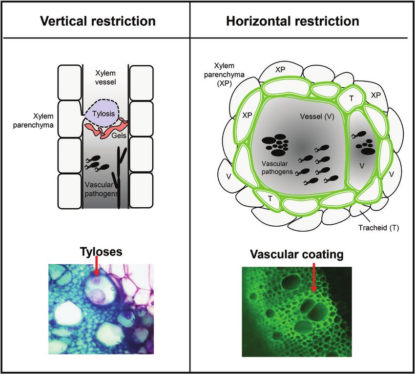
Fig. 1. The two dimensions of plant physico-chemical barriers induced against xylem vascular wilt pathogens. To counter invasion by
xylem vascular wilt pathogens, resistant plants induce two-dimensional physico-chemical defenses that restrict vertical and horizontal movement of
the pathogen. Vertical spread within the vessel lumen is mainly restricted by tyloses and gels (left). In contrast, horizontal spread of the pathogen to
surrounding healthy vessels is prevented by reinforcement of the walls of colonized vessels (V) and the surrounding xylem parenchyma (XP) and tracheids
(T), through vascular coating with mainly lignin and suberin (shown as a green color in the diagram). Synchronized formation of vertical and horizontal
barriers early after pathogen invasion results in compartmentalization of the pathogen inoculum at the site of infection, thereby preventing wilt, and
constitute a major component of resistance. To visualize tyloses and vascular coating (panels below), tomato root cross-sections were obtained after
R. solanacearum soil soak inoculation, and fixed in 70% ethanol. For tyloses, cross-sections were stained with 0.1% toluidine blue and observed using
a Leica DM6B-Z microscope under bright field conditions, and images were recorded through a MC190-HD-0518131623 camera. To visualize phenolic
vascular coating, the cross-sections were illuminated by UV using a Leica DM6B-Z microscope, and the auto-florescence emitted from phenolic deposits
was observed using a HC PL FLUOTAR objective. Images were captured using a Leica-DFC9000GT-VSC07341 camera. In the left panel, the arrow
points towards R. solanacearum-induced formation of tyloses inside vessel lumen, which appear pink to violet color upon staining with 0.1% toluidine
blue. In the right panel, the arrow points towards R. solanacearum-induced auto-fluorescence emitted from phenolics, deposited in the walls of vessels
and the surrounding tracheids and parenchyma cells. Scale bar=120 µm.
grapevines with differing resistance to X. fastidiosa. Excessive are key (Sun et al., 2013). Interestingly, the anatomy of xylem
tylosis formation in response to X. fastidiosa infection in the vessels also plays a role in augmenting pathogen compartmen-
susceptible grapevine cultivar led to heavy blockage of vessels talization by tyloses. Cultivars of grapevine having larger vessel
and development of wilting symptoms, and did not signifi- size are known to be susceptible to vascular wilt pathogens
cantly affect pathogen spread. In contrast, in resistant grape- such as X. fastidiosa, Eutypa lata, Phaeoacremonium aleophilum,
vines, tylosis development was specific and mainly limited to Phaeomoniella chlamydospora, Diplodia seriata, and Neofusicoccum
a few internodes close to the point of inoculation, impacting parvum (Pouzoulet et al., 2017, 2019, 2020; Deyett et al., 2019).
less of the vessels and indicating that timing and localization Likewise, Dutch elm cultivars having larger vessel diametersInducible barriers against plant vascular wilt pathogens | 189
Table 1. List of plant pathosystems in which (I) vertical or (II) horizontal restriction of pathogen movement inside the plant has been
shown.
I. Vertical restriction
Structure Host Pathogen Reference
Tylose formation Banana Fusarium oxysporum f.sp. cubense (Vander Molen et al., 1987)
Butternut Ophiognomonia clavigignenti-juglandacearum (Rioux et al., 2018)
Cotton Fusarium oxysporum f. sp. vasinfectum (Shi et al., 1991)
Cucurbits Fusarium oxysporum f. sp. melonis (Seo and Kim, 2017)
Elm Ophiostoma novo-ulmi (Plichta et al., 2016)
Grapevine Xylella fastidiosa (Sun et al., 2013)
Phaeomoniella chlamydospora (Pouzoulet et al., 2013)
Downloaded from https://academic.oup.com/jxb/article/72/2/184/5911728 by guest on 08 September 2021
Potato Rasltonia solanacearum (Ferreira et al., 2017)
Tomato Rasltonia solanacearum (Grimault et al., 1994)
Verticillium albo-atrum (Hutson and Smith, 1980)
Verticillium dahliae (Tjamos and Smith, 1975)
Fusarium oxysporum f. sp. lycopersici (Hutson and Smith, 1980)
Gel deposition Banana Fusarium oxysporum f.sp. cubense (Vander Molen et al., 1987)
Butternut Ophiognomonia clavigignenti-juglandacearum (Rioux et al., 2018)
Carnation Fusarium oxysporum f.sp. dianthi (Baayen and Elgersma, 1985)
Elm Ophiostoma novo-ulmi (Plichta et al., 2016)
Grapevine Xylella fastidiosa (Sun et al., 2013)
Pea Fusarium oxysporum f. sp. pisi (Bishop and Cooper, 1983)
Plane tree Ceratocystis fimbriata f. sp platani (Clérivet et al., 2000)
Tomato Rasltonia solanacearum (Grimault et al., 1994; Kim et al., 2016)
Verticillium albo-atrum (Hutson and Smith, 1980)
Fusarium oxysporum f. sp. lycopersici (Hutson and Smith, 1980)
II. Horizontal restriction
Structure Host Pathogen Reference
Lignin deposition Banana Fusarium oxysporum f. sp. cubense (De Ascensao and Dubery, 2000)
Cotton Verticillium dahliae (Xu et al., 2011; Bu et al., 2014)
Dutch elm Ophiostoma novo-ulmi (Martin et al., 2007)
Flax Fusarium oxysporum f. sp. lini (Galindo-González and Deyholos, 2016)
Oilseed rape Verticillium longisporum (Eynck et al., 2009)
Olive Xylella fastidiosa (Sabella et al., 2018)
Pepper Verticillium dahliae (Novo et al., 2017)
Potato Rasltonia solanacearum (Ferreira et al., 2017)
Tomato Verticillium dahliae (Street et al., 1986; Hu et al., 2019)
Rasltonia solanacearum (Ishihara et al., 2012; Ferreira et al., 2017)
Suberin deposition Alfalfa Verticillium albo-atrum (Newcombe and Robb, 1988)
Butternut Ophiognomonia clavigignenti-juglandacearum (Rioux et al., 2018)
Dutch elm Ophiostoma novo-ulmi (Martín et al., 2008)
Grapevine Phaeomoniella chlamydospora (Pouzoulet et al., 2013)
Tomato Verticillium albo-atrum (Street et al., 1986; Robb et al., 1991)
are susceptible to Ophiostoma novo-ulmi (Pouzoulet et al., 2014; Deposition of gels
Venturas et al., 2014). Across the grapevine genotypes, it has
been observed that the extent of P. chlamydospora compart- Deposition of electrodense material corresponding to gels or
mentalization is a function of the diameter of the host xylem gums in the lumen of the xylem vessels is another feature com-
vessels. Genotypes with increased number of xylem vessels monly observed during xylem invasion by vascular pathogens,
above 100 µm in diameter resulted in increased infection of and acts as one of the multiple factors that contribute to in-
host tissue (Pouzoulet et al., 2020). Though numerous tyloses duced structural defense (Vander Molen et al., 1977). Besides
are formed in large vessels of susceptible grapevine cultivars in its role in immunity, these occluding structures also form in re-
response to the wilt pathogen P. chlamydospora, the compart- sponse to other stimuli such as wounding or aging (Ratnayake
mentalization process is not as efficient as in narrow diameter et al., 2013).
vessels, due to the presence of large escape routes (Pouzoulet Gels are commonly secreted by xylem parenchyma cells and
et al., 2017). they are transported across pit membranes into vessel elements190 | Kashyap et al.
(Fig. 1; Bishop and Cooper, 1984). Tyloses have also been ob- pathogens, resistant plants with altered composition and struc-
served to secrete gels into the lumen of vessels, thereby multi- ture of homogalacturonans (HGs) and xyloglucans (XyGs) in
plying the clogging effect (Bonsen and Kucera, 1990; Rioux pit membranes have evolved; however, these compounds are
et al., 1998). Gels appear fibrillar, forming thin networks of potential targets of the pathogen’s cell wall degrading enzymes.
varying electron density that ultimately fill and clog the vessel Grapevine genotypes resistant to X. fastidiosa lacked fucosylated
lumen (Sun et al., 2008). Gels initially appear as translucent XyGs and weakly methylesterified HGs (ME-HGs), and con-
fibres arising from several places along lateral walls of vessels, tained a small amount of heavily ME-HGs. In contrast, pit
and later form a continuous layer with wavy edges toward the membranes of susceptible genotypes all had substantial amounts
vessel lumen. Subsequently, gels turn yellow and are inter- of fucosylated XyGs and weakly ME-HGs, but lacked heavily
spersed with small particles, coinciding with vessel occlusions ME-HGs (Sun et al., 2011).
(Sun et al., 2008). Although the main component of gels are In addition, reinforcement occurs at vessel walls, paren-
pectic substances such as partially esterified pectic polysac- chyma cells and pit membranes, to confine the spread of vas-
Downloaded from https://academic.oup.com/jxb/article/72/2/184/5911728 by guest on 08 September 2021
charides (Rioux et al., 1998; Clérivet et al., 2000), they may cular pathogens. Ultra-microscopic studies showed that the
also accumulate antimicrobial compounds such as elemental pit membranes, as well as vessels walls and parenchyma cells,
sulfur and phytoalexins (Cooper and Williams, 2004; Sun et al., form a conspicuously thick coating in the form of an elec-
2008). Furthermore, these gels are strengthened by deposition tron dense amorphous layer, as part of the defense response
of lignin and other phenolic compounds, which make these against vascular pathogens (Street et al., 1986; Benhamou, 1995;
plugs strong physical barriers (Kpemoua et al., 1996; Rioux Daayf et al., 1997; Nakaho et al., 2000; Araujo et al., 2014). Such
et al., 1998). reinforcement acts to limit the horizontal movement of the
Formation of vascular gels is considered an important part of pathogen from the protoxylem or the primary xylem to the
resistance towards several wilt diseases (Table 1). For example, surrounding cells (Street et al., 1986; Benhamou, 1995; Daayf
F. oxysporum f. sp. dianthi colonization is restricted due to the et al., 1997; Nakaho et al., 2000; Araujo et al., 2014). Besides,
formation of gels in the vascular lumen of carnation plants its deposition acts as a shield against pathogen-derived metab-
(Baayen and Elgersma, 1985). Moreover, in most resistant culti- olites such as toxins and enzymes, and makes water and nutri-
vars these gels are often observed together with tyloses (Vander ents inaccessible for pathogens, thereby impeding their growth
Molen et al., 1987; Grimault et al., 1994). In banana plants re- (Araujo et al., 2014).
sistant to F. oxysporum f. sp cubense, formation of vascular oc- Importantly, the timing of synthesis of the vascular coating
clusions including both gels and tyloses have been observed plays a crucial role in immunity. Even if vascular coating can
(Vander Molen et al., 1977). Gel formation in the xylem lumen be observed in response to vascular pathogens in susceptible
is also a trait of tomato cultivars resistant towards the vascular plants, these structures form at late time points, compared with
bacterium R. solanacearum (Grimault et al., 1994). In other their induction in resistant plants (Shi et al., 1991; Daayf et al.,
cases, such as pea plants resistant to F. oxysporum f. sp. pisi, vas- 1997). The specific composition of vascular deposits varies de-
cular gels, but not tyloses, are observed after infection (Bishop pending on the particular host-pathogen interaction. However,
and Cooper, 1984). phenolics are the most important compounds, as they act as
building blocks of the secondary cell wall, and they also have
Horizontal restriction of vascular colonization direct antimicrobial activity (Eynck et al., 2009). Among the
phenolic polymers constituting vascular coating structures, the
The mechanisms mentioned above are part of host responses principal players are lignin and suberin, described in further
that restrict vertical movement of vascular pathogens to detail below.We also outline the role of callose, a non-phenolic
healthy regions of the host. In addition, resistant plants can compound that plays an important role in the formation of
often develop a protective vascular coating upon invasion by horizontal vascular barriers in certain interactions.
vascular pathogens, posing a horizontal barrier to further col-
onization of adjacent healthy tissues. Vascular coating involves
Deposition of lignin
physico-chemical structural modifications in the cell walls of
xylem tissues that result in confinement of the pathogen to the Lignin is a complex phenolic polymer that constitutes a
infected vessels (Figs 1, 2A). major component of secondary cell walls in vascular plants.
Xylem pits are the primary routes of vessel-to-vessel and Lignin imparts strength to secondary cell walls, being depos-
vessel-to-parenchyma cell water transport. Pits are covered by ited in spaces between cellulose, hemicellulose and pectin
a pit membrane, which is impermeable to particulate matter (Kang et al., 2019; Fig. 2A). The building blocks of the lignin
like bacteria and other pathogens (Choat et al., 2008). For a polymer are monolignols, synthesized from phenylalanine via
pathogen to achieve successful horizontal transfer into ves- the phenylpropanoid pathway, where numerous enzymes are
sels, it has to either form openings in vessel walls or degrade involved (Fig. 2B). Monolignols are then transported from the
pit membranes, thereby reaching the adjacent parenchyma cytosol into the apoplast, where they are polymerized to lignin
cells and vessels (Nakaho et al., 2000). To avoid the breach by units by the oxidative activities of laccases and peroxidases.Inducible barriers against plant vascular wilt pathogens | 191
A Vessel Parenchyma
B
Secondary wall
Middle lamella
Plasma membrane
Primary wall
Shikimate
Phenylalanine
pathway
PAL
Invasion
Cinnamic acid Phenylpropanoid
pathogens
pathway
Vascular
C4H
p-coumaric acid
C4H
4CL Caffeic acid
C3H
p-coumaroyl-CoA
COMT
Downloaded from https://academic.oup.com/jxb/article/72/2/184/5911728 by guest on 08 September 2021
HCT
4CL
p-coumaroyl shikimate
Suberin Lignin C3H
HCT
Hemi-cellulose Cellulose Caffeoyl shikimate Caffeoyl-CoA
CCoAOMT
4CL
Acyl lipid Feruloyl-CoA Ferulic acid
Suberin Fatty acids
pathway
pathway CYP86A1 FAR
ω-hydroxyacids Primary Alcohols
Lignin pathway
CCR CCR
F5H
p-coumaraldehyde Coniferaldehyde Sinapaldehyde
Tyrosine COMT
CAD CAD CAD
TyDC
p-coumaryl alcohol Coniferyl alcohol Sinapyl alcohol
Tyramine
FHT Lignin
THT monomers
PRX PRX PRX
Feruloyl tyramine Suberin Feruloyl esters
p-coumaroyl tyramine p-coumaroyl esters p-hydroxyphenyl (H) Guaiacyl (G) Syringyl (S)
monomers
Caffeoyl tyramine Caffeoyl esters unit unit unit
PRX
H LIGNIN G LIGNIN S LIGNIN
SUBERIN
Fig. 2. Lignin and suberin have a major role in vascular coating induced by xylem vascular wilt pathogens. A. Schematic structure of
reinforced cell walls of xylem vessels and parenchyma cells in resistant plants upon infection with xylem vascular wilt pathogens. B. The phenypropanoid
pathway provides precursors for both lignin and suberin biosynthesis. Phenylalanine, derived from the shikimate pathway, undergoes several enzymatic
reactions as part of the phenylproanoid pathway. The resulting precursors yield the monolignols p-coumaryl alcohol, coniferyl alcohol and sinapyl
alcohol, which are the building blocks of lignin. In parallel, the phenypropanoid metabolites feruloyl-CoA, caffeoyl-CoA, and p-coumaroyl-CoA bifurcate
into the suberin pathway. In the suberin pathway, these metabolites can be conjugated to aromatic amine compounds such as tyramine by the action
of THT, or can be linked to aliphatic compounds by the action of FHT, to yield suberin monomers. Lignin and suberin monomers are then transported
to the cell wall, where they are subsequently polymerized into the reinforcing matrices that constitute vascular coating structures. Abbreviations: PAL:
phenylalanine ammonia–lyase; C4H: cinnamate–4–hydroxylase; C3H: coumarate 3-hydroxylase; 4CL: 4–coumarate–CoA ligase; HCT: hydroxycinnamoyl–
CoA shikimate/quinate hydroxycinnamoyl transferase; COMT: caffeic acid 3-O-methyltransferase; CCOMT: caffeoyl CoA 3-O-methyltransferase; CCR:
cinnamoyl CoA reductase; CAD: cinnamoyl alcohol dehydrogenase; PRX: peroxidase; CYP86A1: fatty acid cytochrome P450 oxidases; FAR: fatty acyl-
CoA reductase; TyDC: tyrosine decarboxylase; FHT: feruloyl transferase; THT: tyramine hydroxycinnamoyl transferase.
Lignin is a particularly important element in cell walls of reinforcements that further restrict pathogen colonization.This
xylem tissue cells, not only because of its structural function, inducible deposition of lignin as a vascular coating can be an
but also because it facilitates water retention in vascular bun- important component of resistance towards certain pathogens
dles due to its hydrophobic nature. In addition, lignin is re- (Table 1).
sistant to biodegradation and acts as a potent structural barrier Restriction of pathogen colonization by lignin has been ele-
against pathogens. Although lignin is an integral part of pre- gantly shown using a non-vascular pathogen, the leaf-infecting
existing structural barriers in plants, its deposition can also be bacterium Pseudomonas syringae pv tomato (Pto) on Arabidopsis
induced in the xylem upon pathogen attack to form additional thaliana (Lee et al., 2019). It was shown that infection with192 | Kashyap et al.
either virulent or avirulent strains of the pathogen induced Nevertheless, the mechanisms that orchestrate timely and
localized lignification at the site of pathogen attack. However, effective induction of lignin deposition in resistant plants
lignin deposition was more conspicuous upon avirulent Pto upon vascular colonization remain vastly unknown. Some
recognition, leading to confinement of the pathogen, and re- progress has been made using cotton plants infected with
stricting hypersensitive response cell death to the infection site. V. dahliae. In this pathosystem, two proteins potentially regu-
In contrast, virulent strains could overcome induced lignifica- lating V. dahliae-induced vascular lignin deposition have been
tion, as the deposition in this case was milder, leading to unre- identified. GhUMC1, a copper-binding protein, is involved
stricted disease progression. in resistance mediated by lignin deposition and jasmonate
Interestingly, Casparian strip membrane domain proteins signaling (Zhu et al., 2018). GhUMC1 knock-down plants are
(CASPs), were shown to be involved in pathogen-induced more susceptible to the pathogen, and lignin vascular coating
lignin deposition (Lee et al., 2019). The Casparian strip acts is drastically reduced. On the other hand, the proline-rich
as a diffusion barrier that controls the uptake of water and protein GhHyPRP1 acts as a negative regulator of defense
Downloaded from https://academic.oup.com/jxb/article/72/2/184/5911728 by guest on 08 September 2021
molecules from the soil into the water-conducting tissues, and against V. dahlia, and was shown to induce lignin deposition
prevents the entry of pathogens and other harmful substances. (Zhu et al., 2018). In accordance, GhHyPRP1 knock-down
Both the Casparian strip and the above-mentioned pathogen- plants displayed more lignin deposition upon infection and
induced lignification mechanism involve lignin-containing were more resistant to the pathogen. Interestingly, PevD1, a
structures that function as anti-pathogenic physical barriers, V. dahliae secreted protein, has been shown to activate defenses
serving parallel functions. In addition to lignin, suberin (dis- in cotton, triggering the expression of phenylpropanoid genes
cussed in detail later) is a chief component of the Casparian and lignin accumulation (Bu et al., 2014). This may indicate
strip (Doblas et al., 2017). Recent findings show that the that PevD1 acts as an avirulence effector in cotton triggering
Casparian strip possesses a barrier surveillance pathway com- a defense reaction that includes lignin deposition in the vascu-
prised of the receptor-like cytoplasmic kinase SCHENGEN1 lature, which may lead to pathogen confinement into infected
and the LRR-RLK SCHENGEN3, hence bearing a striking vessels. It remains to be determined whether the differential
resemblance to signaling pathways for perception of pathogen- lignin deposition phenotypes observed in resistant cultivars is
associated molecular patterns (Alassimone et al., 2016; Fujita a direct consequence of pathogen effector recognition via its
et al., 2020). These receptors in the Casparian strip domain secreted effectors, and if so, to what degree the mechanisms are
interact with peptides (CASPARIAN STRIP INTEGRITY conserved between different pathosystems. Moreover, the mo-
FACTORS, CIF1/2) expressed in the stele, leading to lecular players involved also need to be identified.
Casparian strip formation. This spatial separation of receptor
and ligand constitutes a surveillance system where interaction Deposition of suberin
stops after effective sealing by the strip, but any breach in
the barrier leads to re-interaction and further strengthening Suberin is a heteropolymer that deposits as a poly-lamellar
(Doblas et al., 2017). structure between the plasma membrane and the cell wall,
Lignin deposits seem to also play a crucial role in spa- forming a hydrophobic protective barrier (Fig. 2A). Suberin
tial growth restriction of vascular pathogens. There are is deposited in specialized tissues such as root and tuber epi-
several examples showing a pronounced transcriptional dermis, root endodermis, and seed coats. In addition, suberin
upregulation of genes involved in lignin biosynthesis in is formed in response to several stresses such as wounding, salt
resistant plants following infection with various vascular injury and pathogen attack (Dixon and Paiva, 1995; Bernards,
pathogens (Table 1). This occurs, for example, in cotton after 2002). Besides providing strength to the cell wall, suberin pre-
V. dahliae infection, flax after infection with F. oxysporum, vents water loss and pathogen entry by sealing off the layer of
tomato after infection with R. solanacearum, or olive tree suberized cells.
after X. fastidiosa infection (Xu et al., 2011; Ishihara et al., Suberin consists of a polyphenolic and a polyaliphatic domain.
2012; Galindo-González and Deyholos, 2016; Sabella et al., The polyphenolic domain is predominantly formed by esters
2018). Furthermore, there are several examples showing and amides of ferulic acid, as well as other hydroxycinnamic
that resistance/tolerance to vascular pathogens is accom- acids, such as caffeic acid and p-coumaric acid (Negrel et al.,
panied by an increase in lignin content and enhanced cell 1995; Lashbrooke et al., 2016; Woolfson et al., 2011; Woolfson,
wall lignification upon infection. This includes examples of 2018). The aliphatic domain consists of a glycerol-based fatty
different pathosystems such as V. dahliae—pepper, O. novo- acid-derived polyester comprised primarily of ω-hydroxyacids,
ulmi—Ulmus minor, V. longisporum—rapeseed, F. oxysporum— α, ω-dicarboxylic acids, fatty alcohols, and small amounts of
banana or tomato, and R. solanacearum—potato (Street et al., hydroxycinnamic acids (mainly alkyl ferulates; Beisson et al.,
1986; De Ascensao and Dubery, 2000; Martín et al., 2005; 2012). Ferulic esters composed of ferulic acid esterified to
Martin et al., 2007; Eynck et al., 2009; Ferreira et al., 2017; fatty acids are considered as one of the monomers of suberin
Novo et al., 2017). polymer (Negrel et al., 1995).Inducible barriers against plant vascular wilt pathogens | 193
Significant progress elucidating suberin biosynthesis has little is known about its regulation. Similar to lignin, the effect-
been achieved in the last two decades using molecular gen- iveness of vascular suberization as a structural barrier against
etics approaches, especially in the model species A. thaliana and horizontal colonization by vascular pathogens largely depends
in potato tuber periderm (Ranathunge et al., 2011). Suberin on the spatio-temporal control of its deposition, i.e. formation
shares the phenypropanoid pathway with lignin (Fig. 2B). This of suberin deposits early after pathogen detection at the site
pathway provides the precursors for its polyphenolic domain, of vascular invasion. The phytohormones abscisic acid (ABA)
which are used by downstream suberin-specific enzymes such and ethylene have both been shown to regulate suberization
as tyramine N-feruloyltransferase (THT) and feruloyl trasferase (Soliday et al., 1978; Cottle and Kolattukudy, 1982; Barberon
(FHT; Fig. 2B; Serra et al., 2010; Woolfson, 2018; Woolfson et al., 2016). Since these two hormones are involved in de-
et al., 2011). Similarly, several genes involved in the aliphatic fense against various vascular pathogens, a possibility exists that
metabolism of suberin have been described, such as fatty acid pathogen-induced vascular suberization correlates with an in-
cytochrome P450 oxidases (CYP86A1), fatty acyl-CoA re- crease of hormone concentrations during immune responses.
Downloaded from https://academic.oup.com/jxb/article/72/2/184/5911728 by guest on 08 September 2021
ductase (FARs), β-ketoacyl-CoA synthases (KCS2/Daisy and However, the mechanistic links between these hormonal path-
KCS20), glycerol-3-phosphate acyltransferase5 (GPAT5), as ways and suberin biosynthesis during pathogen-triggered su-
well as ATP-BINDING CASSETTE G (ABCG) genes in- berin vascular coating remain to be established.
volved in the delivery of suberin monomers to the site of su-
berization (ABCG2, ABCG6, and ABCG20; Beisson et al., Deposition of callose
2007; Höfer et al., 2008; Franke et al., 2009; Vishwanath et al.,
2013;Yadav et al., 2014). In addition, a few upstream regulators Callose is a linear amorphous cell wall polysaccharide formed
of the suberin biosynthetic pathway have been identified in by hundreds of glucose units linked by β-1,3 glucosidic bonds
A. thaliana, such as the MYB transcription factors MYB41, (Stone, 2009). This homopolysaccharide is synthesized from
MYB107 and SUBERMAN (MYB39; Kosma et al., 2014; uridine diphosphate glucose by callose synthases (also known
Gou et al., 2017; Cohen et al., 2020). as CalS or GSL for glucan synthase-like), large multisubunit
Suberin reinforcement in the xylem vascular tissue has been complexes at the plasma membrane (Ellinger and Voigt, 2014).
long recognized as a potent barrier to colonization by patho- Callose is not a particularly abundant polymer in the cell wall,
gens (Robb et al., 1991). There are numerous studies reporting but has very relevant regulatory roles in development, plasmo-
that suberin deposition in the xylem tissue upon infection desmata function, as well as in immunity (Schneider et al.,
contributes to resistance (Table 1). For example, vascular de- 2016). Pathogen-induced callose deposition has been shown
posits have been observed after infection with V. albo-atrum to be localized to callosic papillae, providing structural defense
in resistant tomato and alfalfa (Street et al., 1986; Newcombe against various pathogens (Schneider et al., 2016). In addition,
and Robb, 1988; Robb et al., 1991). Similarly, induction of su- callose can constitute a matrix for accumulation of antimicro-
berin deposition is an important line of defense in Ulmus minor bial compounds, thereby providing targeted delivery of chem-
against O. novo-ulmi (Martín et al., 2008). Interestingly, ex- ical defenses at the sites of pathogen attack (Luna et al., 2011).
ogenous application of phenolic compounds further increased In the vasculature, callose has also been shown to act as a
resistance of trees to this pathogen through formation of structural barrier against fungal wilt pathogens, restricting
suberin-like compounds in xylem tissues (Martín et al., 2008). their horizontal vessel-to-vessel movement. Tomato plants re-
Another example of xylem tissue suberization induced by a sistant to F. oxysporum f. sp. lycopersici form callose deposits in
vascular pathogen is provided by the histological characteriza- paravascular parenchyma cells and at pit membranes in response
tion of grapevine infection by the wilt fungus Phaeomoniella to infection by this pathogen (Beckman et al., 1982; Mueller
chlamydospora (Pouzoulet et al., 2013, 2017). It was shown that and Beckman, 1988). In addition, application of the microbe-
deposition of suberin in paravascular parenchyma cells is an associated molecular pattern chitosan, a derivative of chitin, re-
effective barrier against horizontal P. chlamydospora coloniza- stricts colonization of F. oxysporum f.sp. lycopersici by inducing a
tion from one vessel to the adjacent vessel. Suberin was also vascular coating composed of callose and phenolic compounds
shown to form deposits in tyloses induced by infection with (Benhamou et al., 1994). Furthermore, cotton roots infected
this pathogen (Pouzoulet et al., 2013). Interestingly, inhibition with V. dahliae showed reinforcement with callose deposits
of tylose formation in grapevine by P. chlamydospora led to vas- (Daayf et al., 1997). In contrast, infection by the bacterial wilt
cular coating of surrounding parenchyma cells with phenolic pathogen R. solanacearum caused deposition of callose in both
compounds, including suberin (Pouzoulet et al., 2017).This in- tolerant and susceptible potato plants, indicating that in this
dicates that sequential/superimposed defense mechanisms are interaction callose may not be as important for resistance to-
in place to restrict pathogen progression to the infection site, wards the pathogen (Ferreira et al., 2017). Additional research
once it has reached the vasculature. is needed to clarify the precise role and regulation of callose
Although suberin deposition seems to be an important com- as a structural defense mechanism induced upon perception of
ponent of defense responses against vascular pathogens, very vascular wilt pathogens.194 | Kashyap et al.
Concluding remarks and future prospects Raman spectroscopy or MALDI (matrix-assisted laser desorp-
tion ionization) spectrometry imaging, both of which can be
During the last few decades, much evidence has accumulated extremely useful in zonal responses such as structural resistance.
showing the importance of physico-chemical barriers as a cru- All this knowledge could lead in the future to the engineering
cial component of resistance towards xylem vascular patho- of metabolic pathways of vascular coating compounds in spe-
gens. From all the research until now in this field, it becomes cific cells, to produce resistant plants against xylem colonizers.
clear that the localization and timing of formation of these With this article we hope to contribute towards raising
vascular structures is key for their effectiveness as barriers for awareness of the importance of attaining a better understanding
pathogen confinement. Resistant plants are able to form ver- of the structural physico-chemical barriers as a crucial compo-
tical and horizontal barriers quickly upon pathogen invasion of nent of resistance towards xylem vascular pathogens. Disease
the vasculature, confining them to infected vessels and avoiding management through host resistance is the most efficient and
spread to the rest of the plant. In susceptible plants, formation eco-friendly approach to control pathogens. However, a lot
Downloaded from https://academic.oup.com/jxb/article/72/2/184/5911728 by guest on 08 September 2021
of the same vascular structures is observed, but not targeted to has to be learnt about the complex genetic interactions which
infected vessels, and later in time, once the pathogen has spread govern induced structural resistance in various hosts, to be able
throughout the plant they have no effect on disease progression. to deploy this trait in future cultivars and fight vascular patho-
However, the mechanisms regulating the spatial and temporal gens, agents of the most devastating plant diseases in the field.
formation of vascular structures leading to effective pathogen
confinement, and their precise composition, are old questions
that remain unanswered. In fact, it remains unclear as to how Acknowledgements
vascular wilt pathogens are perceived at the vasculature, and
how this perception is transduced into timely and restricted The authors would like to thank all members of the Bacterial plant dis-
formation of structural defenses. Since effective mechanisms of eases and cell death lab for helpful comments.We apologize to all authors
whose work has been omitted because of space limitations. Research in the
resistance are very much sought after in breeding programs, in
lab is funded by the Spanish Ministry of Economy and Competitiveness
the coming years it will be important to make an effort to ad- with grants 2016-78002-R (AGL) and RyC 2014–16158 (NSC), by the
vance knowledge in this area, even though inducible structural Ministry of Science and Innovation / Spanish State Research Agency
defenses are governed by complex polygenic traits. PID2019-108595RB-I00 / AEI / 10.13039/501100011033, and
Major technological advances in the last few years have through the “Severo Ochoa Programme for Centres of Excellence in
placed plant molecular biologists in a privileged position to R&D” (SEV-2015-0533). AK is the recipient of a Netaji Subhas—Indian
make significant advances. Of particularly relevance is the Council of Agricultural Research (ICAR) International Fellowship. This
CRISPR-Cas9 technology, which has proven extremely ef- work was also supported by the CERCA Programme / Generalitat de
ficient for Solanaceous crops such as tomato, pepper and Catalunya.
eggplant, which are severely affected by wilt diseases caused
by xylem vascular pathogens (Hu et al., 2019; Li et al., 2019;
Wang et al., 2019). Importantly, an array of technologies have Author contribution
emerged that allow the study of specific processes in a cell, or AK, MP-M, MC, MV, and NSC conceptualized and wrote the article,
in a tissue-specific manner. These techniques will become in- and obtained funds.
strumental in the study of plant-pathogen interactions, which
constitute a localized phenomenon by its very nature. This is
particularly the case for colonization of vascular cells by vas- References
cular pathogens, since the ability to confine the invading agent Alassimone J, Fujita S, Doblas VG, et al. 2016. Polarly localized kinase
is a key feature of resistant plants. With the advent of single- SGN1 is required for Casparian strip integrity and positioning. Nature Plants
cell technologies it will be possible to attain astonishing reso- 2, 16113.
lution when investigating the processes occurring at infected Álvarez B, Biosca EG, López MM. 2010. On the life of Ralstonia
solanacearum, a destructive bacterial plant pathogen. In: Méndez-Vilas A,
cells and surrounding areas. For instance, RNA sequencing ed. Technology and education topics in applied microbiology and micro-
of laser-dissected areas or single cells allows profiling of the bial biotechnology. Current Research, Technology and Education Topics in
transcriptomic landscape after infection at relevant sites. In Applied Microbiology. Badajoz: Formatex, 267–279.
turn, this will allow the identification of marker genes associ- Antanaviciute L, Šurbanovski N, Harrison N, McLeary KJ,
Simpson DW, Wilson F, Sargent DJ, Harrison RJ. 2015. Mapping QTL
ated with the formation of structural defenses and the subse- associated with Verticillium dahliae resistance in the cultivated strawberry
quent generation of transgenic marker lines, to be able to track (Fragaria × ananassa). Horticulture Research 2, 15009.
relevant cells/tissues at early time points after infection for their Araujo L, Bispo WM, Cacique IS, Moreira WR, Rodrigues FÁ.
analysis. In addition, extremely sensitive analytical techniques 2014. Resistance in mango against infection by Ceratocystis fimbriata.
Phytopathology 104, 820–833.
have been developed in recent years that allow the identifica-
Baayen RP, Elgersma DM. 1985. Colonization and histopathology of sus-
tion and quantification of proteins, small molecules and me- ceptible and resistant carnation cultivars infected with Fusarium oxysporum
tabolites, and the interactions between them. This includes f. sp. dianthi. Netherlands Journal of Plant Pathology 91, 119–135.You can also read