Breathing coordinates limbic network dynamics underlying memory consolidation - bioRxiv
←
→
Page content transcription
If your browser does not render page correctly, please read the page content below
bioRxiv preprint first posted online Aug. 16, 2018; doi: http://dx.doi.org/10.1101/392530. The copyright holder for this preprint
     (which was not peer-reviewed) is the author/funder, who has granted bioRxiv a license to display the preprint in perpetuity.

bioRxiv                                 All rights reserved. No reuse allowed without permission.

                     Breathing coordinates limbic network dynamics
                           underlying memory consolidation
                                              Nikolaos Karalis1,2,∗ and Anton Sirota1,∗

                1
                    Faculty of Medicine, Ludwig-Maximilian University Munich, 82152, Planegg-Martinsried, Germany.
                     2
                       Present address: Friedrich Miescher Institute for Biomedical Research, 4058, Basel, Switzerland.
                                  ∗ Correspondence:   nikolaskaralis@gmail.com (N.K.), sirota@bio.lmu.de (A.S.)

                                                                   15 August 2018

            The coordinated activity between remote brain regions underlies cognition and memory function. Although
            neuronal oscillations have been proposed as a mechanistic substrate for the coordination of information trans-
            fer and memory consolidation during sleep, little is known about the mechanisms that support the widespread
            synchronization of brain regions and the relationship of neuronal dynamics with other bodily rhythms, such as
            breathing. Here we address this question using large-scale recordings from a number of structures, including
            the medial prefrontal cortex, hippocampus, thalamus, amygdala and nucleus accumbens in mice. We identify
            a dual mechanism of respiratory entrainment, in the form of an intracerebral corollary discharge that acts
            jointly with an olfactory reafference to coordinate limbic network dynamics, such as hippocampal ripples
            and cortical UP and DOWN states, involved in memory consolidation. These results highlight breathing, a
            perennial rhythmic input to the brain, as an oscillatory scaffold for the functional coordination of the limbic
            circuit, enabling the segregation and integration of information flow across neuronal networks.

Over the past century, cortical and subcortical structures of the             remain elusive and a global pacemaker that ties together distinct
limbic circuit and the medial temporal lobe have been identified              network dynamics has not been identified. Recently, a number of
as critical elements of the memory circuit, involved in emo-                  studies have identified signatures of respiration in the cortical
tional regulation and the formation, consolidation, and retrieval             and hippocampal LFP of rodents20–23 and humans24,25 , which
of episodic memories1–3 . Although the anatomical substrate of                has been attributed to reafferent olfactory activity. However, the
these circuits has been elaborated in detail, mechanisms that                 function of these phenomena is not understood and the mecha-
enable the processing and transfer of information across these                nism and consequences of respiratory modulation have not been
distributed circuits are not well understood.                                 established, given the limitations in interpreting LFP signals.
   Neuronal dynamics are characterized by oscillatory activity                    Here we address the hypothesis that the breathing rhythm
associated with distinct behavioral states and functional roles4 .            modulates the activity of the limbic brain and underlies the
During active states, hippocampal theta oscillations dynami-                  coordination of network dynamics across limbic systems during
cally coordinate local activity and information flow between the              offline states. To this end, using high-density silicon probes we
hippocampus and entorhinal cortex5–7 , as well as other limbic                performed a large-scale in vivo functional anatomical character-
structures such as the medial prefrontal cortex (mPFC)8,9 .                   ization of the medial prefrontal cortex (mPFC), hippocampus,
   During slow-wave sleep, the cortex is in a bistable state, char-           basolateral amygdala (BLA) and nucleus accumbens (NAc).
acterized by spontaneous alternations between UP and DOWN                     Using this approach, we identified an intracerebral centrifugal
states in the membrane potential and action potential firing of               respiratory corollary discharge that acts in concert with a respira-
neurons10,11 . In parallel, the hippocampal neurons are engaged               tory reafference and mediates the inter-regional synchronization
in transient, fast oscillatory events termed sharp-wave ripples               of limbic memory circuits. The respiratory modulation is act-
(SWR), during which awake activity is replayed12 . Such non-                  ing as a functional oscillatory scaffold, that together with the
linear dynamics are coordinated between regions13–15 and their                underlying anatomical substrate, organizes information flow and
interaction is believed to support memory consolidation16,17 and              systems memory consolidation processes.
the transfer of memories to their permanent cortical storage18,19 .
   While the importance and role of the cortical slow oscillation
(SO), hippocampal ripples, and their coordination during sleep
have been established, the mechanisms that support this coordi-
nation across distributed cortical and limbic circuits during sleep

                                                                                                                                                1
bioRxiv preprint first posted online Aug. 16, 2018; doi: http://dx.doi.org/10.1101/392530. The copyright holder for this preprint
     (which was not peer-reviewed) is the author/funder, who has granted bioRxiv a license to display the preprint in perpetuity.
                                        All rights reserved. No reuse allowed without permission.

bioRxiv                                                                                                                        Karalis & Sirota, 2018

Results
Respiratory entrainment of prefrontal cortex across brain
states

To investigate the role of breathing in organizing neuronal
dynamics in the medial prefrontal cortex (mPFC), we recorded
simultaneously the local electrical activity (electroolfactogram;
EOG)26 of the olfactory sensory neurons (OSNs) and single-
unit and LFP in the mPFC in freely-behaving mice (Fig. 1a).
The EOG reflected the respiratory activity and exhibited reli-
able phase relationship to the respiratory cycle, as established
by comparing this signal to the airflow from the nostrils (Supple-
mentary Fig. 1a-d), and was reflected in rhythmic head-motion
(Supplementary Fig. 1b). We then segmented behavioral states
based on the head micro-motion (Supplementary Fig. 1e-f), dif-
ferentiating slow wave sleep (SWS), REM sleep, quiescence and
awake exploration. Behavioral state changes were associated with
changes in the breathing frequency (Fig. 1c, Supplementary
Fig.2a) which are accompanied by autonomic changes con-
ferred upon the heart by central regulation and respiratory sinus
arrhythmia (RSA), indicative of a generalized role of breathing
in coordinating bodily rhythms (Supplementary Fig. 2b-f ).
    Examination of the spectrotemporal characteristics of respira-
tory activity and the prefrontal LFP revealed a faithful reflection           Figure 1 Prefrontal oscillations are related to respiration
of the respiratory activity in the prefrontal LFP (Fig. 1b),                  throughout behavioral states.
suggesting a potential relationship between these oscillations.               (a) Example traces of simultaneously recorded respiratory EOG and
                                                                              medial prefrontal local field potentials (LFP). (b) Example time-fre-
The two oscillations were comodulated across a wide frequency                 quency decomposition of respiratory and mPFC LFP signals, revealing
range (Fig. 1f ) and this relationship was preserved through-                 the reliable relationship between the two signals. (c) Distribution of
                                                                              peak frequency bins of the spectrally decomposed respiration (left;
out many active (online) as well as inactive (offline) states in              darker colors) and mPFC LFP (right; lighter colors) during slow-wave
freely-behaving mice (Fig. 1c-e). Coherence and Granger causal-               sleep, quiescence, exploratory behavior and self-initiated wheel running
ity analysis of the respiratory and LFP signals suggested that                (n = 9 mice). (d, e) Averaged normalized power spectral density of
                                                                              respiration (d) and mPFC LFP (e) across states as in (c). (f ) Frequen-
the respiratory oscillation is tightly locked and likely causally             cy-resolved comodulation of respiration and mPFC LFP oscillation
involved in the generation of the prefrontal LFP oscillation signal           power, across mice and behaviors (n = 9 mice). (g) Example coherence
(Fig. 1g-i). During fear behavior, the mouse mPFC is dominated                spectrum between respiration and mPFC LFP during offline states.
                                                                              Inset, average coherence value in the 2-5 Hz band (n = 9 mice). (h)
by a prominent 4 Hz oscillation27 . The similarity in frequency               Phase shift of 2-5 Hz filtered respiration and mPFC LFP signals dur-
suggests that respiration is the origin of fear-related 4 Hz oscil-           ing offline states for an example animal (blue histogram) and overlaid
                                                                              magnitude of phase modulation (logZ) and average phase shift for all
lations, To explicitly test this, we exposed mice to auditory,                animals (red dots; n = 9 mice). Black arrow depicts the average phase
contextual and innate fear paradigms (Supplementary Fig. 3a,b).               and logZ of the phase shift for the example and the red arrow for the
During freezing, the respiratory rhythm changed in a stereotypic              population. (i) Example spectral Granger causality between respira-
                                                                              tion and mPFC LFP for both causal directions. Inset, average Granger
manner and matched the rhythmic head-motion and the pre-                      causality for the 4 Hz band (2–5 Hz) between respiration and mPFC
frontal LFP oscillation (Supplementary Fig. 3c,d). Interestingly,             LFP for both causality directions (n = 9 mice, Wilcoxon signed-rank
the respiratory peak frequency was distinct for different types of            test, resp → mPFC versus mPFC → resp, ** P
bioRxiv preprint first posted online Aug. 16, 2018; doi: http://dx.doi.org/10.1101/392530. The copyright holder for this preprint
     (which was not peer-reviewed) is the author/funder, who has granted bioRxiv a license to display the preprint in perpetuity.
                                        All rights reserved. No reuse allowed without permission.

                                                         Breathing coordinates limbic network dynamics underlying memory consolidation

Figure 2 Topography of prefrontal neuronal entrainment by respiration.
(a) Respiration phase histogram of the spiking activity of an example prefrontal neuron. Top, associated average respiration (red) and mPFC
LFP (blue) traces. (b) Color-coded normalized phase histograms of all prefrontal neurons, ordered by phase modulation magnitude (n = 1640
neurons, n = 13 mice). The horizontal dashed line indicates the significance threshold for the logZ. (c) Cumulative distribution of the logZ
for all prefrontal PNs (blue, n = 1250 neurons) and INs (red, n = 390 neurons). Phase modulation is assessed in relation to the respiration
(solid lines) and the local prefrontal LFP (dashed lines). (d) Distribution of the preferred phase for PNs (blue) and INs (red). The height
of each bar corresponds to the relative number of units. (e) Schematic depiction of a typical recording using a high-density silicon polytrode
inserted in the deep layers of the mPFC, overlaid on an example depth- and frequency-resolved power spectrum spanning all medial prefrontal
subregions. (f ) Example depth- and frequency-resolved coherence between the respiration and local prefrontal LFP spanning all medial prefrontal
subregions. (g) Average depth-resolved normalized power (red) and coherence in the 2-5 Hz band (black) (n = 11 mice). (h) Depth-resolved
average phase modulation statistics (logZ) (n = 1263 cells, n = 11 mice). (i) Example of 2D coherence between respiration and local LFP
throughout the frontal subregions. (j) Schematic depiction of a 16-shank probe (50µm shank spacing) inserted in the prelimbic region of
the mPFC to record simultaneously from all cortical layers and an example inspiration-triggered current-source density profile. (k) Example
average inspiration-triggered LFP traces and overlaid corresponding translaminar current-source density profile from the dorsal mPFC. (l)
Average inspiration-triggered translaminar normalized current-source density profile from the dorsal mPFC (n = 6 mice). (m) Average cortical
layer-resolved profile of the normalized 2-5 Hz band local LFP power (red) and coherence with respiration (coherence) (n = 6 mice). (n) Cortical
layer-resolved phase modulation statistics (logZ) (n = 58 cells, n = 6 mice). Shaded areas, mean ± s.e.m. a.u., arbitrary units; s.d., standard
deviations; L1, layer 1; L6, layer 6. Stars indicate significance levels (* P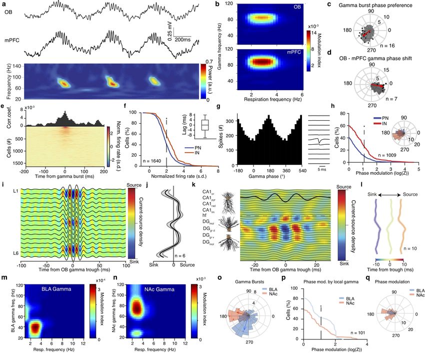
bioRxiv preprint first posted online Aug. 16, 2018; doi: http://dx.doi.org/10.1101/392530. The copyright holder for this preprint
     (which was not peer-reviewed) is the author/funder, who has granted bioRxiv a license to display the preprint in perpetuity.
                                        All rights reserved. No reuse allowed without permission.

bioRxiv                                                                                                                        Karalis & Sirota, 2018

Figure 3 Breathing modulates BLA, NAc, hippocampal and thalamic neuronal activity.
(a) Color-coded respiration phase histograms of the normalized firing rate of all hippocampal cells (n = 650 cells). (b) Cumulative distribution
of the modulation strength for all CA1 and DG PNs (CA1, n = 226 cells; DG, n = 206 cells) and INs (CA1, n = 98 cells; DG, n = 120
cells). (c) Distribution of the preferred phase for all significantly phase locked CA1 (top) and DG (bottom) cells. (d) Schematic depiction
of CA1 pyramidal and DG granular cell somato-dendritic domains aligned to the example inspiration-triggered high-density CSD profile of
the dorsal hippocampus. Horizontal dashed line indicates the hippocampal fissure. (e) Average normalized inspiration-triggered CSD profile of
dorsal hippocampus at different lags from inspiration (n = 10 mice). Stars mark the middle molecular layer sink and circles the outer molecular
layer sink (f, h) Left, schematic of recording configurations. Right, example simultaneously recorded respiration and BLA (f) or NAc (h) LFP
trace. (g, i) Example frequency-resolved comodulation of respiration and BLA (g) or NAc (i) LFP oscillation power. (j, l) Respiration phase
histogram of the spiking activity of one example BLA (j) or NAc (l) neuron. Insets, the spatio-temporal spike waveform for the respective units.
(k, m) Cumulative distribution of the logZ for all BLA (k) or NAc (m) cells (BLA: n = 101 cells, NAc: n = 233 cells). Insets, distribution of
the mean preferred respiration phases of all significantly modulated cells. (n) Schematic of recording configuration for sensory thalamus. (o)
Respiration phase histograms of the spiking activity (left) and spatio-temporal spike waveforms of respective example units from the midline
(top) and sensory (bottom) thalamus. (p) Cumulative distribution of the modulation strength for all thalamic neurons (n=802 cells). (q) As in
(e) but for sensory and midline thalamus. Stars indicate significant phase modulation levels (* P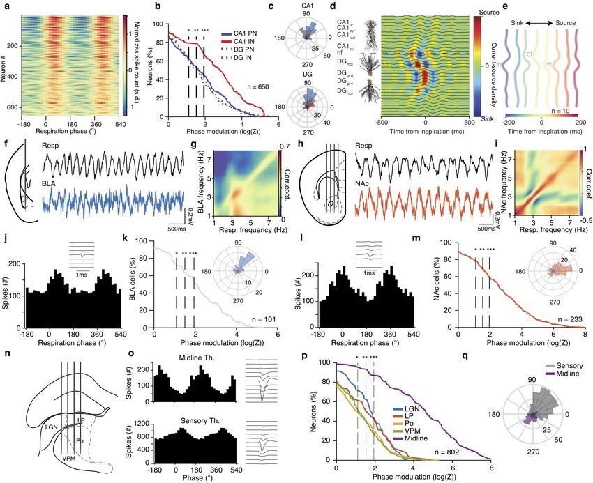
bioRxiv preprint first posted online Aug. 16, 2018; doi: http://dx.doi.org/10.1101/392530. The copyright holder for this preprint
     (which was not peer-reviewed) is the author/funder, who has granted bioRxiv a license to display the preprint in perpetuity.
                                        All rights reserved. No reuse allowed without permission.

                                                         Breathing coordinates limbic network dynamics underlying memory consolidation

For this, we turned our attention to other limbic structures,                 phase of the local LFP (Fig. 4c). The simultaneously occur-
reciprocally connected to the mPFC that are known to interact                 ring OB and mPFC gamma oscillations match in frequency and
with prefrontal networks in different behaviors. Using large-scale            exhibit reliable phase relationship with a phase lag suggesting
single-unit and laminar LFP recordings from the dorsal hip-                   directionality from the OB to the mPFC (Fig. 4d, Supple-
pocampus, we identified that in both dorsal CA1 and dentate                   mentary Fig. 6b). To examine the underlying synaptic inputs
gyrus (DG), ∼60% of PNs and 80% CA1 INs were modulated by                     mediating the occurrence of these oscillations in the mPFC, we
the phase of respiration, firing preferentially after the inspiration         calculated CSD across mPFC layers, triggered on the phase of
(Fig. 3a-c), in line with previous reports of respiratory entrain-            the OB gamma bursts. This analysis revealed a discrete set of
ment of hippocampal activity21,22,37,38 . A separation of CA1                 sinks across prefrontal layers associated with OB bursts (Fig.
PNs based on their relative position within the pyramidal layer               4i,j). Similar to the slow time scale LFP signals, these results
into populations with known distinct connectivity patterns39,40               suggest that fast gamma oscillations in mPFC are generated by
did not reveal particular differences in their modulation by res-             OB-gamma rhythmic polysynaptic inputs to mPFC and are not
piration, suggesting a generality of this entrainment throughout              a locally generated rhythm.
the CA1 sub-populations (Supplementary Fig. 5b).                                 Examining the OB ∼80 Hz gamma-triggered dorsal hip-
    To understand what afferent pathways are responsible for                  pocampal CSD reveals a DG outer molecular layer sink, indica-
breathing-related synaptic currents that underlie the modulation              tive of an OB gamma propagating to DG via the LEC LII input
of spiking activity, we calculated finely-resolved (23µm resolu-              (Fig. 4k,l), a profile distinct from the similar frequency CA1lm
tion) laminar profile of inspiration-triggered dorsal hippocampal             gamma (Supplementary Fig. 5c). In parallel, slow BLA gamma
current-source density, enabling the identification of synaptic               (∼40 Hz) and fast NAc gamma (∼80 Hz) oscillations are mod-
inputs into dendritic sub-compartments. Although the LFP pro-                 ulated by the phase of breathing, occurring predominantly in
file only highlights the prominence of the respiratory band in the            the trough and ascending phase of breathing respectively (Fig.
DG hilus region (Supplementary Fig. 5a), high-resolution CSD                  4m-o).
analysis revealed the presence of two distinct and time-shifted                  To examine whether these respiration-modulated OB-
respiratory-related inputs in DG dendritic sub-compartments                   mediated gamma oscillations have a functional role in driving
(Fig. 3d, Supplementary Fig. 5d). Inspiration was associated                  local neuronal activity, we quantified coupling of local single
with an early sink in the outer molecular layer of DG, indicative             units to mPFC gamma signals, revealing that ∼40% of princi-
of an input from the layer II (LII) of the lateral entorhinal cor-            pal cells and ∼55% of interneurons increased their firing rate in
tex (LEC), followed by a sink in the middle molecular layer of                response to local gamma oscillations (Fig. 4e,f ). Interestingly,
DG, indicative of an input from the layer II of medial entorhinal             ∼10% of PN and ∼30% of IN were significantly phase modulated
cortex (MEC) (Fig. 3d,e).                                                     by gamma oscillations, firing preferentially in the trough of the
    To explore the extent of limbic entrainment by respiration,               local oscillation (Fig. 4g,h). Similarly, ∼40% of BLA and ∼25%
we further recorded LFP and single-unit activity in the BLA,                  of NAc cells fire preferentially in the trough of the local gamma
NAc as well as somatic and midline thalamus (Fig. 3f-q).                      oscillations (Fig. 4p, Supplementary Fig. 6d,f ). Thus OB
Similar to mPFC, LFP in both BLA and NAc was comodu-                          gamma propagates
lated with breathing across a range of frequencies, with most
prominent modulation at ∼4 Hz, the main mode of breathing
frequency during quiescence (Fig. 3f-i), and exhibited reliable
cycle-to-cycle phase relationship with the respiratory oscillation            Efferent and reafferent mechanisms of respiratory entrainment
(Supplementary Fig. 6c,e). Given the nuclear nature and lack
of lamination of these structures, which obfuscates the interpre-             These results suggest a mechanistic picture in which the OB reaf-
tation of slow LFP oscillations, we examined the modulation of                ferent gamma and respiration-locked currents are responsible for
single-units by the phase of breathing. Phase-modulation anal-                the observed respiration-associated LFP patterns in the mPFC,
yses of the spiking activity revealed that a large proportion of              consistent with disruption of these LFP patterns after OB lesion
BLA, NAc, and thalamic neurons are modulated by respiration,                  or tracheotomy20,23,29,46 . However, the distributed and mas-
firing in distinct phases of the breathing cycle (Fig. 3j-q).                 sive modulatory effect that respiration had on unit activity in
                                                                              these regions is at odds with the anatomically-specific synap-
                                                                              tic pathways that we identified as responsible for slow and fast
Reafferent origin of limbic gamma oscillations                                currents.
                                                                                 To causally test whether OB reafferent input is the sole ori-
A prominent feature of prefrontal cortex LFP are fast gamma                   gin of the LFP patterns and unit entrainment, we resorted
oscillations (∼80 Hz)41,42 (Fig. 4a). To investigate the rela-                to a pharmacological approach, that enables selective removal
tionship of prefrontal gamma oscillations to the breathing                    of the reafferent input. A well-characterized effect of systemic
rhythm and well-known OB gamma oscillations of similar fre-                   methimazole injection is the ablation of the olfactory epithelium
quency34,43–45 (Supplementary Fig. 6a), we recorded simultane-                that hosts the olfactory sensory neurons47 , known to express
ously from the two structures and calculated the phase-amplitude              mechanoreceptors35 . Effectively, this deprives the OB of olfac-
coupling between breathing and gamma oscillations (Fig. 4a,b,                 tory and respiratory input, while leaving the bulbar circuits
Supplementary Fig. 6a). Both OB and mPFC fast gamma                           intact, enabling us to study the activity of the de-afferentiated
oscillations are modulated by respiratory phase and gamma                     brain in freely-behaving mice. This manipulation eliminated the
bursts occur predominantly simultaneously and in the descending               respiration-coherent prefrontal oscillatory LFP component (Fig.

                                                                                                                                             5
bioRxiv preprint first posted online Aug. 16, 2018; doi: http://dx.doi.org/10.1101/392530. The copyright holder for this preprint
     (which was not peer-reviewed) is the author/funder, who has granted bioRxiv a license to display the preprint in perpetuity.
                                        All rights reserved. No reuse allowed without permission.

bioRxiv                                                                                                                        Karalis & Sirota, 2018

Figure 4 Reafferent gamma entrainment of limbic circuits.
(a) Example simultaneously recorded LFP traces (top) from OB and mPFC LFP and its spectral decomposition in the gamma range (bottom).
(b) Color-coded modulation strength of OB (top) and mPFC (bottom) gamma power by respiration phase for an example animal. (c) Phase
distribution of mPFC gamma bursts for an example animal (gray histogram) and average preferred phase and phase modulation strength
(logZ) for all animals (n = 16 mice). The red arrow indicates the population average preferred phase and log(Z). (d) Distribution of the phase
shift between OB and mPFC gamma filtered traces for one example animal (gray histogram) and average phase shift and phase-coupling
strength (log(Z), red dots) for all animals (n = 7 mice). (e) Gamma-burst triggered time histogram for one example mPFC cell and color-coded
normalized time histograms for all mPFC cells (n = 1640 cells). (f ) Cumulative distribution of the gamma-triggered normalized firing of mPFC
PNs (n = 1250 cells) and INs (n = 390 cells). Inset, time lag between time from gamma burst and peak firing probability for all significantly
responsive cells. (g) Gamma phase histogram of one example mPFC unit (left) and the respective unit spike spatio-temporal waveform (right).
(h) Cumulative distribution of the modulation strength (logZ) for all PNs (blue, n = 685 neurons) and INs (red, n = 324 neurons). Phase
modulation is assessed in relation to the phase of the locally recorded prefrontal gamma oscillation. Inset, distribution of the mean preferred
phases of all significantly modulated PN and IN cells. (i, j) Example (i) and average zero-lag (j) OB gamma-triggered translaminar CSD of
the dorsal mPFC LFP profile. (n = 6 mice). (k) Example OB gamma-triggered CSD profile of dorsal hippocampus. Horizontal dashed line
indicates the hippocampal fissure. (l) Average normalized OB-triggered current-source density profile of dorsal hippocampus at different time
lags from OB gamma trought (n = 10 mice). (m, n) Example phase-power modulation of BLA (m) and NAc (n) gamma activity by respiration.
(o) Example distribution of the respiratory phase of BLA and NAc gamma bursts (histogram) and mean preferred phase of gamma occurrence
and modulation strength (dots; BLA, blue, n = 3 mice; NAc, red, n = 4 mice). (p) Cumulative distribution of modulation strength for local
gamma phase entrainment of spikes of all BLA (blue, n = 25 cells) and NAc cells (red, n = 76 cells). (q) Distribution of the mean preferred
gamma phase for each significantly modulated BLA and NAc cell. Star indicates significance (* P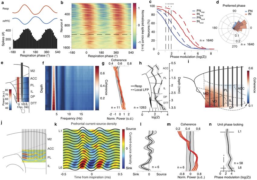
bioRxiv preprint first posted online Aug. 16, 2018; doi: http://dx.doi.org/10.1101/392530. The copyright holder for this preprint
     (which was not peer-reviewed) is the author/funder, who has granted bioRxiv a license to display the preprint in perpetuity.
                                        All rights reserved. No reuse allowed without permission.

                                                         Breathing coordinates limbic network dynamics underlying memory consolidation

Figure 5 Reafferent respiratory input accounts for LFP but not neuronal entrainment.
(a,b) Left, schematic of the manipulation strategy. Right, example time-frequency decomposition of power and coherence between respiratory
and mPFC LFP signals during quiescence before (a) and after (b) OD. (c) Average normalized power spectra before and after OD (n = 7).
(d) Coherence spectrum between respiration and mPFC LFP before and after OD (n = 7 mice). (e) Left, example inspiration-triggered CSD
of the mPFC LFP during quiescence and sleep after OD. Right, average normalized CSD at zero lag (n = 6 mice). (f ) Example power-phase
modulation of mPFC gamma oscillations before (top) and after (bottom) OD. (g) Average mPFC power-phase modulation strength of ∼80
Hz gamma oscillations (n = 7 mice; paired t-test: before vs. after OD). (h) Cumulative distribution of modulation strength for all mPFC
neurons pre and post OD (Pre: n = 511 cells; Post: n = 431 cells). Inset, percentage of significantly phase-modulated cells before and after
OD. (i) Cumulative distribution of modulation strength for CA1, DG and somatic thalamus neurons before and after OD. (j) Percentage of
significantly phase-modulated cells before and after OD. (k) Example inspiration-triggered CSD of the dorsal hippocampus LFP before (left)
and after (right) OD. (l) Average outer molecular layer sink depth (n = 6 mice; paired t-test: before vs. after OD). Shaded areas, mean ± s.e.m.
a.u., arbitrary units; s.d., standard deviations; n.s., not significant. Shaded areas, mean ± s.e.m. Stars indicate significance levels (* P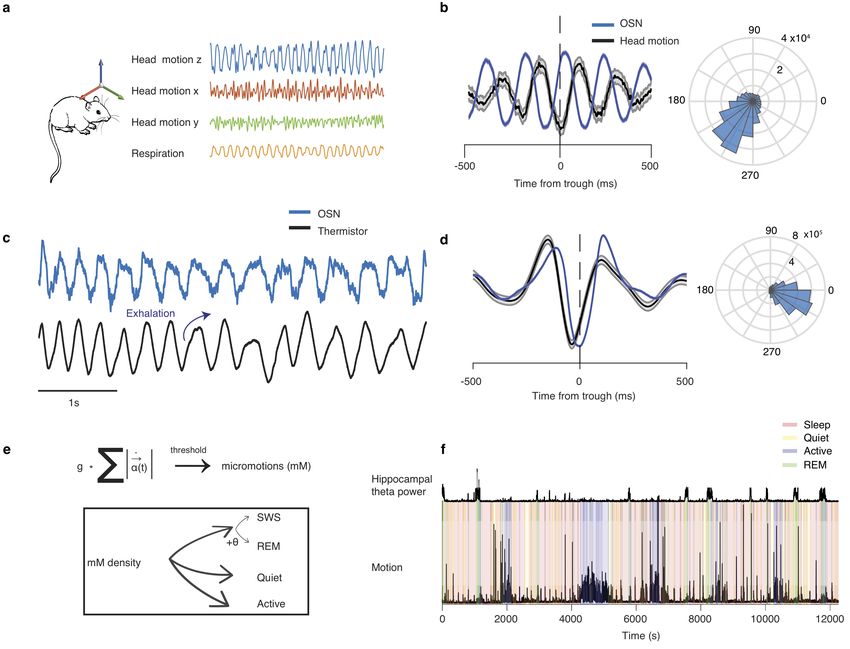
bioRxiv preprint first posted online Aug. 16, 2018; doi: http://dx.doi.org/10.1101/392530. The copyright holder for this preprint
     (which was not peer-reviewed) is the author/funder, who has granted bioRxiv a license to display the preprint in perpetuity.
                                        All rights reserved. No reuse allowed without permission.

bioRxiv                                                                                                                        Karalis & Sirota, 2018

Figure 6 Breathing modulates hippocampal ripples and dentate spikes.
(a) Example traces of the respiratory signal and CA1 pyramidal layer LFP. In the magnified LFP signal, ripple events and the associated spiking
activity can be observed. (b) Average ripple-triggered time-frequency wavelet spectrogram of the CA1 pyramidal layer LFP from one example
animal. (c) Schematic of the CA1 pyramidal and granular cells somato-dendritic domains aligned to the average ripple-triggered CSD profile of
the hippocampal LFP activity for one example animal. (d) Cumulative distribution of the ripple-triggered normalized firing of CA1 and DG
PNs (CA1, n = 220 cells; DG, n = 202 cells) and INs (CA1, n = 76 cells; DG, n = 119 cells). (e) Average cross-correlation between inspiratory
events and ripple occurrence (n = 22 mice). Dashed horizontal lines indicate the significance levels. (f ) Distribution of the respiratory phase of
occurrence of individual ripple events for one example animal (n = 4813 ripples). (g) Distribution of average phase and modulation strength
for ripples (n = 22 mice). (h) Phase modulation of ripples before and after OD. (i) Color-coded LFP power depth profile and overlaid LFP
traces of an example dentate spike. (j) Average dentate spike triggered normalized spiking activity for all CA1 (top) and DG (bottom) PNs and
INs. (k) Cumulative distribution of the dentate spike-triggered normalized firing of CA1, DG and mPFC PNs (CA1, n = 44 cells; DG, n = 175
cells; mPFC, n = 239 cells) and INs (CA1, n = 31 cells; DG, n = 82 cells; mPFC, n = 64 cells). (l) Respiration phase histogram for dentate
spikes in one example animal. (m) Preferred respiratory phase of dentate spike occurrence and phase modulation strength for all animals (dots)
and population average (arrows) before and after OD (npre = 10 mice,npost = 6 mice). s.d., standard deviations; a.u., arbitrary units; n.s., not
significant; OD, olfactory de-afferentiation. Shaded areas, mean ± s.e.m. Stars indicate significance levels (* P
bioRxiv preprint first posted online Aug. 16, 2018; doi: http://dx.doi.org/10.1101/392530. The copyright holder for this preprint
(which was not peer-reviewed) is the author/funder, who has granted bioRxiv a license to display the preprint in perpetuity.
All rights reserved. No reuse allowed without permission.
Breathing coordinates limbic network dynamics underlying memory consolidation
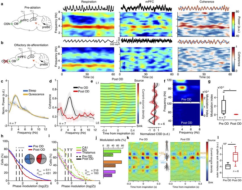
Figure 7 Breathing organizes prefrontal UP states and modulates hippocampal output.
(a) Example traces of respiration signal (top), mPFC LFP (middle trace) and spike trains of 183 simultaneously recorded mPFC units during
sleep. Three typical delta waves and the corresponding DOWN states of the neuronal population are marked with red. (b) Example distributions
of the breathing phase of UP (top) and DOWN (bottom) state onsets. (c) Distribution of preferred breathing phase of UP and DOWN states
(n = 11 mice). (d) Cross-correlation of UP and DOWN state onsets with respect to inspiration. (e) Normalized power (black) and occurrence
probability (blue) for prefrontal UP states (n = 11 mice). (f, g) Example (f) and population (g) distribution of preferred breathing phase of
UP and DOWN state occurrence after OD (n = 7). (h) Example average ripple-triggered CA1pyr and mPFC LFP traces and wavelet spectral
decomposition of mPFC LFP power (upper) and real-part (lower) (n = 3162 ripples). (i) Probability of SWR occurrence as a function of time
from UP or DOWN state onset (n = 8 mice). (j) Example (black) and distribution of preferred breathing phase of SWR occurrence , for
SWRs that are terminating an UP state (red dots, n = 8 mice). (k) Example mPFC unit spiking raster across individual ripples (bottom)
and cross-correlogram of unit firing to ripple (top). (l) Cumulative distribution of the ripple-triggered normalized firing of mPFC PNs (n =
791 cells) and INs (n = 237 cells) in response to ripples occurring in the preferred (in-phase) and in the least-preferred (anti-phase) phases of
respiration (n = 11 mice). Inset, average ripple-triggered firing rate (PN: 791 cells. IN: 237 cells) (Wilcoxon signed-rank test, in-phase versus
anti-phase). (m) Top, pie charts indicating the percentage of all mPFC PNs (left; n = 791 cells) and INs (right; n = 237 cells) that are either
phase modulated by respiration (resp. mod), responding significantly to ripples (ripple resp.), being both significantly modulated by breathing
and significantly responsive to ripples or neither. Bottom, similarly, for all NAc cells (n = 216 cells). (n) Cross-correlation of firing with respect
to ripple time for one example NAc unit (top) and color-coded cross-correlograms for all NAc cells (n = 216 cells). (o) Cumulative distribution
of the ripple-triggered normalized firing of NAc cells (n = 216 cells) in response to, as in (l), “in-phase” and “anti-phase” ripples (n = 4 mice).
Inset, average ripple-triggered firing rate (n = 164 cells, Wilcoxon signed-rank test, in-phase versus anti-phase). Shaded areas, mean ± s.e.m. ,
s.d., standard deviations; a.u., arbitrary units; OD, olfactory de-afferentiation; FR, firing rate. Stars indicate significance levels (* P
bioRxiv preprint first posted online Aug. 16, 2018; doi: http://dx.doi.org/10.1101/392530. The copyright holder for this preprint
      (which was not peer-reviewed) is the author/funder, who has granted bioRxiv a license to display the preprint in perpetuity.
                                         All rights reserved. No reuse allowed without permission.

bioRxiv                                                                                                                         Karalis & Sirota, 2018

activity and characterized their relationship with the phase of
the ongoing breathing rhythm (Fig. 7a). Surprisingly, both UP
and DOWN state onsets were strongly modulated by the breath-
ing phase and time from inspiration (Fig. 7b-d), while the
magnitude of UP states followed the profile of UP state onset
probability (Fig. 7e). In line with the results on ripples and pre-
frontal units, UP and DOWN state modulation was not affected
by olfactory de-afferentiation, suggesting that RCD is the source
of this modulation (Fig. 7f,g).
   Previous observations during sleep in rats identified a corre-
lation between ripple occurrence and cortical SO11,50,51 . Here,
ripples preceded the termination of prefrontal UP states and
onset of the DOWN states both before (Fig. 7i) and after
de-afferentation (Supplementary Fig. 8e), with ripples con-
tributing to UP state termination occurring in the early post-
inspiratory phase (Fig. 7j). This is in line with the RCD-driven
synaptic inputs to the DG middle molecular layer preceding rip-
ple events, which are suggesting an RCD-mediated coordination
of SWR occurrence with the MEC UP states (Supplementary
Fig. 8c,d).
   Ripple output is known to recruit prefrontal neural activ-
ity50,51 . In line with this, hippocampal ripples evoked a response
in prefrontal LFP and gave rise to an efferent copy detected as a
local increase in fast oscillatory power in the PFC LFP52 (Fig.
7h). In response to ripple events, ∼14% of prefrontal PNs and
∼42% of INs exhibited increased firing (Fig. 7k,l, Supplemen-
tary Fig. 8f ). In parallel, ∼69% of NAc cells are significantly
driven by ripple events (Fig. 7n,o, Supplementary Fig. 8f ).
Interestingly, in both mPFC and NAc there is a great overlap
between cells that are phase modulated by breathing and that
are responsive to ripples (Fig. 7m), while ripples occurring in
the preferred post-inspiratory phase of ripple occurrence, drive a
greater fraction of cells from both structures to fire significantly
more compared to anti-preferred phase (Fig. 7l,o).
                                                                               Figure 8 Breathing organizes network dynamics across lim-
                                                                               bic structures.
                                                                               (a) Schematic depiction of the efferent copy pathway carrying the
Discussion                                                                     respiratory corollary discharge (RCD) signal and the reafferent path-
                                                                               way carrying the respiratory olfactory reafferent (ROR) signal. (b)
In this study, we demonstrate that the respiratory rhythm pro-                 Summary schematic of the network dynamics organized by breathing
                                                                               throughout all structures studied. Black traces: LFPs; Black ticks: neu-
vides a unifying global temporal coordination of neuronal firing               ronal spikes; Green traces: Fast (∼80Hz) gamma; Cyan traces: Slow
and nonlinear dynamics across cortical and subcortical limbic                  gamma (∼40Hz); Red traces: CA1 Ripples; Blue traces: Dentate spikes;
networks. Using recordings of the three-dimensional LFP profile                Orange dots: CSD sinks (mPFC deep layers and DG middle molecular
                                                                               layer).
of the mPFC (Fig. 1,2) and large-scale unit recordings from
the hippocampus, amygdala, nucleus accumbens and thalamus
(Fig. 3), we identified that during quiescence and slow-wave                   coordination of hippocampo-cortical nonlinear dynamics (Fig.
sleep limbic LFPs are dominated by breathing-related oscilla-                  7). Finally, we comprehensively characterized the distinct phase
tory activity, while the majority of neurons in each structure are             relationship between different network events within the breath-
modulated by the phase of the respiratory rhythm and reafferent                ing cycle across limbic structures, painting the picture of how this
gamma oscillations (Fig. 4). Using pharmacological manipula-                   organization enables the multiplexing and segregation of infor-
tions paired with large-scale recordings, we causally identified a             mation flow across the limbic system and providing the basis for
joint mechanism of respiratory entrainment (Fig. 8a), consisting               mechanistic theories of memory consolidation processes enabled
of an efference copy of the brainstem respiratory rhythm (res-                 by the respiratory modulation (Fig. 8b).
piratory corollary discharge; RCD) that underlies the neuronal                    Over the years, mounting evidence have suggested that
modulation and a respiratory olfactory reafference (ROR) that                  breathing can entrain frog53 and human EEG54,55 , as well as
contributes to the modulation and accounts for LFP phenomena                   LFP and spiking activity in the hedgehog OB and cortex32 ,
(Fig. 5). Hippocampal SWR and dentate spikes (Fig. 6), as                      rodent OB33–35,56 , cortex20,23,29,57–59 and the hippocampus in
well as prefrontal UP and DOWN states (Fig. 7), were strongly                  cats60 , rodents21,22,37,38,61 , and humans24,25 . With this study,
modulated by the respiratory phase with RCD being a sufficient                 we contribute to the ongoing effort to understand the mecha-
source of modulation. This modulation accounts partially for the               nism and role of the respiratory entrainment of brain circuits.

10
bioRxiv preprint first posted online Aug. 16, 2018; doi: http://dx.doi.org/10.1101/392530. The copyright holder for this preprint
(which was not peer-reviewed) is the author/funder, who has granted bioRxiv a license to display the preprint in perpetuity.
All rights reserved. No reuse allowed without permission.
Breathing coordinates limbic network dynamics underlying memory consolidation
Our results extend and provide a mechanistic explanation and in fear-associated enhanced breathing, the ROR is not neces-
interpretation of the previous studies that described respiration- sary for the expression of innate or conditioned fear behavior, in
related LFP oscillations in different brain regions. Here, we agreement with a recent report23 . This suggests the potential suf-
leverage large-scale recordings from multiple limbic regions and ficiency of RCD for the behavioral expression. Interestingly, the
thousands of cells to comprehensively characterize and uncover optogenetic induction of such oscillations is sufficient to drive
the underlying mechanisms of the limbic respiratory entrain- fear behavior in naïve animals27 , raising the possibility that this
ment and understand the role of this phenomenon in organizing effect is mediated by the bidirectional interaction of prefrontal
neuronal activity and coordinating network dynamics between networks with the RCD via top-down projections to PAG. This
remote regions. sets the stage for future investigations of the interaction between
In this study, we expanded the characterization of respiratory the RCD and ROR in limbic networks and in turn the top-down
entrainment to the NAc, BLA, and thalamus for the first time, modulation of breathing and emotional responses.
while we comprehensively characterized the neuronal entrain- We extend previous work on the hippocampal entrain-
ment across prefrontal layers and subregions. These results ment21,22,38 and provide new evidence for the mechanistic under-
highlight the extent and significance of this modulation, given pinnings of CA1 and DG modulation, by means of joint RCD and
the crucial role of the interaction between these structures for ROR inputs. We demonstrate robust modulation of hippocam-
emotional processing and regulation. We further report the reaf- pal ripple occurrence by breathing, in agreement with a previous
ferent OB origin of local gamma dynamics and their modulation report61 , and its effect on the response of prefrontal50,70 and
by breathing, as well as the relation between local gamma oscil- accumbens neurons71 to SWR. Importantly, we provide the first
lations and neuronal activity in all structures. This sheds new evidence for the role of this global limbic circuit modulation by
light onto the origin and role of prefrontal41,42,59 , BLA62 , and the respiratory rhythm in coordinating the interaction between
NAc63 gamma oscillations, that might provide a temporally- the hippocampus and the downstream structures (i.e. mPFC and
optimized privileged route for olfactory reafferent input to affect NAc), thought to underlie memory consolidation72,73 .
the ongoing activity, in line with recent reports in humans24 . Our results suggest that the respiratory rhythm orchestrates
Using a pharmacological olfactory de-afferentiation approach, the hippocampo-cortical dialogue, by jointly biasing the neuronal
paired with large-scale recordings, we identified that although firing and temporally coupling network dynamics across regions.
cortical LFP signatures of respiration and gamma are mediated We report for the first time the modulation of prefrontal UP and
by bulbar inputs in the form of a ROR, the bulk entrainment of DOWN states as well as dentate spikes by breathing, which sug-
limbic neuronal activity by breathing is mediated by an intrac- gests a novel potential mechanism for the large-scale entrainment
erebral RCD originating in the brainstem rhythm generators and of thalamocortical excitability during sleep74 . Along these lines,
being unaffected by olfactory de-afferentiation. We suggest that an OB-mediated pacing of slow oscillations in olfactory cortices
this joint modulation of limbic circuits by respiration is analogous has been demonstrated in the past58 . Importantly, prefrontal
to the predictive signaling employed in a wide range of neural cir- SO entrainment appears to be dominated by the intracerebral
cuits64 , such as those underlying sensory-motor coordination65 . RCD, while still receiving synchronous olfactory inputs via the
Although the pathway of RCD remains unknown, we speculate ROR pathway. This highlights olfaction as a royal path to the
that ascending long-range somatostatin-expressing interneuron sleeping brain that via synchronous ROR input reaches the
projections from the preBötzinger complex to the thalamus, limbic system in sync with RCD-coordinated UP-DOWN state
hypothalamus and basal forebrain66 or the locus coeruleus67 are dynamics during slow-wave sleep and could explain the efficacy
probable pathways for this widespread modulation. The global of manipulations that bias learning75 , consolidation76 or sleep
and powerful nature of the RCD calls for future tracing and depth77 using odor presentation during sleep. The intrinsic RCD-
activity-dependent labeling studies to identify its anatomical mediated comodulation of both SWR and SO by the respiratory
substrate. A disinhibition-mediated mechanism of RCD would phase brings into perspective the mechanistic explanation of
be consistent with the lack of prominent LFP sources in the studies that improve consolidation using stimulation conditioned
absence of ROR, similar to the mechanism of disinhibitory pac- on the ongoing phase of the cortical SO15,78,79 . Understanding
ing by the medial septum of the entorhinal-hippocampal system the causal role of respiratory entrainment in the formation, con-
during theta oscillations5 . We predict that the functional role solidation, and retrieval of memories will require fine time scale
of RCD in coordinating activity across the limbic system during closed-loop optogenetic perturbation.
offline states extends to other brain structures and brain states. While a causal model of the role of respiration in temporally
The global outreach of RCD to higher order areas suggests that coordinating hippocampal ripples, dentate spikes, entorhinal cor-
it might play an important role in the coordination of multi- tex inputs, and prefrontal UP and DOWN states remains to
sensory processing, in sync with orofacial motor output during be elucidated, their temporal progression and phase relationship
both passive and active orofacial sampling, thus providing a cen- with the ongoing respiratory cycle hints to a possible mecha-
trifugal component synchronized with reafferent sensory inputs nism. ROR-driven gamma-associated waves and RCD lead to
and respiratory efference copies to orofacial motor centers68 . differential recruitment of the entorhinal cortex, consistent with
We provide causal evidence that fear-related 4Hz oscilla- the sinks in the dentate molecular layer and the entrainment of
tions27,69 are a state-specific expression of the limbic respira- dentate spikes. Depending on the strength of the inputs, either
tory entrainment and originate from the reafferent respiratory feed-forward inhibition of the CA380 or excitation during UP
entrainment of olfactory sensory neurons by passive airflow35 . states14 can suppress or promote respectively SWRs. In parallel,
Importantly, although prefrontal 4Hz LFP oscillations originate the ripple-driven recruitment of prefrontal neurons likely triggers
the resetting of the ongoing UP states by tilting the bias between
11bioRxiv preprint first posted online Aug. 16, 2018; doi: http://dx.doi.org/10.1101/392530. The copyright holder for this preprint
(which was not peer-reviewed) is the author/funder, who has granted bioRxiv a license to display the preprint in perpetuity.
All rights reserved. No reuse allowed without permission.
bioRxiv Karalis & Sirota, 2018
excitation and inhibition81 and results in a feedback re-entrance of limbic circuits, this common reference acts in tandem with the
to the entorhinal-hippocampal network14 . Further analysis of the direct anatomical links between brain regions to pace the flow of
SO dynamics across the cortical mantle and their relationship information.
with hippocampal ripples, as well as causal manipulation of the
two nonlinear dynamics is required to validate and elucidate the
physiological details of this model. Acknowledgments
While we show here that the respiratory dynamics bias the
prefrontal SO via ROR and RCD, slow oscillations can emerge We thank G. Schwesig, E. Blanco Hernandez and E. Resnik
in isolated cortical slubs82 or slices83 . Furthermore, from a mech- for valuable input, R. Ahmed for technical assistance, J. Lu
anistic perspective, the generative mechanism of the two oscilla- for assistance in the experiments and all the members of the
tions is potentially comparable. Leading models of the generation Sirota laboratory for helpful discussions and comments on the
of neocortical UP states from DOWN states84 or inspiratory manuscript. This work was supported by grants from Munich
bursts from expiratory silence in preBötzinger circuits85 suggest Cluster for Systems Neurology (SyNergy, EXC 1010), Deutsche
that both phenomena rely on regenerative avalanches due to Forschungsgemeinschaft Priority Program 1665 and 1392 and
recurrent connectivity, that are followed by activity-dependent Bundesministerium für Bildung und Forschung via grant no.
disfacilitation. Given that neocortical slow oscillations can be 01GQ0440 (Bernstein Centre for Computational Neuroscience
locally generated11,86 , are globally synchronized by the thalamic Munich) and European Union Horizon 2020 FETPOACT pro-
input87 and propagate across the neocortex14,88 , ROR and RCD gram via grant agreement no.723032 (BrainCom) (A.S.).
biasing of the cortical SO could be considered as an extension of
a global system of mutually-coupled nonlinear oscillators. The
persistent synchronous output of the respiratory oscillator and Author Contributions
its marginal independence of the descending input might provide N.K and A.S. designed the experiments and data analysis, inter-
a widespread asymmetric bias to the slow oscillatory dynamics preted the data and wrote the manuscript, N.K. performed the
in the cortical circuits and SWR complexes in the hippocampus experiments and analyzed the data.
across offline states of different depth. It is likely, however, that
via descending cortical projections, cortical SO provides feed-
back to the pontine respiratory rhythm-generating centers (e.g. Competing Financial Interests
via mPFC projections to PAG) and thus the interaction between
respiratory dynamics and slow oscillations could be bidirectional. The authors declare no competing financial interests.
This perpetual limbic rate comodulation by respiration also
suggests a potential framework for memory-consolidation pro-
References
cesses that do not rely on deep sleep and the associated syn-
chronous K-complexes. This could explain the mechanism and 1.Papez, J. W. A proposed mechanism of emotions. Archives
distinctive role of awake replay in memory consolidation89,90 . of Neurology & Psychiatry 38, 725–743 (1937).
Given the substantial cross-species differences in respiratory fre- 2.Maclean, P. D. Psychosomatic disease and the visceral brain;
quency, as well as the effects of sleep depth and recent experience recent developments bearing on the Papez theory of emotion.
on cortical and hippocampal dynamics, it is conceivable that Psychosomatic medicine 11, 338–353 (1949).
more synchronized and generalized UP states or awake vs. sleep 3.Scoville, W. B. & Milner, B. Loss of recent memory after
ripples are differentially modulated by breathing. Further work bilateral hippocampal lesions. Journal of Neurology, Neuro-
is required to investigate the role of these parameters on the surgery & Psychiatry 20, 11–21 (1957).
respiratory biasing of network dynamics and its role in memory 4.Buzsaki, G. & Draguhn, A. Neuronal oscillations in cortical
consolidation. networks. Science 304, 1926–1929 (2004).
Finally, in light of the wide modulation of limbic circuits by 5.Buzsaki, G. Theta oscillations in the hippocampus. Neuron
breathing during quiescence, we suggest that breathing effec- 33, 325–340 (2002).
tively modulates the default mode network (DMN). To examine 6.Mizuseki, K., Sirota, A., Pastalkova, E. & Buzsaki, G. Theta
this hypothesis future work will be needed to carefully exam- oscillations provide temporal windows for local circuit com-
ine the fine temporal structure of neuronal assemblies and their putation in the entorhinal-hippocampal loop. Neuron 64,
modulation by the RCD and ROR copies of the breathing rhythm 267–280 (2009).
throughout cortical and subcortical structures, an endeavor that 7.Fernández-Ruiz, A. et al. Entorhinal-CA3 dualinput con-
might uncover functional sub-networks of the DMN. trol of spike timing in the hippocampus by theta-gamma
In summary, the data provided here suggest that respiration Coupling. Neuron 93, 1213–1226.e5 (2017).
provides a perennial stream of rhythmic input to the brain. In 8.Siapas, A. G., Lubenov, E. V. & Wilson, M. A. Prefrontal
addition to its role as the condicio sine qua non for life, we phase locking to hippocampal theta oscillations. Neuron 46,
provide evidence that breathing rhythm acts as a global pace- 141–151 (2005).
maker for the brain, providing a reference signal that enables 9.Benchenane, K. et al. Coherent Theta Oscillations and Reor-
the integration of exteroceptive and interoceptive inputs with ganization of Spike Timing in the Hippocampal- Prefrontal
the internally generated dynamics of the limbic brain during Network upon Learning. Neuron 66, 921–936 (2010).
offline states. In this emergent model of respiratory entrainment
12bioRxiv preprint first posted online Aug. 16, 2018; doi: http://dx.doi.org/10.1101/392530. The copyright holder for this preprint
(which was not peer-reviewed) is the author/funder, who has granted bioRxiv a license to display the preprint in perpetuity.
All rights reserved. No reuse allowed without permission.
Breathing coordinates limbic network dynamics underlying memory consolidation
10.Steriade, M. M., McCormick, D. A & Sejnowski, T. J. Tha- 28.Bartho, P. Characterization of neocortical principal cells
lamocortical oscillations in the sleeping and aroused brain. and interneurons by network interactions and extracellular
Science 262, 679–685 (1993). features. Journal of Neurophysiology 92, 600–608 (2004).
11.Sirota, A. & Buzsaki, G. Interaction between neocortical and 29.Biskamp, J., Bartos, M. & Sauer, J. F. Organization of pre-
hippocampal networks via slow oscillations. Thalamus and frontal network activity by respiration-related oscillations.
Related Systems 3, 245–259 (2005). Scientific Reports 7, 45508 (2017).
12.Wilson, M. & McNaughton, B. Dynamics of the hippocampal 30.Hoover, W. B. & Vertes, R. P. Anatomical analysis of afferent
ensemble code for space. Science 261, 1055–1058 (1993). projections to the medial prefrontal cortex in the rat. Brain
13.Sirota, A., Csicsvari, J., Buhl, D. L. & Buzsaki, G. Commu- Structure and Function 212, 149–179 (2007).
nication between neocortex and hippocampus during sleep 31.Herry, C. & Johansen, J. P. Encoding of fear learning
in rodents. Proceedings of the National Academy of Sciences and memory in distributed neuronal circuits. Nature Neu-
100, 2065–2069 (2003). roscience 17, 1644–1654 (2014).
14.Isomura, Y. et al. Integration and Segregation of Activity 32.Adrian, E. D. Olfactory reactions in the brain of the
in Entorhinal-Hippocampal Subregions by Neocortical Slow hedgehog. The Journal of Physiology 100, 459–473 (1942).
Oscillations. Neuron 52, 871–882 (2006). 33.Macrides, F & Chorover, S. L. Olfactory bulb units: activity
15.Maingret, N., Girardeau, G., Todorova, R., Goutierre, M. & correlated with inhalation cycles and odor quality. Science
Zugaro, M. Hippocampo-cortical coupling mediates memory 175, 84–87 (1972).
consolidation during sleep. Nature Neuroscience 19, 959–964 34.Fukunaga, I., Herb, J. T., Kollo, M., Boyden, E. S. &
(2016). Schaefer, A. T. Independent control of gamma and theta
16.Girardeau, G. et al. Selective suppression of hippocampal activity by distinct interneuron networks in the olfactory
ripples impairs spatial memory. Nature Neuroscience 12, bulb. Nature Neuroscience 17, 1208–1216 (2014).
1222–1223 (2009). 35.Grosmaitre, X., Santarelli, L. C., Tan, J., Luo, M. & Ma, M.
17.Rothschild, G., Eban, E. & Frank, L. M. A cortical – hip- Dual functions of mammalian olfactory sensory neurons as
pocampal – cortical loop of information processing during odor detectors and mechanical sensors. Nature Neuroscience
memory consolidation. Nature Neuroscience 20, 1–12 (2016). 10, 348–354 (2007).
18.Maviel, T., Durkin, T. P., Menzaghi, F. & Bontempi, B. 36.Buzsaki, G. Neural syntax: cell assemblies, synapsembles,
Sites of neocortical reorganization critical for remote spatial and readers. Neuron 68, 362–385 (2010).
memory. Science 305, 96–99 (2004). 37.Vanderwolf, C. H. Hippocampal activity, olfaction, and sniff-
19.Kitamura, T. et al. Engrams and circuits crucial for systems ing: An olfactory input to the dentate gyrus. Brain Research
consolidation of a memory. Science 356, 73–78 (2017). 593, 197–208 (1992).
20.Ito, J. et al. Whisker barrel cortex delta oscillations and 38.Nguyen Chi, V. et al. Hippocampal respiration-driven
gamma power in the awake mouse are linked to respiration. rhythm distinct from theta Oscillations in awake mice.
Nature Communications 5, 3572 (2014). Journal of Neuroscience 36, 162–177 (2016).
21.Yanovsky, Y., Ciatipis, M., Draguhn, a., Tort, a. B. L. & 39.Valero, M. et al. in Nature Neuroscience 9, 1281–1290
Branka k, J. Slow Oscillations in the Mouse Hippocampus (Nature Publishing Group, 2015).
Entrained by Nasal Respiration. Journal of Neuroscience 34, 40.Mizuseki, K., Diba, K., Pastalkova, E. & Buzsaki, G. Hip-
5949–5964 (2014). pocampal CA1 pyramidal cells form functionally distinct
22.Lockmann, A. L. V., Laplagne, D. A., Leão, R. N. & Tort, sublayers. Nature Neuroscience 14, 1174–1183 (2011).
A. B. L. A respiration-coupled rhythm in the rat hippocam- 41.Sirota, A. et al. Entrainment of neocortical neurons and
pus independent of theta and slow oscillations. Journal of gamma oscillations by the hippocampal theta rhythm. Neu-
Neuroscience 36, 5338–5352 (2016). ron 60, 683–697 (2008).
23.Moberly, A. H. et al. Olfactory inputs modulate respiration- 42.Stujenske, J. M., Likhtik, E., Topiwala, M. A. & Gordon,
related rhythmic activity in the prefrontal cortex and freez- J. A. Fear and safety engage competing patterns of theta-
ing behavior. Nature Communications (2018). gamma coupling in the basolateral amygdala. Neuron 83,
24.Zelano, C. et al. Nasal respiration entrains human limbic 919–933 (2014).
oscillations and modulates cognitive function. Journal of 43.Adrian, E. D. The electrical activity of the mammalian
Neuroscience 36, 12448–12467 (2016). olfactory bulb. Electroencephalography and Clinical Neuro-
25.Herrero, J. L., Khuvis, S., Yeagle, E., Cerf, M. & Mehta, physiology 2, 377–388 (1950).
A. D. Breathing above the brainstem: Volitional control and 44.Freeman, J. A. & Nicholson, C. Experimental optimization
attentional modulation in humans. Journal of Neurophysiol- of current source-density technique for anuran cerebellum.
ogy 119, 145–159 (2018). Journal of neurophysiology 38, 369–382 (1975).
26.Ottoson, D. Analysis of the electrical activity of the olfactory 45.Lepousez, G. & Lledo, P.-M. Odor discrimination requires
epithelium. Acta physiologica Scandinavica. Supplementum proper olfactory fast oscillations in awake mice. Neuron 80,
35, 1–83 (1955). 1010–1024 (2013).
27.Karalis, N. et al. 4-Hz oscillations synchronize prefrontal- 46.Tort, A. B., Brankačk, J. & Draguhn, A. Respiration-
amygdala circuits during fear behavior. Nature Neuroscience entrained brain rhythms are global but often overlooked.
19, 605–612 (2016). Trends in Neurosciences 41, 186–197 (2018).
13You can also read