Magnetic Nanowires Review - applied sciences
←
→
Page content transcription
If your browser does not render page correctly, please read the page content below
applied
           sciences
Review
Magnetic Nanowires
Luc Piraux
 Institute of Condensed Matter and Nanosciences, Université catholique de Louvain, Place Croix du Sud 1,
 1348 Louvain-la-Neuve, Belgium; luc.piraux@uclouvain.be

 Received: 19 December 2019; Accepted: 20 January 2020; Published: 6 March 2020

 Abstract: Magnetic nanowires are attractive materials because of their morphology-dependent
 remarkable properties suitable for various advanced technologies in sensing, data storage, spintronics,
 biomedicine and microwave devices, etc. The recent advances in synthetic strategies and approaches
 for the fabrication of complex structures, such as parallel arrays and 3D networks of one-dimensional
 nanostructures, including nanowires, nanotubes, and multilayers, are presented. The simple
 template-assisted electrodeposition method enables the fabrication of different nanowire-based
 architectures with excellent control over geometrical features, morphology and chemical composition,
 leading to tunable magnetic, magneto-transport and thermoelectric properties. This review article
 summarizing the work carried out at UCLouvain focuses on the magnetic and spin-dependent
 transport properties linked to the material and geometrical characteristics.

 Keywords: magnetic nanowires; nano-magnetism; spintronics; spin-caloritronics; nanowire
 networks; electrodeposition; nanoporous templates

1. Introduction
     Over the last 25 years, the group at UCLouvain has developed a strong expertise in the
template-assisted electrodeposition of elongated magnetic nanostructures such as homogeneous
and multilayered nanowires (NWs) and nanotubes (NTs). Arrays of magnetic nanofibers with desired
aspect ratio, composition, structure, morphologies and density were successfully synthesized by
electrochemical deposition in nano-templates with well-defined pore architecture (see Figure 1).
This template-assisted synthesis of nanowires has been demonstrated as a very powerful and versatile
technique and has the merits of simplicity, high efficiency and low cost. These advantages are
key elements compared to more complex and restrictive synthesis methods such as electron-beam
lithography [1] and interference lithography [2,3]. Magnetic nanowires provide interesting physics
and innovative ideas for fundamental studies in nanomagnetism and spintronics as well as for
various device applications [4–9]. The group at UCLouvain has performed pioneering work on giant
magnetoresistance (GMR) phenomena in magnetic multilayered NWs [5,10–13]. Extensive studies
were realized on many ferromagnetic NW arrays using various experimental techniques for a
deeper understanding of the magnetic properties and spin-dependent transport properties in
relation with their nanoscopic dimensions [14–45]. The research activity at UCLouvain on magnetic
NWs were also motivated by related applications in microwave devices [46–54] and multiferroic
nanocomposites [55–59]. Recently, we demonstrated the suitability of three-dimensional (3D)
nanoporous polymer templates to form dense interconnected magnetic NW networks with tunable
geometrical parameters in terms of nanowire size, density, and orientation opens up the possibility for a
controlled synthesis of a large variety of complex 3D networks of high aspect-ratio nanostructures with
different geometries and materials [60–67]. Their unique architectures and high degree of nanowire
interconnectivity facilitate the ability to perform magneto-transport measurements and to investigate
the interplay between electronic and thermoelectric transport and magnetic properties.

Appl. Sci. 2020, 10, 1832; doi:10.3390/app10051832                             www.mdpi.com/journal/applsci
Appl. Sci. 2020, 10, 1832                                                                                   2 of 25

                                                 shape
                                                control

                                                                          density/spatial
                                                                             control

                                 composition
                                   control

               Figure 1. Illustration of the versatility of the template-assisted synthesis of nanowires.

      The intent of this article is to provide a review of the most relevant results obtained by the group at
UCLouvain on the fabrication, characterization and properties of ferromagnetic nanowires. The paper
is organized as follows: first, the template-assisted electrodeposition methods which have successfully
grown high quality parallel arrays and complex networks of nanowires, nanotubes and multilayers
are briefly recalled in Section 2. The experimental investigations and analysis of the magnetic,
magneto-transport and thermoelectric properties of single magnetic nanowires, nanowire arrays
and nanowire networks are presented in Section 3. Finally, prospects of this field are outlined in
Section 4.

2. Materials and Methods
     Polymer membranes are made using the nuclear track-etch technology in which a film
(typically 5–50 µm thick) of polymer (polycarbonate, PET, polyimide, . . . ) is bombarded by heavy
ions of high energy [68]. The bombarded film is then irradiated with UV light to increase the track
etching selectivity and finally etched in an adequate solution where tracks are revealed, leading to
the formation of pores with a diameter defined by the etching time. Pore size and pore density can
be controlled over wide ranges, between 15 nm and a few microns and in the range 105 –1010 cm−2 ,
respectively. For such randomly distributed pores array, this implies that the mean separation distance
between nanomaterials can be tuned between ∼100 nm up to several tens of microns, with a maximum
porosity close to 30%. Porous alumina films formed by anodic oxidation of aluminum in an acidic
solution have also been intensively used to fabricate nanostructured materials [69]. These templates
contain cylindrical pores of uniform diameter oriented perpendicular to the surface and approximately
arranged in a hexagonal array. The interpore distance and pore length increase in proportion to the
applied anodization voltage and time, respectively. The pore diameter can be controllably increased
by a pore widening treatment. Porous alumina with pore density as large as 1012 cm−2 and pore
diameters ranging from approximately 5 nm up to >300 nm can be produced. Typical porosities are
in the range 10–65%. It has also been shown that a two-step anodization process can lead to highly
ordered two-dimensional pores arrays with a hexagonal pattern over large areas [70]. Figure 2a,b show
the Scanning Electron Microscopy (SEM) images of the surface of the two types of porous templates.
Electrochemical deposition of a material within the pores of polymer and alumina membranes is
done by coating one face of the membrane with a sufficiently thick metal film to cover the pores
and serve as a cathode for electrodeposition [5,10]. The nanoporous membrane is then placed in a
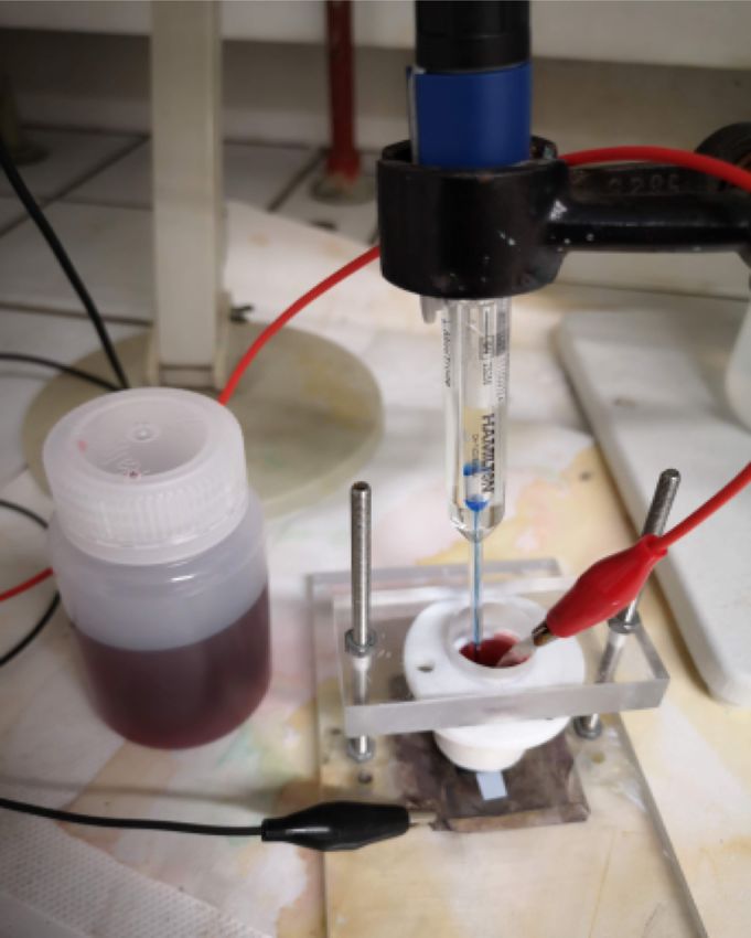
home-made Teflon-cell and exposed to the electrolyte (see Figure 2c,d). A standard three-electrode
setup is employed to reduce ions in solution at the cathode and make the nanowires. When necessary,
the pH values of the as-prepared electrolytes were adjusted to the desired pH by addition of a
diluted HCl solution or by addition of NaOH. Different types of parallel NW arrays made from pure
Appl. Sci. 2020, 10, 1832                                                                                             3 of 25

metals, alloys of controlled composition and multilayered nanowires were successfully grown by
electrodeposition at room temperature (RT) [5,10,23].

                                                                            Reference electrode
               (a)

                                                                                           Anode (Pt)
                                                                                                              A

                             400 nm                          V
                                                                                  Electrolytic
             (b)                                                                   solution
                                                                                                                  E

                                                                                                   Membrane
                                                                 Au layer

                                           (c)                   (d)                Cathode (Pt)

      Figure 2. SEM images of (a) track-etch polycarbonate membrane and (b) porous alumina;
      (c) photograph and (d) schematic of the electrodeposition cell used to grow nanowire arrays.

      Typical SEM images of metal nanowires obtained via the simple potentiostatic mode after
dissolution of the host nanoporous media are shown in Figure 3a,b. Note that, after the template
is filled with magnetic wires, the term packing factor is used instead of the membrane porosity,
since interest is focused on the properties of nanowire networks.

                                  500 nm                                       20 µm

                            (c)                                                           (d)
                                                                       10 nm

                             100 nm
                            (e)                            500 nm                         (f)

      Figure 3. (a,b) SEM images of metal nanowires; (c) schematic of Co/Cu nanowires embedded in a
      polymer template; (d,e) TEM image of Ni80Fe20/Cu and Co/Cu multilayered nanowires; (f) SEM
      image of Ni nanotubes.

     The technique mostly used for the fabrication of multilayered nanowires (see Figure 3c) consists
in a pulse-plating method in which two metals are deposited from a single solution (in which the
nobler element is kept in dilute concentration) by switching between the deposition potentials of
the two constituents. Following a procedure described elsewhere [5,10], the deposition rates of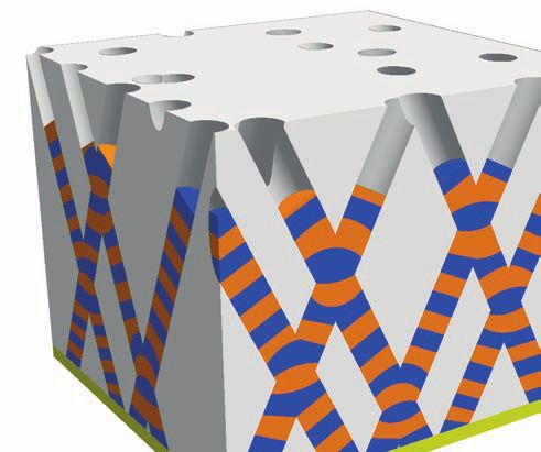
Appl. Sci. 2020, 10, 1832                                                                             4 of 25

each metals were determined from the pore filling time. This single-bath method was successfully
used to prepare various multilayered nanowires including Co/Cu, NiFe/Cu, CoNi/Cu and Ni/Cu.
Transmission Electron Microscopy (TEM) images of permalloy (Py = Ni80 Fe20 )/Cu and Co/Cu
multilayered nanowires are shown in Figure 3d,e. Over the last decade, several groups also reported
on the electrochemical synthesis of parallel arrays of Ni nanotubes within nanopores of anodic alumina
oxide and polymer templates [42,44,71,72]. In this method, core-shell Cu/Ni nanocables are first
grown in porous templates at a constant deposition potential, followed by the selective electrochemical
etching of the Cu-rich core at an oxidation potential (see Figure 3f).
      Most of the magneto-transport measurements were performed by simply contacting a small
number of nanowires parallel to each other embedded in the porous matrix. However, measurements
on single nanowires were also realized in previous studies [17,21,30,41,73]. Various experimental
approaches have been developed for this purpose. For polymer templates with low pore density,
a self-contacting technique [17,72] has been used successfully for contacting a single wire. For anodic
porous alumina supported on Si substrate, nanolithography-based contacting techniques have been
developed to allow the electrical connection of one single nanowire in a large assembly of wires [30,41].
Another approach is based on the use of electron beam lithography to connect a single nanowire after
dissolution of the matrix that contains the nanowires [21].
      Recently, 3D nanoporous templates were obtained by performing a sequential multi-step
exposure of energetic heavy ions, at various angles with respect to the normal of polycarbonate
film surface [60,74]. Then, the latent tracks within the polymer film were chemically etched
following the previously reported protocol to obtain membranes with distinct porosities and pores
sizes [68]. Hereafter, the as-prepared polymer membranes containing networks of interconnected
cylindrical nanopores were designed with pores of well-defined diameters between 40 and 230 nm
and with different porosity characteristics (P = 3% and P = 20%, with P the void volume
fraction of the membrane). Next, the template-assisted electrodeposition method was used for the
synthesis of well-defined 3D architecture consisting of crossed nanowire (CNW) and nanotube (CNT)
networks [60–63,66]. In addition, magnetic and non-magnetic layers were electrochemically stacked to
make interconnected multilayered nanowires [64,65,67]. Figure 4 illustrates the various 3D nanofiber
architectures which have so far been investigated. After electrodeposition, the complete dissolution of
the PC template leads to an interconnected metallic self-standing structure, as the one observed for the
40 nm and 230 nm diameter Ni CNW network shown in Figure 4e–g. The CNW network exhibits the
replicated complex nanowires branching morphology of the porous template.

        (a)                   (b)
                                                       (e)                     (f)

        (c)                   (d)                      (g)                     (h)

      Figure 4. (a) schematics of 3D nanoporous polymer template, (b) crossed nanowire and (c) crossed
      nanotube networks, and (d) crossed multilayered nanowire network; (e–h) SEM images of
      self-supported interconnected nanowire and nanotube networks; (e,f) Ni NW network film with
      230 nm diameter; (g) Ni NW network film with 40 nm diameter; (h) Ni crossed nanotube network with
      230 nm diameter.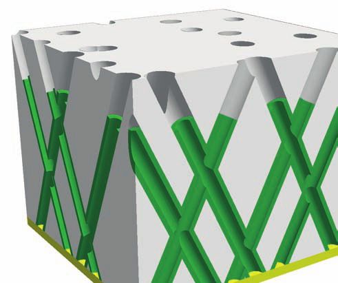
Appl. Sci. 2020, 10, 1832 5 of 25
The feasibility of using the same electrochemical method to fabricate large-scale 3D Ni CNT
networks from Cu/Ni-core/shell nanocables was recently demonstrated [63]. This process results in
Ni CNT networks as the one shown by the SEM micrograph reported in Figure 4h after dissolution of
the host PC membrane. In addition, since the geometrical features of the crossed core(Cu)/shell(Ni)
nanocables can be carefully modified via the control over the reduction or deposition potential,
Ni CNT networks with different wall thickness in the range 10–50 nm were obtained after carrying
out the dealloying step of the Cu core [63]. In addition, CNWs with multilayer structure of FM/Cu,
with FM = Co, CoNi and NiFe alloys were fabricated in the host 3D porous templates [64,65,67].
Interestingly, this fabrication method appears as a very convenient approach for large scale production
of current-perpendicular to-plane (CPP) giant magnetoresistance (GMR) films based on CNW networks,
as shown in Sections 3.3 and 3.5.
3. Results
3.1. Magnetization Reversal Processes in Magnetic Nanowires
Anisotropic magnetoresistance (AMR) arising from the relativistic spin-orbit coupling is a
powerful tool for studying the magnetization reversal in magnetic nanowires. AMR is related to
the dependence of the resistance on the angle between the magnetization of the ferromagnet and the
electrical current direction. The change in resistance between current parallel and perpendicular to
magnetization is often less than a few percent. However, as a high accuracy in the electrical resistance
measurement can be obtained, AMR was used to investigate the reversal of magnetization of magnetic
nanowires at different temperatures [17,73] and quantitative analysis of the results was conducted
using classical models describing magnetization reversal. The magnetoresistive curve R( H ) is related
to the magnetization M( H ) by
M( H )
R( H ) = R0 + (∆R)max , (1)
Ms
where M ( H ) = Ms cos[ ϕ( H )] is uniform and measured along the wire axis, Ms is the saturation
magnetization and ϕ is the angle between the applied field and the current (which is along the
wire axis). In Equation (1), the quantity (∆R)max /R0 defines the AMR ratio. Figure 5a shows the
magnetoresistive hysteresis loops obtained at room temperature on a single 75 nm Ni nanowire for the
angles ϕ = 10◦ and ϕ = 75◦ . Low density Ni nanowire arrays constitute an attractive model system in
which the magnetization easy axis is simply determined by shape anisotropy.
15 January 2000 826 J. Appl. Phys., Vol. 87, No. 2, 15 JanuaryPignard
2000 et al. 825 Pignard et al.
zoom in the region where the discontinuity occurs. Two
jumps of resistance are evidenced at two fields close to each
other. The first jump of resistance refers to the nucleation of
the magnetization reversal and propagation of the domain
wall until it is trapped at a pinning center. The domain wall
is evacuated only at a larger field !giving rise to a second
jump" where the magnetization is completely reversed. Such
a behavior, in which the magnetization reversal is achieved
in several steps, has been observed in most investigated
samples. This particular behavior is probably related (a)to struc- (b)
tural defects acting as pinning centers.
membrane used for the electrodeposition. The values of the nucleation field can be analyzed in
used as cathode; the top Au strip is used to
view of micromagnetic theories. For a single domain crystal
t nanowire. Inset: zoom on the deposition
with a FIG.
cylindrical or a prolate
3. AMR characteristics atspheroidal shape,
300 K of a single for '$10°
three rever- 75° FIG. 4. Nucleation field at 300 K vs # for a single Ni wire (d"75 nm).
contacting the top gold strip. Figure 5. (a) AMR characteristics atwire
300 K of aandsingle 75 nm Ni nanowire for 2 angles between the wire
(d$75 nm). Inset: zoom showing the irreversible jumps of the resistance as Dashed line: expected nucleation field for an infinite cylinder with d
sal modes the
of the magnetization
magnetization is reversed.
can be observed: the coherent
axis and magnetic field direction.
rotation, the curling mode, and the buckling one. The ob- Inset: zoom of R(T)
"75 at lowreversal
nm !curling fields (from
mode". [17]);
Solid line: (b) nucleation
expected nucleationfield forfield
a
volume of aspect ratio 4.3:1 !curling reversal mode".
olycarbonate membranes produced served
Hn atmode 300depends
K vs. the on the radius
angle of the studied
between wirewire.
axis Forand field direction for a single 75 nm Ni wire (from [17]).
membranes exhibit improved prop- R!R c avoid the emergence
the reversal of other nanowires
of the magnetization and electrical
is expected to fol-con-
pe, minimal size, size control, andDashed line: expected nucleation field for an infinite cylinder 75 nm in diameter (curling reversal
low thetacts are
curling then
mode;established
here, R is on the
the radius
two circular
of themetallic
cylinderareas case, D and k are the only independent adjustable param-
he pores. As a consequence, thesemode). Solid line: expected nucleation for a volumez of aspect ratio 4.3:1 (curling reversal mode).
located
and R is the at the extremities
critical radius of the strip.
which depends on thefield
material
s are almost perfectly cylindrical, c Transport measurements
15 were weperformed on Nithewires eters. The solid line corresponds to the adjustment of Eq. !1"
!R
less than !5°" and present a veryFigure c "20 nm for
adapted nickelfrom ". In this case,
ref. [17] with assume that
permission of the author.
fabricated
stable state before from
and after twothedifferent nanoporous
reversal has membranes with the experimental values and shows that the curling
a magnetization
urface. The thickness and the pore samples, with pore diameters of d$75!5 nm and d$35 mode fairly well reproduces the angular dependence of the
used in this study are 22 #m and direction given by the Stoner–Wohlfarth model of coherent
!5
As shown 16 nm. The in total
Figurelength5a, of the wires
the AMRis 22 #curves
m. The pore are nucleation field
sizecomposed of for the whole range
continuous of # angle.
variations ofThethegeo-resistance
Prior to electrodeposition, a gold rotation. The reversal itself occurs according to the curling
was determined after dissolution of the membranes and metrical parameters obtained are D x "0.454, D z "0.092, and
side of the membrane; this mode for a nucleation
corresponding
elec- to a field whichrotation
reversible can be expressed
of the as a
magnetization and of discontinuous jumps of resistivity at
SEM-FEG observation of the nanowires. At room tempera- k"1.2. These values of D z and k both correspond to an
hode during the electrodeposition functionture,of the angle #, thearesaturation
the resistances 595 and 2500magnetization M s andwith
$ for the samples aspect ratio of 4.3:1 showing that the magnetization reversal
is also deposited on the other side geometrical
d%75parameters.
nm and d%35 From nm,Ref. 17, we get:
respectively. By measuring these is initiated in a small volume of the wire: the nucleation
this film is thin enough in order to resistances at two temperatures, say 300 and 77 K, we derive volume can be roughly estimated to 1/65 of the whole wire
ver the pores. In a second step, the H n ! # " "2
& 300$KM/&s77 K%6 and 2.5 for d%75 nm and d%35 nm, respec- volume. These results are consistent with those obtained by
! "! "
ectrolyte on a small area of 1 mm2 tively. On the other k hand, we k know that & 300 K – Wegrowe et al. who found a nucleation volume 50 times
2#
on of Ni ions into the pores is &77 K$6.5 #$ cm,2D 13
so that 2D we $ straightforwardly derive smaller than the entire wire for a Ni wire of 60 nm in
z$ 2 x 2
rement of plating current versus & 300 K$7.8 #$ cm and S& 300 K$10.8 S#$ cm for d%75 nm
xed to "1.1 V. The emergence of # , diameter.19 However, in their work the agreement between
and d%35 nm, respectively.
2
These resistivity
2
values are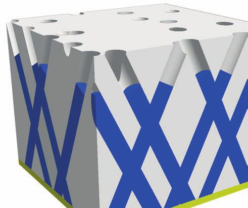
Appl. Sci. 2020, 10, 1832                                                                         6 of 25

fields corresponding to magnetization reversal. It involves processes such as nucleation of small
reversed domains at the end of the wire, before their possible expansion through domain-wall
motion [14]. The jumps in AMR curves refer to successive pinning and depinning of the domain wall.
The inset of Figure 5a shows a zoom in the region where the discontinuity occurs. Two resistance
jumps appear for two field values that are close to each other. The first one refers to the nucleation
of the magnetization reversal and propagation of a domain wall until it is trapped at a pinning
center due to the presence of structural defects. The domain wall is evacuated when the external
magnetic field reaches a higher field (giving rise to a second jump) where the magnetization of the
wire is completely reversed. The nucleation field was measured on individual nanowires as a function
of the angle between the applied field and the wire axis. The results were then compared with
predictions of the micromagnetic theories. Figure 5b shows the dependence of the nucleation field
Hn with the angle ϕ for the 75 nm-Ni nanowire. The angular dependence of the nucleation field
is consistent with a process controlled by a curling rotational mode [14]. The results show that the
magnetization switching is initiated by reversal inside some nucleation volume much smaller than
the total volume of the nanowire [17,73]. For Ni nanowires, the measurements of the nucleation field
as a function of temperature evidence an extra uniaxial anisotropy adding to shape anisotropy at
low temperatures. By combining X-ray diffractometry, magnetization and ferromagnetic resonance
measurements at different temperatures, this additional contribution was demonstrated to find its
origin in magnetoelastic effects conjointly induced by the Ni and polycarbonate thermal-expansion
coefficients mismatch and by the low volume fraction of Ni in the nanocomposite samples [16,31,40].
This additional magnetic anisotropy, whose magnitude is comparable to that of shape anisotropy,
reinforces the wire axis as an easy axis for magnetization.

3.2. Tunable Magnetic Anisotropy of Magnetic Nanowire Arrays
     The effective magnetic anisotropy in ensembles of parallel nanowires is determined by different
contributions, namely shape anisotropy, dipolar interwire coupling, magnetocrystalline (MC)
anisotropy and magnetoelastic terms. Magnetoelastic effects were found to be important only at
low temperature in low porosity polymer membranes filled with Ni [16,40] and will be neglected
hereafter. Consequently, the effective anisotropy field (HEF ) with a uniaxial symmetry parallel to the
wire axis is given by:
                                       HEF = HS − HDIP ± HMC ,                                       (2)

which includes the shape anisotropy HS = 2πMS for an infinite long cylinder, the dipolar coupling
between the wires which lowers the magnetostatic energy and, in a mean field approximation, can be
written as HDIP = 6πMS P [19] (with P the packing factor of the nanowire array), as well as the possible
contribution of a magneto-crystalline anisotropy HMC that adds or competes with the shape anisotropy.
Among the different approaches that have been used to investigate the static and dynamic properties
of arrays of magnetic nanowires, ferromagnetic resonance (FMR) has proved to be a very powerful
technique [19,23,24,29,32,34,35]. Indeed, at a given constant frequency f , the ferromagnetic resonance
condition when the magnetic field is applied parallel to the wires is [19]:

                                                   f
                                            HR =     − HEF ,                                         (3)
                                                   γ

where HR is the resonance field and γ the gyromagnetic ratio provides a direct and precise
quantification of the effective field HEF . Figure 6a shows typical microwave absorption curves
measured on an array of 100 nm Ni nanowires (P ≈ 27%) as a function of the intensity of the static
field applied parallel to the wires [19]. From these results, it can be seen that the absorption curves
present an intense absorption peak which varies in position and intensity as the amplitude of the
magnetic field is varied. Using porous membranes, the inter-wire spacing can be controlled over a
wide range. This allows to explore the properties and dipolar interactions between the wires from
Appl. Sci. 2020, 10, 1832 7 of 25
the limit of isolated towards the limit of almost touching wires [19]. In Figure 6b–d, we present the
dispersion relation and the corresponding hysteresis loops, measured with the magnetic field applied
parallel (closed symbols) and perpendicular to the wires (open symbols) for three different Ni samples
having a diameter and packing density of (b) 180 nm and 4%, (c) 100 nm and 27% and (d) 115 nm
and >35%. The data were compared to the calculated ones for isolated wires, shown as solid lines.
For low packing densities, the measured resonance frequencies in the saturated states agree well with
the values expected for an array of isolated wires, see Figure 6b. At higher wire density for both field
directions, the measured frequencies are almost identical, as shown in Figure 6c. In addition, above a
critical value of the packing (estimated around 33% for an ordered nanowire array), there is a tilt of the
easy axis of magnetization between directions parallel to perpendicular to the wire axis, as shown in
Figure 6d. Therefore, by increasing the density of the NW array, it is possible to modulate the effective
magnetic anisotropy, from the case of isolated wires with the easy axis parallel to the wires to dense
NW arrays in which the easy axis is perpendicular to the wires, passing through the particular case of
quasi-isotropic magnetic systems. The observed deviations from the frequency-field dispersion curves
calculated in weak field conditions are due to the fact that the system is no longer single-domain.
40
//
(b)
(a) 1
//
30 ⊥
F (G Hz )
S
M/M
20 0
⊥
10
0 -1
0
40 1
-10 (c)
(b )
//
// ⊥
30
F (G Hz )
S
M/M
-20
2 20 0
db
3
-30 4 10
⊥
5
6 -1
-40 7 0
(a) 8
40
-50 (d)
(c) 1
0 5 10 15 20 25 30 35 40 // ⊥
30
F (G Hz )
Frequency(GHz)
S
//
M /M
20 0
⊥
10
-1
0
0 2 4 6 8 -5 0 5
H(kO e) H (kO e)
Figure 6. (a) microwave absorption spectra as a function of the applied field parallel to the wires of an
array of Ni nanowires of diameter 100 nm and packing density of 27%. Continuous lines correspond to
measurements made with the applied field value indicated by the numbers; (b–d) dispersion relations
and the corresponding hysteresis loops measured in arrays of Ni nanowires with the field applied
parallel (closed symbols) and perpendicular (open symbols) to the wires. The wire diameter and
packing density are (b) 180 nm, 4%, (c) 100 nm, 27%, and (d) 115 nm, >35% (from [19]).
A control of the effective magnetic anisotropy can be achieved not only by changing the packing
factor nanowire arrays but also by tailoring the magnetocrystalline anisotropy, as demonstrated for
Co [24,29] and CoPt [28] nanowire systems. From our previous studies, the electrolytic bath acidity,
or pH, was demonstrated to be a key parameter in determining the structure of electrodeposited cobalt
FIGURE 2
nanowires.
A. Encinas-Oropesa, et. al. Indeed, it was found that face-centered cubic (fcc) Co is favored at low pH (typically less
J. Appl. Phys.
than 2.5) while hexagonal close-packed (hcp) Co is formed at pH larger than ∼3.5 [24,29]. Moreover,
it was shown that an abrupt transition in crystallographic orientation takes place for pH in the range
4.0–6.5, with a preferred in plane orientation at low pH and a reorientation of the c-axis normal to
the film at large pH. These structural changes have been attributed to the influence of evolution of
hydrogen at the cathode, which are known to depend on the pH [75,76]. Another relevant parameter
FIGURE 3
of the operative conditions in cobalt electrocrystallization
A. Encinas-Oropesa, et. al. is the current density.
J. Appl. Phys.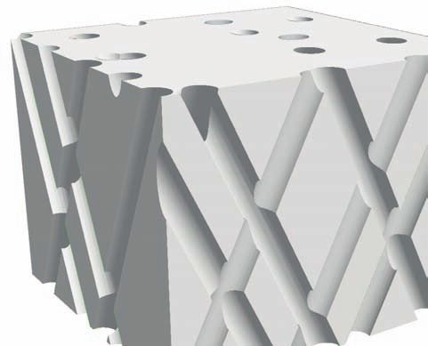
Appl. Sci. 2020, 10, 1832                                                                                      8 of 25

     Figure 7a shows the variation of the effective field for a 30 nm Co nanowires array as a function
of the pH in the range 2.0–6.6 for two different plating current densities, around 50 mA·cm−2 and
5 mA·cm−2 , respectively. These data were extracted from FMR experiments [24,29]. Let’s first consider
the results obtained at 50 mA·cm−2 . For reference, the effective field HEF = 2πMs (1 − 3P) ≈ 8 kOe
expected when there is no contribution from the MC anisotropy is indicated by the dotted line in
Figure 7a (P ≈ 3% is for the array samples in this study). This value agrees with that of the low-pH
samples, for which only the shape anisotropy and dipolar coupling should be considered due to the
lack of texture in the electrodeposited Co microstructure and possible presence of fcc phase. In contrast,
for pH values of 3.8–4.0, it was found that the microstructure of the deposited Co nanowires contains a
dominant fraction of large hcp grains with the c-axis oriented perpendicular to the wire. A similar
result was observed for all the diameters considered in the range 30–120 nm [24,29]. For such samples,
the effective anisotropy field is weaker than for the low-pH samples due to the competition between
shape anisotropy and MC anisotropy (which has an upper bound of HMC = 6.4 kOe for bulk hcp Co).
Moreover, a re-orientation of the hcp c-axis along the wire axis is also observed for pH values ≥ 6.0 [24],
resulting in an additive MC contribution to the total anisotropy field.

                                           (a)                              (b)                          (c)

      Figure 7. (a) effective field as a function of the pH for 30 nm Co nanowires electrodeposited at current
      densities of 5 mA/cm2 (squares) and 50 mA/cm2 (circles). The dashed line shows the expected value
      of HEF for an array of Co nanowires (P ≈ 3.5%) assuming no contribution from magnetocrystalline
      anisotropy; (b,c) TEM images and corresponding diffraction patterns of two 30 nm Co nanowires
      electrodeposited at pH 5.2 at current densities of (b) 5 mA/cm2 and (c) 50 mA/cm2 (from [29]). Figure
      adapted from ref. [29] with permission of the author.

      For both low and high plating currents, the overall variation of the effective field with bath acidity
is similar, see Figure 7a. However, the transition between the low and high effective fields takes place
around different pH values depending on the plating current density. Therefore, for a limited pH
range, it is possible to selectively obtain both structural phases (c-axis parallel and perpendicular
to the wires) from the same electrolyte simply by changing the plating current (or deposition rate).
These results are corroborated by TEM investigations as shown in Figure 7b,c [29]. In the case of low
current density, the TEM bright field images and corresponding diffraction patterns clearly show an
orientation of the c-axis parallel to the wire axis, while, in the case of high current density, the images
clearly show a perpendicular orientation of the c-axis.
      CoPt alloy nanowires constitute another system whose magnetic anisotropy can be changed
drastically by appropriate annealing treatment after electrodeposition. These interesting properties of
Cox Pt1− x alloys are related to its ordered face centered tetragonal (fct) phase (L10 ) which exhibits very
large magnetocrystalline anisotropy energy (up to 5 × 10⁷ erg·cm−3 around the equiatomic region).
For this purpose, we used a thin anodized aluminum oxide film template (1.5 µm thick) on a doped
silicon wafer, which affords the thermal durability of the nanoholes for high temperature process after
electrodeposition. The as-made electrodeposited 80 nm Co47 Pt53 nanowires were magnetically soft as
shown by the hysteresis loops in Figure 8a. The large packing in the array (close to 50%) induces a
strong interwire dipolar coupling that overcomes the shape anisotropy, thus resulting in an easy axis
Appl. Sci. 2020, 10, 1832 9 of 25
perpendicular to the wire axis. Appropriate thermal treatment at 700 ◦ C has contributed to ordering the
Electrodeposition of L1 Co Pt nanowires 0 x 1−x
alloy nanowires. X-ray diffraction patterns have confirmed that the L10 ordered phase was successfully
obtained in such CoPt nanowires [28]. Hysteresis loops of the annealed sample measured parallel
and perpendicular to the wires are displayed in Figure 8b. Magnetic hardening is developed after
annealing at 700 ◦ C with coercive fields as large as 11 kOe at room temperature. No anisotropy exists
between the two directions, although the length/diameter ratio of the wires is larger than 10. This is
because the L10 CoPt has a strong MC anisotropy, much higher than the shape anisotropy. In addition,
the nanowires are textured along the direction which makes an angle of 55◦ with the (001) easy
axis, resulting in an absence of anisotropy.
(a) (b)
Figure 3. 80 nm diameter Co0.47 Pt0.53 ordered nanowires after
annealing at 700˚C; (a) XRD pattern; the superstructure peaks, as
(001) or (110), reveal an ordered phase of the alloy; (b) room
temperature hysteresis loops. The curves were obtained with the
magnetic field parallel (——) and perpendicular (- - - -) to the
wire’s axis.
from 70 to 300 K. All the hysteresis loops of L10 ordered
samples exhibit very high coercive fields. The highest
equal to 1.087 T (10.870 kOe) has been obtained with 80 nm
diameter Co0.47 Pt0.53 samples at 300 K. The hysteresis loops of
such samples measured at room temperature are displayed in
figure 3(b), where external fields were applied both parallel
and perpendicularly to the wire axis. No large difference
Figure 2. As-deposited nanowires: (a) cross-sectional SEM image Figure 3. 80 nm diameter Co0.47 Pt0.53 ordered nanowires after
Figure 8. Hysteresis
of 80 nm diameterloops
CoxPt1−xmeasured at 300 K
nanowires electrodeposited in arrays
in the in the hysteresis
of atCo
annealing 700˚C;Pt
47 (a) 53 loops
XRD has been
nanowires
pattern; observed
withpeaks,
the superstructure between
theasfieldthe two
applied
(001) or (110), although
directions reveal an ordered phase ratio
the aspect of the of
alloy;
the(b) roomis much larger
wires
supported alumina template; (b) XRD patterns of the nanoporous temperature hysteresis loops. The(a) curves were obtained with the sample and
parallel (solid line) and perpendicular (dashed
substrate and the as-deposited CoxPt1−x nanowires showing an fcc magneticline) to
than 10. the
field This
wires.
parallelis(——)
because As-deposited
the L10 Co(-xPt
and perpendicular -) tohas
- -1−x the very strong
structure with a preferred ⟨111⟩growth orientation and
(b) annealed at 700 ◦ C.
sample magnetization
(c) corresponding Note
curves measuredthe withdifferent
magnetocrystalline
wire’s
the magnetic magnetic
axis. fieldanisotropy,
scales in much (a)higher
andthan (b)the(from
shape [28]).
anisotropy. In addition, the as-deposited nanowires were
field parallel (——) and perpendicular (- - - -) to the wire’s axis at from 70 to 300 K. All the hysteresis loops of L1 ordered
Figure adapted from ref. [28] with permission of the author.
room temperature.
textured along the ⟨111⟩direction, which makes 0 an angle of
samples
55.45˚ exhibit
with thevery [001]high coercive fields. The highest
fct easy magnetocrystalline axis.
(This figure is in colour only in the electronic version) equal toThe1.087 value of the first magnetocrystalline nm
T (10.870 kOe) has been obtained with 80 anisotropy
3.3. Spin Transport in Multilayered Nanowires diameter Co0.47 Pt0.53 samples at 300 K. The hysteresis loops of
constant K1 of the L10 phase has been estimated by integrating
such samples measured at room temperature are displayed in
the Co0.47 Pt0.53 sample is found to be a tetragonal lattice the virgin curves measured parallel and perpendicular to the
figure 3(b), where external fields were applied both parallel
Multilayered nanowires
(L10 phase) areofideal
with a c/aratio structures
0.979 (table 1). to investigate
andwire’s theto giant
axis, respectively.
perpendicularly the wire magnetoresistance
Usually,
axis. classical
No large methods, (GMR) and
difference such as
Figure 2. As-deposited
The magnetic nanowires:
properties and(a)magnetocrystalline
cross-sectional SEM imageaniso- in integration of the virgin curveobserved
or torquebetween
determination,
the hysteresis loops has been the two are not
its temperaturetropy dependence
of 80 nm diameter CoxPtin
of L1alumina
supported 0 CoxPt
the
1−x nanowires
template;
Current
1−x nanowires
have ofbeen
(b) XRD patterns
Perpendicular
electrodeposited in the
investigated directions
the nanoporous
to
suitablealthoughthe layer
for calculating Plane
1 of of
the aspectKratio
(CPP)
thetheL1wires
0 samples.
geometry.
is muchThe reason is The first
larger
substrate and the as-deposited Co Pt nanowires showing an fcc than 10. This is because the L10 CoxPt1−x has very strong
measurements were performed in 1994 x 1−x
structure with a preferred ⟨111⟩growth orientation and
on Co/Cu nanowires by Piraux et al. [10], Blondel
magnetocrystalline anisotropy, much higher than the shape 911
et al. [77]
(c) corresponding magnetization curves measured with the magnetic anisotropy. In addition, the as-deposited nanowires were
and Liu et al. [78]. Nanowire
field parallel systems
(——) and perpendicular (- - - have key
-) to the wire’s axisadvantages
at over conventional magnetic
textured along the ⟨111⟩direction, which makes an angle of
multilayers
room temperature.
made by vacuum (Thisdeposition. Firstly,
figure is in colour only in the electronicthe
version)electrodeposition method
55.45˚ with the [001] into porous
fct easy magnetocrystalline axis. templates is much
The value of the first magnetocrystalline anisotropy
more convenient than vacuum deposition techniques forKmaking
constant multilayers
1 of the L10 phase has been estimated with layer thicknesses
by integrating
the Co0.47 Pt0.53 sample is found to be a tetragonal lattice the virgin curves measured parallel and perpendicular to the
close to the micron without
(L10 phase) degrading
with a c/aratio of 0.979 (tablethe 1). quality wire’s of the axis, interfaces.
respectively. Usually,As a result,
classical measurements
methods, such as on
The magnetic properties and magnetocrystalline aniso- integration of the virgin curve or torque determination, are not
nanowires have tropy
been very useful to study the regime with layer thicknesses
of L10 CoxPt1−x nanowires have been investigated suitable for calculating K1 of the L10 samples. The reason is above the spin diffusion
length (SDL), to test the theoretical predictions in this regime and to determine the911SDL of various
materials. Secondly, most of the measurements in CPP geometry on planar multilayers were performed
with superconducting niobium contacts. Such measurements are therefore limited to the helium
temperature range. On the other hand, measurements on nanowire systems can be carried out up
to the ambient temperature, which has made it possible to study the temperature dependence of
CPP-GMR [79]. Third, a high aspect ratio of the multilayered nanowires leads to large signals and
precise measurements. Up to now, most measurements have been performed on Co/Cu, CoNi/Cu,
Py/Cu systems and FeCoNi/Cu [10,11,77–83]. In Figure 9a, we show an example of CPP-GMR curves
obtained for a Py/Cu multilayered nanowires sample. The MR ratio reaches 67% at 77 K and 71%
at 4.2 K. Another example of experimental results for Co/Cu nanowires is also shown in Figure 9a.
In both cases, the thickness of each FM/Cu bilayers is about 15 nm.
Most measurements on multilayered nanowires have been analyzed using the Valet–Fert
model [84,85]. This model takes into account the important effects due to the spin accumulation
occurring at the interfaces when the conduction in the ferromagnetic layers is spin dependent.
In addition to the usual relaxation term for the momentum, the Boltzmann equation of the Valet–Fert
model also includes a spin-lattice relaxation term describing the relaxation of spin accumulation by
spin-flip scattering. In the case of multilayers with thick ferromagnetic layers, the GMR contribution
Appl. Sci. 2020, 10, 1832                                                                                                   10 of 25

from bulk scattering dominates that from interface scattering. For FM/Cu multilayers, if FM layers
are thicker than the spin diffusion length in FM, and Cu layer much thinner than the one in Cu,
the CPP-GMR ratio can be expressed as [5]:

                                                           ∆R     2pβ2 `FM
                                                                        sf
                                                              =                                                                 (4)
                                                           RP   (1 − β2 )tFM

with ∆R = RAP − RP (RAP and RP are the corresponding resistances for antiparallel and parallel
arrangements, respectively), β the bulk scattering spin asymmetry coefficient (β = (ρ↓ − ρ↑ )/(ρ↓ + ρ↑ )),
`FM
 sf the spin diffusion length in the FM material, tFM the thickness of the FM layers and p the proportion
of antiparallel orientations of the magnetization in consecutive magnetic layers. The spin asymmetry
coefficients are usually determined from analysis of the experimental results in the long SDL limit
(tFM
Sweep (H↑) Sweep (H↓)
the low packing factor Co/Cu sample obtained while with
and negative
sweeping theslope
cross each for state
external
other V"/V#)
at zero are symmetric
bias magnetic ¯eld.
Sweep (H↑) Sweep (H↓)
magnetic field in the IP (blue curve) and OOP (red curves)
and directions.
Furthermore,
cross eachthe As seen,
oscillating
other at zerofrequency versus ¯eld.
bias magnetic mag- ΔR
netic ¯eldappears
features obtainedfrequency
are linear and mag-
their 4 mΩ
almost isotropic behaviour is observed, although theFurthermore,
IP curves the to be
oscillating versus ΔR
slopes are directly related to the vortex polarity of ΔR
saturated at slightly lower magnetic field.
3.3.Besides,
Giant the small¯eld
netic difference
magnetoresistance in between
features obtained
interconnected are magnetoresistance
linear
3.3. multilayered
Giant and networks
nanowire their
in interconnected
153 5multilayered
mΩ 4 mΩ
nanowire networks 153
the
andthick
the resistance state obtained at saturation in the IPslopes OOP magnetic
is ascribed layer
to that composes the STVO. 3.3. Giant
ΔR magnetoresistance in interconnected multilayered nanowire networks 153
are directly related to the vortex polarity of
5 mΩ −6 −4 −2 0 2 4 6
the anisotropic magnetoresistance contribution, andthe appears
thick to be marginal.
magnetic layer that composes the STVO. Out of plane field (kOe)
GMR ratio will hereAppl.
be defined
Sci. with
2020,respect of the saturated
10, 1832 resistance state inemission from single
3.2. Microwave 11 of 25
reasonable agreement with previous reasonable
results from agreement
the CPP-GMR with previous
experiments results from the CPP-GMR experiments
−6 −4 −2 0 2 4 6
the IP direction. nanowire
3.3. Giant magnetoresistance spin-torque
in interconnected oscillator
multilayered nanowire 151 reasonable
networks[221]. agreement with Out of plane results
previous field (kOe)from the CPP-GMR experiments
performed on3.2.Py/CuMicrowave
multilayers [221]. performed on Py/Cu multilayers −6 −4 −2 0 2 4 6
As detailed in Sec. emission from single
3.1, micromagnetic simulations performed on In Py/Cu multilayers
plane field (kOe) [221].
were nanowire
performed to spin-torque
predict andoscillator characterize the Fig. 10.−6 GMR −4 curves−2 measured0 2by contacting
4 6 a single NW
ground magnetic states of metallic SVs. Further- (made of a single Co(22 In plane field (kOe)
nm)/Cu(12 nm)/Co(6 nm) SV) with the
(a) As detailed in Sec. 3.1, micromagnetic
more, simulations also o®er the opportunity to (b) simulations (c)
magnetic ¯eld applied in the plane of the layers (IP ¯eld). The
were
identify performed
the current to predict
and magnetic and characterize
¯eld conditions the inset10.
Fig. shows GMRthe GMRcurvescurve obtained
measured by by sweepingathe
contacting external
single NW
ground magnetic states of metallic
for the observations of steady state oscillations. As SVs. Further- ¯eld along
(made the wire
of a single Co(22axisnm)/Cu(12
(out of plane (OOP) nm)
nm)/Co(6 ¯eld).
SV) with the
Supported alumina template more,Electrodeposition
shownsimulations
hereafter, microwave of Cu/Co NWs
also o®er signals the opportunity
have been ob- to magnetic ¯eld applied in the plane of the layers (IP ¯eld). The
pore diameter identify the current andonmagnetic
served experimentally single Co/Cu/Co ¯eld conditions nano- insetMicrowave
shows the GMR emission
curve measurements
obtained by sweepingare performed
the external
~ 90 - 120 nm interpore distance wire Pt Ag/AgCl withalong
¯eld a spectrum
the wire axis analyzer after(OOP)
(out of plane a 42-dBm
¯eld). ampli¯-
~ 120 - 150 nm for theSVs even at zero
observations magnetic
of steady ¯eld.
state Interestingly,
oscillations. As cation of the output oscillating GMR signal.
high frequency
shown hereafter, versus magnetic
microwave signals ¯eld have tunability
been ob-
was demonstrated, electrolyte in the range 0.4–2 MHz/Oe, Selected experimental results of spin-transfer-driven
served experimentally on single Co/Cu/Co nano- Microwave emission measurements are performed
depending on the orientation of the applied mag- vortex excitation obtained by measuring a single SV
wire Co2+ even Cu at2+zerotomagnetic with a spectrum analyzer
11. after a 42-dBm ampli¯-
neticSVs ¯eld relative the magnetic ¯eld. Interestingly,
layers of the are displayed in Fig.
high
pseudo frequency
spin-valve. versus
However, magnetic ¯eld tunability
it is stressed that the
cation of the the
To stabilize outputmagnetic oscillating
vortex GMR state, asignal.
satu-
was demonstrated,
individual layer thicknessesin the and range wire0.4–2
diameter MHz/Oe, in the Selected experimentalmagnetic
rating perpendicular results of¯eldspin-transfer-driven
is ¯rst applied to
pore simulationson(24/13/7
depending the orientation nmalumina
thickness
of field for the mag-
the applied spin- vortex excitation obtained by measuring a single SV
thickness
10. (a) CPP-GMR
Figure alumina valves¯eld andversus80 nm wire magneticAu
diameter) di®erlayers applied
slightly from in the plane ofinthe
are displayed Fig.crossed
11. NW networks
netic relative
Si to the magnetic of the 1.55
~ 1.2 - 1.4 μm at T = (Al2O33)
15 K and pseudo T = 300
the experimental STVOs
K forHowever,(a) Co/Cu, (22/12/6 nm thickness
(b) CoNi/Cu and To stabilize the magnetic vortex state, a satu-
spin-valve. it is stressed that the and (c)1.50Py/Cu multilayered nanowires
100 nm diameter). (b) The di±culties in controlling
(GHz)Frequency (GHz)
(a) individual layer thicknesses and wire diameter in the rating perpendicular magnetic ¯eld is ¯rst applied to
(adapted from [64,65,67]). precisely the Figure adapted
individual layer from thicknessesref. [65] and wire with permission 1.45 of the author.
simulations
Large array of NWs containing
diameter spin-valves (24/13/7
while nm thickness
fabricating the porous for thealumina spin-
1.40
NWs valves
template andand 80 nm wire diameter)spin-valve
electrodeposited di®er slightly nanowiresfrom 1.55
the
areexperimentalalumina for
responsible STVOs these(22/12/6
di®erences. Hnm = -50 Oe
thickness
However, and
de- 1.35
spite these (Al2O3)
small disagreement, the frequency values 1.50
1.30
100 nm diameter). The di±culties in controlling
and the variation
precisely the individual of the layeremitted signal as aand
thicknesses function
wire 1.45
dV/dI (Ω)
of the external magnetic ¯eld are in good agreement
Frequency
(a)
Figure 3.16 diameter
–with while
(a-b)micromagnetic
Giant fabricating
magnetoresistance
simulations.
the with
curve porous alumina
the applied field along 1.40
(MHz)
Figure 3.15 – (a-b) Giant magnetoresistance curve obtained
the in-planetemplate with
and the applied nanowire
electrodeposited spin-valve
direction
Electric of transport
Co 50 Ni50 /Cu measurements werenanowires
networks with
then(a) per- 40 nm in 30
field along the in-plane direction of Co/Cu nanowire
diameter
Figure 3.18and–are
networks
⇠20%
(a) Giant
with
responsible
into (a)for
packing 40 nm
these
factor
magnetoresistance Figure
and (b)
curve 3.18
di®erences.
80with
nm – (a)
in Giant
However,
the diameter de- ⇠3% in Figure
magnetoresistance
and
1.35 3.18 – (a) Giant magnetoresistance curve with the applied field along
curve with the applied field along
APapplied
state field along
formed characterize the resistance and magnetic 20
Linewidth
in diameter and ⇠20% in packing factor and Au(b) 80 nm in diameter and ⇠3% in
packing factor at room the in-plane direction
withof80permalloy/Cu
nm in diam- nanowire networks with 80 nm in diam-
the in-plane spite these
con¯guration
direction of temperature
permalloy/Cu(blue
small of disagreement,
the thecurves)
single in-plane
nanowire NW. theanddirection
frequency
T =with 15 of
Magnetoresistance
networks K80(green
values
nm0.2 in curves).
permalloy/Cu
Ωdiam- nanowire
1.30
10
networks
packing factor at room temperature (blueThe curves) and
inset⇠3%in andT shows
(a) =the
curves 15 obtained
Kthe(green
variation curves).
variationfor
of of
a the
the giant
eter
positive
emittedand(b)
magnetoresistance
current
signal in of
aspacking
4.0
a mA ratio
function factor
are withat the
room
eter and (c)
temperature
⇠3% in packing factor at room temperature (blue curves) and T =
(blue curves) and T =
(c) Comparison between the(a)
eter and in Si
packing Spin-Valve
factor at room temperature ⇠3% (blue curves)
P stateis about and T =
giant magnetoresistance
pulse duration measurements
TP of the Fig.performed 15 K 0 (green curves). (b) Comparison between the giant magnetoresistance
(b)Co Ni50The layer compared togiant
the theoretical prediction between
shown
ofcurves).
the in
external 5010.
magnetic single
15 K NW
¯eld (green
arethe resistance
in curves).
good (b) Comparison
agreement the giant magnetoresistance
with the applied field along the in-plane 15 Kblue
(green Comparison between magnetoresistance -400 -300 -200 -100 0
(a) 100 200 300 400
(IP, (c)
curve)
of the Valet-Fert and
18 !.modelout-of-plane
The in GMR (OOP,
value
the limit of is
thin"R -4
Cu¼layer -2
21 m! and 0
while
large Cosweep-2
Ni 4
layers. measurements performed
Out ofthe plane
with
magnetic
the applied field along the in-plane (IP, blue
measurements with micromagnetic
performed with the appliedmeasurements
simulations. field along performed
the
I (mA)in-plane with
50 50 the
(IP, blueapplied field along in-plane (IP,field
blue(Oe)
Linewidth (MHz)
red curve) directions for the Co/Cu nanowire networking
(c) Comparison with the 80in-plane
between nmthe in giant
diameter
applied magnetic ¯eld
magnetoresistance and "R =12
measurements performed curve)
30
and out-of-plane (OOP, red curve) directions for the network in (a) at
curve) and (d) Electric
out-of-plane transport
(OOP, red ¯eld
curve) curve)
measurements and out-of-plane
directions were
offor the then
network(OOP,
per- redatcurve) directions for the network
in (a) (b) in (a) at
and ⇠3% in packing factor at room temperature.
with the applied m!magnetization
in out
field along curves
of the
planein-planein (IP,(see inset
blue curve) Fig.
and10). These
out-of-plane room temperature. (c) magnetization curves in the IP and OOP directions
(OOP, curves
room(a) temperature.
formed
illustration (c)
to magnetization
characterize
of the spin room
curves
the temperature.
in the
resistance
transfer IPand
torque (c)
andmagnetic
OOP magnetization
device directions
with Fig.spin inEvolution
valves the IP and
stacked theOOP in directions
each nanowire
the IP and OOP directionsFigure 11.
corresponding to (c). results indicate
red curve) directions for the Co/Cu nanowire that an anti-parallel
network to magnetization
with 11. 20(a)
corresponding to (b).
of emitted signal frequency as a
corresponding to (b).
con¯guration
con¯guration ofcanthebesingle
morecorresponding
NW.
easily Magnetoresistance
obtained (b).80 nm in diameter
when the function of the perpendicular applied magnetic ¯eld for the two
and ⇠3% inin
electrodeposited packing
a factor at room alumina
nanoporous temperature.template; (d) magnetization (b) curvesmagnetization
in
differential 10 resistance
con¯gurations as a
V#/V" function
(in blue) ofV"/V#
and bias(in
curves
¯eld isobtained
orientedfor in athe positive
plane current
of the layers.of 4.0 Also,
mA are as
the IP and OOP
direct current at H =directions
expected,
shown −in 50 the
Fig. Oecorresponding
for
easy
10. The asingle
axis Py(30 to (c).nanomagnet was set
of theNW nm)/Cu(10
resistance
Figurethe3.16b is nm)/Py(6
about
shows the comparison
red). The Figure
nm) signal was
0 spin
of 6.0between the
3.16b showsby
obtained
valve
The room-temperature
the comparison
injecting
nanowire GMR withbetween
a positive
the the room-temperature GMR
dc current
Figure 3.16b by!.shows
the the comparison
shape anisotropy between
to be room-temperature
perpendicular to the GMR
mA.-400
curves (b) -300
obtained -200 -100sweeping
linewidth
while 0
versus 100 200 ¯eld
magnetic
the 300 for
magnetic 400both
field in the IP and OOP directions.
18 The GMR value is "R
curves ¼ 21
obtainedm! while
IPwhile sweep-
and sweeping the magnetic The field inOut
theof IPplane
and OOP directions.
magnetization con¯gurations in the corresponding colors
magneticcurves fieldobtained
in the plane
while sweeping of inthe
thethe layer.
magnetic Arrows
field in the mark OOP thedirections.
scan direction; (c)
anisotropic evolution magneticof field
the (Oe)
emittedis found to be of few percent,
wire axis, i.e., plane of the layers. [Corresponds to Phase Bmagnetoresistance contribution
1 in Fig. 7] (color online).
ing the in-plane applied magnetic ¯eld and "R =12
Theisanisotropic
is found magnetoresistance
to high
The
signal frequency packing
anisotropic
by
m!
networks. The
magnetoresistance
injecting
in
difference
contribution
a positive exacerbated
dcinset
current inbe6.0
toof alloys,
of few
mA where acontribution
as the
percent, is found
as expected
function theto
offrom the be of(b)few percent,
measurements
perpendicular performed
applied on homogeneous Py CNWs (see
asincrease is up
expected theout
to more
from of plane
than ¯eld
twice. The
measurements (see
as
mainexpected
performed differenceoffrom
Fig.
between
on homogeneous 10).
the These
measurements
the
Py high
CNWs and(seeperformed
low
1740007-9 Sectionson 3.1.1
homogeneous
and 3.1.2). Py Moreover,
CNWs (seea smaller saturated field is observed for
magneticSections
field for
packing the
and istwo
results
networks
3.1.1 magnetization
indicate
the ratio
3.1.2). that an
between
Moreover, a the configurations
anti-parallel
Sections
volume
smaller 3.1.1 magnetization
and
fraction
saturated 3.1.2).
of
field NW Vdown
Moreover,
intersections
is observed /V
Fig.
for a 11. (in
smaller
the (a)
up OOP blue)
curve, and
saturated
Evolution theV
offield
indicating up /Vdown
isemitted
observed
that the for
signal (in
axisred)
frequency
easy of as
thea multilayered NW network
(adaptedtheand NW[30,41]).
OOP
from segments. Therefore,
con¯guration
curve, indicating
Figure theeasy
canadapted
that be
the difference
morethe of between
OOP
easily
axis
from thecurve,
obtained highwhen
multilayered
ref. [41] and
indicating low
NW
with that
the packing
thefunction
network
permission
of the
easy axis of perpendicular
the
themultilayered
ofcon¯gurations
author.
applied
NWmagnetic
network¯eld for the two
factor samples
¯eldcan be ascribed
is oriented in to
thethe unreliable
plane of theformation of multilayered
layers. Also, as magnetization V#/V" (in blue) and V"/V# (in
structure in expected,
the interconnections
the easyduring
axis ofthethe
pulse electro-deposition,
nanomagnet was setbecausered).
of The signal was obtained by injecting a positive dc current
of 6.0 mA. (b) The linewidth versus magnetic ¯eld for both
In Figure 11b, we show
by the shapethe variation
anisotropy to be of the differential
perpendicular to the resistance dV/dI versus injected direct
magnetization con¯gurations in the corresponding colors
wire axis,field.
current at small magnetic i.e., in Starting
the plane offrom
the layers.
the P state at zero current,
[Corresponds to Phase the injected
B1 in Fig. current is first
7] (color online).
decreased toward negative values. We first observed a 1740007-9 sharp increase of the resistance at −2.5 mA
due to the magnetization switching of the thin Py layer. The curve is hysteretic because the system
remains in this high resistance state until the current is swept to a positive value. At a critical positive
current of about +0.4 mA, the resistance flips back to the P state level. The system remains in this
low resistance state at higher positive current. In addition, under specific magnetic field and injected
current conditions, emission of microwave current was detected in Co/Cu/Co pseudo spin-valves
with frequency in the GHz range and linewidth as low as 1.8 MHz [37,41,45]. Microwave signals
have been obtained even at zero magnetic field and high frequency versus magnetic field tunability
was demonstrated. From micromagnetic simulations, it appeared that the microwave emission is
generated by the vortex gyrotropic motion which occurs in, at least, one of the two magnetic layers of
our spin-valve structures. To stabilize the magnetic vortex state, a saturating perpendicular magnetic
field is first applied to the sample before a decreasing ramp of magnetic field. The results reported in
Figure 11c for the measured spin-transfer nano-oscillator containing a single spin-valve are related
to the two-vortex state with vortices of opposite polarity present in both the magnetic layers of the
spin-valve. The magnetic states responsible for the microwave emission were found to be consistent
with micromagnetic simulations [37,41,45]. This bottom-up approach is promising for microwave
device applications since it allows connection of dense arrays of stacked metallic spin valves in series
and/or parallel, which could lead to high-quality coherent emission by synchronization.You can also read


