Lawrence Berkeley National Laboratory - Recent Work
←
→
Page content transcription
If your browser does not render page correctly, please read the page content below
Lawrence Berkeley National Laboratory
Recent Work

Title
Metabolic Capacity of the Antarctic Cyanobacterium Phormidium pseudopriestleyi That
Sustains Oxygenic Photosynthesis in the Presence of Hydrogen Sulfide.

Permalink
https://escholarship.org/uc/item/94h0p69v

Journal
Genes, 12(3)

ISSN
2073-4425

Authors
Lumian, Jessica E
Jungblut, Anne D
Dillion, Megan L
et al.

Publication Date
2021-03-16

DOI
10.3390/genes12030426

Peer reviewed

 eScholarship.org                            Powered by the California Digital Library
                                                             University of California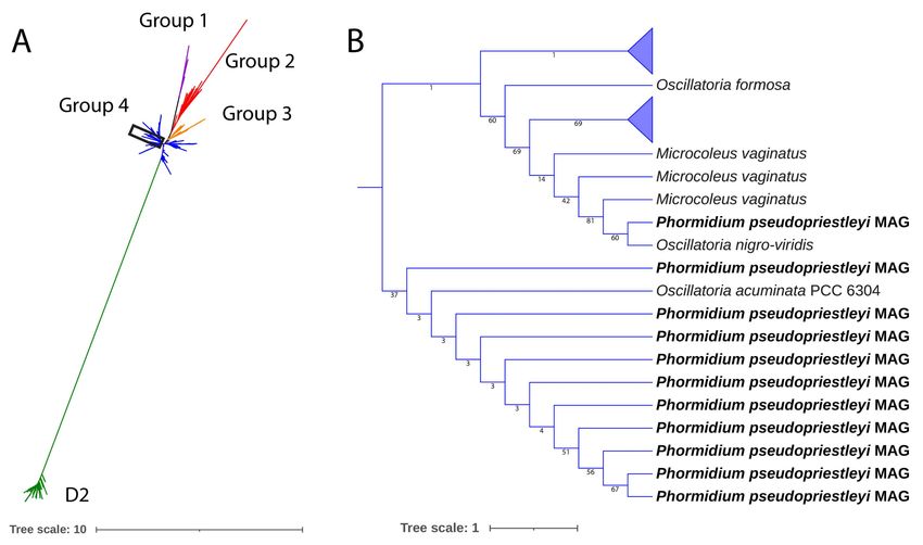
G C A T
T A C G
G C A T
               genes
Article
Metabolic Capacity of the Antarctic Cyanobacterium
Phormidium pseudopriestleyi That Sustains Oxygenic
Photosynthesis in the Presence of Hydrogen Sulfide
Jessica E. Lumian 1 , Anne D. Jungblut 2 , Megan L. Dillion 3 , Ian Hawes 4 , Peter T. Doran 5 , Tyler J. Mackey 6 ,
Gregory J. Dick 7 , Christen L. Grettenberger 8 and Dawn Y. Sumner 8, *

                                          1   Microbiology Graduate Group, University of California, Davis, CA 95616, USA; jemizzi@ucdavis.edu
                                          2   Life Sciences Department, The Natural History Museum, London SW7 5BD, UK; a.jungblut@nhm.ac.uk
                                          3   Genomics and Bioinformatics, Novozymes, Inc., Davis, CA 95616, USA; mled@novozymes.com
                                          4   Coastal Marine Field Station, University of Waikato, Tauranga 3110, New Zealand; ian.hawes@waikato.ac.nz
                                          5   Geology and Geophysics, Louisiana State University, Baton Rouge, LA 70803, USA; pdoran@lsu.edu
                                          6   Department of Earth and Planetary Sciences, University of New Mexico, Albuquerque, NM 87131, USA;
                                              tjmackey@unm.edu
                                          7   Department of Earth and Environmental Sciences, University of Michigan, Ann Arbor, MI 48109, USA;
                                              gdick@umich.edu
                                          8   Department of Earth and Planetary Sciences, University of California, Davis, CA 95616, USA;
                                    clgrettenberger@ucdavis.edu
                                   *   Correspondence: dysumner@ucdavis.edu; Tel.: +1-530-752-5353
Citation: Lumian, J.E.; Jungblut,
A.D.; Dillion, M.L.; Hawes, I.; Doran,    Abstract: Sulfide inhibits oxygenic photosynthesis by blocking electron transfer between H2 O and
P.T.; Mackey, T.J.; Dick, G.J.;           the oxygen-evolving complex in the D1 protein of Photosystem II. The ability of cyanobacteria to
Grettenberger, C.L.; Sumner, D.Y.         counter this effect has implications for understanding the productivity of benthic microbial mats in
Metabolic Capacity of the Antarctic       sulfidic environments throughout Earth history. In Lake Fryxell, Antarctica, the benthic, filamentous
Cyanobacterium Phormidium                 cyanobacterium Phormidium pseudopriestleyi creates a 1–2 mm thick layer of 50 µmol L−1 O2 in
pseudopriestleyi That Sustains
                                          otherwise sulfidic water, demonstrating that it sustains oxygenic photosynthesis in the presence
Oxygenic Photosynthesis in the
                                          of sulfide. A metagenome-assembled genome of P. pseudopriestleyi indicates a genetic capacity for
Presence of Hydrogen Sulfide. Genes
                                          oxygenic photosynthesis, including multiple copies of psbA (encoding the D1 protein of Photosystem
2021, 12, 426. https://doi.org/
                                          II), and anoxygenic photosynthesis with a copy of sqr (encoding the sulfide quinone reductase
10.3390/genes12030426
                                          protein that oxidizes sulfide). The genomic content of P. pseudopriestleyi is consistent with sulfide
Academic Editors: Ilka Axmann,            tolerance mechanisms including increasing psbA expression or directly oxidizing sulfide with sulfide
Denis Baurain and Luc Cornet              quinone reductase. However, the ability of the organism to reduce Photosystem I via sulfide quinone
                                          reductase while Photosystem II is sulfide-inhibited, thereby performing anoxygenic photosynthesis
Received: 18 January 2021                 in the presence of sulfide, has yet to be demonstrated.
Accepted: 12 March 2021
Published: 16 March 2021                  Keywords: cyanobacteria; cryosphere; genomics; sulfide; photosynthesis; lake; Antarctica

Publisher’s Note: MDPI stays neutral
with regard to jurisdictional claims in
published maps and institutional affil-   1. Introduction
iations.
                                               Cyanobacterial production of O2 from oxygenic photosynthesis (OP) oxidized the
                                          Earth’s atmosphere during the Great Oxidation Event 2.4 billion years ago, which changed
                                          various elemental cycles, including the sulfur cycle [1]. Specifically, the Great Oxidation
                                          Event increased oxidative weathering, leading to a large flux of sulfate to the ocean,
Copyright: © 2021 by the authors.         allowing more microbial sulfate reduction and increased sulfide concentrations in high
Licensee MDPI, Basel, Switzerland.
                                          productivity environments [2]. The biogeochemistry of OP is influenced by the presence
This article is an open access article
                                          of sulfide, which normally inhibits OP, presenting a challenge for cyanobacteria living in
distributed under the terms and
                                          the presence of sulfide in diverse environments since the Great Oxidation Event. About
conditions of the Creative Commons
                                          750 million years ago, a global “Snowball Earth” glaciation is associated with significant
Attribution (CC BY) license (https://
                                          sulfate reduction [3–5], suggesting that cyanobacteria may have sustained OP in cold,
creativecommons.org/licenses/by/
                                          sulfidic environments during at least one global glaciation. The only cold environment
4.0/).

Genes 2021, 12, 426. https://doi.org/10.3390/genes12030426                                                    https://www.mdpi.com/journal/genes
Genes 2021, 12, 426 2 of 21
where sulfide-tolerant OP has been described is Middle Island Sinkhole in Michigan, USA
(8–10 ◦ C) [6], even though cyanobacteria are the dominant primary producers in many
extreme cold ecosystems [7]. Sulfide-tolerant O2 production has also been documented in
springs and sink holes in warmer environments, including Frasassi springs in Italy [8,9]
and Little Salt Spring sinkhole in Florida, USA [10,11].
Prior work identified a cyanobacterium, P. pseudopriestleyi in ice-covered Lake Fryxell,
Antarctica [12,13], that creates a thin layer of O2 in sulfidic pore water, an “oxygen oa-
sis” [12,13]. Compared to other environments where sulfide-tolerant OP has been observed,
Lake Fryxell is colder (2.4–2.7 ◦ C), and its high latitude leads to months of continuous
winter darkness. Based on sulfide and O2 fluxes, the oxygen oasis is expected to disappear
during the dark winter months when photosynthesis does not occur. The increasing avail-
ability of light in spring allows benthic mats dominated by P. pseudopriestleyi to transition
from sulfidic to oxic conditions due to photosynthetic O2 production [12]. However, the
mechanism by which P. pseudopriestleyi tolerates sulfide and initiates this redox change
is unknown.
Sulfide inhibits OP by blocking electron donation interaction between H2 O and the
Mn4 CaO5 cluster at the oxygen-evolving complex (OEC) in the D1 protein of Photosystem
II (PS II). Cyanobacteria respond to sulfide in one of four ways: (1) complete inhibition
of OP, (2) continued but partial inhibition of OP, (3) simultaneous OP and anoxygenic
photosynthesis (AP), or (4) shutting down of OP and use of AP until enough sulfide is
oxidized that OP can start again [14–16] (Figure 1). Response 1 results in a cessation of the
electron flow by blocking the OEC in PS II with no source of electrons for Photosystem
I (PS I). Response 2 consists of the sulfide quinone reductase (SQR) protein oxidizing
sulfide to elemental sulfur and providing electrons to PS I to perform AP. Response 3
involves modification of the D1 protein by increasing psbA expression or switching to an
alternate variant of the D1 protein allowing reduced O2 production and electron flow to
and functioning of PS I. Response 4 involves the mechanism from response 3 with SQR
providing additional electrons to PS I [10,17,18].
The direct oxidation of sulfide to sulfur by SQR removes the OP inhibiting chemical
from the surrounding environment whether or not it provides electrons to fuel AP. Thus,
its activity can eventually allow OP. Alternatively, a cyanobacterium that can maintain OP
in sulfidic conditions produces O2 , which can react with the sulfide in its environment
through the abiotic reaction:
H2 S + 2O2 → SO4 2− + 2H+ . (1)
SO4 2− does not interfere with the OEC and does not affect OP. Therefore, depending
on the rate of OP and kinetics of abiotic sulfide oxidation, which may be slow at low
temperatures, the production of a small amount of O2 might lead to a positive feedback
loop by reducing OP inhibition.Genes 2021, 12, 426 3 of 21
Figure 1. (A) standard electron flow in oxygenic photosynthesis. (B1) inhibition of oxygenic photosynthesis (OP) by sulfide
blocking the oxygen-evolving complex (OEC) in the D1 protein, prohibiting H2 O from interacting with the OEC and from
electrons to flow through the system. (B2) anoxygenic photosynthesis (AP) occurs when sulfide quinone reductase (SQR)
extracts electrons from sulfide and passes them along the photosynthetic electron transport chain. Sulfide is oxidized to S0 ,
which does not interact with the OEC. (B3) partially inhibited OP occurs when some D1 proteins are blocked by sulfide
while others extract electrons from H2 O and pass them along to carry out OP. Excess O2 produced from OP will oxidize
sulfide to sulfate, removing sulfide from the environment. Some cyanobacteria increase psbA expression to replace the D1
protein in response to stress, which may support this response. (B4) some cyanobacteria can do OP and AP at the same time,
or alternate between the two processes, until sulfide is fully depleted. Oxygenic photosynthesis protein complex image
modified from the Kyoto Encyclopedia of Genes and Genomes (KEGG) [19,20].
The D1 protein is directly affected by sulfide, and many cyanobacteria have multiple
psbA genes encoding this protein. One model for reducing sulfide inhibition is altering the
production of the D1 protein, which is essential for extracting electrons from H2 O for OP.
The effects of sulfide on D1 protein production have not been studied in cyanobacteria,
although the effects of low light and 3-(3,4-dichlorophenyl)-1,1-dimehtylurea (DCMU)
have [21–23]. DCMU blocks the electron flow from PS II to PS I, which is similar to sulfide
due to preventing electron flow to PS I by blocking the extraction of electrons from H2 O.
Understanding how DCMU affects OP may provide insights as to how cyanobacteria deal
with sulfide. In response to inhibition from light or DCMU, some cyanobacteria increaseGenes 2021, 12, 426 4 of 21
their expression of psbA to support replacement of the D1 protein [22–24]. Cyanobacteria
can also respond to some environmental stressors by using different types of D1 proteins.
The D1 proteins are divided into four groups: One standard group present in all cyanobacte-
ria that is used under normal conditions, a version specialized for microaerobic conditions,
a version for red light, and a nonfunctional version to support nitrogen fixation [25]. To
allow flexible responses to environmental conditions, most cyanobacteria contain multiple
copies of psbA, which allows cyanobacteria to increase expression or use the version of the
protein most appropriate for the environmental conditions [22].
Instead of oxidizing sulfide with excess O2 , some cyanobacteria directly oxidize sulfide
to sulfur with SQR [26,27]. Cyanobacteria able to do AP pass these electrons through the
quinone pool to PS I [28,29]. Some cyanobacteria can switch between OP and AP, while
others can do both simultaneously, with both H2 O and sulfide donating electrons [16,29].
However, some cyanobacteria, such as Aphanothece halophytica, that have sqr can survive in
the presence of sulfide but not grow, suggesting that the electrons are not shuttled to an
energy-producing pathway [26,28].
If P. pseudopriestleyi can create an oxygen oasis in the sulfidic benthic environment in
Lake Fryxell [12], then it must be able to perform photosynthesis in the presence of sulfide
or oxidize the sulfide before producing O2 . To evaluate the genomic potential for tolerance
mechanisms, we obtained a metagenome-assembled genome (MAG) from a natural sample
and an enrichment culture of P. pseudopriestleyi. We use this MAG to evaluate the genomic
potential for survival in elevated sulfide, low light, and cold temperatures and build on
the phylogenetic characterization of the P. pseudopriestleyi 16S rRNA gene sequence in
Jungblut et al. [13]. This study presents an investigation into the metabolic potential of this
MAG to gain a better understanding of the connection between metabolic potential and
environmental function with implications for primary productivity in sulfidic environments
throughout Earth history.
Site Description
Lake Fryxell is a perennially ice-covered lake located at 77◦ 360 S 162◦ 60 E in the
McMurdo Dry Valleys of east Antarctica. It is 5 times 1.5 km with a maximum depth of
~20 m [30]. The floor of Lake Fryxell is covered with photosynthetically active microbial
mats to depths of ~10 m [12]. At the 9.8 m sampling depth, the lake floor is covered by flat
prostrate mats dominated by a single diatom species and P. pseudopriestleyi.
The lake receives water from thirteen glacial meltwater streams [31]. Evaporation and
ablation from the surface allow for water balance, as no streams flow out from the lake [32].
Salts remaining in the lake water and the historical balance of inflow and sublimation have
led to density stratification of the lake water [13,33]. At the 9.8 m sampling depth, the
salinity is approximately 4 mS cm−1 with 1.2 M NaCl [34–36], and sulfide (H2 S + HS− )
was present based on diver observations (rotten egg smell). Above 9 m, sulfide was
undetectable, and below 10 m, sulfide concentration was 69.9 µM and increased to 1210 µM
at the bottom of the lake [30].
Water temperature varies from 2.4 to 2.7 ◦ C, and pH varies from 7.50 to 7.52 along
a dive transect established from 8.9 m to 11.0 m in depth [13]. The water column has a
sharp oxycline; dissolved oxygen is super saturated below the ice cover to a depth of 9.1 m
where it decreases rapidly [33,37]. At 9.8 m depth, there is no O2 in the water column,
but a microlayer of 50 µmol O2 L−1 is at least transiently present in the top ~1 mm of
the mat [12].
Irradiance is highly seasonal. The lake experiences four months of darkness in the win-
ter, followed by two months of diurnal light variations in spring, four months of continuous
summer illumination, and two months of autumn diurnal light variations [31]. Even at
peak illumination during the summer, only 0.5–3% of incident light penetrates the ice cover,
and light is further attenuated by planktonic communities in the water column [12,38]. A
daily average of 1–2 µmol photons m−2 s−1 reaches the mat at ~10 m water depth [12]. The
most penetrating waveband at ~10 m in Lake Fryxell is 520–580 nm [39].Genes 2021, 12, 426 5 of 21
2. Materials and Methods
2.1. Sulfide Concentrations
Water samples (12 mL) for total sulfide analysis were collected on 17 January 2020
from within 10 mm of the lake floor by a diver using syringes. On return to the surface,
samples were immediately injected into glass tubes and preserved for later analysis with
0.2 mL of 2 M zinc acetate. On return to New Zealand, samples were analyzed using
the methylene blue method from Standard Methods for the Examination of Water and
Wastewater (21st Edition) from the American Public Health Association [40].
2.2. Field Work
Samples of the microbial mat were collected in November 2012. Divers accessed
the lake through a hole melted in the ice cover and sampled the microbial mat at 9.8 m
depth [41]. Sampling and dissection of samples were performed using sterile technique.
Briefly, divers used spatulas to cut samples of the mat and transfer them to plastic boxes
underwater. In the field lab, mat samples were dissected according to morphology and
pigmentation of layers. A blue-green biofilm from 9.8 m water depth was dominated by
a single cyanobacterial morphotype based on field microscopy. Samples of this biofilm
were peeled off the top of a prostrate microbial mat using sterile forceps [13]. Samples for
metagenomic sequencing were preserved in the field within a few hours of collection with
Xpedition Soil/Fecal DNA MiniPrep kit (Zymo Research, Irvine, CA, USA) and stored
on ice. They were shipped to UC Davis where they were stored at −80 ◦ C until they
were processed for sequencing. One subsample for culturing was transferred to a sterile
plastic vial filled with lake water that was filtered through a sterile syringe and 0.2 µm
syringe filter. The cyanobacteria culture was stored at ambient indoor light at the lakeside
laboratory for approximately ten days and then shipped to the Natural History Museum,
London, UK where it was grown in BG11 liquid medium at 10 ◦ C, and 24 h light at an
average of 9.25 µmol photons m−2 s−1 [42].
2.3. DNA Extraction and Sequencing
DNA from the blue-green biofilm subsample was extracted from frozen samples using
an Xpedition Soil/Fecal DNA MiniPrep kit (Zymo Research, Irvine, CA, USA) as per
manufacturer instructions. Metagenomic sequencing on mat samples was performed at the
Genome Center DNA Technologies Core at the University of California using the Illumina
HiSeq 2500, PE250 platform. Illumina’s Nextera DNA Kit was used for library preparation
(Oligonucleotide sequences (c) 2007–2013 Illumina, Inc., San Diego, CA, USA)
DNA was extracted from the cyanobacteria enrichment culture from Lake Fryxell
using the MoBio Powerbio DNA extraction kit according to the manufacturer’s instructions.
The culture was sequenced on the Illumina HiSeq platform (2000 PE 100, Illumina, Inc.,
San Diego, CA, USA) at the University of Michigan DNA Sequencing Core.
2.4. Bioinformatics Analysis
The sequencing of the biofilm sample from 9.8 m depth resulted in 3,911,904 reads.
The biofilm sample data were quality filtered to Q20, and forward and reverse reads were
joined using PEAR v0.9.6 [43]. Singletons and replicates with fewer than 10,000 reads
were removed from downstream analysis. Sequencing of the culture resulted in 47,243,886
reads. For the lab culture data, trimmomatic v0.36 was used to trim sequencing adaptors
with a LEADING and TRAILING parameter of 3, a SLIDINGWINDOW parameter of 4:15,
and a MINLEN parameter of 25 [44]. The interleave-reads.py script from khmer v2.1.2
was used to interleave the reads [45]. The biofilm sample and lab-cultured sample were
assembled separately and by coassembly with MEGAHIT v1.1.2 [46]. QUAST v4.4 was
used to generate assembly statistics [47]. Mapping of both sets of reads to the coassembly
was done with bwa v2.3 and samtools v1.9 [48,49]. Anvi’o v2.2.2 was used to bin and
visualize the samples with the CONCOCT binning algorithm [50,51]. CheckM v1.0.7 was
used to assess the quality of the bins and assign phylogeny based on marker genes ofGenes 2021, 12, 426 6 of 21
interest [52]. Taxonomy of the bins was assigned using the Genome Taxonomy Database
(GTDB-Tk) on KBaseGhostKOALA v2.2 and Prokka v1.11 were used to annotate genes in
the cyanobacterial bin of interest [53–64]. To refine the bin, spacegraphcats was used to
extract additional content of the bin with a k size of 21 [65]. The code used for the analyses
presented here is available at https://github.com/jessicalumian/fryxell-phormidium,
acessed on 1 March 2021.
A custom Basic Local Alignment Search Tool (BLAST) database containing a reference
amino acid sequence was constructed using the makeblastdb command in BLAST+, and D1
protein sequences from the P. pseudopriestleyi MAG were retrieved by using a blastx search
with an e value of 1e-20 [66]. Subsequent analysis was performed on XSEDE Cipres Science
Gateway [67]. D1 protein sequence fragments were aligned to D1 and D2 protein sequences
compiled by Cardona et al. [25] with ClustalW v2.1 using standard parameters [68]. A best-
fit model of evolution of LG + G4 was selected with ModelTest-ng v0.1.5 using maximum
likelihood for the tree topology parameter and the discrete gamma rate categories option
was selected for the candidate model’s rate heterogeneity parameter [69]. A phylogenetic
tree was generated with RAxML-HPC2 v8.2.12 using a protein gamma model with an LG
substitution matrix and 1000 bootstrap iterations [70].
To determine if the SQR in the P. pseudopriestleyi is type I or type II, the SQR amino
acid sequence from the MAG was aligned to type I and type II references from Shahak
and Hauska 2008 [71]. The sequences were aligned with ClustalW v2.1 and then trimmed
with TrimAl v1.2.59 using standard parameters [68,72]. A best-fit model of evolution of
WAG + G4 was selected with ModelTest-ng v0.1.5 using maximum likelihood for the tree
topology parameter and the discrete gamma rate categories option was selected for the
candidate model’s rate heterogeneity parameter [69]. A phylogenetic tree was generated
with RAxML-HPC2 v8.2.12 using a protein gamma model, WAG substitution matrix and
1000 bootstrap iterations [70].
The average nucleotide identity (ANI) was calculated between the P. pseudopriestleyi
MAG and available Antarctic cyanobacteria genomes (Leptolyngbya sp. BC1307, accession
number NRTA00000000.1, Aurora vandensis, accession number JAAXLU010000000, Syne-
chococcus sp. CS-601, accession number CP018091, and Phormidesmis priestleyi ULC007,
accession number MPPI01000000) [73–76]. The ANI was also calculated between the
MAG and an Arctic cyanobacterium closely related to an Antarctic strain (Phormidesmis
priestleyi BC1401, accession number LXYR01000000) [77], and the closest related genome
according to 16S rRNA gene sequence (Oscillatoria acuminata PCC 6304, accession number
CP003607.1) [78]. Calculations were performed using the ANI calculator from the Kostas
lab using the default parameters [79]. The alignment options required a 700 bp minimum
length, 70% minimum identity, and 50 minimum alignments. The fragment option window
size was set to 1000 bp with a step size of 200 bp.
To identify the presence P. pseudopriestleyi in other locations, the 16S rRNA gene
sequence reported in Jungblut et al. [13] (accession number KT347094) was aligned with
blastn to sequences from Jungblut et al. [80] (accession numbers AY541534 and AY541575)
and Taton et al. [81] (accession number DQ181670). The phylogenetic tree of the 16S rRNA
gene sequence of P. pseudopriestleyi and the most closely related operational taxonomic
units (OTUs) in Jungblut et al. [13] was used for context.
3. Results
3.1. Sulfide Concentrations
Total sulfide ([H2 S] + [HS− ]) was measured to beGenes 2021, 12, 426 7 of 21
Table 1. Total sulfide measurements on Lake Fryxell water samples collected on 17 January 2020.
Depth (m) Total Sulfide (mg L−1 )
8Genes 2021, 12, 426 8 of 21
Table 3. Quality metrics of the P. pseudopriestleyi metagenome-assembled genome (MAG). All statistics
are from QUAST v4.4, except for completion and contamination statistics which were generated from
CheckM v1.0.7 and the number of protein coding genes from Prokka v1.11.
Metric MAG
Total number of contigs 678
Longest contig length 44,245
Total length (bp) 5,965,908
GC content (%) 47.43
N50 10,908
Completion (%) 91.73
Contamination (%) 1.35
Number of protein coding genes 4738
Number of contigs ≥ 0 bp 678
Number of contigs ≥ 1000 bp 678
Number of contigs ≥ 5000 bp 458
Number of contigs ≥ 10.000 bp 203
Number of contigs ≥ 25.000 bp 20
Number of contigs ≥ 50.000 bp 0
3.3. Taxonomic Assignment of MAG
The MAG obtained from P. pseudopriestleyi was classified as family Oscillatoriaceae
and genus Oscillatoria based on comparison with GTDB-tk on KBASE database [13]. The
P. pseudopriestleyi MAG and O. acuminata PCC 6304 had an ANI of 88.99%, with a standard
deviation of 3.25% based on 14,129 fragments. The P. pseudopriestleyi MAG and P. priestleyi
BC1401 had an ANI of 75.69% with a standard deviation of 5.94% based on 75 fragments.
The ANI score between the P. pseudopriestleyi MAG and P. priestleyi ULC007 was 72.03%
with a standard deviation of 3.65% based on 72 fragments. Each of the ANIs between
the P. pseudopriestleyi MAG and A. vandensis, Synechococcus sp. CS-601, and Leptolyngbya
sp. BC1307 were less than 70%. These results suggest that the P. pseudopriestleyi MAG
was most similar to O. acuminata [57,79,82]. However, additional cyanobacteria strains in
Oscillatoriaceae need to be isolated and sequenced from Antarctica to allow phylogenomic
interference to better resolve the relationship between Phormidium and Oscillatoria.
3.4. Photosynthetic and Electron Transport Machinery
The P. pseudopriestleyi MAG contains genes for the pigments phycoerythrocyanin,
phycocyanin, and allophycocyanin, which have absorption peaks at 575, 620, and 650 nm,
respectively, and lacks the gene for phycoerythrin, the light-harvesting protein that absorbs
wavelengths from 495 to 560 nm [83] (Table 4). The genes for the D1/D2 protein cluster in
PS II (psbA and psbD) are present along with two genes for proteins that hold the Mn4 CaO5
OEC cluster in place (psbP and psbO) [25]. The MAG contains four of the eight subunits
that make up the cytochrome b6 f complex (petB, petD, petA, and petC). Notably, petA codes
for apocytochrome f and petC codes for the iron-sulfur subunit within the b6 f complex.
The genes for the remaining subunits, petL, petM, petN, and petG, were not identified in
the bin (Table 4). The majority of PS I genes are present in the MAG, including the core
chlorophyll dimer made up of psaA and psaB (Table 4). Genes for both ferredoxin (petF) and
ferredoxin-NADP+ reductase (petH) are present. The MAG does not contain petE, which
codes for the electron transport protein plastocyanin that connects PS II to PS I, however
cytochrome c6 encoded by petJ can perform the same function [84]. The MAG has all the
genes necessary to create an F-type ATPase. Notably, the MAG has a type II sqr (Figure 2),
which is necessary for sulfide oxidation in AP. The MAG also contains genes encoding
arsenic resistance (arsR and acr3) that may be involved with transcriptional regulation
of sqr [85].Genes 2021, 12, 426 9 of 21
Table 4. Phycobilisome, photosynthesis, and respiratory machinery genes present in the P. pseudo-
priestleyi MAG generated with GhostKoala v2.2.
Complex Gene Presence Function
apcA Yes Allophycocyanin α subunit
apcB Yes Allophycocyanin β subunit
apcC Yes Phycobilisome core linker protein
Allophycocyanin
apcD Yes Allophycocyanin-B
apcE Yes Phycobilisome core-membrne linker protein
apcF Yes Phycobilisome core component
cpcA Yes Phycocyanin α chain
cpcB Yes Phycocyanin β chain
cpcC Yes Phycocyanin-associated rod linker protein
Phycocyanin/
cpcD No Phycocyanin-associated, rod
Phycoerythrocyanin
cpcE Yes Phycocyanobilin lyase α subunit
cpcF Yes Phycocyanobilin lyase β subunit
cpcG Yes Phycobilisome rod-core linker protein
cpeA No Phycoerythrin α chain
cpeB No Phycoerythrin β chain
cpeC No Phycoerythrin-associated linker protein
cpeD No Phycoerythrin-associated linker protein
cpeE No Phycoerythrin-associated linker protein
Phycoerythrin cpeR No Phycoerythrin-associated linker protein
cpeS No Phycoerythrin-associated linker protein
cpeT No CpeT protein
cpeU No Billin biosynthesis protein
cpeY No Billin biosynthesis protein
cpeZ No Billin biosynthesis protein
psbA Yes Photosystem II P680 reaction center D1 protein
psbD Yes Photosystem II P680 reaction center D2 protein
psbC Yes Photosystem II CP43 chlorophyll apoprotein
psbB Yes Photosystem II CP47 chlorophyll apoprotein
psbE Yes Photosystem II cytochrome b559 subunit α
psbF Yes Photosystem II cytochrome b559 subunit β
psbL Yes Photosystem II PsbL protein
psbJ Yes Photosystem II PsbJ protein
psbK Yes Photosystem II PsbK protein
pskM Yes Photosystem II PsbM protein
psbH Yes Photosystem II PsbH protein
psbI Yes Photosystem II PsbI protein
Photosystem II oxygen-evolving enhancer
psbO Yes
Photosystem II protein 1
Photosystem II oxygen-evolving enhancer
psbP Yes
protein 2
Photosystem II oxygen-evolving enhancer
psbQ No
protein 3
psbR No Photosystem II 10 kDa protein
psbS No Photosystem II 22kDa protein
psbT Yes Photosystem II PsbT protein
psbU Yes Photosystem II PsbU protein
psbV Yes Photosystem II cytochrome c550
psbW No Photosystem II PsbW protein
psbX Yes Photosystem II PsbX protein
psbY Yes Photosystem II PsbY protein
psbZ Yes Photosystem II PsbZ protein
Psb27 Yes Photosystem II Psb27 protein
psb28 Yes Photosystem II 13kDa protein
psb28-2 No Photosystem II Psb28-2 proteinGenes 2021, 12, 426 10 of 21
Table 4. Cont.
Complex Gene Presence Function
Photosystem I P700 chlorophyll a
psaA Yes
apoprotein A1
Photosystem I P700 chlorophyll a
psaB Yes
apoprotein A2
psaC Yes Photosystem I subunit VII
psaD Yes Photosystem I subunit II
psaE Yes Photosystem I subunit IV
psaF Yes Photosystem I subunit III
Photosystem I psaG No Photosystem I subunit V
psaH No Photosystem I subunit VI
psaI Yes Photosystem I subunit VIII
psaJ No Photosystem I subunit IX
psaK Yes Photosystem I subunit X
psaL Yes Photosystem I subunit XI
psaM Yes Photosystem I subunit XII
psaN No Photosystem I subunit PsaN
psaO No Photosystem I subunit PsaO
psaX No Photosystem I 4.8kDa protein
petB Yes cytochrome b6
petD Yes cytochrome b6 f complex subunit 4
petA Yes apocytochrome f
petC Yes cytochrome b6 f complex iron-sulfur subunit
Cytochrome b6 f Complex
petL No cytochrome b6 f complex subunit 6
petM No cytochrome b6 f subunit 7
petN No cytochrome b6 f complex subunit 8
petG No cytochrome b6 f complex subunit 5
petE No plastocyanin
Photosynthetic Electron petF Yes ferredoxin
Transport Chain petH Yes ferredoxin-NADP+ reductase
petJ Yes cytochrome c6
atpD Yes H+ /Na+ transporting ATPase subunit β
atpA Yes F-type H+ /Na+ transporting ATPase subunit α
atpG Yes H+ transporting ATPase subunit γ
atpH Yes F-type H+ transporting ATPase subunit δ
F-type ATPase
atpC Yes F-type H+ transporting ATPase subunit ε
atpE Yes F-type H+ transporting ATPase subunit c
atpB Yes F-type H+ transporting ATPase subunit a
atpF Yes F-type H+ transporting ATPase subunit b
ndhC Yes NADH-quinone oxidoreductase subunit 3
ndhK Yes NADH-quinone oxidoreductase subunit K
ndhJ Yes NADH-quinone oxidoreductase subunit J
ndhH Yes NADH-quinone oxidoreductase subunit H
ndhA Yes NADH-quinone oxidoreductase subunit 1
ndhI Yes NADH-quinone oxidoreductase subunit I
ndhG Yes NADH-quinone oxidoreductase subunit 6
ndhE Yes NADH-quinone oxidoreductase subunit 4L
NADH Dehydrogenase ndhF Yes NADH-quinone oxidoreductase subunit 5
ndhD Yes NADH-quinone oxidoreductase subunit 4
ndhB Yes NADH-quinone oxidoreductase subunit 2
ndhL Yes NADH-quinone oxidoreductase subunit L
ndhM Yes NADH-quinone oxidoreductase subunit M
ndhN No NADH-quinone oxidoreductase subunit N
bidirectional [NiFe] hydroganse
hoxE Yes
diaphorase subunit
bidirectional [NiFe] hydroganse
hoxF Yes
diaphorase subunit
bidirectional [NiFe] hydroganse
hoxU Yes
diaphorase subunitGenes 2021, 12, 426 11 of 21
Table 4. Cont.
Complex Gene Presence Function
sdhC Yes H+ /Na+ transporting ATPase subunit β
Succinate sdhD No F-type H+ /Na+ transporting ATPase subunit α
Dehydrogenase sdhA Yes H+ transporting ATPase subunit γ
sdhB Yes F-type H+ transporting ATPase subunit δ
ctaC Yes cytochrome c oxidase subunit 2
Cytochrome c ctaD Yes cytochrome c oxidase subunit 1
oxidase ctaE Yes cytochrome c oxidase subunit 3
ctaF No cytochrome c oxidase subunit 4
cydA Yes cytochrome bd ubiquinol oxidase subunit I
Cytochrome bd
cydB Yes cytochrome bd ubiquinol oxidase subunit II
complex
cydX No cytochrome bd ubiquinol oxidase subunit X
Figure 2. A maximum likelihood tree of type I and II SQR amino acid sequences from Shahak and Hauska, 2008. Homo
sapiens (accession number AAH16836), Schizosaccharomyces pombe (accession number CAA21882), Phormidium pseudopriestleyi
(presented in paper), Synechocystis SQR-type II (accession number WP_010872226), Synechococcus strain sp. JA-3-3Ab
(accession number ABD00861), Synechococcus sp. RS 9917 (accession number EAQ69368), Synechococcus strain WH 5701
(accession number EAQ74835), Rhodobacter capsulatus (accession number CAA66112), Aphanothece halophytica (accession
number AAF72963), Synechocystis SQR-type I (accession number WP_011153573), Thermosynechococcus elongatus (accession
number WP_011056143), Geitlerinema sp. PCC 9228 (formerly known as Oscillatoria limnetica, accession number AAF72962),
Trichormus variabilis ATCC 29413 (formerly known as Anabaena variabilis accession number ABA22985), and Nostoc PCC 7120
(accession number WP_010998645). A type I SQR is indicated by (I) after the organism name, while SQR type II is indicated
by (II).
The MAG contains all genes for a type 1 NADH dehydrogenase except for ndhN,
subunit n of the complex. The succinate dehydrogenase gene sdhD, encoding a membrane
anchor subunit, is absent but sdhA, shdB, and sdhC are present. The genes ctaC, ctaD,
and ctaE for aa3- type cytochrome c oxidase genes are present but ctaF is not. The genes
for cytochrome bd-quinol oxidase cydA and cydB are present, but cydX is not. Genes for
alternative respiratory terminal oxidase and plastid terminal oxidase were not found in the
MAG [86,87].
Because of the importance of the D1 protein to OP, we examined psbA sequence in
more detail. Although spacegraphcats software was used to refine the MAG, a full copy of
the gene for the D1 protein could not be obtained. Fragments of the D1 protein sequence
are present, including the C-terminal and N-terminal portions ranging between 80 and 264
amino acids out of the full length 360 amino acids protein sequence ( ). Additionally present
are all seven of the amino acids involved in ligating with the Mn4 CaO5 cluster in the OEC:
Asp170, Glu189, His332, Glu333, His337, Asp342, and Ala344 [88,89]. Each of these amino
acids were present in fragments that contained the appropriate part of the protein sequenceGenes 2021, 12, 426 12 of 21
for psbA. A phylogenetic tree was constructed of the MAG’s D1 fragments and reference
sequences from all four D1 groups, and all fragments grouped closely with group 4 D1
proteins, demonstrating that the MAG does not contain an alternative version of the D1
protein (Figure 3). Where overlapping, the sequences of the fragments are not identical,
indicating the presence of at least two copies of the D1 gene in the MAG.
Figure 3. (A) a maximum likelihood tree of D1 and D2 proteins of sequences presented in Cardona et al. [25] and the 11 D1
protein fragments in the P. pseudopriestleyi MAG. All P. pseudopriestleyi fragments (enclosed in the rectangle) grouped with
group 4 D1 proteins. (B) region of the tree showing P. pseudopriestleyi and the most closely related D1 proteins.
3.5. Metabolic Pathways
The MAG contains genetic capacity for the Calvin cycle for carbon fixation. The
MAG also contains genes for glycolysis via the Embden–Meyerhof pathway except for tpiA
encoding for triosephosphate isomerase. Capacity for the tricarboxylic acid cycle is present
except for genes for 2-oxoglutarate dehydrogenase, which is absent in cyanobacteria,
and fumarate hydratase. The MAG contains genes for the pentose phosphate pathway
and glycogen and trehalose biosynthesis pathways. It also contains full capacity for the
initiation, elongation, and β-oxidation of fatty acids (Table 5).Genes 2021, 12, 426 13 of 21
Table 5. Number of genes present in the P. pseudopriestleyi MAG in functional categories based on KEGG annotations. A full
list of these genes is available in supplemental materials.
Total Number of Genes in
Category Complex or System Number of Genes in MAG
KEGG Category
Allophycocyanin 6 6
Phycobilisome Antenna Proteins Phycocyanin/Phycoerythrin 6 7
Phycoerythrin 0 11
Photosystem II 22 27
Photosystem I 10 16
Photosynthesis Machinery Cytochrome b6 f complex 4 8
Photosynthetic electron
3 4
transport
F-type ATPase 8 8
Dissimilatory Nitrate
0 4
Reduction
Nitrogen Metabolism
Assimilatory Nitrate
2 5
Reduction
All Nitrogen Metabolism 9 35
Assimilatory Sulfate
4 7
Reduction
Sulfur Metabolism
Dissimilatory Sulfate
1 3
Reduction and Oxidation
All Sulfur Metabolism 10 54
Carbon Fixation in
Carbon Fixation 9 23
Photosynthetic Organisms
Methane Metabolism Methane Metabolism 19 79
All genes necessary for assimilatory sulfate reduction are present in the MAG (sat,
cysNC, cysH, and sir), but essential genes for dissimilatory sulfur metabolism are not (aprAB
and dsrAB). Additionally, the MAG has the genes necessary for a sulfate ion transport sys-
tem through a membrane (cysPUWA, sbp). The presence of narB and nirA indicate capacity
for assimilatory nitrate reduction, but the MAG does not contain genes for dissimilatory
nitrogen metabolism or nitrogen fixation [90]. Although 19 genes associated with methane
metabolism were found in the MAG, most of them are involved with various biosynthesis
pathways, and there is not a full pathway for methanogenesis or methanotrophy. Notably,
hdrA2, hdrB2, and hdrC2, are present, which code for heterodisulfide reductase, an enzyme
typically found in methanogens. Previous work has found these genes at 9.8 m depth in
Lake Fryxell, and consistent with the content of the MAG, the capacity for methanogenesis
(hdrD) was absent [91]. The presence of hdrB may indicate capacity for flavin-based electron
bifurcation [92].
3.6. Genes Implicated in the Adaption to Environmental Stress
P. pseudopriestleyi encodes some genes related to osmotic stress. Specifically, the MAG
contains several genes related to sodium and potassium antiporters (nhaS2, nhaS3, mrpA,
mrpC, trk, and ktr). Additionally, the MAG contains treZ and treY, which support a trehalose
biosynthesis pathway, a compatible solute that has been found to have membrane protective
features, particularly in filamentous, mat-forming cyanobacteria strains [93]. Trehalose
has also been found in cyanobacteria tolerant of desiccation [94–96]. The MAG does not
contain genes related to glucosylglycercol or glycine betaine, which are compatible solutes
that have been identified in halotolerant and halophilic cyanobacteria strains [97]. Sucrose
can also be used as an osmolyte in cyanobacteria, and the MAG contains spsA, encoding
sucrose phosphate synthase which supports the production of sucrose 6-phosphate, but not
spp, encoding sucrose phosphate phosphatase, which is necessary to produce sucrose [98].Genes 2021, 12, 426 14 of 21
Some cyanobacteria synthesize the compounds scytonemin and mycosporine to over-
come the harmful effects of long-term UV radiation exposure [99]. The MAG contains no
genes related to the biosynthesis of these compounds, though the pathways are not fully
understood [100,101]. The photoprotective proteins such as orange carotenoid protein (ocp)
and fluorescence recovery protein (frp) protect against high light stress by converting excess
excitation energy to heat [87], but the MAG does not contain either of these genes. Another
method of dealing with high light stress is to divert electrons away from the photosynthetic
electron transport chain using electron valves to prevent over-reduction of photosynthetic
machinery. The MAG contains genetic capacity for cyanobacterial electron valves flavodi-
iron proteins Flv1-4 and another cyanobacterial bidirectional hydrogenase. Additionally,
the MAG contains genes for terminal oxidases cytochrome bd-1 and cytochrome c oxidase,
which can act as electron valves [87]. Carotenoids are another important molecule for pho-
tooxidative stress. The MAG contains crtE, crtB, crtP, crtH, crtQ, cruF, cruG, crtR, and crtO,
allowing for carotenoid biosynthesis of myxol-20 -dimehtylfucoside, β-carotene, zeaxanthin,
echinenone, and 3’-hydroxyechinenone. It is missing cruE and cruH, and thus does not
demonstrate a capacity to produce the carotenoid synechoxanthin [102–104]. Additional
carotenoid biosynthesis genes crtI_1, crtI_2, and crtI_3 for lycopene and neurosporene
phytoene desaturase are present. Although chlorophyll F has been shown to support
near-infrared OP, the gene for chlorophyll F synthase (chlf ) is not present in the MAG [105].
The MAG contains genes relating to replication, transcription, and translation asso-
ciated with cold-tolerant organisms. It has the gene for DNA gyrase (gyrA) that helps
uncoil DNA that is tightly wound at cold temperatures [106]. The MAG contains genes
associated with cold-adapted ribosomal function, including ribosome-binding factor A
(rbfA) as well as genes for translational factors Initiation Factor 1 and 2 (IF1 and IF2) (infA,
infB) [106]. Genes for a ribosomal rescue system ssrA and smpB are also present. Genes for
delta (12)-fatty-acid desaturase (desA) and NADPH-dependent stearoyl-CoA 9-desaturase
(desA3) are present in the MAG. These produce unsaturated and branched fatty acids,
which help organisms maintain membrane integrity at lower temperatures. None of the
csp cold shock proteins are present in the MAG.
Cyanophycin is a copolymer of aspartic acid and arginine that stores nitrogen for
when environmental nitrogen levels become deficient [107]. The MAG contains genes for
both cyanophycin synthetase to build the polymer (cphA) and cyanophycinase (cphB) to
break it down.
4. Discussion
4.1. Sulfide Resistance in P. pseudopriestleyi
Knowing the concentration of sulfide present in the mat at 9.8 m depth is important for
understanding the extent of OP inhibition experienced by P. pseudopriestleyi in Lake Fryxell
during the winter to spring transition. The early spring concentration was likely higher
than the 0.091 mg L−1 sulfide measured at this depth (Table 1), because water samples
were collected in mid-January, several months after initiation of oxygenic photosynthesis,
which results in sulfide oxidation. Thus, the sulfide concentrations reported here represent
minimums for early spring OP inhibition.
Both prior research [12,13] and our field work demonstrate that P. pseudopriestleyi
sustains OP in an environment with at least 0.091 mg L−1 sulfide which suggests the
presence of a tolerance mechanism for sulfide. One option for a sulfide tolerance mechanism
is that the D1 protein is repaired and replaced while OP occurs (response 3 or 4 in Figure 1).
Another possible mechanism is that SQR production supports AP in the presence of sulfide
(response 2 or 4 in Figure 1). Additionally, the low irradiance in Lake Fryxell may contribute
to P. pseudopriestleyi’s sulfide tolerance by minimizing photodamage to the D1 protein and
allowing OP to occur at low rates (response 2 in Figure 1).
Sulfide inhibits water from interacting with the water-splitting Mn4 CaO5 cluster of the
OEC in the D1 protein of PS II (encoded by psbA), preventing the use of H2 O as an electron
donor and consequently OP [15]. Expression of psbA is increased in cyanobacteria exposedGenes 2021, 12, 426 15 of 21
to light stress or DCMU, which inhibits the quinone binding site of PS II and thus electron
transfer to PS I [21–23]. Similar increased expression of psbA may also occur in response to
sulfide stress, although this effect has not been studied in cyanobacteria. P. pseudopriestleyi
has multiple copies of the D1 protein, which may assist with increasing expression of psbA.
Even though P. pseudopriestleyi grows in a low O2 environment, the MAG appears to only
contain genetic capacity for the standard group 4 D1 protein, and there is no evidence
that the organism has a microaerobic D1 protein. Thus, although the MAG is incomplete,
P. pseudopriestleyi does not appear to use an alternative D1 as part of its sulfide tolerance
strategy. If its tolerance mechanism is related to the D1 protein, P. pseudopriestleyi likely
overcomes sulfide inhibition by either increasing psbA expression (response 2 in Figure 1)
or activating a mechanism that has not been identified. In this scenario, O2 produced from
OP will oxidize sulfide in the abiotic reaction presented in reaction 1 at a quick enough rate
to prevent sulfide inhibition. This oxidation is passive and may be kinetically very slow
at Lake Fryxell temperatures (2 ◦ C), but includes positive feedback between decreasing
sulfide and increasing capacity for O2 production.
Alternatively, P. pseudopriestleyi may employ AP by directly oxidizing sulfide to S0 with
the membrane protein SQR (response 3 or 4 in Figure 1). This process would deplete sulfide,
allowing OP. Cyanobacteria capable of AP pass electrons from SQR oxidation through the
quinone pool to PS I to harvest energy [16]. If P. pseudopriestleyi transfers electrons from
SQR oxidation to PS I, it gains energy through AP, while sulfide is being depleted. The
MAG contains the sqr gene and may be capable of this response. However, laboratory
incubation experiments are necessary to determine if the cyanobacterium performs AP,
regardless of sqr in the genome [10,26,108].
4.2. Ecology of P. pseudopriestleyi in Lake Fryxell
The seasonality of Lake Fryxell controls how P. pseudopriestleyi shapes the redox
potential of its environment by producing O2 through OP. Complete darkness from mid-
April to mid-August allows sulfide to accumulate in the mats, creating a reduced, sulfidic
environment [12]. As light becomes available starting in mid-August, OP or AP may
initiate, leading to the oxidation of sulfide. Constant summer irradiance starts at the end
of October, with irradiance sufficient to allow OP, resulting in the accumulation of O2 the
mats [12]. OP slows down as light levels fall from mid-February to mid-April and then
ceases in total darkness, and sulfide reaccumulates.
The low irradiance in P. pseudopriestleyi’s spring and summer environment may con-
tribute to its sulfide resistance. Even during peak irradiance in the summer, the daily mean
photon flux at 9.8 m depth is 1–2 µmol m−2 s−1 . Previous research demonstrates that
low irradiance allowed the hot spring cyanobacteria Planktothrix str. FS34 to perform OP
uninhibited in up to 230 µM sulfide (or 7.83 mg L−1 sulfide) even though sulfide inhibited
photosynthesis at higher light fluxes [18]. Lower light levels may reduce photo-damage on
the photosensitive D1 protein, allowing more D1 proteins in the thylakoid membrane to
perform sulfide-tolerant OP. If sulfide levels are below the threshold for OP inhibition, the
O2 from OP will oxidize sulfide, eventually allowing O2 to accumulate. If this mechanism is
happening in Lake Fryxell, the balance of irradiance and sulfide levels in Lake Fryxell plays
an important role in P. pseudopriestleyi’s ability to perform OP in an extreme environment.
The effect of low irradiance on P. pseudopriestleyi is amplified by a partial mismatch
between the wavelengths of light available and the pigments it can produce. The most
penetrating waveband at 9 m in Lake Fryxell is 520–580 nm because the ice cover transmits
blue light and absorbs wavelengths longer than 600 nm [38,109]. The MAG contains genes
for phycoerythrocyanin, phycocyanin, and allophycocyanin, which have peak absorptions
at 575, 620, and 650 nm, respectively. Without the genes for phycoerythrin, absorption of
wavelengths between 495–560 nm is limited. The combination of pigments and available
light suggests that phycoerythrocyanin harvests most of the light for photosynthesis in
the Lake Fryxell environment. Although there is a mismatch between available light andGenes 2021, 12, 426 16 of 21
pigments for photosynthesis, the low irradiance of the environment is consistent with the
absence of UV exposure genes in the MAG.
Continuous illumination during summer, even at low levels, requires a persistent
nutrient source. The MAG has genes to create and break down cyanophycin, a nitrogen
storage molecule synthesized by cyanobacteria. This may aid P. pseudopriestleyi in meeting
peak nitrogen demands during the summer in Lake Fryxell, which is nitrogen limited [110].
4.3. P. pseudopriestleyi in Other Environments
In addition to dominating a narrow sulfide-rich photic benthic zone of Lake Fryx-
ell, P. pseudopriestleyi has been reported from several ponds and lakes across Antarctica,
based on 16S rRNA gene analyses [80,81]. P. pseudopriestleyi’s 16S gene sequence reported
in Jungblut et al. [13] has a 99.45%, 91.28%, and 99.9% identity to 16S rRNA gene se-
quences from Antarctic Salt Pond, Fresh Pond, and Ace Lake, respectively (accession
numbers AY541534, AY541575, and DQ181670) according to a blastn search. Salt and
Fresh Ponds are meltwater ponds on the McMurdo Ice Shelf that experience high UV
radiation [111]. Salt Pond is hypersaline with a conductivity of up to 52.9 mS cm−1 , with
high salinity originating from diluted seawater or sulfate salts from chemical weathering
of sedimentary material [80,112,113]. Ace Lake is considered hyposaline with conductivity
of 25.4–26.4 mS cm−1 and is permanently stratified [81,114]. P. pseudopriestleyi is present
in both locations, suggesting it is adapted to varying levels of salinity, UV, and high light
stress in addition to sulfide. Future DNA sequencing beyond 16S amplicons in these envi-
ronments may reveal whether or not P. pseudopriestleyi possesses additional stress tolerance
genes related to these conditions that are absent in the Lake Fryxell MAG.
5. Conclusions
P. pseudopriestleyi is the first example of a cyanobacterium capable of sulfide-tolerant
OP in a cold, Antarctic environment. The MAG reconstructed for P. pseudopriestleyi revealed
a genome that is consistent with its ability to produce O2 in a sulfidic, cold, low-light
environment of the perennially ice-covered Lake Fryxell, Antarctica. The MAG has a
genomic capacity to deal with sulfide with multiple copies of a psbA, the D1 protein that
is the site of water splitting, or by using SQR to deplete sulfide through AP or through
sulfide oxidation. The low light levels at 9.8 m in Lake Fryxell may also contribute to its
sulfide tolerance. Thus, there are likely several methods for dealing with sulfide stress
on OP in a low light environment. Sulfide tolerance varies widely among cyanobacteria,
and the consideration of light level may have implications on a response to sulfide and
should be studied further. Specifically, microelectrode measurements combined with gene
expression data are likely to uncover the molecular mechanism P. pseudopriestleyi uses to
perform sulfide-tolerant OP.
Besides Lake Fryxell, P. pseudopriestleyi has been found in shallow freshwater and
hypersaline ice shelf melt water ponds and lakes, indicating a widespread distribution
in Antarctica, and ability to thrive in a range of environmental conditions. If genomic
data from P. pseudopriestleyi living in these environments can be obtained, a comparison
with the MAG from Lake Fryxell can provide insight about the effects of various Antarctic
conditions, such as UV exposure, high light levels, or salinity, on a genome. Additionally,
the isolation and sequencing of other Antarctic cyanobacteria will allow for more in-depth
genomic comparisons between Antarctic cyanobacteria genomes beyond the three genomes
currently published.
A deeper understanding of the ecology of cold cyanobacteria ecosystems will provide
insights into the production of O2 through Earth history. Specifically, primary productivity
during “Snowball Earth” glaciations was required to sustain the biosphere through these
climatic crises. In some cases, sulfide appears to have been abundant [3–5,115], suggesting
that P. pseudopriestleyi may provide a model for how OP persisted in some “Snowball
Earth” ecosystems.Genes 2021, 12, 426 17 of 21
Supplementary Materials: The following are available online at https://www.mdpi.com/2073-442
5/12/3/426/s1, Table S1: Full list of KEGG annotated genes in P. pseudopriestleyi MAG. Alignment
S1: Alignment between D1 sequences in P. pseudopriestleyi MAG and D1 and D2 sequences from
Cardona et al. [25].
Author Contributions: Author Contributions: Conceptualization, J.E.L., D.Y.S., A.D.J. and M.L.D.;
methodology, J.E.L., D.Y.S., A.D.J., and C.L.G.; software, J.E.L.; validation, J.E.L.; formal analysis,
J.E.L., M.L.D., and C.L.G.; investigation, J.E.L., D.Y.S., T.J.M., I.H., P.T.D., and M.L.D.; resources,
D.Y.S., P.T.D., I.H., T.J.M., G.J.D., and A.D.J.; data curation, J.E.L., M.L.D., G.J.D., and A.D.J.; writing—
original draft preparation, J.E.L., D.Y.S., A.D.J., and C.L.G.; writing—review and editing, J.E.L., A.D.J.,
M.L.D., I.H., P.T.D., T.J.M., G.J.D., C.L.G., and D.Y.S.; visualization, J.E.L., D.Y.S., A.D.J., and C.L.G.;
supervision, D.Y.S.; project administration, D.Y.S. and J.E.L.; funding acquisition, D.Y.S., P.T.D., G.J.D.,
and J.E.L. All authors have read and agreed to the published version of the manuscript.
Funding: Support for this project was provided by NASA Astrobiology (Grant NN13AI60G), the
National Science Foundation (Grants OPP-1745341 and EAR-1637066), and a NSF Graduate Research
Fellowship to J.E.L. Funding for sample collection and field logistics was provided by the McMurdo
Long Term Ecological Research project (NSF Grants OPP-115245 and OPP-1637708) and Antarctic
New Zealand Program.
Institutional Review Board Statement: Not applicable.
Informed Consent Statement: Not applicable.
Data Availability Statement: The high-throughput sequencing data are available via NCBI’s Se-
quence Read Archive. Metagenomic reads from the Lake Fryxell microbial mats are available at
the accessions SAMN09937182-SAMN09937191. The P. pseudopriestleyi laboratory culture reads
are available at the accession SAMN17478355. The P. pseudopriestleyi MAG has been deposited at
GenBank under the accession JAFLQW000000000. The version described in this paper is version
JAFLQW010000000.
Acknowledgments: We would like to thank Tanai Cardona for D1 and D2 sequences from
Cardona et al. [25] and Colin Hillmann for field assistance.
Conflicts of Interest: The authors declare no conflict of interest. The funders had no role in the design
of the study; in the collection, analyses, or interpretation of data; in the writing of the manuscript, or
in the decision to publish the results.
References
1. Kasting, J.F. What Caused the Rise of Atmospheric O2 ? Chem. Geol. 2013, 362, 13–25. [CrossRef]
2. Canfield, D.E. A New Model for Proterozoic Ocean Chemistry. Nature 1998, 396, 450–453. [CrossRef]
3. Parnell, J.; Adrian, J.B. Microbial Sulphate Reduction during Neoproterozoic Glaciation, Port Askaig Formation, UK. J. Geol. Soc.
2017, 174, 850–854. [CrossRef]
4. Dahl, T.W.; Canfield, D.E.; Rosing, M.T.; Frei, R.E.; Gordon, G.W.; Knoll, A.H.; Anbar, A.D. Molybdenum Evidence for Expansive
Sulfidic Water Masses in ~750Ma Oceans. Earth Planet. Sci. Lett. 2011, 311, 264–274. [CrossRef]
5. Wang, P.; Algeo, T.J.; Zhou, Q.; Yu, W.; Du, Y.; Qin, Y.; Xu, Y.; Yuan, L.; Pan, W. Large Accumulations of 34S-Enriched Pyrite in a
Low-Sulfate Marine Basin: The Sturtian Nanhua Basin, South China. Precambrian Res. 2019, 335. [CrossRef]
6. Voorhies, A.A.; Biddanda, B.A.; Kendall, S.T.; Jain, S.; Marcus, D.N.; Nold, S.C.; Sheldon, N.D.; Dick, G.J. Cyanobacterial Life
at Low O2: Community Genomics and Function Reveal Metabolic Versatility and Extremely Low Diversity in a Great Lakes
Sinkhole Mat. Geobiology 2012, 10, 250–267. [CrossRef] [PubMed]
7. Quesada, A.; Vincent, W.F. Cyanobacteria in the Cryosphere: Snow, Ice and Extreme Cold. In Ecology of Cyanobacteria II: Their
Diversity in Space and Time; Whitton, B.A., Ed.; Springer: Dordrecht, The Netherlands, 2012; pp. 387–399. [CrossRef]
8. Klatt, J.M.; Meyer, S.; Häusler, S.; Macalady, J.L.; de Beer, D.; Polerecky, L. Structure and Function of Natural Sulphide-Oxidizing
Microbial Mats under Dynamic Input of Light and Chemical Energy. ISME J. 2016, 10, 921–933. [CrossRef] [PubMed]
9. Macalady, J.L.; Lyon, E.H.; Koffman, B.; Albertson, L.K.; Meyer, K.; Galdenzi, S.; Mariani, S. Dominant Microbial Populations in
Limestone-Corroding Stream Biofilms, Frasassi Cave System, Italy. Appl. Environ. Microbiol. 2006, 72, 5596–5609. [CrossRef]
10. De Beer, D.; Weber, M.; Chennu, A.; Hamilton, T.; Lott, C.; Macalady, J.; Klatt, J.M. Oxygenic and Anoxygenic Photosynthesis in a
Microbial Mat from an Anoxic and Sulfidic Spring. Environ. Microbiol. 2017, 19, 1251–1265. [CrossRef]
11. Hamilton, T.L.; Klatt, J.M.; de Beer, D.; Macalady, J.L. Cyanobacterial Photosynthesis under Sulfidic Conditions: Insights from the
Isolate Leptolyngbya Sp. Strain Hensonii. ISME J. 2018, 12, 568–584. [CrossRef]
12. Sumner, D.Y.; Hawes, I.; Mackey, T.J.; Jungblut, A.D.; Doran, P.T. Antarctic Microbial Mats: A Modern Analog for Archean
Lacustrine Oxygen Oases. Geology 2015, 43, 887–890. [CrossRef]Genes 2021, 12, 426 18 of 21
13. Jungblut, A.D.; Hawes, I.; Mackey, T.J.; Krusor, M.; Doran, P.T.; Sumner, D.Y.; Eisen, J.A.; Hillman, C.; Goroncy, A.K. Microbial
Mat Communities along an Oxygen Gradient in a Perennially Ice-Covered Antarctic Lake. Appl. Environ. Microbiol. 2016, 82,
620–630. [CrossRef] [PubMed]
14. Oren, A.; Padan, E.; Malkin, S. Sulfide Inhibition of Photosystem II in Cyanobacteria (Blue-Green Algae) and Tobacco Chloroplasts.
Biochim. Biophys. Acta 1979, 546, 270–279. [CrossRef]
15. Miller, S.R.; Bebout, B.M. Variation in Sulfide Tolerance of Photosystem II in Phylogenetically Diverse Cyanobacteria from Sulfidic
Habitats. Appl. Environ. Microbiol. 2004, 70, 736–744. [CrossRef] [PubMed]
16. Cohen, Y.; Jørgensen, B.B.; Revsbech, N.P.; Poplawski, R. Adaptation to Hydrogen Sulfide of Oxygenic and Anoxygenic
Photosynthesis among Cyanobacteria. Appl. Environ. Microbiol. 1986, 51, 398–407. [CrossRef]
17. Dick, G.J.; Grim, S.L.; Klatt, J.M. Controls on O2 Production in Cyanobacterial Mats and Implications for Earth’s Oxygenation.
Annu. Rev. Earth Planet. Sci. 2018, 46. [CrossRef]
18. Klatt, J.M.; Haas, S.; Yilmaz, P.; de Beer, D.; Polerecky, L. Hydrogen Sulfide Can Inhibit and Enhance Oxygenic Photosynthesis in
a Cyanobacterium from Sulfidic Springs. Environ. Microbiol. 2015, 17, 3301–3313. [CrossRef] [PubMed]
19. Kanehisa, M.; Goto, S. KEGG: Kyoto Encyclopedia of Genes and Genomes. Nucleic Acids Res. 2000, 28, 27–30. [CrossRef] [PubMed]
20. Kanehisa, M. Toward Understanding the Origin and Evolution of Cellular Organisms. Protein Sci. Publ. Protein Soc. 2019, 28,
1947–1951. [CrossRef]
21. Bishop, N.I. The Influence of the Herbicide, DCMU, on the Oxygen-Evolving System of Photosynthesis. Biochim. Biophys. Acta
1958, 27, 205–206. [CrossRef]
22. Mulo, P.; Sicora, C.; Aro, E. Cyanobacterial PsbA Gene Family: Optimization of Oxygenic Photosynthesis. Cell. Mol. Life Sci. 2009,
66, 3697. [CrossRef]
23. Mulo, P.; Sakurai, I.; Aro, E. Strategies for PsbA Gene Expression in Cyanobacteria, Green Algae and Higher Plants: From
Transcription to PSII Repair. Biochim. Biophys. Acta 2012, 1817, 247–257. [CrossRef]
24. Summerfield, T.C.; Toepel, J.; Sherman, L.A. Low-Oxygen Induction of Normally Cryptic PsbA Genes in Cyanobacteria.
Biochemistry 2008, 47, 12939–12941. [CrossRef] [PubMed]
25. Cardona, T.; Murray, J.W.; Rutherford, A.W. Origin and Evolution of Water Oxidation before the Last Common Ancestor of the
Cyanobacteria. Mol. Biol. Evol. 2015, 32, 1310–1328. [CrossRef]
26. Bronstein, M.; Schütz, M.; Hauska, G.; Padan, E.; Shahak, Y. Cyanobacterial Sulfide-Quinone Reductase: Cloning and Heterolo-
gous Expression. J. Bacteriol. 2000, 182, 3336–3344. [CrossRef] [PubMed]
27. Garlick, S.; Oren, A.; Padan, E. Occurrence of Facultative Anoxygenic Photosynthesis among Filamentous and Unicellular
Cyanobacteria. J. Bacteriol. 1997, 129, 623–629. [CrossRef] [PubMed]
28. Cohen, Y.; Padan, E.; Shilo, M. Facultative Anoxygenic Photosynthesis in the Cyanobacterium Oscillatoria Limnetica. J. Bacteriol.
1975, 123, 855–861. [CrossRef]
29. Cohen, Y.; Jørgensen, B.B.; Padan, E.; Shilo, M. Sulphide-Dependent Anoxygenic Photosynthesis in the Cyanobacterium
Oscillatoria Limnetica. Nature 1975, 257, 489. [CrossRef]
30. Green, W.J.; Lyons, W.B. The Saline Lakes of the McMurdo Dry Valleys, Antarctica. Aquat. Geochem. 2009, 15, 321–348. [CrossRef]
31. McKnight, D.M.; Niyogi, D.K.; Alger, A.S.; Bomblies, A.; Conovitz, P.A.; Tate, C.M. Dry Valley Streams in Antarctica: Ecosystems
Waiting for Water. BioScience 1999, 49, 985–995. [CrossRef]
32. Lawrence, M.J.F.; Hendy, C.H. Water Column and Sediment Characteristics of Lake Fryxell, Taylor Valley, Antarctica. N. Z. J. Geol.
Geophys. 1985, 28, 543–552. [CrossRef]
33. Vincent, W.F. Production Strategies in Antarctic Inland Waters: Phytoplankton Eco-Physiology in a Permanently Ice-Covered
Lake. Ecology 1981, 62, 1215–1224. [CrossRef]
34. Lyons, W.B.; Frape, S.K.; Welch, K.A. History of McMurdo Dry Valley Lakes, Antarctica, from Stable Chlorine Isotope Data.
Geology 1999, 27, 527–530. [CrossRef]
35. Roberts, E.C.; Laybourn-Parry, J.; McKnight, D.M.; Novarino, G. Stratification and Dynamics of Microbial Loop Communities in
Lake Fryxell, Antarctica. Freshw. Biol. 2000, 44, 649–661. [CrossRef]
36. Laybourn-Parry, J.; James, M.R.; McKnight, D.M.; Priscu, J.; Spaulding, S.A.; Shiel, R. The Microbial Plankton of Lake Fryxell,
Southern Victoria Land, Antarctica during the Summers of 1992 and 1994. Polar Biol. 1997, 17, 54–61. [CrossRef]
37. Spigel, R.H.; Priscu, J.C. Physical Limnology of the McMurdo Dry Valleys Lakes. In Ecosystem Dynamics in a Polar Desert: The
McMurdo Dry Valleys, Antarctica; American Geophysical Union (AGU): Washington, DC, USA, 2013; pp. 153–187. [CrossRef]
38. Howard-Williams, C.; Schwarz, A.; Hawes, I.; Priscu, J.C. Optical Properties of the McMurdo Dry Valley Lakes, Antarctica. In
Ecosystem Dynamics in a Polar Desert: The McMurdo Dry Valleys, Antarctica; American Geophysical Union (AGU): Washington, DC,
USA, 2013; pp. 189–203. [CrossRef]
39. Lizotte, M.P.; Priscu, J.C. Photosynthesis-Irradiance Relationships in Phytoplankton from the Physically Stable Water Column of
a Perennially Ice-Covered Lake (Lake Bonney, Antarctica). J. Phycol. 1992, 28, 179–185. [CrossRef]
40. Miner, G. Standard Methods for the Examination of Water and Wastewater, 21st Edition. Am. Water Works Assoc. J. 2006, 98,
130. [CrossRef]
41. Hillman, C. Structure of Benthic Microbial Mat Assemblages in Lake Fryxell, Antarctica. 2013. Available online: https://ir.
canterbury.ac.nz/handle/10092/8737 (accessed on 28 November 2018).You can also read