2018 ACC/HRS/NASCI/SCAI/SCCT - EXPERT CONSENSUS DOCUMENT - Heart Rhythm Society
←
→
Page content transcription
If your browser does not render page correctly, please read the page content below
JOURNAL OF THE AMERICAN COLLEGE OF CARDIOLOGY                                                                                   VOL.   -, NO. -, 2018
                    ª 2018 BY THE AMERICAN COLLEGE OF CARDIOLOGY FOUNDATION
                    PUBLISHED BY ELSEVIER

                    EXPERT CONSENSUS DOCUMENT

                   2018 ACC/HRS/NASCI/SCAI/SCCT
                   Expert Consensus Document
                   on Optimal Use of Ionizing
                   Radiation in Cardiovascular Imaging—
                   Best Practices for Safety and Effectiveness,
                   Part 1: Radiation Physics and
                   Radiation Biology
                    A Report of the American College of Cardiology Task Force on
                    Expert Consensus Decision Pathways

                    Developed in Collaboration With Mended Hearts

Writing             John W. Hirshfeld, JR, MD, FACC, FSCAI, Chair                           Geoffrey D. Rubin, MD, MBA, FNASCI{
Committee           Victor A. Ferrari, MD, FACC, Co-Chair                                   Donnette Smith#
Members                                                                                     Arthur E. Stillman, MD, PHD, FNASCI
                    Frank M. Bengel, MD*                                                    Suma A. Thomas, MD, MBA, FACC
                    Lisa Bergersen, MD, MPH, FACC                                           Thomas T. Tsai, MD, MSC, FACC
                    Charles E. Chambers, MD, FACC, MSCAIy                                   Louis K. Wagner, PHD
                    Andrew J. Einstein, MD, PHD, FACC                                       L. Samuel Wann, MD, MACC
                    Mark J. Eisenberg, MD, MPH, FACC
                    Mark A. Fogel, MD, FACC
                                                                                            *Society of Nuclear Medicine and Molecular Imaging
                    Thomas C. Gerber, MD, FACC
                                                                                            Representative. ySociety for Cardiovascular Angiography and
                    David E. Haines, MD, FACCz                                              Interventions Representative. zHeart Rhythm Society
                    Warren K. Laskey, MD, MPH, FACC, FSCAI                                  Representative. xAmerican Society of Nuclear Cardiology
                    Marian C. Limacher, MD, FACC                                            Representative. kSociety for Cardiovascular Computed Tomography
                                                                                            Representative. {North American Society for Cardiovascular
                    Kenneth J. Nichols, PHDx
                                                                                            Imaging Representative. #Mended Hearts Representative.
                    Daniel A. Pryma, MD
                    Gilbert L. Raff, MD, FACCk

                        This document was approved by the American College of Cardiology Clinical Policy Approval Committee in November 2017 and the
                    approval bodies of the Heart Rhythm Society, North American Society for Cardiovascular Imaging, Society for Cardiovascular Angiography
                    and Interventions, and Society of Cardiovascular Computed Tomography in January 2018.
                        The American College of Cardiology requests that this document be cited as follows: Hirshfeld JW Jr, Ferrari VA, Bengel FM, Bergersen L,
                    Chambers CE, Einstein AJ, Eisenberg MJ, Fogel MA, Gerber TC, Haines DE, Laskey WK, Limacher MC, Nichols KJ, Pryma DA, Raff GL, Rubin
                    GD, Smith D, Stillman AE, Thomas SA, Tsai TT, Wagner LK, Wann LS. 2018 ACC/HRS/NASCI/SCAI/SCCT expert consensus document on
                    optimal use of ionizing radiation in cardiovascular imaging—best practices for safety and effectiveness, part 1: radiation physics and radi-
                    ation biology: a report of the American College of Cardiology Task Force on Clinical Expert Consensus Documents. J Am Coll Cardiol
                    2018;71:XXXX–XXXX.
                        Copies: This document is available on the World Wide Web site of the American College of Cardiology (www.acc.org). For copies of this
                    document, please contact Elsevier Reprint Department via fax (212) 633-3820 or e-mail (reprints@elsevier.com).
                        Permissions: Multiple copies, modification, alteration, enhancement, and/or distribution of this document are not permitted without the
                    express permission of the American College of Cardiology. Requests may be completed online via the Elsevier site (http://www.elsevier.
                    com/about/our-business/policies/copyright/permissions).

ISSN 0735-1097/$36.00                                                                                                            https://doi.org/10.1016/j.jacc.2018.02.017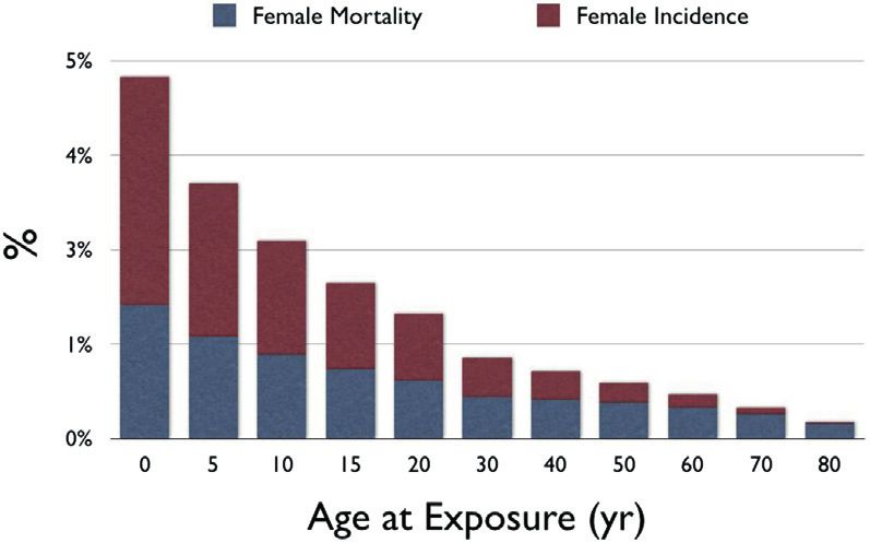
2   Hirshfeld Jr. et al.                                                                                                                             JACC VOL.      -, NO. -, 2018
    Radiation Safety ECD Part 1: Physics and Biology                                                                                                                  -, 2018:-–-

    ACC Task Force            James L. Januzzi, JR, MD, FACC                                              Pamela Bowe Morris, MD, FACC
    on Expert                 Luis C. Afonso, MBBS, FACC                                                  Robert N. Piana, MD, FACC
    Consensus                 Brendan Everett, MD, FACC                                                   Karol E. Watson, MD, FACC
    Decision                  Adrian F. Hernandez, MD, MHS, FACC                                          Barbara S. Wiggins, PHARMD, AACC
    Pathways**                William Hucker, MD, PHD
                              Hani Jneid, MD, FACC
                                                                                                          **Formerly named ACC Task Force on Clinical Expert Consensus
                              Dharam Kumbhani, MD, SM, FACC
                                                                                                          Documents.
                              Joseph Edward Marine, MD, FACC

    TABLE OF CONTENTS

    ABSTRACT . . . . . . . . . . . . . . . . . . . . . . . . . . . . . . . . . . . . . . -         5.1. Mechanism of Radiation-Induced
                                                                                                        Biological Effects . . . . . . . . . . . . . . . . . . . . . . . . . . .         -

    PREAMBLE . . . . . . . . . . . . . . . . . . . . . . . . . . . . . . . . . . . . . . -        5.2. Types of Radiation-Induced Health Effects . . . . . .                             -

                                                                                                         5.2.1. Tissue Reactions (Formerly Called
     1. INTRODUCTION . . . . . . . . . . . . . . . . . . . . . . . . . . . . . . . -                            Deterministic Effects) . . . . . . . . . . . . . . . . . . .             -

                                                                                                         5.2.2. Stochastic Effects: Cancer . . . . . . . . . . . . . . .                 -
    2. PURPOSE . . . . . . . . . . . . . . . . . . . . . . . . . . . . . . . . . . . . . -               5.2.3. Stochastic Effects: Heritable Effects in
        2.1. Document Purpose . . . . . . . . . . . . . . . . . . . . . . . . . .          -
                                                                                                                Offspring . . . . . . . . . . . . . . . . . . . . . . . . . . . . .      -

        2.2. The Radiation Safety Issue . . . . . . . . . . . . . . . . . . .              -      5.3. Tissue Reactions: Dose-Effect Relationships . . . . .                             -

                                                                                                         5.3.1. Skin Injury . . . . . . . . . . . . . . . . . . . . . . . . . . .        -
        2.3. The Need for Physician Radiation
             Safety Education . . . . . . . . . . . . . . . . . . . . . . . . . . . .      -             5.3.2. Bone Injury . . . . . . . . . . . . . . . . . . . . . . . . . . .        -

                                                                                                         5.3.3. Cataracts . . . . . . . . . . . . . . . . . . . . . . . . . . . . .      -
       2.4. Appropriateness of Medical Radiation . . . . . . . . . .                       -
                                                                                                         5.3.4. Tissue Reactions: Managing Skin Injuries . .                             -

    3. CURRENT TRENDS IN AND CONSEQUENCES OF                                                      5.4. Stochastic Effects: Radiation-Induced Cancer . . . .                              -
       PATIENT AND MEDICAL PERSONNEL RADIATION                                                           5.4.1. Stochastic Effects: Attribution Challenges . .                           -
       EXPOSURE FROM CARDIOVASCULAR
                                                                                                         5.4.2. Stochastic Effects: Risk Metrics . . . . . . . . . .                     -
       PROCEDURES . . . . . . . . . . . . . . . . . . . . . . . . . . . . . . . . . -
                                                                                                         5.4.3. Stochastic Risk: Dose-Risk Relationships . . .                           -

                                                                                                         5.4.4. Incremental Cancer Risk Attributable to
    4. THE MANY MEASURES OF RADIATION . . . . . . . . . . . -
                                                                                                                Radiation Exposure for Occupationally
        4.1. Radiation Exposure and Dose Metrics . . . . . . . . . .                       -                    Exposed Healthcare Workers . . . . . . . . . . . .                       -

        4.2. Challenges in Relating Radiation Exposure and
                                                                                               REFERENCES . . . . . . . . . . . . . . . . . . . . . . . . . . . . . . . . . . . -
             Dose to Risk of Detrimental Effects . . . . . . . . . . . .                   -

        4.3. Types of Ionizing Radiation Used in                                               APPENDIX A
             Medical Imaging . . . . . . . . . . . . . . . . . . . . . . . . . . . .       -
                                                                                                  Author Relationships With Industry and Other Entities
               4.3.1. X-Rays and Gamma Rays . . . . . . . . . . . . . . .                  -
                                                                                                  (Relevant) . . . . . . . . . . . . . . . . . . . . . . . . . . . . . . . . . . . . .   -
              4.3.2. Positrons . . . . . . . . . . . . . . . . . . . . . . . . . . . . .   -

       4.4. Relationships Between Exposure and                                                 APPENDIX B
            Absorbed Dose . . . . . . . . . . . . . . . . . . . . . . . . . . . . .        -
                                                                                                  Peer Reviewer Information . . . . . . . . . . . . . . . . . . . . . . .                -
               4.4.1. Measures of Exposure From
                      External Beams . . . . . . . . . . . . . . . . . . . . . . .         -
                                                                                               APPENDIX C
              4.4.2. Exposure From Radionuclides . . . . . . . . . . .                     -
                                                                                                  Abbreviations . . . . . . . . . . . . . . . . . . . . . . . . . . . . . . . . . .      -
        4.5. Estimating Effective Dose . . . . . . . . . . . . . . . . . . . .             -

       4.6. Synopsis of Measures of Radiation Exposure                                         ABSTRACT
            and Dose . . . . . . . . . . . . . . . . . . . . . . . . . . . . . . . . . .   -

                                                                                               The stimulus to create this document was the recognition
    5. HOW RADIATION CAN HARM PEOPLE . . . . . . . . . . . -                                   that ionizing radiation-guided cardiovascular procedures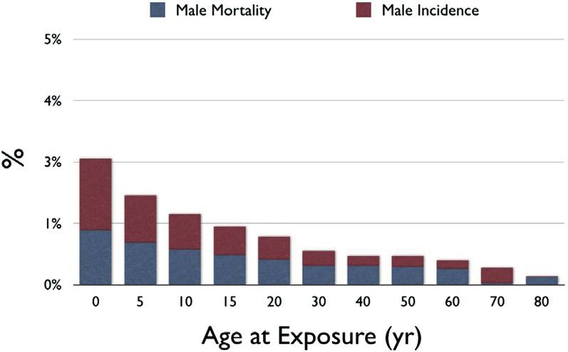
JACC VOL.   -, NO. -, 2018                                                                                                   Hirshfeld Jr. et al.   3
-, 2018:-–-                                                                                    Radiation Safety ECD Part 1: Physics and Biology

are being performed with increasing frequency, leading to              Thus, the reader should view the Expert Consensus
greater patient radiation exposure and, potentially, to                Document as the best attempt of the ACC and document
greater exposure for clinical personnel. Although the                  cosponsors to inform and guide clinical practice in areas
clinical benefit of these procedures is substantial, there is           where rigorous evidence may not yet be available or evi-
concern about the implications of medical radiation                    dence to date is not widely applied to clinical practice.
exposure. The American College of Cardiology leadership                  To avoid actual, potential, or perceived conflicts of
concluded that it is important to provide practitioners                interest that may arise as a result of industry relationships
with an educational resource that assembles and in-                    or personal interests among the writing committee, all
terprets the current radiation knowledge base relevant to              members of the writing committee, as well as peer
cardiovascular procedures. By applying this knowledge                  reviewers of the document, are asked to disclose all
base, cardiovascular practitioners will be able to select              current healthcare-related relationships, including those
procedures optimally, and minimize radiation exposure                  existing 12 months before initiation of the writing effort.
to patients and to clinical personnel.                                 The ACC Task Force on Expert Consensus Decision Path-
   Optimal Use of Ionizing Radiation in Cardiovascular                 ways (formerly the ACC Task Force on Clinical Expert
Imaging: Best Practices for Safety and Effectiveness is a              Consensus Documents) reviews these disclosures to
comprehensive overview of ionizing radiation use in                    determine which companies make products (on the mar-
cardiovascular procedures and is published online. To                  ket or in development) that pertain to the document un-
provide the most value to our members, we divided the                  der development. Based on this information, a writing
print version of this document into 2 focused parts. Part              committee is formed to include a majority of members
I: Radiation Physics and Radiation Biology addresses the               with no relevant relationships with industry (RWI), led by
issue of medical radiation exposure, the basics of radi-               a chair with no relevant RWI. Authors with relevant RWI
ation physics and dosimetry, and the basics of radiation               are not permitted to draft or vote on text or recommen-
biology and radiation-induced adverse effects. Part II:                dations pertaining to their RWI. RWI is reviewed on all
Radiological Equipment Operation, Dose-Sparing Meth-                   conference calls and updated as changes occur. Author
odologies,    Patient     and     Medical   Personnel     Protection   and peer reviewer RWI pertinent to this document
covers the basics of operation and radiation delivery for              are disclosed in Appendixes A and B, respectively.
the 3 cardiovascular imaging modalities (x-ray fluoros-                 Additionally, to ensure complete transparency, authors’
copy,     x-ray     computed          tomography,   and     nuclear    comprehensive     disclosure    information—including              RWI
scintigraphy).                                                         not pertinent to this document—is available online.
                                                                       Disclosure information for the ACC Task Force on
                                                                       Clinical Expert Consensus Documents is also available
PREAMBLE
                                                                       online, as is the ACC disclosure policy for document
                                                                       development.
This document has been developed as an Expert
                                                                                                        James L. Januzzi, MD, FACC
Consensus Document by the American College of Cardi-
                                                                         Chair, ACC Task Force on Expert Consensus Decision Pathways
ology (ACC) in collaboration with the American Society of
Nuclear Cardiology, Heart Rhythm Society, Mended
Hearts, North American Society for Cardiovascular Imag-
                                                                       1. INTRODUCTION
ing, Society for Cardiovascular Angiography and In-
terventions,       Society      for    Cardiovascular   Computed
                                                                       1.1. Document Development Process and Methodology
Tomography, and Society of Nuclear Medicine and Mo-
lecular Imaging. Expert Consensus Documents are inten-                 1.1.1. Writing Committee Organization
ded to inform practitioners, payers, and other interested              The work of the writing committee was supported
parties of the opinion of ACC and document cosponsors                  exclusively by the ACC without commercial support.
concerning evolving areas of clinical practice and/or                  Writing committee members volunteered their time to
technologies that are widely available or new to the                   this effort. Conference calls of the writing committee
practice community. Topics chosen for coverage by expert               were confidential and attended only by committee
consensus documents are so designed because the evi-                   members and ACC staff.
dence base, the experience with technology, and/or clin-                 The writing committee consisted of a broad range
ical    practice    are   not     considered    sufficiently    well    of members representing 9 societies and the following
developed to be evaluated by the formal ACC/American                   areas of expertise: interventional cardiology, general
Heart Association practice guidelines process. Often the               cardiology, pediatric cardiology, nuclear cardiology,
topic is the subject of considerable ongoing investigation.            nuclear medicine, electrophysiology, cardiac computed
4 Hirshfeld Jr. et al. JACC VOL. -, NO. -, 2018
Radiation Safety ECD Part 1: Physics and Biology -, 2018:-–-
tomography, cardiovascular imaging, and the con- ionizing radiation-based procedures. The goal is to
sumer patient perspective. Both a radiation safety enhance cardiovascular practitioners’ ability to select the
biologist and physicist were included on the writing optimal imaging technique for a given clinical circum-
committee. stance while balancing a technique’s risk and benefits,
This writing committee met the College’s disclosure and to apply that technique optimally to generate high-
requirements for RWI as described in the Preamble. quality diagnostic images of greatest clinical value and
minimal radiation exposure.
1.1.2. Document Development and Approval
The Writing Committee convened by conference call and 2.2. The Radiation Safety Issue
e-mail to finalize the document outline, develop the Cardiovascular procedures that employ ionizing radiation
initial draft, revise the draft per committee feedback, and have great value for diagnosis and treatment of properly
ultimately sign off on the document for external peer selected patients with known or suspected cardiovascular
review. All participating organizations participated in disease. However, ionizing radiation has molecular-level
peer review, resulting in 21 reviewers representing 299 detrimental effects on tissue, with potential for injury
comments. Comments were reviewed and addressed by both to patients and to exposed medical personnel. It is
the writing committee. A member of the ACC Task Force desirable to minimize radiation exposure both to patients
on Expert Consensus Decision Pathways served as lead and to medical personnel while achieving optimal bene-
reviewer to ensure that all comments were addressed fits to health. This principle requires that clinicians
adequately. Both the writing committee and the task force employ judicious use and conduct of radiation-employing
approved the final document to be sent to the ACC Clin- procedures.
ical Policy Approval Committee. This committee reviewed Currently, cardiovascular diagnostic and therapeutic
the document, including all peer review comments and procedures are a major source of patient exposure to
writing committee responses, and approved the docu- medical ionizing radiation, accounting for approximately
ment in November 2017. The Heart Rhythm Society, North 40% of total medical radiation exposure (exclusive of ra-
American Society for Cardiovascular Imaging, Society for diation oncology) (1,2). Among occupationally exposed
Cardiovascular Angiography and Interventions, and So- healthcare workers, interventional cardiologists and
ciety of Cardiovascular Computed Tomography endorsed clinical electrophysiologists are among the most highly
the document in January 2018. This document is consid- exposed, and there is potential for exposure to support
ered current until the Task Force on Expert Consensus personnel as well (3,4).
Decision Pathways revises or withdraws it from
publication. 2.3. The Need for Physician Radiation Safety Education
Cardiovascular specialists have a responsibility to:
2. PURPOSE
1. Apply knowledge of the radiation safety knowledge
2.1. Document Purpose base to make appropriate case selection choices.
This print-published document is part 1 of an abbreviated 2. Conduct radiation-assisted procedures optimally,
version of a larger, more comprehensive document that is minimizing exposure to patients and personnel.
published concurrently online. The online version con-
There is evidence that many cardiovascular special-
tains additional technical detail for readers who wish to
ists who order and conduct radiation-employing pro-
understand a topic in greater depth. The online published
cedures are not fully informed about the radiation
document, in addition to covering the topics in the 2
doses that accompany the procedure or the associated
print-published documents in greater depth, also covers
health implications for their patients and for them-
additional topics not covered in the print-published doc-
selves (5,6).
uments including: 1) dose reduction strategies; 2) oper-
ator education and certification; 3) quality assurance; and 2.4. Appropriateness of Medical Radiation
4) patient radiation tracking.
The balance between a procedure’s risk and benefit de-
This document covers radiation physics, radiation
termines its appropriateness. The hazard associated with
dosimetry and its determinants, and radiation harm. The
ionizing radiation is a potentially important determinant
document’s purpose is to provide a comprehensive in-
of a procedure’s risk-benefit relationship. Physicians who
formation source about ionizing radiation use in cardio-
either order or conduct such procedures need to:
vascular procedures. The writing group has assembled
this information to assist cardiovascular practitioners to 1. Know the magnitude of a patient’s risk associated with
provide optimal cardiovascular care when employing a procedure’s radiation exposure.JACC VOL. -, NO. -, 2018 Hirshfeld Jr. et al. 5
-, 2018:-–- Radiation Safety ECD Part 1: Physics and Biology
TABLE 1 Typical Effective Doses for Cardiac Procedures TABLE 1 Continued
Typical Effective Typical Effective
Modality Protocol Dose (mSv) Modality Protocol Dose (mSv)
82
MDCT Coronary CT angiography: 8–30 PET 50 mCi Rb rest/ 4
82
helical, no tube current modulation 50 mCi Rb stress
13
MDCT Coronary CT angiography: 6–20 PET 15 mCi N ammonia rest/ 2
13
helical, tube current modulation 15 mCi N ammonia stress
18
MDCT Coronary CT angiography: 0.5–7 PET 10 mCi F FDG 7
prospectively triggered axial 99m
Planar 30 mCi Tc-labeled erythrocytes 8
MDCT Coronary CT angiography:6 Hirshfeld Jr. et al. JACC VOL. -, NO. -, 2018
Radiation Safety ECD Part 1: Physics and Biology -, 2018:-–-
levels. In addition, increasing complexity of cardiovas- Potential Consequences of Patient and Medical
TABLE 2
cular interventional procedures requires longer fluoro- Personnel Radiation Exposure
scopic times and, accordingly, larger radiation exposures. Individual Patient Although many individual patients receive little or no
Natural background radiation averages 3.0 millisieverts medical radiation exposure, some receive lifetime
doses in excess of 100 mSv. Doses in excess of 100
(mSv) per person per year in the United States—equivalent mSv are associated with a detectable increased
to 150 posteroanterior chest radiographs (a poster- cancer risk
oanterior chest-x-ray dose is 0.02 mSv; combined post- Population Increased total exposure incurred by total population of
patients has the potential to increase the population
eroanterior and lateral is 0.06 mSv) (8). At the population
incidence of cancer and other radiation-related
level, between 1987 and 2006 estimated per person total disorders
medical radiation exposure grew from 0.6 mSv/year Occupationally Occupationally exposed physicians and support staff
(0.2 background) to 3.2 mSv/year (1.07 background) Exposed Workers may receive doses as large as 10 mSv per year over a
career that may span 30–40 years. The implications
(9). Consequently, currently, patients are receiving, on of this level of exposure at the level of the individual
average, more radiation from medical sources than from practitioner are uncertain.
natural background sources. 2006 is the latest year for
which compiled data are available. (The National Council
on Radiation Protection is currently compiling contem- which is the amount of energy released by the interac-
porary data—expected availability 2019—and it is likely tion of the radiation with a unit mass of air. Its unit of
that current average medical exposure will be found to measure is the gray (Gy) (J/kg). One Gy is the quantity of
have increased further.) The 2006 medical exposure is radiation that when interacting with 1 kg of air releases 1
equivalent to 160 posteroanterior chest x-rays per person J of energy.
per year. Risks associated with this exposure must be Absorbed Dose:
weighed in relation to the health status benefits achieved Absorbed radiation dose is a measure of the energy that
by these procedures. radiation deposits in an exposed tissue through in-
Physicians who are invasive cardiovascular procedure teractions with its molecular constituents. It differs from
operators are among the most highly occupationally exposure in that the radiation present at a given location
exposed healthcare workers. Measurements of interven- does not deposit all of its energy there. The fraction of its
tional cardiologist operator exposure using current energy that a given radiation exposure will deposit in the
equipment and protection practices demonstrate an exposed tissue varies with the type and energy of the
exposure range of 0.2 to >100 microsieverts ( m Sv) per radiation, the tissue composition, and the exposure
procedure with a per-procedure average of 8 to 10 m Sv duration.
(10). Thus, an active interventional cardiologist perform- Absorbed dose is a measure of the intensity of energy
ing 500 procedures/year employing current technology deposition (energy deposited per unit mass of tissue) and
may be expected to receive, in addition to background is expressed in Gy—joules of energy deposited per kilo-
exposure, a dose of as much as 10 mSv/year or, in a most gram of tissue.
extreme scenario, 300 mSv over a 30-year active profes- Equivalent Dose:
sional career. Different types of ionizing radiation cause varying
The potential implications of this level of medical ra- degrees of tissue injury for a given absorbed dose.
diation exposure are summarized in Table 2. Equivalent dose is a construct used to account for
differences in tissue injury caused by different radiation
4. THE MANY MEASURES OF RADIATION types. X-rays and gamma rays are the benchmarks
against which particle radiation types such as protons,
4.1. Radiation Exposure and Dose Metrics neutrons, and beta particles are compared. To adjust for
Ionizing radiation exposure and dosimetry are not easily this variability, each radiation type is assigned a radia-
characterized by simple metrics. For clarity, in this tion weighting factor by which the absorbed dose (in Gy)
document the interaction of radiation with tissue will be is multiplied to yield a measure of the expected tissue
characterized from the perspective of 5 inter-related injury caused by that dose. The unit of measure is the
frames of reference: exposure, absorbed dose, equiva- sievert (Sv), which is the absorbed dose in Gy multiplied
lent dose, effective dose, and injected dose. For this by the radiation weighting factor. All radiation types
document’s purpose, these metrics have specific mean- used in cardiovascular medicine have a radiation
ings as defined in the following text: weighting factor of 1.
Exposure: Effective Dose:
Radiation exposure refers to the presence of ionizing Effective dose is a measure of the estimated potential for
radiation at the location of the exposed tissue. The a stochastic biological effect (such as cancer induction)
typically used measure of radiation quantity is air kerma, caused by a particular absorbed radiation dose. In medicalJACC VOL. -, NO. -, 2018 Hirshfeld Jr. et al. 7
-, 2018:-–- Radiation Safety ECD Part 1: Physics and Biology
radiation exposures, absorbed dose is typically not uni- 4.3.1. X-Rays and Gamma Rays
form throughout all tissues. For x-ray imaging, dose is X-ray and gamma ray photons travel at the speed of light
concentrated in the body region being examined and and have no mass and no charge. Their electromagnetic
varies with depth from the beam entrance port. For nu- energy ranges from a few electron volts (eV) to millions of
clear imaging, dose is concentrated in the tissues that electron volts (MeV). X-rays used in x-ray fluoroscopy and
most avidly take up the tracer or are involved in its x-ray CT have a photon energy spectrum between 30 and
elimination. 140 keV. Thallium-201 releases photons primarily in the
Effective dose is the sum of the equivalent doses 68-80 keV range, similar to diagnostic x-rays.
received by each organ, with each organ equivalent dose Technetium-99m releases photons primarily in the 140
multiplied by a coefficient that reflects that organ’s keV range.
sensitivity to a stochastic effect. The unit of effective dose
is also the Sv. The Sv has the same unit as the Gy (J/kg). 4.3.2. Positrons
The connection between effective dose and absorbed Positrons are positively charged electrons. They have
dose is that an effective dose of 1 Sv (which may be mass and charge. When they travel through a medium,
concentrated in only a few organs) confers the same their electrostatic charge causes them to interact readily
estimated stochastic risk that would be caused by a uni- with electrons in the medium, leaving a trail of ionization.
form total absorbed body dose of 1 Gy of radiation that has Consequently, they have a very short mean free path in
a radiation weighting factor of 1. tissue (6 to 7 mm, with a maximum of 15.2 mm). Positrons
Different tissues have different sensitivities to are annihilated by colliding with an electron of a con-
radiation-induced effects. In the effective dose construct, stituent atom releasing two 511 keV gamma ray photons
each tissue is assigned a tissue-weighting factor that that travel in opposite directions. These high-energy
specifies its sensitivity to radiation effects. To calculate photons are minimally attenuated in tissue, and the ma-
the effective dose in Sv, each exposed tissue’s equivalent jority reach the imaging detector. Rubidium-82 is the
dose is multiplied by its tissue-weighting factor yielding most commonly used positron emitter for myocardial
that tissue’s contribution to the overall risk. The contri- perfusion imaging. Nitrogen-13 ammonia is used less
butions to risk from all exposed tissues are summed frequently for this purpose. Fluorine-18 deoxyglucose is
yielding total risk, which is expressed as the effective used in cardiology for metabolic imaging and to detect
dose in Sv. myocardial sarcoid and other inflammatory conditions.
Injected Dose:
Injected dose describes the quantity of radioactivity 4.4. Relationships Between Exposure and Absorbed Dose
injected into a patient for a nuclear scintigraphy study Medical radiation exposures occur in 2 ways:
(expressed in millicuries). The relationship between an
1. Exposure from an external radiation beam (x-ray fluo-
injected dose and the previously described dose param-
roscopy and x-ray CT)
eters is complex and is discussed in Part II: Radiological
2. Exposure from radioactive decay within the subject
Equipment Operation, Dose-Sparing Methodologies and
(nuclear scintigraphy)
Protection.
4.2. Challenges in Relating Radiation Exposure and Dose to 4.4.1. Measures of Exposure From External Beams
Risk of Detrimental Effects For external radiation beams, the absorbed dose is
Detrimental effects of radiation exposure typically pre- determined by the total incident exposure, the properties
sent weeks to years following exposure. In addition, of the incident radiation, and the volume of tissue
many detrimental effects, principally cancer, have a large exposed.
background frequency. This complicates the attribution Air kerma (“kinetic energy released in material”) is the
of an effect in a particular subject to prior radiation standard unit of measure for x-ray beam exposure. It is an
exposure. energy intensity measured in Gy. 1 Gy ¼ 1 J of energy
released per kilogram of absorbing material. The metric
4.3. Types of Ionizing Radiation Used in Medical Imaging “air kerma” is used because the measurement is made
Radiation in cardiovascular imaging consists of photons using air as the absorbing material.
with energy >10 kiloelectron volts (keV) (x-rays and Absorbed Dose From an External Beam
gamma rays) and positrons. The physical effect of such Radiation absorbed dose, as distinguished from exposure,
radiation is to eject electrons from atoms forming ions is an energy intensity, the concentration of radiation en-
and free radicals. This is the basis for the term “ionizing ergy actually deposited in the exposed tissue. Not all ra-
radiation.” The resulting ions and free radicals react with diation energy that impinges on a tissue is absorbed.
tissue molecules, damaging them. Some radiation (a variable quantity depending on both8 Hirshfeld Jr. et al. JACC VOL. -, NO. -, 2018
Radiation Safety ECD Part 1: Physics and Biology -, 2018:-–-
radiation and tissue characteristics) passes through the Tissue Weighting Factors Used to Calculate
TABLE 3
tissue without interacting with it, depositing no energy (it Effective Dose in Sieverts
is this radiation that contributes to image formation).
Tissue Weighting Factors
Radiation absorbed dose is also measured in Gy. 1 Gy ¼ 1 J Organs (ICRP103–2007)
of energy deposited per kilogram of irradiated tissue. Red bone marrow 0.12
External beam energy deposition in tissue is not uni- Colon 0.12
form. X-ray radiation is attenuated exponentially as it Lung 0.12
passes through tissue, decreasing by approximately a
Stomach 0.12
factor of 2 for each 5 cm of tissue. The incident beam air
Breasts 0.12
kerma is a good measure of dose at the body surface, but
Gonads 0.08
structures deep to the body surface receive smaller doses.
Bladder 0.04
X-Ray Fluoroscopy Kerma-Area Product: Incorporating
Liver 0.04
the Exposed Tissue Volume
Esophagus 0.04
The risk of radiation harm is related both to the intensity
of the radiation dose, and also to the quantity of tissue Thyroid 0.04
that receives the dose. Kerma-area product (KAP) is the Skin 0.01
product of the beam’s kerma and its cross-sectional area Bone surface 0.01
incorporating the volume of tissue irradiated. This Salivary glands 0.01
concept is particularly important in x-ray fluoroscopy, as Brain 0.01
imaging field sizes vary leading to very different KAPs. Remainder of body 0.12
X-Ray Computed Tomography Kerma-Length Product: Total 1.00
Incorporating the Exposed Tissue Volume
Adapted from the International Commission on Radiological Protection (ICRP) (12).
CT delivers radiation to a patient in a manner quite
different from that of projectional imaging or fluoroscopy.
The dose is distributed more uniformly around the abdominal viscera and upper neck. As these organs would
patient. receive smaller exposures, their contribution to the
The total dose delivered by a CT examination is the effective dose calculation would be smaller.
measured kerma multiplied by the axial length of the The International Commission on Radiation Protection
scan. A variety of dose metrics for x-ray CT are derived (ICRP) published the most recent organ sensitivity esti-
from this model. mates in 2007 in ICRP Publication 103 (12). These esti-
mates are listed in Table 3.
4.4.2. Exposure From Radionuclides
Unlike external beam exposures, radionuclide exposures 4.6. Synopsis of Measures of Radiation Exposure and Dose
come from radioactive decay within the subject. Exposure The existence of the many different measures of radia-
is determined by the activity administered, the tracer tion exposure and dose has the potential to cause
distribution, the tracer elimination rate, and the tracer’s confusion leading to misapplication of units of measure.
time-activity relationships. Table 4 contains a synopsis of the principal metrics
described in this section. It should be noted that, because
4.5. Estimating Effective Dose
effective dose in the table is based on gender- and age-
The effective dose construct assigns each organ/tissue a averaged tissue weighting factors, (not accounting for
weighting factor that reflects the tissue’s sensitivity to the fact that children and females are more sensitive), its
radiation-induced stochastic risk. The calculation of practical value is in comparing the effects of different
effective dose involves estimating each organ’s actual exposures rather than in estimating an individual’s sto-
equivalent dose (in Gy). Each organ dose is adjusted by chastic risk.
multiplying it by the organ’s tissue-weighting factor. The
organ sensitivity-adjusted individual organ doses are 5. HOW RADIATION CAN HARM PEOPLE
summed to yield a total effective dose (in Sv) (11).
For a chest exposure, absorbed dose is concentrated in 5.1. Mechanism of Radiation-Induced Biological Effects
the skin, mediastinal structures, lungs, breast, and Radiation-induced tissue injury is due to molecular
thoracic bone marrow. Doses to these organs contribute alteration caused by particles or photons that have suffi-
the largest components to the effective dose calculation. cient energy to induce ionization. Atoms ionized by ra-
Smaller quantities of scattered radiation expose the diation are frequently chemically unstable and transformJACC VOL. -, NO. -, 2018 Hirshfeld Jr. et al. 9
-, 2018:-–- Radiation Safety ECD Part 1: Physics and Biology
TABLE 4 Synopsis of Radiation Exposure and Dose Metrics
Metric Unit Utility
Absorbed Dose-Related Parameters:
Characterize Dose to Organ/Tissue or Whole Body
Absorbed dose Gy Amount of ionizing radiation energy deposited per unit mass of tissue. 1 Gy ¼ 1 Joule of energy deposited per
kg of tissue. This metric is a concentration of energy deposition—not the total quantity of energy
deposited.
Equivalent dose Sv Absorbed dose adjusted by a radiation weighting factor that adjusts for the specific tissue-injuring potential
of the particular radiation type. Photons (x-rays and gamma rays) have a weighting factor of 1. Electrons
also have a weighting factor of 1. Neutrons have larger weighting factors that vary with their energy
level. For medical imaging, because only photons and positrons are used, absorbed dose and equivalent
dose take the same value.
Effective dose mSv Calculated whole-body quantity used to roughly compare potential stochastic risks from different partial-
body exposures. It is expressed as the uniform whole-body dose that would confer the stochastic risk
equivalent to that caused by a regional exposure.
Modality-Specific Parameters
X-ray fluoroscopic air kerma (free-in-air) Gy Used to assess level of radiation present at a location. In x-ray fluoroscopy, cumulative air kerma at the
interventional reference point can be used to approximate beam entrance port skin dose. (For isocentric
C-arms, the reference point is located 15 cm from isocenter in the direction toward the x-ray source. This
point in space approximates the location of beam entry into the patient, but due to variation in table
height and tube angulation, is only an estimate of beam entrance port skin dose).
X-ray fluoroscopic Air-KAP, also referred to Gy,cm2 Used to assess the total quantity of radiation delivered by an external beam. It is the product of the
as dose-area product (DAP) cumulated amount of air kerma and the area of a radiographic or fluoroscopic field. KAP is often used as
the basis for estimating effective dose from a fluoroscopic procedure.
Computed tomographic dose index mGy Used to assess relative level of radiation applied during a CT imaging sequence. This metric is a concentration
(CTDIFDA, CTDI100, CTDIw, and CTDIvol) of energy deposition in the exposed volume. It is not a total deposited energy quantity, as it does not
incorporate the actual exposed volume (See DLP below). Different versions are used for varied purposes.
Computed tomographic dose-length mGy,cm Used to assess integrated amount of radiation applied along an axial length of a patient during a CT
product (DLP) examination. Can be used to estimate effective dose from the procedure.
Radionuclide injected dose mCi A measure of the quantity of radioactivity injected for a nuclear scintigraphy study. The relationship of
injected dose to other dose parameters is complex and includes the nature of the nuclide’s radiation, the
nuclide’s half-life, the distribution in the body, and the elimination kinetics.
CT ¼ computed tomography; CTDI ¼ computed tomographic dose index; KAP ¼ Kerma-Area Product.
into free radicals. A common example is ionization of cell’s ability to repair itself and maintain function. Tissue
water, which, upon interacting with an x-ray photon, reactions only become macroscopically evident if a
decomposes into a free electron, a proton, and a hydroxyl threshold radiation dose is exceeded, causing a sufficient
radical. The hydroxyl radical, because of its unpaired fraction of an exposed tissue’s cells to malfunction or
electron, is highly reactive and interacts avidly with bio- necrose. A dose below the threshold dose may cause un-
molecules (proteins or nucleic acids). Similarly, an x-ray apparent cellular injury but will not cause a detectable
photon can ionize an atom that is a constituent of a reaction (13).
biomolecule. Thus, a biomolecule can be altered by either Tissue reactions typically exhibit dose-related severity
reacting with a radiation-generated free radical or by and occur with a time delay (typically 4 to 8 weeks) be-
direct ionization from radiation. The resulting structural tween exposure and the appearance of tissue injury.
change can alter or degrade its function. Above the threshold dose, a greater dose causes more
extensive injury to a greater fraction of cells in proportion
5.2. Types of Radiation-Induced Health Effects to the dose.
Radiation-induced health effects are divided into 2 Skin injury is the most common tissue reaction
groups that differ in mechanism, the nature of effects, observed in cardiovascular imaging. It occurs almost
relationship to absorbed dose, and time between expo- exclusively from x-ray fluoroscopic exposures. Other
sure and manifestation. tissue reactions include cataract formation, bone
necrosis and, in the heart, damage to myocardium,
5.2.1. Tissue Reactions (Formerly Called Deterministic Effects) cardiac valves, and coronary arteries. In addition, if
Tissue reactions are caused by radiation-induced injury to a fetus incurs sufficient cellular injury at critical
structural and functional molecules in cells. Cell necrosis stages of organogenesis, development will be
will occur if the amount of molecular damage exceeds the impaired (14).10 Hirshfeld Jr. et al. JACC VOL. -, NO. -, 2018
Radiation Safety ECD Part 1: Physics and Biology -, 2018:-–-
Stochastic effects differ from tissue reactions in their
F I G U R E 1 Full Thickness Skin Necrosis Caused By a Large-Dose
X-Ray Fluoroscopic Procedure
dose relationship. Whereas tissue reactions exhibit dose-
related severity and have a definite dose threshold, sto-
chastic events, in contrast, have a probabilistic relation-
ship to dose. They are not known to have a dose threshold
and do not have a quantitative dose-related severity.
Radiation-induced cancer either does or does not occur
(or may not present within the subject’s lifetime). A single
critically located DNA damage event can create an onco-
gene (15). This is the theoretical basis for the concept that
there is no threshold dose below which stochastic risk is
zero (16).
5.2.3. Stochastic Effects: Heritable Effects in Offspring
Theoretically, radiation injury to DNA in germ cells could
cause a clinically important mutation that would not
affect the exposed individual, but would be transmitted
to that individual’s offspring. Such effects have been
demonstrated in animal models but have not been
observed in humans with statistical significance (17).
5.3. Tissue Reactions: Dose-Effect Relationships
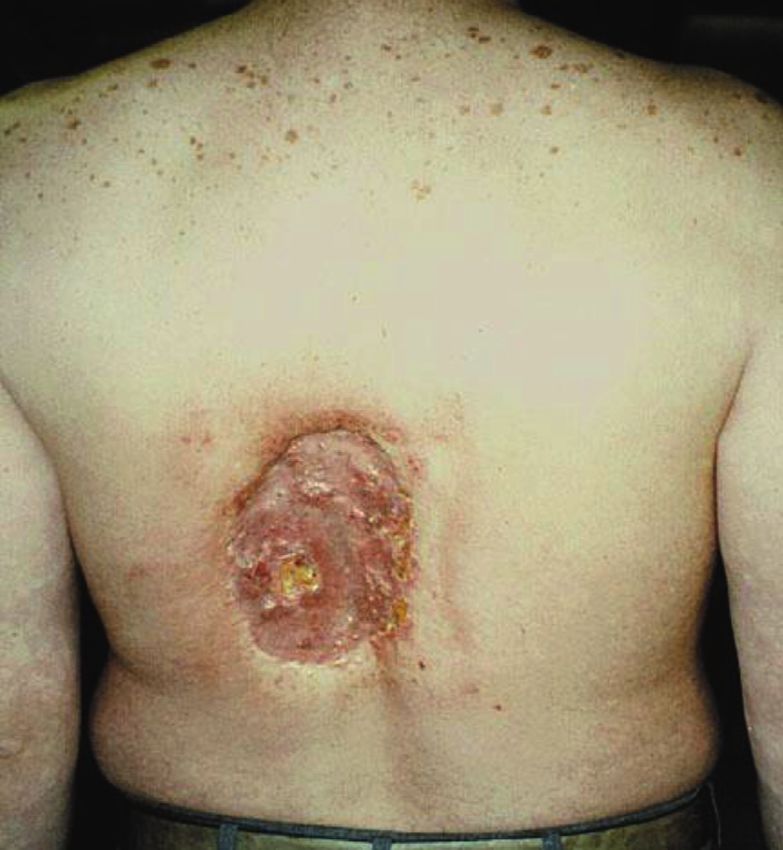
An example of full thickness skin necrosis (underlying muscle and fat 5.3.1. Skin Injury
are exposed) caused by a large-dose x-ray fluoroscopic procedure (90
The most common radiation-induced tissue reaction is
minutes of fluoroscopy time). Note the rectangular area of skin
discoloration surrounding the area of skin necrosis. The injury is on the skin injury at the beam entrance port (typically on the
left side of the subject’s back indicating that the exposure was con- patient’s back) following an x-ray fluoroscopic exami-
ducted in the right anterior oblique projection (17). (This image is nation. Skin entrance port injuries are rectangular,
available on the U.S. Food and Drug Administration Web site and is in reflecting the beam shape. These injuries vary in severity
the public domain.)
from erythema to desquamation to ulceration and
necrosis.
Skin injury typically appears 4 to 8 weeks following the
exposure. In extreme cases, the ulceration can become
5.2.2. Stochastic Effects: Cancer confluent and full thickness necrosis of skin may develop,
Stochastic effects are caused by radiation-induced dam- exposing underlying fat, muscle, and even bone (Figure 1).
age to a cell’s genetic material that reprograms the The skin injury threshold dose is variable, as is the
damaged cell’s deoxyribonucleic acid (DNA) into relationship between dose and injury severity. A proced-
dysfunctional operation. The principal stochastic event of ure’s cumulative air kerma can be used to estimate a pa-
clinical importance is radiation-induced cancer. tient’s skin injury risk.
TABLE 5 Radiation-Induced Skin Injuries—Relationship of Severity to Dose
Skin Reaction
Single Exposure
Dose Range (Gy) 0–2 Weeks 2–8 Weeks 8–40 Weeks Long-Term (>40 weeks)
0–2 No observable effects
2–5 Transient erythema Possible epilation Recovery of hair loss Complete healing
5–10 Transient erythema Erythema epilation Recovery or permanent hair loss At higher doses dermal atrophy or
induration
10–15 Transient erythema Epilation, possible Prolonged erythema, permanent Dermal atrophy or induration
desquamation hair loss
>15 Transient erythema, after very Epilation, moist Dermal atrophy, secondary Dermal atrophy, possible late skin
high doses ulceration desquamation ulceration, necrosis breakdown, ulceration, and necrosis of
subcutaneous tissues
Adapted from Balter et al. (13).JACC VOL. -, NO. -, 2018 Hirshfeld Jr. et al. 11
-, 2018:-–- Radiation Safety ECD Part 1: Physics and Biology
General guideline values for the ranges of threshold injury potential. The 2011 ACCF/AHA/SCAI Guideline for
values for a single first-time exposure for absorbed doses Percutaneous Coronary Intervention state that it is a
associated with degrees of skin injury severity are tabu- Class I recommendation for all patients who receive an
lated in Table 5. Injury thresholds for a subsequent air kerma at the interventional reference point >5 Gy to
exposure are lower. be counseled about the possibility of a skin injury and
Fluoroscopic entrance skin doses vary greatly because instructed how to react to the earliest signs should they
of variations in procedure complexity and duration and occur (19).
variations in patient radiological characteristics. Skin
dose is strongly affected by the patient’s characteristics 5.4. Stochastic Effects: Radiation-Induced Cancer
and procedural techniques. Body habitus is the most Note: Considerable additional detail for this section is
important patient characteristic. Larger patients require a provided in the online version of this document.
greater skin entrance port dose. Dose is also determined Radiation-induced cancer is potentially the most
by equipment calibration and imaging protocol settings. important consequence of medical radiation exposure. It
The prototypical patient at risk for a skin injury is an is an important determinant of a cardiovascular proced-
obese diabetic who has undergone 1 or more long- ure’s risk-benefit relationship and an occupational hazard
duration procedures within the past several months. to healthcare workers who work in a radiation
environment.
5.3.2. Bone Injury
In addition to skin injury, on occasion, incident radia- 5.4.1. Stochastic Effects: Attribution Challenges
tion can cause necrosis of superficial bones such as ribs. It is difficult to attribute a particular cancer to medical
Although the dose to bone needed to cause osteonec- radiation exposure. The large background cancer preva-
rosis is greater than the dose that causes skin necrosis, lence (the lifetime risk of developing cancer is roughly
bone’s high calcium content imparts a greater capacity 46% (16) and the risk of developing fatal cancer is about
to absorb x-ray photons, causing a greater absorbed 23%) and the latent period (2 years to decades) between
dose to bone. exposure and presentation present challenges to efforts
to construct evidence-based models that relate dose to
5.3.3. Cataracts
risk.
The single dose threshold that will cause vision-impairing
Population-based studies have demonstrated a statis-
cataracts in humans is not well characterized but is
tical association between leukemia and other childhood
believed to be on the order of 500 mGy with a minimum
cancers in children exposed to large medical radiation
latency of approximately 1 year (18). Cataracts are also
doses (20,21). Pearce et al. (21) found a 3.18-fold increase
increasingly being observed in physician operators with
in incidence of leukemia in a large cohort of children
long career experience. This area is currently a subject of
exposed to a mean dose of 51 mGy from CT scanning. In a
ongoing study.
cohort of 674 children who underwent cardiac catheteri-
zation with a mean follow-up of 28.6 years (12,978
5.3.4. Tissue Reactions: Managing Skin Injuries
patient-years), Modan et al. (20) found a 4.75 times
Less-severe degrees of skin injury have the potential to
increased risk of malignancies, with a 6.3 times increase
heal if managed with good supportive dermatological
in lymphomas, and a 4.9 times increased risk of
care.
melanoma.
The cornerstone of optimizing the outcome of a skin
injury is mechanical protection of the affected skin. X-ray 5.4.2. Stochastic Effects: Risk Metrics
injured skin is fragile. Mechanical trauma to the skin can
At the population level, stochastic risk can be quantified
aggravate the injury. Dressings and other mechanisms
as an increased cancer incidence in an exposed popula-
that help the patient avoid applying pressure or friction to
tion compared with the background incidence in a com-
the affected area is important.
parable unexposed population. This risk is measured
Early recognition of a radiation-induced skin injury is
using by 2 related but different metrics:
essential to initiate protection and early treatment. The
inherent delay of weeks between exposure and the 1. Excess relative risk. The rate of disease in an exposed
initial signs of skin injury may interfere with recognition population divided by the rate of disease in an unex-
of the cause, delaying appropriate treatment. The best posed population minus 1.0.
strategy to facilitate prompt recognition is to warn the a. Excess relative risk is a ratio derived from the disease
patient, family, and primary care physician of the skin incidence in exposed and unexposed populations.12 Hirshfeld Jr. et al. JACC VOL. -, NO. -, 2018
Radiation Safety ECD Part 1: Physics and Biology -, 2018:-–-
2. Excess absolute risk. The rate of disease in an exposed 3. Dose-response risk for solid organ cancer correlates
population minus the rate of disease in an unexposed loosely with the organ’s intrinsic mitotic activity. The
population. most radiation-sensitive solid organs are: lung, female
breast, colon, bladder, and thyroid.
Excess absolute risk is an incidence.
4. Hematopoietic tissues have a higher dose sensitivity
and a shorter latent period for leukemia induction than
5.4.3. Stochastic Risk: Dose-Risk Relationships
solid organs have for primary cancers.
Understanding of dose-risk relationships in humans is 5. Women have greater risk and a steeper dose-risk rela-
derived from epidemiological studies of exposed human tionship than men. Some, but not all, of this difference
populations. These studies have clearly identified a
is attributable to breast sensitivity.
dose-related risk for cancers including both leukemias
6. Risk and dose-risk relationships have a strong rela-
and solid tumors. The LSS (Life Span Study), conducted
tionship with age, with subjects younger than 30 years
by the Radiation Effects Research Foundation in resi-
of age having greater dose sensitivity (20). Beyond
dents of Hiroshima and Nagasaki, provides some of the age 30 years, dose sensitivity is less strongly age-
best quantitative data relating dose to future cancer related (26).
risk (22). 7. The length of the latent period for clinical presentation
Stochastic Risk: Qualitative Dose-Risk Relationships of an induced cancer decreases the importance of
Most models derived from epidemiological data find a
radiation-related risk for elderly patients who have
linear relationship between dose and increased future
limited natural life expectancies.
cancer risk, with no dose threshold below which there is
no risk. This is the basis of the “linear-no threshold” Stochastic Risk: Quantitative Dose-Risk Relationships
theory, which is the basis for the concept that radiation The quantitative relationship between radiation expo-
exposure should always be minimized (ALARA: “As Low sure and increased cancer risk has implications both for
As Reasonably Achievable”) (16). a patient undergoing a medical procedure and for
Children and young adults are more sensitive to ra- occupationally-exposed healthcare workers.
diation and, accordingly, for a given exposure have a
greater risk of radiation-induced cancer than the Background Cancer Risk in the Overall Population
elderly. Children born with congenital heart disease are An unexposed subject’s lifetime risk of developing solid
at greater risk, compared with other children, for cancer or leukemia is approximately 46% and lifetime risk
increased radiation exposure given their ongoing need of cancer mortality is approximately 23% (16).
for cardiac catheterization and other radiation-based Incremental Cancer Risk Attributable to Patient Medical
procedures. In addition, because radiation-induced Radiation Exposure
cancer has a latent period for induction, young people The BEIR VII models calculate coefficients that estimate
are more likely to live long enough for a stochastic the excess relative risk and excess absolute risk per Sv of
event to present. exposure. Because subject age and gender are important
The Committee to Assess Health Risks from Exposure risk determinants, different age ranges and genders have
to Low Levels of Ionizing Radiation of the National different coefficients (larger coefficients for younger
Research Council has examined a number of statistical subjects and females).
models that relate incremental cancer risk to absorbed The lifetime attributable risk for cancer incidence and
radiation dose for individual solid organ cancers and mortality is the percent of exposed patients who are
leukemia. These models also incorporate important pa- projected to develop a cancer attributable to an expo-
tient characteristics including age and gender. The sure. Figures 2 and 3 display the model-predicted inci-
models were published in the 2006 report, Biological Ef- dence and mortality estimates for a whole-body100 mGy
fects of Ionizing Radiation (BEIR) VII (16). (100 mSv) exposure (a moderately large, but plausible,
The models have several common features that are of medical exposure dose). The impact of gender and age at
pragmatic importance. exposure is highly evident. Children age 15 years and
1. Risk has a graded relationship to total dose. younger are projected to have incremental incidence
2. Excess cancer incidence is statistically detectable in rates in the range of 2% for males and 4% for females
population studies at a dose of 100 mSv in adults and in (27). In older patient groups, the predicted incremental
smaller doses in children (23–25). rates are substantially smaller, but not negligible, withJACC VOL. -, NO. -, 2018 Hirshfeld Jr. et al. 13
-, 2018:-–- Radiation Safety ECD Part 1: Physics and Biology
smaller gender differences than in the pediatric age
F I G U R E 2 Estimated Cancer Incidence and Mortality for Females
range. Attributable to a 100-mGy Radiation Exposure as a Function of Age
These data are displayed graphically in Figures 2 and 3.
5.4.4. Incremental Cancer Risk Attributable to Radiation
Exposure for Occupationally Exposed Healthcare Workers
Occupationally exposed healthcare workers typically
incur very small doses on a daily basis that can accumu-
late over time into a significant exposure. Healthcare
workers in x-ray environments employ protective gar-
ments. Consequently, their exposures are heterogeneous
for different body parts. Healthcare workers in nuclear
cardiology incur exposure when handling radioactive
materials and are at risk of exposure from radiopharma-
ceutical spills or accidents. Stacked bar graph depicts the lifetime attributable risk for cancer
There are few observational human data that assess incidence and mortality for women attributable to a 100-mGy total
body (100 mSv) exposure as a function of age at exposure. Note the
cancer risk from long-term daily small exposures. Most of
strong relationship between age at exposure and risk. Adapted from
the available data comes from studies of nuclear plant
BEIR VII (12).
operators (28). These data have not identified an
increased cancer incidence in this cohort of occupation-
ally exposed workers.
Applying the BEIR VII models to dose levels and Implications of Fetal Radiation Exposure
occupational exposure durations that are typical for The human embryo and fetus are more sensitive to radi-
healthcare workers working in a medical radiation envi- ation effects than adults. This phenomenon has implica-
ronment calculates a small but measurable increase in tions for the impact of radiation exposure both to patients
future cancer risk. Example findings from 2 extremes of and to occupationally exposed workers who are known to
exposure include: be or who may be pregnant.
1. A very low dose (1 mGy/year) throughout life such as Knowledge of the effects of ionizing radiation on the
one might experience living at high altitude. This human embryo and developing fetus is derived from
would result in a lifetime incremental exposure of 80
mGy that would confer an incremental cancer mortality
risk of 0.33% in males and 0.50% in females. F I G U R E 3 Estimated Cancer Incidence and Mortality for Males
2. An extreme occupational dose for a person working Attributable to a 100-mGy Radiation Exposure as a Function of
in an x-ray fluoroscopic environment for his/her Age
entire adult working life (16 mSv/year for 40 years ¼
640 mSv). This would confer an incremental cancer
mortality risk of 1.70% in males and 2.39% in
females.
Implications of Occupational Exposure in
Healthcare Workers
The ALARA principle applies both to patients undergoing
radiation-employing procedures and healthcare workers
who conduct them.
Based on the risk estimates, the current recommended
exposure limits for occupationally exposed workers pub-
lished by the ICRP are in Table 6 (12).
Stacked bar graph depicts the lifetime attributable risk for cancer
It should be noted that the ICRP standards (Europe)
incidence and mortality for males attributable to a total body 100-mGy
are more stringent than the National Council on Radia- (100 mSv) exposure as a function of age at exposure. Note the strong
tion Protection standards (United States). Historically, relationship between age at exposure and risk. Note also the smaller
standards have become more stringent over time. incidence and mortality rates in men compared with women at each
Consequently, the most stringent standards are age range. Adapted from BEIR VII (12).
presented.14 Hirshfeld Jr. et al. JACC VOL. -, NO. -, 2018
Radiation Safety ECD Part 1: Physics and Biology -, 2018:-–-
Recommended Exposure Limits for
organogenesis phase has the potential to cause fetal
TABLE 6
Occupationally Exposed Workers malformations. Later exposure during the fetogenesis
Total body 20 mSv/yr averaged over defined periods of 5 yrs with
phase can cause growth retardation and impaired neuro-
no individual annual exposure to exceed 50 mSv. logical development, and can potentially increase the
Lens of the eye 100 mSv/5 yrs (20 mSv/yr) fetus’ future cancer risk.
Skin 500 mSv/yr In considering these risks, it is important to link the
Hands and feet 500 mSv/yr risk to threshold radiation doses. This knowledge base has
been summarized by the Centers for Disease Control and
Adapted from the International Commission on Radiological Protection (12).
Prevention (35). In this document, dose ranges are
expressed in Gy rather than in Sv, as the Sv construct is
multiple sources, including the Hiroshima, Nagasaki, and not applicable to embryos and fetuses.
Chernobyl experiences as well as radiation of pregnant The increased childhood cancer risk caused by fetal
experimental animals (29). Detrimental radiation effects radiation exposure is less well characterized, and
include embryonic death, fetal malformations, impaired whether fetal radiation exposure might confer a life-
fetal development (particularly neurological), and long increased cancer risk is not known. Estimates of
increased risk of future cancer (16,30,31). The type of childhood cancer risk are summarized in Table 8. The
event and the dose-risk relationship for them is variable available data indicate minimal detectable childhood
throughout the stages of pregnancy and is summarized in risk at fetal doses 50 mGy.
The principal risk of radiation exposure to the early A general synthesis of the fetal radiation dose data
embryo during the blastogenesis phase of development is indicates that fetal doses 15 points, depending n Reduction in IQ dose§
neurological and on dose) possible, depending
motor deficiencies n Incidence of severe on dose
n Growth retardation mental retardation n Severe mental retar-
likely >20%, depending on dation possible,
dose depending on dose
n Incidence of major mal- n Incidence of major
formations will probably malformations may
increase increase
Note: This table is intended only as a guide. The indicated doses and times post conception are approximations. *Acute dose: dose delivered in a short time (usually minutes).
Fractionated or chronic doses: doses delivered over time. For fractionated or chronic doses the health effects to the fetus may differ from what is depicted here. †Both the gray (Gy)
and the rad are units of absorbed dose and reflect the amount of energy deposited into a mass of tissue (1 Gy ¼ 100 rads). In this document, the absorbed dose is that dose received by
the entire fetus (whole-body fetal dose). The referenced absorbed dose levels in this document are assumed to be from beta, gamma, or x radiation. Neutron or proton radiation
produces many of the health effects described herein at lower absorbed dose levels. ‡A fetal dose of 1 Gy (100 rads) will likely kill 50% of the embryos. The dose necessary to kill
100% of human embryos or fetuses before 18 weeks’ gestation is about 5 Gy (500 rads). §For adults, the LD50/60 (the dose necessary to kill 50% of the exposed population in 60
days) is about 3 to 5 Gy (300 to 500 rads) and the LD100 (the dose necessary to kill 100% of the exposed population) is around 10 Gy (1,000 rads). Reproduced with permission from
the Centers for Disease Control and Prevention (35).You can also read