Protective Mechanisms Against DNA Replication Stress in the Nervous System - MDPI
←
→
Page content transcription
If your browser does not render page correctly, please read the page content below
G C A T
T A C G
G C A T
           genes
Review
Protective Mechanisms Against DNA Replication
Stress in the Nervous System
Clara Forrer Charlier and Rodrigo A. P. Martins *
 Programa de Biologia Celular e do Desenvolvimento, Instituto de Ciências Biomédicas,
 Universidade Federal do Rio de Janeiro (UFRJ), Rio de Janeiro 21941-902, Brazil; clara.forrer.charlier@gmail.com
 * Correspondence: rodrigo.martins@icb.ufrj.br

 Received: 27 May 2020; Accepted: 25 June 2020; Published: 30 June 2020

 Abstract: The precise replication of DNA and the successful segregation of chromosomes are essential
 for the faithful transmission of genetic information during the cell cycle. Alterations in the dynamics
 of genome replication, also referred to as DNA replication stress, may lead to DNA damage and,
 consequently, mutations and chromosomal rearrangements. Extensive research has revealed that
 DNA replication stress drives genome instability during tumorigenesis. Over decades, genetic
 studies of inherited syndromes have established a connection between the mutations in genes
 required for proper DNA repair/DNA damage responses and neurological diseases. It is becoming
 clear that both the prevention and the responses to replication stress are particularly important
 for nervous system development and function. The accurate regulation of cell proliferation is
 key for the expansion of progenitor pools during central nervous system (CNS) development,
 adult neurogenesis, and regeneration. Moreover, DNA replication stress in glial cells regulates CNS
 tumorigenesis and plays a role in neurodegenerative diseases such as ataxia telangiectasia (A-T).
 Here, we review how replication stress generation and replication stress response (RSR) contribute
 to the CNS development, homeostasis, and disease. Both cell-autonomous mechanisms, as well
 as the evidence of RSR-mediated alterations of the cellular microenvironment in the nervous system,
 were discussed.

 Keywords: genome stability; neurologic disease; DNA damage; neurodevelopment; neurodegeneration;
 ATR; CNS; replication stress; DDR

1. Relevance of Genomic Stability for the Nervous System
      The maintenance of genomic stability is crucial for human health. In proliferating cells, precise
DNA replication and the successful segregation of chromosomes are essential for the accurate
transmission of genetic information to daughter cells. Not only cell-exogenous genotoxic agents can be
deleterious to genomic maintenance, but the duplication of the genome itself can create—in the wrong
circumstances—a burden that can impact the genomic integrity [1,2]. Alterations in the dynamics
of genome replication, also referred to as DNA replication stress, may lead to DNA damage and,
consequently, mutations and/or chromosomal rearrangements. Extensive research work has established
that replication stress is a source of genomic instability that may compromise the transmission of
genetic information [3,4]. Appropriate replication stress response (RSR) is relevant in various biological
contexts of cell proliferation—in particular, during development and in cancer [5–7]. In non-replicating
cells, a vast machinery of genome maintenance is required to prevent and repair DNA lesions that
frequently occur. Defective DNA repair pathways lead to genomic instability in postmitotic cells
and are associated with aging and various neurological diseases [8–10].
      Genetic studies of inherited syndromes have established a clear connection between mutations
in DNA damage response (DDR) genes and several human diseases [11]. The immune and nervous

Genes 2020, 11, 730; doi:10.3390/genes11070730                                       www.mdpi.com/journal/genes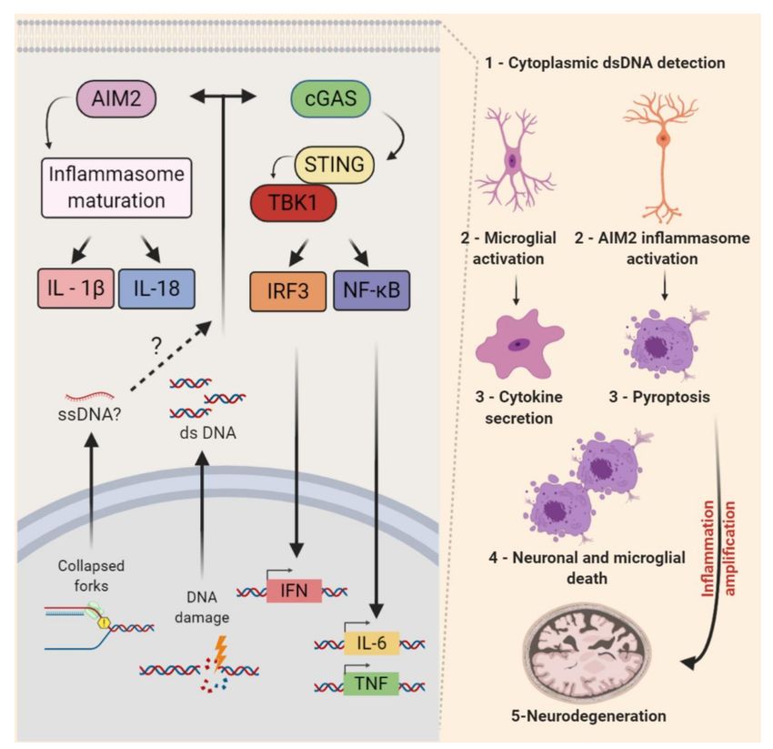
Genes 2020, 11, 730                                                                                  2 of 38

systems are particularly susceptible to defective DDR, and the central nervous system (CNS) is
severely affected when responses to threats to the genome are inadequate. Appropriate DDR is
critical for developmental processes, physiological homeostasis, and for the prevention of maladies
related to aging, including cancer and neurodegenerative diseases [12]. Ataxia-telangiectasia (A-T)
is the most classical example of a genome instability disorder that links defective DDR and CNS
diseases. Mutations in ataxia-telangiectasia-mutated (ATM) cause a severe syndrome characterized by
a hypersensitivity to ionizing radiation, neurodegeneration, and ataxia. Highlighting the importance
of genomic integrity, several other inherited syndromes that affect the CNS development and function
are caused by mutations in genes involved in the generation or in the responses to replication stress [3].
In addition, studies in animal models have confirmed the relevance of the mechanisms that protect
the nervous system against DNA replication stress [12].
      Replication stress may generate genomic instability in the developing nervous system,
and the importance of the replication stress response (RSR) pathways for the CNS formation
has been demonstrated by many studies [13–18]. In addition, evidence that the RSR alters the cellular
microenvironment and regulates immune responses has also accumulated rapidly [19,20]. Interestingly,
recent research indicates that this non-cell-autonomous arm of the RSR may also contribute to
the progression of neurodegenerative diseases [21].
      While other reviews have broadly discussed genomic stability in the nervous system [10,12,22],
here, we will focus on studies that have contributed to our current understanding about how replication
stress is generated across the diverse cell types in the CNS, what players mediate RSR, and what
the consequences are for CNS development and homeostasis when the RSR is defective.

2. Overview of DNA Replication Stress: How Cells Prevent and Respond
      DNA replication occurs during the S-phase of the cell cycle. Importantly, before the actual synthesis
of nascent DNA strands, key events must occur: origin licensing and replisomes formation [23].
Origin licensing takes place during late mitosis (M-phase) and the early G1 phase. Then, during
the transition between the G1 and S phases, licensed origins are activated, and replisomes are
formed. During the S-phase, origin firing occurs, and DNA polymerases initiate the incorporation
of deoxyribonucleoside triphosphates (dNTPs) complementary to the parental DNA molecule
as replication forks progress bidirectionally in opposite directions in thousands of chromosomal
sites. Importantly, in normal conditions, a major fraction of licensed replication origins is not activated
and remains dormant. In special conditions, such as DNA replication stress, these dormant origins
may be activated [24–26].
      DNA replication stress, also known as replicative stress, is defined as the slowing or stalling of
replication fork progression during the synthesis of DNA [27]. Continued replication stress may lead to
replication fork collapse and DNA damage that may cause mutations, amplifications, deletions, and/or
chromosomal rearrangements. DNA replication stress may be caused by both cell-endogenous or
cell-exogenous sources [2–4,7,28], and, importantly, DNA replication stress is now widely recognized
as a cancer hallmark [6].
      Obstacles to replication fork progression, limiting or unbalanced metabolic conditions, conflicts
between DNA replication and transcription machineries, and inappropriate origin firing due to
oncogene activation are among the best-characterized causes of replication stress [4]. Known obstacles
to replication fork progression include intrinsic characteristics of DNA sequences such as microsatellites,
minisatellites, and long terminal repeats. Complex structural arrangements of the DNA molecule
may also constitute a challenge (e.g., intramolecular triplex DNA, hairpins, and G-quadruplexes) to
replication fork movement. DNA lesions, such as interstrand crosslinks (ICL), apurinic/apyrimidinic
(AP) sites, and bulky adducts, may also slow or stall replication [2,29] (Figure 1a).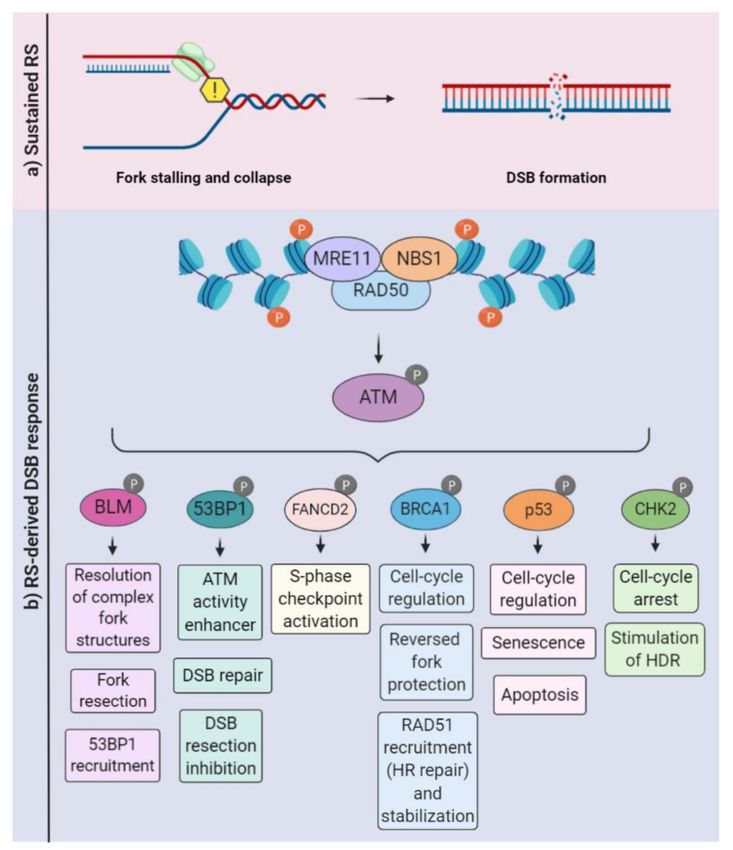
Genes 2020, 11, 730 3 of 38
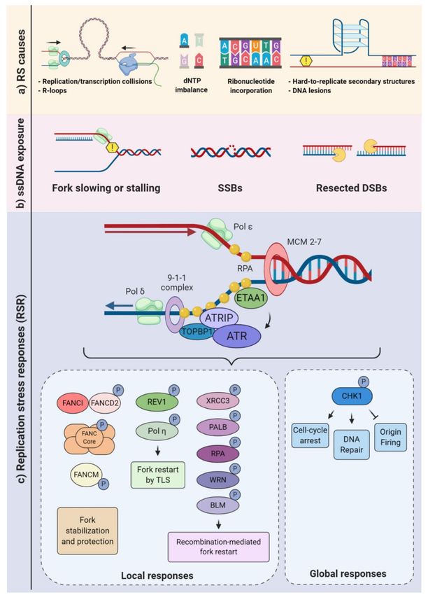
Figure 1. Replication stress causes and responses. (a) Different endogenous sources of replication
stress may either slow or block the progression of the replication forks. Transcription-replication
collisions (TRCs) and hard to replicate DNA are examples of obstacle challenges, and a decreased pool
of nucleotides (dNTPs) is a type of metabolic imbalance. Repair of misincorporated ribonucleotides may
also impair fork progression. (b) Replisome slowing or stalling may lead to single-stranded (ss)DNA
exposure, single-strand breaks (SSB), and fork collapse. Such constraints, as well as double-strand break
(DSB) resections, can result in ssDNA overhangs. (c) Replication protein A (RPA) binds to ssDNA,
triggering the recruitment of the ATR-interacting protein (ATRIP)-ataxia-telangiectasia and Rad3-related
kinase (ATR) complex, either through an interaction with DNA topoisomerase 2-binding protein 1
(TOPBP1) or Ewing’s tumor-associated antigen 1 (ETAA1). Once activated, ATR can phosphorylate
various substrates generating local “at the forks” responses (e.g., fork stabilization, protection,
and restart) and global responses that broadly affect the cell physiology, aimed at replication stress
mitigation and/or resolution (lower panels).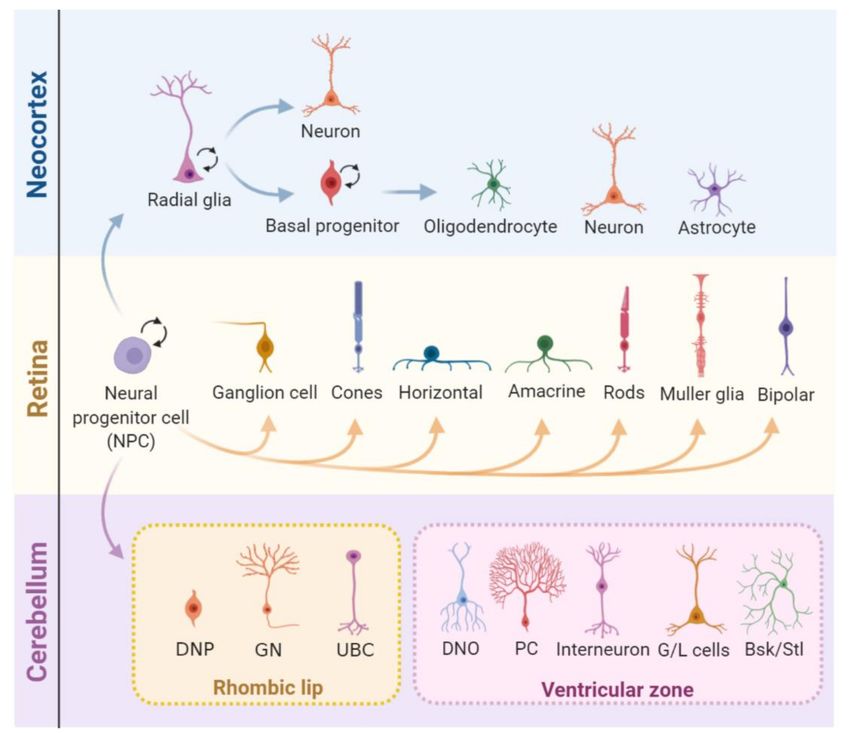
Genes 2020, 11, 730                                                                                 4 of 38

      Limiting or unbalanced metabolic conditions can also be a contributing factor in replication stress.
For example, an unbalanced availability of dNTP can greatly affect the fork progression [30]. During
the S-phase, the cell needs to tightly control the dNTP usage. Defective scavenging and/or de novo
production may deplete the dNTP pools and very rapidly impact the fork progression [31,32] (Figure 1a).
Ribonucleotide reductase (RNR) converts ribonucleotide diphosphate (NDP) to deoxyribonucleotide
diphosphate (dNDP), and the NDP kinase (NDPK) converts it to dNTP [33] Conversely, the rate of
degradation of the dNTPs may also be impacted by DNA replication dynamics. For example, the dNTP
hydrolase SAMHD1 (Sterile alpha motif [SAM] and histidine-aspartic [HD] domain containing
deoxynucleoside triphosphate triphosphohydrolase 1) is a major regulator of dNTP pools and, also,
plays an important role in the RSR, with relevant implications to human health. SAMHD1 mutations
were associated with cancer development and Aicardi-Goutières syndrome, a severe congenital
inflammatory disease [34–36]. In addition, deoxynucleoside kinases and 50 -deoxynucleotidases
contribute to regulate dNTP pools [33,37].
      When transcription and replication machineries encounter, transcription-replication collisions
(TRCs) occur. TRCs and the consequent topological stress can also be hindrances that physically impair
replisome progression. At the interface of transcription and replication, one structure of particular
relevance to replication stress generation is the R-loops. These nucleic acid structures are formed when
a RNA strand invades the double-strand (ds)DNA, forming a DNA:RNA hybrid and a nonpaired
single-stranded DNA (ssDNA) [38–41] (Figure 1a). In addition, R-loops may also occur when a RNA
strand invades a single-strand break (SSB) or a double-strand break (DSB), potentially impairing
the repair process [42–44]. Physiologically, such structures are thought to regulate gene transcription,
its termination, and gene silencing [45–47]. However, when the R-loop formation is deregulated,
it may stall or even collapse the replication fork [48–50]. Interestingly, TRCs can be exacerbated
by R-loops [51–53]. Increased fork pausing and higher genomic instability were associated with
head-on TRCs (when the machineries are moving towards one another) [54–56]. TRCs are especially
relevant for genome regions that are highly transcribed during the S-phase [52,57–59]. Importantly,
it has been suggested that TRCs are a relevant contributing factor for the occurrence of recurrent DNA
double-strand breaks (DSBs) following replicative stress in neural progenitor cells (NPC) [17].
      Finally, seminal studies demonstrated that inappropriate origin firing due to oncogene activation
directly disturbs DNA replication, being a relevant source of replication stress [60–63]. Several
mechanisms for oncogene-induced replication stress have been described. For example, oncogene
activation may directly interfere with nucleotide biosynthesis, depleting dNTP pools and ceasing fork
progression [64,65]. Defective origin firing and the induction of DNA re-replication are also among
the outcomes of replication stress caused by oncogenic stimulus [66,67].
      The replication stress response is broadly defined as a branch of the DNA damage
response that specifically reacts to DNA replication stress, embracing multiple signaling pathways
and the downstream cellular responses. As previously characterized for other subdivisions of DDR,
the cellular responses to replication stress are diverse and may be subdivided into local and global
responses. Local responses are the mechanisms that take place at the replication forks, whereas global
responses include pan-nuclear and cytoplasmic processes, as well as functional responses that regulate
the cellular microenvironment [19,68]. The stabilization of stalled forks, promotion of fork restarts,
and regulation of origin firing are examples of processes that directly regulate replication forks
or dormant origins. The activation of DNA repair mechanisms and checkpoints that inhibit cell
cycle progression, as well as cell death, senescence, and cytokine production pathways, are among
the well-characterized global responses to replication stress [3,4] (Figure 1).
      Upon fork slowing or stalling, one of the first molecular consequences is the exposure of ssDNA.
A junction of ssDNA and double-strand DNA (dsDNA) creates a platform for the recruitment
of various proteins. ssDNA tracts are coated by replication protein A (RPA) and other proteins
(e.g., TOPBP1, ETAA1, and 53BP1), leading to the activation of the ATR-ATRIP complex (composed
of the ataxia-telangiectasia and Rad3-related kinase and the ATR-interacting protein) (Figure 1c).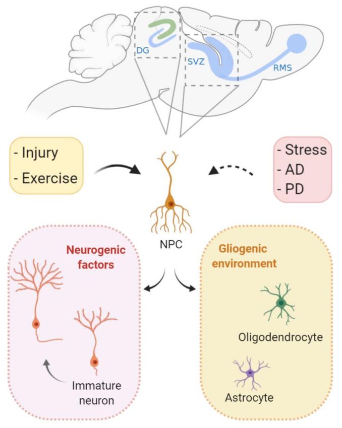
Genes 2020, 11, 730                                                                                               5 of 38

Then, the ATR kinase may phosphorylate more than 700 target proteins, activating multiple signaling
networks. Consequently, complex responses that range from cell cycle arrest, the regulation of
origin firing, replication fork stabilization, replication fork restart, and the control of dNTP availability
are elicited. Notably, ATR-mediated phosphorylation and the activation of checkpoint kinase 1 (CHK1)
are extremely relevant to regulate both local and global responses [69–72].
      Replication fork stabilization and fork restart, as well as fork reversal, contribute to prevent
the replication fork collapse, which is a known source of DNA double-strand breaks (DSBs), an extremely
cytotoxic lesion. The generation of DSBs triggers the activation of other signaling kinases, including
ataxia-telangiectasia-mutated (ATM) and DNA-PKcs (DNA-dependent protein kinase, catalytic
subunit). It is well-established that there is a considerable overlap in the activation of these kinases
following continued replication stress and the consequent DSBs (Figure 2). For example, depending on
the DNA repair pathway elicited, the resection of DSBs exposes ssDNA that fall under the purview
of ATR [11,73]. Classical and alternative DNA repair pathways, such as homologous recombination,
nonhomologous end joining (NHEJ), or break-induced replication repair, may be employed to allow
fork restart [74–76]. When repair is not possible, the mechanisms of damage tolerance may be activated
to restart the fork, such as the lesion bypass (where the replisome “skips” the lesion and resumes
replication downstream), template switch (a homologous recombination (HR)-mediated response that
involves fork regression and the use of the newly synthesized DNA strand as a template) and translesion
synthesis (TLS), in which specialized polymerases capable of bypassing the lesion are recruited [77–79].

      Figure 2. Ataxia-telangiectasia-mutated (ATM)-mediated signaling in the context of replication stress.
      (a) Sustained replication stress can induce fork collapse and DSBs, leading to the recruitment of the MRN
      (MRE11, RAD50, and NBS1) complex. (b) The MRN complex associates with the DSB, recruiting
      and activating ATM. Depending on specific scenarios (e.g., intensity and duration), ATM phosphorylates
      a variety of substrates and coordinates a variety of cellular responses, including DSB repair, the inhibition
      or stimulation of DNA resection, the activation of cell-cycle checkpoints, and cell death programs.
      HDR: homology-directed repair.
Genes 2020, 11, 730 6 of 38
There is a plethora of factors involved in the attenuation or repair and rescue of replication stress.
In addition to these autonomous cellular responses, it is now well-documented that replication stress
may also induce dramatic alterations in the cellular microenvironment. This non-cell-autonomous
paracrine arm of RSR occurs through the regulation of proinflammatory cytokines [19,80]. In fact, a series
of elegant studies provided evidence for replication stress-induced inflammation as a driver of neuronal
degeneration in models of ataxia telangiectasia. Studies about the immune response in At−/− -mice
revealed that the accumulation of cytoplasmic DNA in microglial cells triggered the release of neurotoxic
cytokines, leading to chronic neuroinflammation and neurodegeneration [81,82]. Understanding
the contributions of RSR to nervous system development, degeneration, and regeneration is of
fundamental biomedical importance. In the next section, we provide an overview of biological
contexts when cell proliferation and replication stress may occur in the nervous system physiology
and pathology, focusing on humans and mouse models.
3. Cell Proliferation in Nervous System Development, Homeostasis, and Diseases
The process of DNA replication is essential for cell proliferation, and the dynamics of cellular
renovation vary significantly in multicellular organisms [83]. While some tissues exhibit a high
rate of cell proliferation throughout life, in various CNS tissues, progenitors undergo a burst of cell
proliferation during development and have a very limited generation of new cells afterwards [84,85].
It is estimated that, during development, the human brain gives rise to 160 billion neuronal and glial
cells [83–86].
Developmental neurogenesis relies on the rapid expansion of neural progenitor cell (NPC)
pools [87]. Murine cortex neurogenesis occurs during the embryonic stages and is already finished at
birth. First, neural progenitor cells (NPCs) undergo symmetric divisions (generate two stem cells) that
expand the progenitor pool until embryonic day 10 (E10). Upon the start of neurogenesis, asymmetric
divisions of NPCs with astroglial features (also known as radial glia) initiate in the ventricular zone
(VZ) [88]. Depending on the environmental cues, these NPCs can either generate a postmitotic neuron
and a NPC or intermediate progenitors that locate to the subventricular zone (SVZ) and can further
divide into neurons, astrocytes, or oligodendrocytes [89]. Later, a switch from neurogenesis to gliogenesis
generates astrocytes and, after birth, oligodendrocytes [90–94]. The retina is another well-studied CNS
tissue that is also affected in human diseases caused by mutations in DDR genes [95,96]. In mice,
retinogenesis starts during embryonic development and extends until postnatal ages. Importantly,
all neuronal cell types and the Muller glial derive from a pool of multipotent retinal progenitor
cells [97,98], but retinal astrocytes derive from a distinct progenitor pool that migrates into the retina
through the optic nerve after birth [84,99]. Finally, different from the neocortex and the retina,
the cerebellum originates from two distinct populations of NPCs that occupy different germinative
areas: the ventricular zone (VZ) and the rhombic lip (RL). In mice, its development also expands from
embryonic stages to later postnatal developments [84,100,101] (Figure 3).
Even though cell proliferation is largely limited to development in mammal CNS, it can also
occur during adulthood. The two main sites of adult neurogenesis are the subventricular zone (SVZ)
and the dentate gyrus of the hippocampus [102,103]. The functions of these adult-born neurons are still
a highly contentious subject, and potential functions include memory acquisition and/or loss [104–106].
Nevertheless, despite the putative functions, the presence of proliferation into adulthood exposes
proliferating cells to the risk of replication stress and its possible deleterious consequences (Figure 4).
Replication stress is a driver of genomic alterations that are required for cellular transformation [6],
and, regardless of the cellular origin, unscheduled replication is a hallmark of tumorigenesis [107].
Glioblastoma, one of the most lethal human cancers, medulloblastoma, the most common pediatric
brain tumor, and the pediatric cancer retinoblastoma are among the most studied CNS tumors [108–111].
The progressions of these CNS tumors depends on unrestricted cell proliferation, and replication stress
response pathways are often deregulated in these diseases [112–114]. Absent or defective checkpoints
contribute to enhanced cell proliferation, the prevention of apoptosis, and other relevant transformationGenes 2020, 11, 730 7 of 38
processes. For example, it was proposed that replication stress is responsible for the upregulation of
DDR factors and, consequently, for the radiation resistance of glioma stem cells [115–117]. Recent
therapies that combine DNA-damaging agents and target replication stress mediators have been
intensively studied as a therapeutic strategy, including in CNS tumors [118–120].
Figure 3. Cell proliferation during the central nervous system (CNS) development. In the developing
neocortex, first, the neural progenitor cells (NPCs) undergo expansion and, later, shift identity to
radial glia (RG) cells). These can divide either symmetrically, expanding its pool, or asymmetrically,
generating either a combination of a radial glial cell and a neuron or a radial glial cell and a basal
progenitor (BP). BPs can further divide symmetrically into two BPs or asymmetrically into neurons or
glia (oligodendrocytes and astrocytes). After embryonic day 15.5 (E15.5), the radial glia progressively
loses its neurogenic potential in favor of gliogenesis. The mature retina is composed of seven major
cell types that derive from multipotent progenitor cells. These undergo unidirectional shifts in their
competence in tightly controlled timeframes, generating multiple neuron types and Müller glia.
In the cerebellum, two pools of NPCs that originate from different regions of the embryo give rise to
different types of neurons and glia. Cerebellar NPCs that originate in the upper rhombic lip (RL) form
the external granule layer and give rise to glutamatergic neurons, and NPCs from the ventricular zone
(VZ) originate GABAergic neurons. DNP: deep nuclear neurons, GN: granule neurons, UBC: unipolar
brush cells, DNO: deep nuclei olivary neurons, PC: Purkinje cells, G/L cells: Golgi and Lugaro cells,
and Bsk/Stl: basket/stellate cells.Genes 2020, 11, 730 8 of 38
Figure 4. Cell proliferation during adult neurogenesis. In specific brain regions, neural stem cells give
rise to functional neurons in the mature brain. The subventricular zone (SVZ) and the dentate gyrus
(DG) of the hippocampus are the two main sites of adult neurogenesis. Following environmental cues,
gliogenic programs can lead to terminal differentiation in glial cells (oligodendrocytes and astrocytes).
In specific occasions, neurogenic factors direct proliferating stem cells to differentiate into neurons
that migrate and integrate with previously established neuronal circuits. It is believed that, during
Alzheimer’s and Parkinson’s disease, cell proliferation and neurogenesis are inhibited in the mature
brain. In contrast, physical activity can induce neurogenesis. Interestingly, even though injury itself
leads to cell loss, it may also induce cell proliferation both locally and nonlocally. RMS: rostral migratory
stream, AD: Alzheimer’s disease, and PD: Parkinson’s disease.
4. Replication Stress-Causing Factors in the Nervous System
4.1. DNA Polymerases
The fidelity of DNA replication depends on the selection of correct and undamaged nucleotides for
incorporation by DNA polymerases. DNA polymerases exhibit differences in processivity and fidelity,
and at least nine of them are involved in the replication of the nuclear genome. Polymerases α,
delta e, and epsilon perform the vast majority of nuclear DNA replication and nearly always insert
correct dNTPs [25]. When replicating forks encounter DNA lesions that distort the double helix,
the above-mentioned polymerases involved in regular DNA replication are blocked. In this context,
DNA-damage bypass pathways, such as translesion synthesis, template switching, homologous
recombination, and repriming, can be employed [77]. Polymerases zeta, eta, iota kappa, and Rev1 are
among the best-characterized translesion synthesis (TLS) polymerases, but others such as pol β also
exhibit translesion activity. In addition to its TLS activity, DNA polymerase β is a key enzyme for
the base excision repair (BER) pathway [121].Genes 2020, 11, 730 9 of 38
Polβ gene-null mice die perinatally and exhibit severe nervous system defects mainly due to
apoptosis in the CNS and PNS, but the cell deaths of replicating NPCs were, at first, not evaluated [122].
Later, the conditional inactivation of Polβ using distinct forebrain-specific Cre lines revealed frequent
DSBs in cortical NPCs during the S-phase, likely due to defective BER in these progenitors [123].
The possible contributions of Polβ-mediated translesion activities and replication stress to the death
of these progenitor cells were not considered. While the roles of translesion DNA polymerases
in replication stress have been studied in multiple biological contexts [124], including DNA repair [125],
their contributions to protective mechanisms against DNA replication stress in the nervous system
remain unexplored.
4.2. RecQ Family of DNA Helicases
DNA helicases of the RecQ family have been shown to play a role in replication stress [126],
particularly in the resolution of replication intermediates and arrested forks. Mutations in genes of
this family cause three related syndromes: Bloom (BLM), Werner (WRN), and Rothmund-Thomson
(RECQL4), which lead to phenotypes in the nervous system.
4.2.1. WRN
Werner syndrome (OMIM 277700) is a rare autosomal recessive progeroid disorder in which
patients exhibit accelerated aging, bilateral cataracts, diabetes mellitus, osteoporosis, and a
predisposition to rare cancers [127,128]. Although a neurological disease is not a classical feature of
Werner’s syndrome patients, brain atrophy (~40% of the patients), altered memory, and neuropathies
were reported [129,130] (Table 1).
Table 1. Human neurologic syndromes and mutations in genes related to replication stress (RS).
Syndrome OMIM Mutated Genes Mechanisms Described Neuropathology
Microcephaly,
Seckel Syndrome (SS) 210600 ATR, ATRIP Replication stress response
cortical and retinal malformations
Ataxia telangiectasia (A-T) 208900 ATM DSB signaling, Replication stress Neurodegeneration
Ataxia, neurodegeneration,
Ataxia-telangiectasia-like DSB signaling,
604391 MRE11 dysarthria,
disorder 1 (ATLD1) Fork resection
oculomotor apraxia
Nijmegen Breakage Syndrome
251260 NBN DSB signaling Microcephaly
(NBS)
DNA crosslink repair Medulloblastoma,
Fanconi Anemia (FA) 605724 227646 BRCA2, FANCD2
Fork protection microcephaly and hydrocephalus
225750 TREX1
610181 RNASEH2B Removal of DNA:RNA hybrids,
Microcephaly,
Aicardi-Goutières Syndrome 610329 RNASEH2C Ribonucleotide excision,
cerebral atrophy,
(AGS) 610333 RNASEH2A dNTP hydrolysis,
demyelination
612952 SAMHD1 RNA editing
615010 ADAR1
Complex fork structure
Werner Syndrome (WRN) 277700 WRN Brain atrophy, memory deficits
resolution
Brain atrophy, hypomyelination,
Immunodeficiency 26 (IM26) 615966 PRKDC DSB signaling
visual impairment, hearing loss
Spinocerebellar ataxia,
Ocular motor apraxia,
autosomal recessive 26 617633 XRCC1 Scaffold protein
progressive cerebellar atrophy
(SCAR26)
Spinocerebellar ataxia,
Cerebellar ataxia, axonal neuropathy,
autosomal recessive, with 606002 SETX (AR) * DNA/RNA helicase
oculomotor apraxia
axonal neuropathy 2
Spinal cord atrophy, hyperreflexia,
Amyotrophic lateral sclerosis 4 602433 SETX (AD) * DNA/RNA helicase
axonal neuropathy
* AD—autosomal dominant and AR—autosomal recessive.
The WRN gene (or RECQL2) encodes a RecQ DNA helicase that possesses both exonuclease and 30
to 50 helicase activities and has reported roles in replication fork functions, either in the prevention
or resolution of the fork collapse, as suggested by DNA fiber studies [128]. The prevention of
excessive resection in replication stress-induced stalled forks was also reported [131–133]. In addition,
WRN interacts with class I histone deacetylase (HDAC1), protecting cells from hydroxyurea-induced
fork arrest [134] (Figure 1).Genes 2020, 11, 730 10 of 38
At the cellular level, mitochondrial dysfunction with excessive ROS production [135], increased
gene methylation [136,137], premature telomere shortening [138], and the decreased proliferation of
stem cells were observed [136]. An analysis of WRN mice models suggested that microglial dysfunction,
altered levels of inflammatory cytokines, and neuronal oxidative stress may account for some of
the neurological symptoms [135–139].
4.2.2. BLM
Bloom syndrome (OMIM 210900) is an autosomal recessive disorder, also named microcephaly,
growth restriction, and increased sister chromatid exchange-1 (MGRISCE1) [140]. Bloom syndrome
patients’ features include growth defects, microcephaly, decreased intellectual ability, immunodeficiency,
retinopathies, skin abnormalities, infertility, and a predisposition to hematological malignancies.
The disease is caused by homozygous or compound heterozygous mutations in BLM, the gene
encoding DNA helicase RecQ protein-like-3 (RECQL3) [140,141].
BLM presents multiple functions in genome maintenance and replication. Depending on the phase
of HR, it can have pro- or anti-recombinogenic activity (e.g., stimulating a RAD51 homology search
and strand invasion or the dissolution of the D-loop in later stages) [142–144] (Figures 1 and 2).
It has a unique function amongst the RECQ helicases: the ability to resolve ultra-fine bridges (UFBs)
following chromatid segregation [145]. The maintenance of fork stability during replication, dealing
with structures such as G quadruplexes, was also described [146]. Another proposed function of BLM
in the prevention of replication stress came from the observation that BLM-deficient cells have an
increase in fork stalling and are hypersensitive to replication stress induction [147,148]. In addition,
during replication stress, BLM is targeted to non-centromeric abnormal structures and cooperates with
the FANC pathway proteins to prevent and resolve sister chromatid bridging, avoiding micronuclei
and aneuploidy [149]. In mice, the inactivation of Blm is early-embryonic lethal [150], and its
heterozygosity or hypomorphism predisposes to tumorigenesis. No CNS phenotype was described,
and no CNS-specific inactivation has yet been published.
4.2.3. RECQL4
Rothmund-Thomson syndrome (OMIM 268400) is a very rare recessive autosomal disease [151].
Clinical manifestations of the syndrome include skin depigmentation, hypogonadism, alopecia,
short stature, juvenile cataracts, microphthalmia, microcornea, glaucoma, cognitive deficits,
and, eventually, cerebral atrophy [152,153]. A high predisposition to neoplasias—specially,
osteosarcoma—was also reported [154] (Table 1).
The RECQL4 protein participates in HR and NHEJ DSB repair [155,156] and telomere
maintenance [157]. Its roles in origin activation during the S-phase and chromosome alignment
during replication were also described [158–161]. Different mice models of Recql4 inactivation have
been generated, but no CNS phenotypes were found [162–164].
4.3. Senataxin, Spinocerebellar Ataxia with Axonal Neuropathy 2, and Amyotrophic Lateral Sclerosis 4
Mutations in the SETX gene that encodes the protein senataxin are associated with two rare
autosomal diseases of distinct inheritance: spinocerebellar ataxia, autosomal-recessive, with axonal
neuropathy 2 (SCAN2, formerly known as AOA2, OMIM #606002), and amyotrophic lateral sclerosis
4 (ALS4, OMIM #602433), a juvenile form of ALS (Table 1). Despite sharing the same gene
as a cause, SCAN2 is an autosomal-recessive disease characterized by loss-of-function mutations,
while ALS4 is an autosomal-dominant trait associated with gain-of-function alterations [47,165–168].
SCAN2 clinical manifestations include progressive cerebellar atrophy, ataxia, and sensorimotor
peripheral neuropathy [169–173]. In contrast, ALS4 mainly affects the motor neurons and the spinal
cord, leading to muscle dysfunction [174–176].
Senataxin is an RNA/DNA helicase, with several roles in transcription dynamics.
Senataxin-deficient cells present a decreased association of RNA polymerase II with several geneGenes 2020, 11, 730 11 of 38
loci and undergo premature termination [177–179]. An increased sensitivity to genotoxic agents
and increased DSB formation were also reported, indicating defective DNA repair following
senataxin LOF [180,181]. In yeast, senataxin associates with replication forks and promotes
their progression across RNA polymerase II-transcribed genes, coordinating the transcription
and replication [182]. Consistently, it was observed that the formations of senataxin and 53BP1
foci were proportional to the degree of replication stress induced [183]. Recently, it was shown that
senataxin is recruited to DSB formed in transcriptionally active genes. Even though it did not seem to
be involved in the resolution of R-loops, the promotion of the Rad51 foci formation and the inhibition
of translocation following DSB induction were reported [184].
Setx knockout mice failed to replicate the neurological phenotypes found in SCAN2 patients
but revealed interesting insights into senataxin functions. In vivo LOF led to infertility, the failure
in meiotic sex chromosome inactivation (MSCI), R-loop accumulations, DSBs, and the defective
dissociation of Rad51 filaments [185]. Later, it was suggested that spermatogenesis defects were caused
by reduced SUMOylation and the impaired recruitment of ATR and CHD4 to the XY body following
senataxin loss [186]. Notably, the R-loop accumulations were not observed in the brain. Other mice
models aimed to recapitulate the Setx gain-of-function as found in ALS4, showing the progressive
degeneration of motor neurons and other neuromuscular phenotypes [176]. We did not find studies
about the CNS-specific inactivation of senataxin or brain organoids models of these diseases.
4.4. Aicardi-Goutières’ Syndrome-Causing Genes
Aicardi-Goutières syndrome (AGS) is a genetically heterogeneous encephalopathy. AGS patients’
clinical manifestations include cerebral atrophy, intracranial calcification, and leukodystrophy, as well
as increased interferon alpha (α-IFN, IFNA1) and leukocytosis in the cerebrospinal fluid [187].
Progressive microcephaly, psychomotor retardation associated with the demyelination of motor
neurons, and death in early childhood are also common. These neurological manifestations are
associated with mutations in seven different genes (TREX1, SAMHD1, RNASEH2A, RNASEH2B,
RNASEH2C, ADAR1, and IFIH1) [187–190] (Table 1). The severity and onset of the disease correlate
with the gene mutated, the TREX1 usually being the most severe manifestation [191,192].
4.4.1. TREX1 Exonuclease
TREX1 (Three prime Repair Exonuclease 1), previously designated DNase III, is a dsDNA
and ssDNA 30 -50 exonuclease that has important roles in DNA repair and the degradation of foreign
DNA that reaches the cytoplasm [193]. TREX1 loss-of function (LOF) leads to accumulation of
self-DNA and RNA, which can trigger a potent immune reaction [189,194,195]. Trex1-null mice
show a reduced lifespan due to the development of serious inflammatory cardiomyopathy but are
viable [196]. Subsequent work has revealed that much of the chronic immune response triggered by
cytoplasmic DNA in Trex1 knockout (KO) was due to the reverse transcription of the retrotransposon
LINE-1 (Long INterspersed Element 1). As shown in TREX1-deficient cell lines, Trex1-null mice,
and patient-derived organoids, TREX1 inhibits LINE-1 translocation across the genome [197–199].
In fact, such suppressions of LINE-1 retrotransposition prevent an increased interferon I secretion, a
hallmark of AGS [199]. Morita and colleagues reported no neurological symptoms in Trex1-null mice, but
later inflammatory signatures were reported in many different organs, including the brain [196,198,200].
Cell-type-specific inactivations of Trex1 in either NPCs or the microglia did not cause the mild
brain inflammation of the full KO, but the microglia-specific inactivation of Trex1 caused a spontaneous
interferon response in the CNS [201].
4.4.2. SAMHD1
SAMHD1 (Sterile alpha motif [SAM] and histidine-aspartic [HD] domain containing
deoxynucleoside triphosphate triphosphohydrolase 1) is a dNTP hydrolase that depletes dNTP
pools in the cytoplasm [202]. SAMHD1 also exhibits 30 exonuclease activity against RNA and DNA,Genes 2020, 11, 730 12 of 38
promotes end resection, and facilitates DSB repair by HR [203–205]. Primary SAMHD1-deficient
fibroblasts from AGS patients present chronic DDR activation and elevated type I IFN levels [206].
Recently, it was shown that SAMHD1 acts directly in stalled forks, stimulating the exonuclease activity
of MRE11 and limiting the accumulation of cytoplasmic ssDNA, which may induce proinflammatory
type I interferons [36]. In fact, Samhd1-null mice do present the constitutive IFN production, but no
evidence of brain inflammation, as observed for Trex1 knockout, or neurological phenotypes were
reported [207].
4.4.3. RNAse H Ribonucleases and RNA Deaminase (ADAR1)
Mutations in RNase H2 (RNA:DNA hybrid-specific ribonuclease H2 subunit) are the most
common causes of AGS. RNASEH2 is a ribonuclease that cleaves the 50 -phosphodiester bond of
ribonucleotides embedded in a dsDNA (RNA:DNA hybrids), mediating the excision of a single
ribonucleotide embedded in genomic DNA (gDNA) and the removal of R-loops [41,208]. As observed
in AGS patients harboring TREX1 mutations, RNASEH2-mutated cells also accumulate cytoplasmic
DNA. However, its origin is not clear, because, in contrast to TREX1, RNASEH2 is thought to
facilitate retrotransposon mobility [209]. Rnase2b-null mice had either embryonic or perinatal deaths
and accumulated ribonucleotides in the gDNA, activating the DNA damage response, but did not
recapitulate the nervous system impairments of AGS patients [210–212]. In contrast to the severe
phenotype associated with the human disease, mice models coding hypomorphic RNase H2 [213,214] or
a brain-specific inactivation of Rnase2b did not display neuroinflammation or other clinical signs [215].
ADAR1 (Adenosine Deaminase Acting on RNA) deaminates specific adenosines to inosines
in dsRNA. It is well-established that ADAR1 modifies the host RNA and modulates the sensing of self-
versus nonself RNA, allowing pathogen detection and preventing an autoimmune response [216,217].
However, edition-independent functions of ADAR proteins also have been demonstrated [218,219].
Disease-related ADAR1 mutations have been associated with a type 1 interferon gene expression
signature [220]. Although Adar1-null mice are embryonically lethal and the RNA-editing activity
of ADAR1 is crucial for the aberrant innate immune response, no neurological symptoms were
observed [221,222]. Recent studies have shed light on the potential mechanisms of the nervous system
dysfunction following ADAR1 deficiency. In cultured human cells, the differentiation of Adar1-deficient
neuronal progenitor cells (NPCs) induced a spontaneous upregulation of IFN and IFN-stimulated genes
that were mediated by the dsRNA sensor MDA5 [223]. Finally, flies expressing an editing-incapable
point mutant of ADAR display locomotor deficits and neurodegeneration, indicating that the ADAR
function in the CNS is editing-dependent [224].
4.5. CTC1 and Telomere Maintenance
Cerebroretinal microangiopathy with calcifications and cysts (CRMCC), also known as Coats
Plus syndrome (OMIM 612199), is a rare autosomal multisystem disease characterized by intracranial
calcifications and brain cysts, leukoencephalopathy, retinal vascular abnormalities, and other
non-neurological manifestations [225,226]. Mutations in CTC1 (CST telomere replication complex
component 1) cause this syndrome [227,228]. CTC1 is one of the members of the CST complex,
also composed of STN1 and TEN1, that regulates telomere replication and maintenance by facilitating
the restart of stalled forks at telomeres [229]. In mice, Ctc1 inactivation led to the loss of leading
C-strand telomeres, the accumulation of single-strand telomeric DNA, and sustained ATR-mediated
G2/M arrest due to an impaired fork restart [230]. A CTC1-RAD51 functional interaction was proposed
as the mechanism for fork restart that would facilitate replication under stressed and unstressed
conditions [231,232]. Further research is needed to determine whether CTC1 and the CST complex
play a role in proliferating cells of the nervous system.Genes 2020, 11, 730 13 of 38
4.6. Fanconi Anemia-Causing Genes
Fanconi anemia (FA) is a genetically heterogeneous inherited disease resulting from mutations
in the regulators of genomic stability. It is characterized by congenital abnormalities, bone marrow
failures, and cancer predispositions. The nervous system features include microcephaly, brain and spinal
cord abnormalities, and a medulloblastoma predisposition. However, recent clinical studies show that
the incidence of CNS abnormalities in FA is higher than initially thought, and among the described
alterations are pituitary and corpus callosum malformations, as well as cerebellar atrophy [233–236]
(Table 1).
Pathogenic variants have been identified in at least 22 genes, including FANCA, FANCB,
FANCC, FANCD1/BRCA2, FANCD2, FANCE, FANCF, FANCG, FANCI, FANCJ/BRIP1, FANCL,
FANCM, FANCN/PALB2, FANCO/RAD51C, FANCP/SLX4, FANCQ/ERCC4/XPF, FANCR/RAD51,
FANCS/BRCA1, FANCT/UBE2T, FANCU/XRCC2, FANCV/REV7, and FANCW/RFWD3. These genes
code for a network of proteins referred to as the FA/BRCA DNA repair pathway. The canonical
function described for this pathway is the removal of interstrand crosslinks (ICL) that compromise
DNA replication and transcription [237,238]. Its direct roles in the replication of fork stability
and the prevention of replication stress are known. Nascent DNA strands are protected by FA pathway
proteins, including monoubiquitinated FANCD2, RAD51, BRCA1, and BRCA2. The regulation
of dormant origin firing by FANCI, the prevention of MRE11-mediated resection on stalled forks,
and contributions to the resolution of RNA:DNA hybrids (R-loops) have also been reported [48,239–242]
(Figures 1 and 2). In addition, it appears to be involved in chromosome maintenance (the prevention of
DNA under-replication and subsequent ultra-fine bridge formation, regulation of the spindle assembly
checkpoint, and the protection of fragile sites). It also cooperates with BLM to successfully separate sister
chromatids [243–245]. Mice models for FA genes contributed to the understanding of their relevance
to the nervous system development [246]. While most models do not exhibit gross developmental
defects [247], some display neurodevelopmental issues: Fanca−/− , Fancc−/− , and Fancd2−/− . Fancg−/−
and Fanci−/− present microphthalmia, and Fanca−/− and Fancg−/− display microcephaly, while
Fancp−/− has hydrocephalus and ocular abnormalities [248–251]. The most commonly affected
gene is FANCD1/BRCA2, which is mutated in up to 5% of FA cases. The germline mutation of Brca2 is
lethal [252]. The CNS-specific inactivation of Brca2 profoundly affected developmental neurogenesis
due to DNA damage-induced apoptosis and caused severe microcephaly and cerebellar defects.
Consistent with the occurrence of brain tumors in FANCD1-mutated patients [253], mice deficient for
both Brca2 and p53 developed medulloblastoma, highlighting the importance of BRCA2 to neural
development and CNS tumor suppression [254]. More recently, evidence has been shown that FANCD2
protein levels are strongly associated with the glioblastoma tumor grade, and the inhibition of the FA
pathway sensitizes gliomas to chemotherapeutic agents [255].
4.7. XRCC1 and DNA Single-Strand Break Repair
Mutations in proteins involved in DNA single-strand break (SSB) repair cause neurodegenerative
diseases [256–259] (Table 1). DNA SSBs may be generated by various different mechanisms, such
as lesions caused by reactive oxygen species (ROS), base excision repair (BER) intermediates, or as a
consequence of topoisomerase activity during DNA replication [260]. XRCC1 is a scaffold protein that
interacts and stabilizes protein complexes that are crucial for DNA single-strand break (SSB) repair.
While the germline inactivation of Xrcc1 resulted in embryonic lethality [261], its inactivation specifically
in NPCs induced microcephaly, the loss of cerebellar interneurons, and progressive ataxia [262]. A recent
study showed that biallelic mutations in human XRCC1 are associated with ocular motor apraxia,
axonal neuropathy, and cerebellar ataxia [263]. In addition to defective SSB repair, the cells of this patient
elevated levels of protein ADP ribosylation. Interestingly, the genetic inactivation of Parp1 (poly
(ADP) ribose polymerase 1) rescued ADP ribose levels and reduced the loss of cerebellar neurons
and ataxia in Xrcc1-defective mice, implying that PARP1 hyperactivation was neurotoxic to cerebellarGenes 2020, 11, 730 14 of 38
neurons [263]. Several studies demonstrated the roles of both XRCC1 and PARP1, as well as their
functional cooperation in the generation of replication stress [264–266].
5. Replication Stress Response in the Nervous System
5.1. PI-3 Kinases: ATR, ATM, and DNA-PK
5.1.1. ATR, ATRIP, and Seckel Syndrome
Seckel syndrome is an autosomal recessive disorder characterized by intrauterine growth
retardation, severe dwarfism, microcephaly, and mental retardation [267] (Table 1). Other important
neurological features of Seckel patients include: cortical and retinal malformations and visual
impairments associated with the lack of photoreceptor functions [268–270]. Mutations in distinct genes
have been associated with Seckel. Centriole biogenesis, DNA damage responses, and, more broadly,
genome maintenance are among the described functions of the affected genes [271].
Mutations in ATR (ATM and rad3-related) and in ATRIP (ATR-interacting protein) are among
the causes of Seckel syndrome. The first link between RSR and Seckel syndrome was the identification of
mutations in ATR [272,273]. Later, LOF mutations in ATRIP, which also lead to ATR protein loss, were also
identified [274]. The ATR/ATRIP complex plays a crucial role in cellular responses to single-strand DNA
damage and replication stress and, therefore, maintaining genomic stability [71,73,275]. At the replication
fork, ATR may regulate replication origin firing, stalled fork stability, and restart. Known global responses
downstream of ATR are the control of cell cycle checkpoints and the control of dNTP availability
(Figure 1c). In addition, the ATR/ATRIP signaling pathway is also activated in more specific scenarios of
DDR, such as its activation in telomeres during translesion synthesis, DSB, or ICL repair [3,71,73,275].
The importance of ATR-mediated RSR to the CNS development was deeply studied in mice
models. Not surprisingly, the germline inactivation of Atr led to early embryonic lethality [276,277].
The first Atr Seckel mouse model showed that ATR LOF severely impacted the nervous system
development, inducing the accumulation of DNA damage and apoptosis of NPCs in the embryonic
and postnatal developments of the forebrain [278]. The genetic inactivation of Atr specifically in NPCs of
the developing brain also led to brain growth impairments and cerebellar dysgenesis. In the cerebellum,
Atr loss resulted in proliferation arrest, while, in the ganglionic eminence, extensive DNA damage
and p53-mediated apoptosis was detected [15]. Interestingly, a blockade of apoptotic pathways
through p53 inactivation did not rescue the growth impairment and neuropathology of these neural
tissues [14,15]. Therefore, it remains to be determined which cellular events triggered by defective RSR
are the cause of CNS malformations. In addition, it is not yet clear whether the Seckel malformations
described for other CNS tissues are caused by similar molecular and cellular mechanisms. Moreover,
to date, no studies have addressed the consequences of ATRIP LOF in the CNS development.
5.1.2. ATM
Ataxia telangiectasia (A-T, OMIM #208900) is an autosomal recessive syndrome caused by
a mutation of the ataxia-telangiectasia-mutated (ATM) gene [279]. It is characterized by dilated
blood vessels (telangiectasias), radiosensitivity, immunodeficiency, cancer susceptibility, progressive
cerebellar ataxia, and neurodegeneration [280,281] (Table 1).
Seminal studies on DNA replication revealed that A-T patients’ cells did not stop replicating
their DNA following irradiation due to defective cell cycle checkpoints [282,283]. Decades later,
it became clear that ATM kinase is the master regulator of cellular responses to DSBs, including DNA
repair, cell cycle checkpoints, and apoptosis, among others [73,284,285]. Some examples of ATM
targets in the replication stress context can be seen in Figure 2. Importantly, in addition to its roles
in DDR, alternative mechanisms of ATM activation have been described (e.g., ATM direct oxidation;
see reference [286]. Moreover, ATM regulates a diverse array of cellular processes, including oxidative
stress-induced responses, peroxisome maintenance, and glucose metabolism [287–292]. Therefore,Genes 2020, 11, 730 15 of 38
the cellular and molecular mechanisms that contribute to the neurodegenerative phenotypes associated
with ATM deficiency are still under intense debate (see Section 6).
To some extent, the signaling of DNA DSB and RSR have a significant overlap, and ATM and ATR
kinases may be cooperatively activated. In conditions of continued stress, stalled forks may generate
DSBs; therefore, replication stress may culminate in ATM activation. On the other hand, when
ATM induces a homologous recombination (HR) to repair a DSB, the Mre11-mediated resection
generates RPA-coated ssDNA that leads to ATR activation [293,294]. A recent study revealed that
ATM and ATR cooperate to maintain genomic stability in progenitor cells of the developing brain.
Consistent with a previous analysis of Atr function during unchallenged DNA replication [15], upon
exposure to irradiation, it was observed that ATR mediates the G2/M checkpoint in proliferating NPCs.
Since ATM was only required for DNA damage-induced apoptosis in differentiating progenitor cells,
it was proposed that these kinases may have unique and nonoverlapping functions in the developing
CNS [295].
5.1.3. DNA-PK
Recessive biallelic mutations in the PRKDC gene that expresses the catalytic portion of DNA-PK
(known as DNA-PKcs) cause IMD26 (Immunodeficiency 26, OMIM 615966), a syndrome that can be
accompanied by neurological manifestations, including microcephaly, seizures, and hearing and vision
losses. Imaging studies have revealed widespread brain atrophy, as well as hypomyelination in some
areas [165,296,297] (Table 1).
The best-characterized function of DNA-PK is the repair of DSBs through the NHEJ repair
pathway [73,298]. However, similar to ATR and ATM, DNA-PK interacts with proteins involved
in other DDR processes. For example, DNA-PK may functionally cooperate with regulators of HR,
cell cycle checkpoints, and telomeric maintenance [299–309]. In the context of replication stress,
it was shown that ATR phosphorylates DNA-PK following UV-induced replication stress and that
the DNA-PK function facilitates DNA damage resolutions [310]. The phosphorylation of DNA-PK
can reinforce the ATR-Chk1-mediated DDR by promoting a claspin-Chk1 interaction stability, and it
was proposed that DNA-PK may act as a signal amplifier of ATR [311,312]. Moreover, upon ATR
inhibition, DNA-PK can act as a backup pathway that phosphorylates Chk1 and other targets,
suppressing origin firing [299]. In addition, DNA-PK was also shown to, together with PARP1, recruit
XRCC1, allowing the repair and restart of stalled replication forks [264].
Severe combined immunodeficiency (SCID) mice present spontaneous mutations on the kinase
domain of the DNA-PK (Prkdc) gene that leads to a loss of kinase activity. These animals display
immunodeficiency and DSB repair impairments, premature aging, and telomeric fusions [313–315].
Interestingly, while no significant neural phenotype was observed in Prkdc-null mice [316], DNA-PKcs
(scid/scid) mice exhibit elevated neuronal apoptosis in the embryonic brain [317,318]. The simultaneous
loss of polymerase β and Prdkdc showed increased growth arrest, neuronal apoptosis, and lethality,
indicating an interaction between Polβ and DNA-PK during neurodevelopment [317]. Notably, these
studies did not analyze the direct evidence of replication stress in NPCs.
DNA-PK is also important in retinogenesis. In SCID-mice retinas, proliferating NPCs die after
Prkdc loss. In addition, the pharmacological inhibition of DNA-PK in organotypic cultures induced
caspase-dependent cell death and selectively affected the neurogenesis of early-born retinal types,
indicating a possible role in the prevention of replication stress in retinal progenitor cells [319].
More recently, Enriquez-Rios and colleagues closely studied the cortical neurogenesis of Prkdc-null
mice. While DNA-PK is required for NPC responses to irradiation-induced DNA damage, no role for
DNA-PK in the protective mechanisms against DNA replication was observed [295].
5.2. The MRN Complex: Mre11, Rad50, and Nbs1
The MRN complex, composed of Mre11, Rad50, and Nbs1, is key for the detection and repair of
DSBs and regulates multiple aspects of DDR. In addition to the initial detection of DSBs and stalledGenes 2020, 11, 730 16 of 38
replication forks, this complex contributes to ATM activation and to signaling mediated by both ATM
and ATR. The MRN complex also plays relevant roles in dysfunctional telomeres and in combating
viral DNA. Therefore, crucial cellular responses such as cell cycle progression, the commitment to DNA
repair pathways, and chromatin remodeling are regulated by the MRN complex. Distinct single-gene
disorders illustrate the importance of the MRN complex components to the CNS development
and function. Ataxia-telangiectasia-like disorder 1 (ATLD1) is caused by mutations in MRE11A,
and Nijmegen breakage syndrome (NBS) is caused by NBS1 hypomorphism [320–323] (Table 1).
5.2.1. Ataxia-Telangiectasia-Like Disorder 1 (ATLD1)
ATLD1 (OMIM #604391) is an autosomal recessive disorder that shares common clinical features
with A-T patients (radiosensitivity, microcephaly, progressive ataxia, and cerebellar degeneration),
except for telangiectases or immunodeficiency. It is caused by hypomorphic homozygous or compound
heterozygous mutations in the meiotic recombination 11 homolog 1 gene (MRE11A) [324–326] (Table 1).
The two catalytic components of the MRN complex are MRE11, which encodes a nuclease with
both exo- and endonuclease activities, and the RAD50 ATPase. Nucleolytic actions of MRE11 on dsDNA
depend on the RAD50 activity; however, RAD50-catalyzed ATP hydrolysis is not essential for all MRE11
functions. NBS1, in turn, is a key modulator of MRE11 activities. In replicating forks, MRN-mediated
resection is a key mediator of replication stress generation and RSR. While MRN nuclease activity
helps to solve stalled replication forks, excessive resection can result in fork degradation [320,321].
A MRE11 allele that recapitulated ATLD patient hypomorphism led to a pronounced chromosomal
instability and confirmed the Mre11 relevance for ATM activation and for early embryogenesis but did
not report neurological phenotypes [327]. In cells, it was shown that components of the MRN complex
are required to prevent MYC-induced replication stress in primary cultures of granule cells, the NPCs
of the cerebellum [328].
Hypomorphic mutations in proliferating cell nuclear antigen (PCNA) were reported in one family
with similar neurological features (ATLD2) [329]. An analysis of DNA replication and repair in patient
fibroblasts indicated that DNA replication was not severely impaired, but these cells displayed high
UV sensitivity. It was suggested that defective nucleotide excision repair (NER) caused the observed
phenotypes. Currently, however, we do not know the effects of hypomorphic PCNA in cell proliferation
or RSR in cells of the neural lineage.
5.2.2. Nijmegen Breakage Syndrome (NBS)
Nijmegen breakage syndrome (NBS, OMIM #251260) is an autosomal recessive disorder
caused by hypomorphic mutations of the NBN gene, leading to microcephaly, growth
retardation, immunodeficiency, a predisposition to cancer, premature aging, and neurodegeneration.
With the exception of cerebellar neurodegeneration and ataxia, clinical and cellular features of NBS
overlap with A-T and ATLD [330,331] (Table 1).
The germline inactivation of the Nbn/Nbs1 gene in mice led to an early embryonic lethality [332].
A murine model of the syndrome coding the mutated Nbn [333] confirmed the important role of DDR
to embryogenesis and replicated a few disease phenotypes but did not fully elucidate the NBS1 roles
in the nervous system development. The CNS-specific inactivation of NBS1 led to microcephaly, ataxia,
and, different from human NBS patients, cerebellar degeneration. These phenotypes were caused by a
p53-mediated arrest of progenitor proliferation and neuronal apoptosis. Later, other studies revealed
that Nbn loss also compromised the visual system development, leading to a mild apoptosis of NPCs
of the retina, demyelination of the optic nerves, and impaired retinal functions [334,335]. These studies
also shed light in the functional interplay between NBS1 and ATM, revealing that, in the developing
CNS, these proteins collaborate to prevent DSB accumulation and the apoptosis of progenitor cells
in a tissue- and developmental stage-specific manner [335,336]. None of these studies interrogated
the S-phase-specific or direct roles of NBS1 in the DNA replication of NPCs.You can also read