Targeting Aberrant RAS/RAF/MEK/ERK Signaling for Cancer Therapy - MDPI
←
→
Page content transcription
If your browser does not render page correctly, please read the page content below
cells
Review
Targeting Aberrant RAS/RAF/MEK/ERK Signaling for
Cancer Therapy
Ufuk Degirmenci 1 , Mei Wang 2, * and Jiancheng Hu 1,2, *
 1    Division of Cellular and Molecular Research, National Cancer Centre Singapore, 11 Hospital Crescent,
      Singapore 169610, Singapore; mbgdegirmenci@gmail.com
 2    Cancer and Stem Cell Biology Program, Duke-NUS Medical School, 8 College Road, Singapore 169857,
      Singapore
 *    Correspondence: mei.wang@duke-nus.edu.sg (M.W.); hu.jiancheng@nccs.com.sg (J.H.);
      Tel.: +65-6516-7666 (M.W.); +65-6436-8355 (J.H.)

 Received: 12 December 2019; Accepted: 10 January 2020; Published: 13 January 2020

 Abstract: The RAS/RAF/MEK/ERK (MAPK) signaling cascade is essential for cell inter- and
 intra-cellular communication, which regulates fundamental cell functions such as growth, survival,
 and differentiation. The MAPK pathway also integrates signals from complex intracellular networks
 in performing cellular functions. Despite the initial discovery of the core elements of the MAPK
 pathways nearly four decades ago, additional findings continue to make a thorough understanding
 of the molecular mechanisms involved in the regulation of this pathway challenging. Considerable
 effort has been focused on the regulation of RAF, especially after the discovery of drug resistance
 and paradoxical activation upon inhibitor binding to the kinase. RAF activity is regulated by
 phosphorylation and conformation-dependent regulation, including auto-inhibition and dimerization.
 In this review, we summarize the recent major findings in the study of the RAS/RAF/MEK/ERK
 signaling cascade, particularly with respect to the impact on clinical cancer therapy.

 Keywords: RAS GTPases; RAF family kinases; Ras/RAF/MEK/ERK signaling; BRAF(V600E); RAF
 inhibitors; paradoxical activation; protein–protein interactions; synthetic lethal; neoplasm

1. A Brief History of RAS/RAF/MEK/ERK Signaling Cascade
       The period from 1964 to the 1980s was the era of oncogene discoveries; prominent viral oncogenes
such as Ras (rat sarcoma) and Raf (rapidly accelerated fibrosarcoma) were identified during this
time [1–3]. Shortly after their discovery, these viral oncogenes were shown to be altered versions of
normal cellular genes [2,4–6]. Three cellular Ras genes encode the four members of the RAS family of
small GTPases, KRAS4A, KRAS4B, HRAS, and NRAS, whose mutations drive one-third of human
cancers. Instead of point mutations, V-RAF is an N-terminal truncated version of the cellular RAF
gene (CRAF) that encodes a serine/threonine kinase [7,8]. These milestone discoveries instigated our
current understanding of the dominant cancer signaling pathway: RAS/RAF/MEK/ERK.
       In 1984, it was first suggested that epidermal growth factor (EGF) stimulated activation of RAS,
i.e., increased its GTP bound state [9], which linked RAS to the upstream receptor tyrosine kinase
(RTK) signaling. The subsequent finding of RAS requirement for serum-stimulated DNA replication
further solidified its role at the plasma membrane as a signal transducer [10,11]. In the effort to unravel
the function of RAF proteins, an early study showing that v-RAF could stimulate S-phase entry in the
absence of RAS activity [11] suggested that RAF functions as either downstream of RAS or in parallel
to RAS to promote cell division. Studies of RAF in Drosophila [12] and C. elegans [13] confirmed its role
in RTK signaling, which put RAF under RTKs and RAS. In separate studies, the cytoplasmic Ser/Thr
kinases ERK1 and ERK2 were found to promote cell cycling [14–17]; and ERK1/2 activity was shown to

Cells 2020, 9, 198; doi:10.3390/cells9010198                                        www.mdpi.com/journal/cells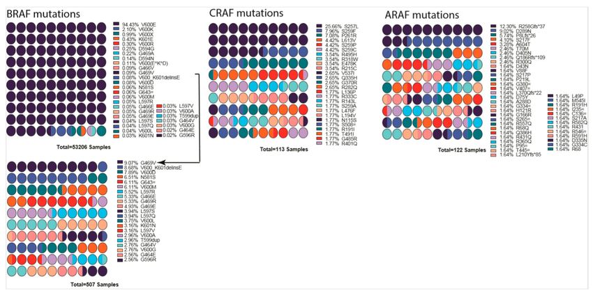
Cells 2020, 9, 198                                                                                          2 of 33

be enhanced by yet other cytosolic kinases, MEK1/2, that phosphorylate the conserved Thr/Tyr in the
activation loop of ERK1/2 [18]. Further investigation of the kinase cascade revealed that CRAF is the
upstream kinase that phosphorylates MEK1 at Ser222 and MEK2 at Ser218 that regulates the activity of
MEK, and through which ERK [19,20], thus rank-ordering the MAPK signaling from RAS, RAF, MEK,
and finally to ERK [21]. The RAS GTPase is “switched on” to the GTP-bound active form by upstream
regulators, such as RTKs, activated Ras can then physically interact with RAF and turn on the signaling
cascade [22–25]. These findings delineated how signals generated from membrane-bound receptors
are conveyed through RAS GTPase and transmitted intracellularly through a kinase cascade, setting a
milestone in understanding of cell communication and signaling (Figure 1).

      Figure 1. The Ras/RAF/MEK/ERK signaling pathway. Epidermal growth factor (EGF) initiates the signal
      on the cell surface through the EGF receptor (EGFR) (receptor tyrosine kinase), which activates guanine
      exchange factor to load RAS with GTP. RAS–GTP dimers/nanoclusters recruit RAFs or RAF/MEK
      heterodimers to plasma membranes, where RAF and MEK assemble transient tetramers that facilitate
      RAF activation through a back-to-back dimerization. MEKs docking on active RAF dimers further
      form face-to-face homodimers that are turned on by RAF. Activated MEKs phosphorylate ERKs, which
      generate response to the signal. CRR; Cys-rich region, RBD; Ras-binding domain.

     For RAF research, the early spotlight on CRAF was shifted to BRAF after the discovery in 2002
that BRAF mutations (especially BRAFV600E) were dominant in cancer [26]. Recent studies, however,
have brought CRAF back to the center stage for its role in the complicated regulation of RAF kinases by
the so called inhibitor-induced paradoxical activation of RAF seen in RAF and RAS mutant cancers [27].
A main therapeutic challenge in treating RAS/RAF-driven cancers is to develop drugs that can inhibit
this pathway without paradoxical activation.
     There are several major reviews in the field that describe the importance of RAS and RAF signaling
and their roles in cellular regulatory processes. In this paper, we refer to these reviews, at times, due
to the abundance of original research articles. However, we do provide short summaries of crucial
aspects of the field, with their primary references, where we feel it enhances the clarity of this review.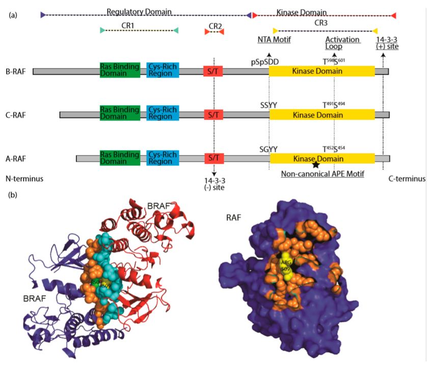
Cells 2020, 9, 198                                                                                   3 of 33

2. RAS GTPases and Their Activation
      The mammalian RAS GTPases consist of three gene isoforms, HRAS, NRAS, and KRAS, and
four protein isoforms (splicing isoforms of KRAS give rise to KRAS4A and KRAS4B proteins).
Whilst the isoforms share most of their sequence, substantial differences appear in the C-terminal
so-called hypervariable regions and in post-translational modifications [28–30]. These differences in
sequence and modification are considered to underlie the findings that RAS isoforms can function
differentially in different biology and pathophysiology [31–35]. From the standpoint of engaging
MAPK signaling, KRAS is more efficient than HRAS for CRAF activation while the opposite is true for
PI3K activation [36]. Furthermore, both KRAS and HRAS have higher activity toward NFκB activation
in contrast to NRAS [37]. While being members of the most frequently mutated oncogene family
in human cancer [38], RAS isoform mutants are clearly not equally prevalent in cancers [30]. KRAS
mutations are overwhelmingly represented in cancers as whole compared to the other two isoforms.
There is also strong tissue predilection of the occurrence of RAS isoform mutations; while KRAS
monopolizes pancreatic cancers, NRAS mutants dominate melanoma and AML. Furthermore, the
RAS isoforms also have different favored mutations, which interplay with cancer types and tissue
origins [38], adding complexity and intrigue [31]. All these differences among RAS isoforms underscore
the limitation of our understanding of RAS proteins and their downstream pathway engagements [33].
      The cellular activities and functions of RAS proteins are regulated at different levels. As a GTPase,
RAS activity is regulated by the GTP/GDP cycle [39]. GTP-bound RAS proteins adopt the so-called
active conformation that allows them to bind and activate downstream effectors, while the GDP-bound
RAS proteins have altered conformations that impede such interactions. The process of dislodging
bound GDP for GTP, thereby activating RAS, is facilitated by guanine exchange factor (GEF) proteins.
The intrinsic GTPase activity, enhanced by RAS GTPase activating protein (GAP) [40], hydrolyses
the bound GTP into GDP and returns the protein to the inactive GDP-bound state, thus completing
the GTP/GDP cycle. As an important signaling process, the GTP/GDP cycle is regulated by various
stimuli, including the cell surface receptors, such as several RTKs. Genetic alterations that affect the
regulation of RAS activation/inactivation cycle, particularly ones that result in the persistent activation
of RAS, can lead to human pathologies—the so called RASopathies [41]. For example, upregulation
or gain-of-function mutations of RTKs stimulate the activity of the RasGEF, Sos [42], which in turn
elevates the cellular level of GTP-bound Ras and oncogenic transformation. On the other hand,
the loss-of-function mutations of RasGAPs, exemplified by NF1 [43], also results in persistent RAS
activation and proliferative diseases. The most common mutations leading to RAS activation are on
RAS itself, which occur in one-third of human malignancies. There are two hot spots of RAS activating
mutations: the mutations at glycines 12 and 13 (G12/13) that impair RAS association with RasGAPs
and at glutamine 61 (Q61) that diminish the intrinsic GTPase activity of Ras [38].
      The proper functions of RAS proteins are also subject to the regulation of their posttranslational
modifications, which are essential for their trafficking, membrane localization, and interaction with
regulators and effectors [28,44–48]. RAS proteins belong to group of proteins that contain the C-terminal
CAAX consensus sequence for the prenylation processing [48,49]. The nascent RAS proteins in the
cytosol are firstly modified by either protein farnesyltransferase or geranylgeranyltransferase on
the cysteine residue of their CAAX box [44,50,51], after which they transiently associate with the
endoplasmic reticulum (ER) [51–53]. On the ER, they are further processed by the endoprotease
RCE1 [54], followed by isoprenylcysteine carboxylmethyltransferase ICMT [55], which converts the
carboxyl-terminus of RAS proteins from a hydrophilic region into a hydrophobic one, facilitating the
insertion of Ras proteins into cellular membranes [56,57]. Subsequently, RAS proteins can undergo
isoform-specific modifications, such as phosphorylation for KRAS4B and palmitoylation for HRAS and
NRAS, which facilitate transport and plasma membrane localization through distinct mechanisms [29].
      Interestingly, the functions of RAS are subject to another layer of regulation—dimerization and
nano-clustering on plasma membrane to trigger and transmit signaling downstream [58]. Lipid rafts,
subdomains of plasma membranes that have distinct chemical composition and properties, have been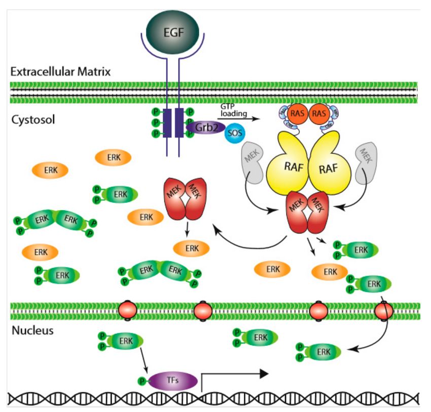
Cells 2020, 9, 198                                                                                        4 of 33

known since 1998 [59] and were observed as lateral heterogeneity, and consequently non-random
distribution, of proteins by proteolipid-based sorting. Earlier works in Ras nano-clustering showed
that Ras proteins gather on lipid rafts differentially among isoforms [60,61]. The current model is that
all Ras isoforms in their GDP and GTP bound state give rise to distinct conformations and interact with
distinct lipid compositions, cholesterol, PS, PA, PIP2 , PIP3 , PI3 P, and PI4 P, and that lipid composition
contributes to the stability of the nanoclusters of Ras [62]. RAS dimerization or nano-clustering is
thought to be a key step in the generation of its ability to couple to RAF [61–63].

3. RAF Isoforms
     RAF proteins, pivotal components of Ras/RAF/MEK/ERK signaling cascade, include three isoforms:
CRAF, BRAF, and ARAF. CRAF (also called Raf-1) is the first RAF protein identified in 1984, followed
by ARAF [64,65] in 1986 and BRAF [66] in 1988. All RAF proteins share three conserved regions: CR1
(conserved region 1) [67,68], which contains a RAS-binding domain [69–72] (RBD) and a Cys-rich
domain [73]; CR2, which is characterized by Ser/Thr-rich sequence; and CR3, which is constituted of a
putative kinase domain with an acidic N-terminus (NTA) [74,75] and a regulatory C-terminus [76,77].
Structures of RAF kinases revealed that the proteins could be divided into two functional regions as
the regulatory domain (CR1 and CR2) and kinase domain (CR3) [78,79]. Although they have similar
molecular structures, RAF proteins have quite different activity and play differential roles in cell function.
BRAF, which is well-known in cancer, as it is a major target of genetic mutations in tumorigenesis,
has the highest activity among three isoforms, likely by virtue of its constitutively-phosphorylated
NTA motif [75,80]. CRAF, which plays an indispensable role in RASopathies [81–83], has intermediate
activity. Lastly, ARAF is rarely seen genetically altered and has the lowest kinase activity due, for the
most part, to its non-canonical APE motif [84,85] (Figure 2a).

      Figure 2. The RAF family kinases. (a) Conserved domains on three RAF proteins are shown in panel (a).
      CR1 contains a Ras binding domain and a Cys-rich region while CR2 includes a S/T phosphorylation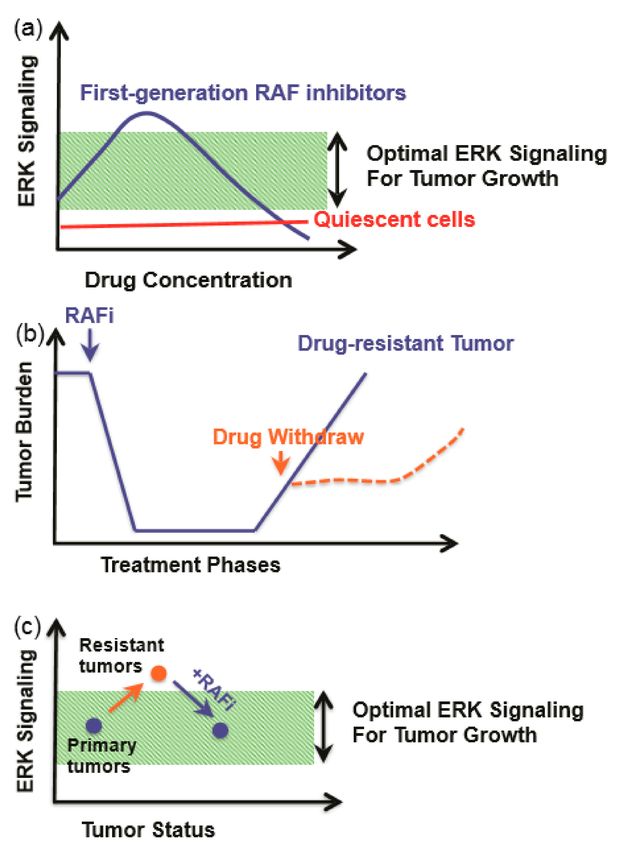
Cells 2020, 9, 198                                                                                          5 of 33

      site. The 14-3-3 binding at this region inhibits RAF. CR3 contains a putative kinase domain adjacent
      to an acidic N-terminus (NTA) and a regulatory C-terminus. At the C terminus, there is a secondary
      14-3-3 binding site which promotes dimerization. The non-canonical APE motif of ARAF is labeled
      with a star. ((b), left) Dimer interface of BRAF is shown, crystallography data was obtained from [86],
      PDB ID: 4MNE. Blue and red color indicates two separate BRAF molecules. Orange (Blue BRAF) and
      turquoise colored (Red BRAF) sphere-shaped amino acids indicate the dimer interface. While R509
      from both RAF molecule located at the center of the dimer interface, green R509 belongs to red BRAF,
      while yellow R509 does to blue BRAF. ((b), right) Crystal structure of dimer interface from plane of
      interaction with only blue BRAF chain is visible. Orange spheres indicate the amino acids at the dimer
      interface, with exception of Arg509, which is labeled with yellow color. Structure was drawn by using
      pyMOL. CR1: conserved region 1; CR2: conserved region 2; CR3: conserved region 3.

     As typical protein kinases, RAFs contain a kinase domain that comprises an N-lobe (amino-terminal
lobe) and a larger C-lobe (Carboxyl-terminal lobe). The N-lobe that includes five antiparallel β-strands
and a signal-integrated helix denoted as αC is connected by a flexible hinge to the C-lobe that
mostly consists of α-helices and a key loop termed activation segment/activation loop. Although
the kinase domains oscillate between closed and open conformations via inter-lobe motions, the
active state is restricted to the closed conformation. The closed conformation is maintained by the
alignment of two parallel hydrophobic cores (spines) of spatially conserved residues spanning N- and
C-lobes [87,88]. The regulatory spine (R-spine) is built upon the alignment of four residues in RAFs,
F516 from β4 strand, L505 from αC, F595 from DFG, and H574 from HRD (BRAF residue numbers used
here) [80,89,90]. The catalytic spine (C-spine) is formed to control kinase activation upon ATP loading.
These hydrophobic spines limit the movement of αC helix and stabilize the active conformation of the
kinase domain [91,92].
     In inactive conformation of RAF, the “Asp-Phe-Gly (DFG)” motif at the N terminus of the activation
loop is flipped “OUT” relative to its conformation in the active state “IN”. The “OUT” DFG blocks
the ATP binding pocket and stabilizes an open inactive conformation [93]. The αC helix adjacent to
N-terminus of the dimer interface is also in an “OUT” position in the inactive kinase, where it binds to
dimer interface residues (504–511 in BRAF) and further stabilizes the inactive conformation [87,94].
In addition, F485 from the β3 strand, L597 on the activation loop, and V600 on helix AS-H1 form
a hydrophobic network that provides further stabilization of inactive conformation [95]. In the
active state, αC assumes the “IN” conformation that helps the formation of a salt bridge between
glutamine from αC and catalytic lysine from VAIK motif (E501-K483 for B-Raf, E393-K375 for C-Raf,
and E354-K336 for A-Raf) [96]. The activation loop AS-H1 is disordered in the active conformation
due to αC being pulled “IN”, which reciprocally enables dimerization [95]. It is important to note
that the hallmark of an active RAF kinase requires the assembly of the two hydrophobic spines in its
core, which supersedes the more dynamic/oscillating elements such as the so-called “open” or “closed”
states or formation of the conserved salt bridge [88].

4. RAF-Knockout Mouse Models
     Gene-knockout mouse models of the three RAF proteins have yielded a vast amount of information
on their functional differences. CRAF-knockout mice are embryonic lethal with poor development
of the placenta, liver, and hematopoietic organs [97,98]; BRAF-knockout mice die in utero at D12.5
with vascular and neuronal defects [99,100]. In contrast, ARAF-knockout mice were born alive, but
presented severe intestinal and neurological abnormalities [101].
     Interestingly, mice that express a kinase-impaired CRAF with the loss of phosphorylation
sites in NTA motif (YY340/341FF) exhibit a different phenotype from that caused by complete
CRAF-deficiency [102].        Several mechanisms have been suggested to explain potential
kinase-independent functions of CRAF. Firstly, Rok-α is hyperactivated and mis-localized to the
membrane in CRAF−/− cells, which blocks Fas death receptor internalization, leading to increased
Cells 2020, 9, 198 6 of 33
Fas on the plasma membrane and Fas-dependent apoptosis [103,104]. Furthermore, CRAF NTA
mutant mice show impaired wound healing and migration of keratinocytes [104]. CRAF-deficient
fibroblasts and keratinocytes exhibit rounded morphology and impaired migration, indicating the
defective cytoskeleton. This phenotype could be rescued with Rok-α double knockout. Biochemical
and functional evidence support a mechanism for these phenotypes of kinase-independent actions
of CRAF being that the autoinhibitory Cysteine-rich domain of CRAF acts in trans to inhibit Rok-α.
This involvement of CRAF in the regulation of apoptosis has been shown to be relevant in several
human diseases. For example, Rok-α inhibition by CRAF is a prerequisite for Ras-induced cellular
transformation [81,82,105–107], and it has been shown that CRAF plays a role in pathogen-mediated
macrophage apoptosis and erythroid differentiation [108,109].
ERK signaling can inhibit apoptosis in various ways, including expression of caspase inhibitors,
neutralization of Bcl-2 family proteins [110,111], and activation of NFκB [112–114]. It is worth
noting, however, that the anti-apoptotic role of CRAF does not depend on its ability to activate ERK.
Additionally, targeting CRAF in KRAS G12V/Trp53 mutant lung tumors triggered massive apoptosis
without blocking ERK phosphorylation, which may explain the acceptable levels of toxicity of this
approach [107]. In addition to its role in regulating Fas, mitochondrial CRAF has been shown to
protect the cell from apoptosis; indeed, some growth factors have been reported to play roles in the
translocation of CRAF to mitochondria where p21-activated kinase (Pak1) phosphorylates it on Ser338
in the NTA motif [115–117]. There is also evidence suggesting that CRAF has a scaffold function for
PKCθ to facilitate phosphorylation of BAD as a way of CRAF regulation [118]. In addition, CRAF
directly interacts with voltage-dependent anion channels and prevents the release of cytochrome C
from mitochondria [119]. Other targets of CRAF include two pro-apoptotic kinases, ASK1 [120–122]
and MST2 [123], which also rely on a kinase-independent activity of CRAF. Deficiency of CRAF
promotes apoptosis of cardiomyocyte, which can be rescued with ASK1 knockdown [124]. Despite
these reports, the definitive mechanism through which mitochondrial CRAF prevents apoptosis
requires further investigation.
Similar to CRAF, ARAF also binds and sequesters MST2 independent of its kinase activity [125].
ARAF interaction with MST2 is dependent on the presence of full-length splicing product controlled
by hnRNP H [126]. In primary tumors and cell lines, ARAF and MST2 co-localize to mitochondria
and prevent apoptosis [126]. In addition to MST2, ARAF was found to bind pyruvate kinase M2
(PKM2), which is an embryonic splice variant of PKM and responsible for aerobic glycolysis [127].
In ARAF-transformed NIH3T3 cells, PKM2 is changed from dimeric to a highly active tetrameric
conformation, linking ARAF to cancer metabolism [128].
Double knockout of RAF proteins generated more severe phenotype which suggested their
additive effects [129,130]. Studies indicated that RAF proteins could compensate for lack of one of
the isoforms up to a certain point but all three are required for regulating the development of the
organism. In addition to knockouts, constitutively-active BRAFV600E mutant mouse also showed a
embryonically lethal phenotype [131].
5. Activation of RAF Proteins
5.1. Auto-Inhibited State of RAF
Once the RAS/RAF/MEK/ERK pathway was identified, research efforts focused on studying
both negative and positive regulators of the signaling cascade. In non-dividing cells, RAF exists
in an auto-inhibited state where its N-terminus docks on the kinase domain, inhibiting its catalytic
activity [132,133]. This is supported by the fact that overexpression of the N-terminal CR1 domain
inhibits kinase activity in trans [132]. The interaction between the two parts of CRAF was further
validated by the study of the N-terminus mutations of CRAF in regulating its kinase activity [132].
Moreover, RAF inhibitors that disrupt the auto-inhibited state trigger paradoxical activation, much to
the disappointment of oncologists [134,135].Cells 2020, 9, 198 7 of 33
5.2. RAF Recruitment to Plasma Membrane by Activated RAS
RAS proteins are anchored on the plasma membrane through their prenylated C-terminal CAAX
motif [136]. Upon GTP-loading, RAS is able to recruit RAF to the plasma membrane via its RAS-binding
domain (RBD) [137], which consists of a ubiquitous fold shared by other RAS effectors such as PI3K
p110 subunits and RAL guanine nucleotide dissociation stimulator (RALGDS) [69,138,139]. Single
residue substitution in the RBD is sufficient to disrupt the association of RAF with RAS and abolish
the activation of RAF [140]. Outside of the RBD, the Cys-rich region has been shown to form zinc
coordinated structures that interact with phospholipids and facilitate membrane translocation of
several kinases, including CRAF [141–146]. Furthermore, the Cys-rich region of RAF can also interact
with the farnesyl group in RAS proteins [143–147]. In addition, the N-terminus before RBD domain
(also called as N’-segment) has been shown to regulate the binding selectivity of both RAF and
Ras isoforms [96,143,148]. Thus, RBD, Cys-rich region and N’-segment are involved in the plasma
membrane recruitment of RAF proteins by active Ras. Despite the large amount of work and progress
made in the understanding of RAS–RAF interaction, there are critical details missing on the major
structural and functional interactions of these two proteins. For example, it is not clear how RAS
triggers the de-phosphorylation of inhibitory Ser residues [149]. In addition, the manner through
which the auto-inhibitory intramolecular interactions of RAF proteins are relieved is also not yet
clear [150], although Phosphatase 2A (PP2A) [151] and PP1 [152,153] have been shown to regulate this
process [150–154].
5.3. Dimerization Is a Key Event in RAF Activation
Despite the differences in their ability to trigger downstream ERK signaling, all RAF isoforms are
activated through dimer-driven transactivation. Under physiological condition, Ras-driven activation
of RAF proteins occurs on the plasma membrane where activated RAS promotes RAF dimerization, a key
event to trigger the kinase activity of RAF proteins. The notion of dimerization-driven transactivation
of RAF proteins arises from an early observation that artificial oligomerization of CRAF triggers its
kinase activity [155,156]. The subsequent observation of RAS-induced heterodimerization of BRAF
and CRAF under physiological conditions further supports the relevance of dimer formation [157,158].
The finding that kinase-dead BRAF was able to activate ERK signaling through dimerizing with and
activating CRAF not only provide further support for the role of RAF dimerization [159], but raised
the awareness that both catalysis-dependent and -independent functions of RAF are functionally
important [158,160,161]. In addition to the BRAF–CRAF heterodimer, respective homodimers of the
two isoforms, i.e., BRAF:BRAF and CRAF:CRAF, have also been detected, but were noted to have
lower kinase activity [159]. ARAF, however, has the lowest affinity for dimer formation due to its
different structural features, most notably its non-canonical APE motif that does not stabilize the dimer
interface as BRAF and CRAF are able to do [84]. However, site-specific mutagenesis of this motif from
AAE to APE enables ARAF to form dimers as strongly as CRAF. In summary, RAF family members can
form physiologically relevant heterodimers and homodimers, resulting in their transactivation [88,162]
(Figure 2b).
The dimeric structure of RAF stabilizes the closed conformation of the kinase domain by limiting
the oscillating motion of the two kinase lobes and promoting key conformational transitions [158].
Conversely, the acquisition of the active conformation also facilitates RAF dimerization. Once RAF
achieves active conformation, its dimer interface becomes further stabilized by the hydrophobic R-spine
residue in the αC- helix (L505 for B-Raf, L397 for C-Raf, and L358 for A-Raf) located adjacent to the
conserved RKTR motif, which is allosterically connected αC-helix and dimer interface [163]. Upon the
relocation of R509 to the center of the dimer interface, αC-helix interacts with the NTA motif of the
trans RAF molecule and adopts the “IN” conformation [80,164]
The dimerization of RAF proteins can be promoted by shortening their β3-αC loop; and in-frame
deletions of β3-αC loop activate ARAF by enforcing homodimer formation, showing that ARAF can
function as a “true” kinase to induce ERK phosphorylation [84], even though it is the weakest kinase inCells 2020, 9, 198 8 of 33
the family. The corresponding deletions in BRAF also ramp up its kinase activity through enhancing its
homodimer formation. A surprising finding from these studies is that the kinase-dead BRAF mutant
with an in-frame deletion of β3-αC loop (∆NVTAP/V471F) can activate ERK signaling in the absence
of active RAS [84] due to its substantial dimer affinity.
The dimerization of RAF proteins can be also improved in other ways. The BRAF splicing variants
lacked exon 4–7 exhibit a stronger dimer affinity and thereby a strong resistance to RAF inhibitors [165].
In addition to alternative splicing products; gene fusions, translocations and deletions that remove the
auto-inhibitory N-terminus allow RAFs to homodimerize with higher affinity [166–169]. Furthermore,
RAF inhibitors, especially the first-generation drugs, enhance RAF dimerization upon their binding,
and thereby paradoxically activate the pathway, which is further discussed below. Understanding the
complete mechanism of dimerization is still a work in progress, with questions remaining about the
scaffolding proteins, activation direction, and the priming of the cells for RTK signaling by increasing
RAS nanoclusters.
5.4. The Role of NTA Motif and Activation Loop Phosphorylation in RAF Activation
RAF proteins undergo multiple phosphorylation events during their activation cycle. Two major
phosphorylation events, the NTA motif and the activation loop, play key roles. The NTA motif contains
divergent loci for phosphorylation among RAFs, allowing them to have distinct regulations [75]. In
BRAF, the NTA motif contains SSDD, in which the D448 and D449 provide the initial negative charge
before serine phosphorylation. In CRAF, this locus contains SSYY and requires phosphorylation of both
serine and tyrosine residues. The constitutively phosphorylated SS and acidic DD in the NTA motif of
BRAF can explain the higher activity of BRAF compared to ARAF and CRAF. Negative charges in
this loci contribute to the relief of autoinhibition [132,150,170] and are critical for dimerization-driven
transactivation [80]. In RAF homo/heterodimers, the phosphorylated NTA motif was suggested by
molecular modeling to form multiple salt bridges that extend and stabilize the dimer interface between
two protomers [116,129]. It has been shown that the phosphorylation status of NTA motif dictates the
direction of transactivation in RAF dimers [80]; the protomer with phosphorylated NTA motif acts as
an activator, while the other protomer with non-phosphorylated NTA motif does as a receiver in RAF
dimers. A ‘receiver’ protomer can be switched into an ‘activator’ protomer upon phosphorylation
in its NTA and thereby amplify the dimerization-driven transactivation of RAF proteins. Protein
kinases that target the NTA motif play an important role not only in the activation of RAF proteins
but also by controlling the receiver–activator switch. SRC family kinases are the main kinase family
targeting YY in ARAF and CRAF [171–175]. Although still controversial [176], p21-activated kinase
(Pak) family members acting downstream of PI3K-CDC42 or RAC signaling cascades have been
suggested to phosphorylate Ser338 [177,178] in CRAF. Other kinases that potentially target the NTA
motif of RAF proteins include the following: (1) Janus Kinase 2 (JAK2), which is able to phosphorylate
CRAF to activate MEK1 [179], (2) Casein Kinase 2 (CK2), which phosphorylates CRAF at Ser338 and
BRAF at Ser446 [180], and (3) Calcium/calmodulin-dependent protein kinase II (CaMKII), which can
phosphorylate CRAF at S338 [181].
The phosphorylation of activation loop is also important for the function of RAFs. The dimerization
of RAF proteins facilitates their transition to an active conformation, which directly induces their
activation loop auto-phosphorylation as a consequence. Like most protein kinases, the activation
loop of RAFs contain two conserved phosphorylation sites relevant to their kinase function; for
ARAF, these are Thr452 and Thr455 [182]; for BRAF, these are Thr599 and Ser602 [183]; and for CRAF,
these are Thr491 and Ser494 [184]. Although studying the phosphorylation of the activation loop is
difficult due to highly dynamic de-phosphorylation, the data from RAF co-activation assay suggest
that cis auto-phosphorylation is the mechanism [80]. A recent study of a mouse model with BRAF
T599A/S602A mutation [185,186] showed that the loss of activation loop phosphorylation could not
duplicate the lethal phenotype of BRAF null mice. However, these mice had an aberrant development
of the hematopoietic system and reduction of p-ERK level in the brain, among other characteristics,Cells 2020, 9, 198 9 of 33
indicating the importance of proper activation loop phosphorylation of BRAF in development, function,
and maintenance of cell populations [187].
6. Regulation of RAF by Accessory Molecules
Activation of RAF is also regulated by other proteins, including heat-shock protein
90(Hsp90) [188,189], CDC37 [188], kinase suppressor of Ras (KSR), and 14-3-3 [190–193] proteins.
6.1. Hsp90/Cdc37 Chaperone Complex
The Hsp90/Cdc37 chaperone complex participates in proper protein folding that stabilizes protein
kinases, including RAFs [194]. The association of RAF proteins with hsp90/cdc37 complex is essential
for their activity towards MEK. Further, the association of BRAF with hsp90/cdc37 complex facilitates
the assembly of high molecular weight BRAF complex and promotes its kinase activity, probably by
increasing dimer affinity [195]. It is not surprising, therefore, that Hsp90 inhibitors can block the
activity of RAF proteins and also induce their degradation, especially that of the constitutively active
RAF mutants such as BRAF(V600E) [188,196,197]. Recently, Hsp90 inhibitors were shown to prevent
development of resistance to RAF inhibitors on BRAF(V600E)-harboring cancers in clinical trials, even
though the underlying mechanism were too complicated to pinpoint a single molecule [198–200].
6.2. KSR
Kinase suppressor of RAS (KSR) was initially identified through screening molecules essential
for Ras function in Drosophila [201]. It had been referred to as pseudo-kinase due to its low kinase
activity [202]. Subsequently, KSR was identified as a scaffold protein that not only brings close
proximity between RAF and MEK1 [203], but also allosterically activates RAF [158,204–206]. It should
be noted that KSR forms not only a side-to-side dimer with RAF proteins but also a face-to-face dimer
with MEK, both of which are critical for its ability to transactivate RAF proteins [158,207,208].
6.3. Proteins 14-3-3
The 14-3-3 proteins were the first identified, and the most well-known, phosphoserine/
phosphothreonine binding proteins, which can interact with a wide variety of proteins including
transcriptions factors, cytoskeletal proteins, apoptosis factors and tumor suppressors. Binding to 14-3-3
can alter the proteins’ stability, localization, conformation, and association with other proteins. The
functions of 14-3-3 has been reviewed extensively [209,210].
The 14-3-3 binds to phospho-serine/threonine residues in two conserved motifs of RAF proteins:
RSXpS/TXP or RXXXpSXP [78,211]. RAF proteins contain two 14-3-3 binding sites: S259 and S621
for CRAF [79,212]; S365 and S729 for BRAF; and S214 and S582 for ARAF (Figure 2a). The 14-3-3
association with these two sites plays opposite roles in RAF activity. Activation of RAF by 14-3-3 occurs
in the event of 14-3-3 binding two RAF molecules at the C-terminal phosphoserine, which promotes
dimerization. Cryo-EM studies have shown that a dimeric 14-3-3 binds two phosphorylated serine
residues on different RAF proteins, such as CRAF at Ser621 and BRAF at Ser 729, and thereby stabilizes
the side-to-side heterodimer or homodimer [77,213]. On the other hand, if a dimeric 14-3-3 binds to the
N- and C-terminus of a single RAF, respectively, it can stabilize autoinhibitory conformation [211,213].
The serine residues in conserved 14-3-3 binding motifs can be phosphorylated by Protein Kinase A
(PKA) [214–216], Akt [216–218], AMPK [219,220], or by LATS1 from the MST2-Hippo pathway [221].
Regulation of phosphorylation events at two 14-3-3 binding sites can change the response drastically
due to their opposite activity. The 14-3-3 binding site phosphorylation by different kinases may
influence the therapeutic efficacy of cancer drugs. For example, in Ras-mutated cancer cells, the CRAF
S621 is phosphorylated redundantly by AMPK and itself. Combination of RAF inhibitors with AMPK
inhibitor could reduce the paradoxical activation [219]. Recognition of the alternative kinases that
could phosphorylate 14-3-3 binding sites and thereby alter RAF activity, could improve clinical success.Cells 2020, 9, 198 10 of 33
7. RAF Function as a Dimer
As mentioned above, the Arg at the center of RAF dimer interface is a key residue for RAF
function, and its mutation to His blocks the dimerization-driven transactivation of RAF proteins [158].
This mutation can also abolish the drug resistance of BRAF(V600E) splicing variants that lack a
part of N-terminus and thereby have a higher dimer affinity than their prototype [165]. Based on
these observations, a monomer hypothesis in which RAF inhibitors bind and inhibit monomeric
BRAF(V600E), but not dimeric variants, has been suggested to explain the drug resistance of BRAF
mutants. However, this hypothesis has been challenged by other findings [80,84,95,195,222,223]. Firstly,
it was shown that BRAF(V600E) has an extended dimer interface in contrast to its wild-type counterpart
and exists as dimer/oligomer when expressed in cells [195,223]. Secondly, the Arg-to-His mutation did
not fully diminish dimer formation of RAF proteins [223], and some RAF mutants with high dimer
affinity such as BRAF(∆NVTAP) and BRAF(∆MLN) were still able to transactivate wild-type RAF in
the presence of Arg-to-His mutation [84]. Thirdly, the Arg-to-His mutation, together with APE motif
alteration, completely dissociated BRAF(V600E) dimers and abolished their activity, which could be
rescued by GST fusion-mediated re-dimerization [84]. Lastly, RAF had been shown to phosphorylate
MEK in a dimer-to-dimer manner in which an active RAF needs the other RAF partner to facilitate
MEK phosphorylation (further discussed below) [84]. Therefore, the active monomer hypothesis is not
supported at its current standing.
8. RAF–MEK Heterodimerization and MEK–MEK Homodimerization, Essential Events
for Signaling
MEK was identified independently by multiple groups as a substrate of RAF in 1992 [19,224,225],
and their interaction was further supported by the yeast two-hybrid assay [226]. Following these
findings, MEK was shown to be phosphorylated by RAF on Ser218 and Ser222 in its activation
loop [227]. Recent studies have provided detailed mechanisms of RAF phosphorylation and activation
of MEK. As a substrate, MEK needs to be recruited to RAF before activation. In quiescent cells, BRAF
and MEK form a heterodimer in the cytosol, while CRAF and ARAF do not interact with MEK under
this condition [86], presenting the question of how MEK is recruited to these RAFs. Crystallography
studies have revealed that BRAF interacts with MEK1 in a face-to-face manner through two contact
sites [86,208]. The first contact site is αG helices, a structural component of kinase–substrate docking
interaction. The second contact site is consisting of their activation loop which generates antiparallel
β-sheet [86,208]. Mutations on both contact sites that disrupt the BRAF/MEK interaction block both
allosteric and catalytic activities of BRAF [84], suggesting that the RAF/MEK association plays an
indispensable role in signal transmission from RAF to MEK.
Given the abilities of RAF proteins to form side-to-side dimers with themselves and face-to-face
dimers with MEK, it is not surprising that RAF and MEK assemble a tetramer complex of
MEK:RAF:RAF:MEK in the process of activation, which has been captured in crystal structures [86].
Although how the RAF dimer phosphorylates MEK in this transient RAF/MEK tetramer is not
completely understood, recent studies suggested that two MEK molecules need form a homodimer
that is further phosphorylated by RAF dimer or itself [84,222], since monomeric MEK cannot be
phosphorylated by RAF, and MEK homodimerization drives autophosphorylation. Moreover,
phosphorylated MEK exerts its activity towards ERK as a dimer [222], suggesting that MEK/MEK
homodimerization plays a critical role, as do RAF/RAF and RAF/MEK dimerizations, in the
pathway activation.
9. Feedback Inhibition and Return to the Inactive State
Cessation of the activated ERK signal is a crucial part of controlled cell division; therefore, the
triggered ERK pathway needs to return to the basal state with regulated feedback cues. As a part
of immediate inhibitory phosphorylation; SOS1, the RAS-GEF, is phosphorylated by ERK, which
inhibits interaction with Grb2 and creates 14-3-3 binding site for inhibitory binding [228,229]. ERKCells 2020, 9, 198 11 of 33
also exerts negative feedback on RAF by phosphorylating multiple Ser/Thr sites on RAF [230–232].
ERK phosphorylation of these Ser/Thr sites breaks the interaction of RAF with Ras, and also RAF-RAF
dimerization [229]. Additionally, some of these Ser/Thr sites can also be phosphorylated by c-Jun
amino-terminal kinases (JNKs) as a protective mechanism under stress conditions [233]. Further, the
autophosphorylation of P-loop residues in RAF proteins serves as another feedback mechanism that
impairs their kinase activity [234]. For MEK, it can be phosphorylated at its N-terminus that intercepts
its activity [235].
Shortly after inhibitory phosphorylation as described above, the components of Ras/RAF/MEK/ERK
signaling cascade require dephosphorylation to return to the inactive state. It has been shown that
PP5 participates in the dephosphorylation of pSer338 in the NTA motif of CRAF [236], while PP2A is
involved in Ser/Thr site dephosphorylation in a PIN1 dependent manner [231]. However, in the area
of dephosphorylation, most underlying molecular mechanisms remain to be elucidated.
Outside of the signaling cascade and immediate inhibitory phosphorylation, Dual-specificity
phosphatases (DUSP) and Sprouty proteins are involved in transcriptional inhibition of the ERK
signaling [237]. DUSP6 is upregulated by its transcription factor that activated by ERK1/2 [238,239].
Sprouty proteins are induced upon growth factor treatment [240], which can inhibit ERK signaling
through different mechanisms. Spry1 and Spry2 are phosphorylated at their N-terminus upon RTK
activation, which enables Spry to dock on Grb2 [241]. Furthermore, Spry4 inhibits ERK signaling by
binding to CRAF [242].
10. Mutations in the Ras/RAF/MEK/ERK Signaling Cascade
The RAS signaling cascade has well-defined role in tumorigenesis. Point mutations across each of
the members within the cascade have been identified as either driver of the tumor formation (RAS
and RAF mutations) or indicator of poor prognosis (MEK and ERK mutations). Interestingly, driver
mutations within the pathway are mutually exclusive. In this section, we describe the mutation
hotspots for each member of the signaling cascade.
10.1. Ras Mutations
RAS mutations in all cancer types occur mostly in KRAS (85%), followed by NRAS (12%) and
HRAS (3%). Interestingly, mutants of different RAS isoforms have tissue/cancer-type-dependent
distributions. For example, NRAS is the most common in melanoma, while HRAS in adrenal glands
and KRAS in pancreas [243]. Moreover, KRAS G12D was shown to promote stronger colon cancer
development than NRAS G12D in Apc-deficient mice [244], and HRAS G12V knock into the KRAS
locus was not tumorigenic in the lungs of mice [245].
RAS proteins have two mutational hotspots, 12–13 and 61. Most mutations changing G12 or
G13, likely to intercept GAP’s Arg finger loop accession to the RAS GTPase site and prevent it from
promoting hydrolysis [246–248]. As a consequence, G12 and G13 mutants trap RAS in a constitutively
active state. Mutations at Q61 inhibits intrinsic GTP hydrolysis and GAP-mediated GTP hydrolysis
(Figure 3) [246]. It is worth noting that, in addition to the isoform-dependent tissue distribution
in RAS-driven cancers, the site of mutation hotspots also comes into play in the tissue/cancer-type
distribution. For example, 90% of KRAS mutations in pancreatic ductal adenocarcinoma are at the
position G12, while 90% of NRAS mutations in melanoma are at Q61. These patterns might indicate
underlying fundamental signaling landscapes and RAS mutant interplay with these landscapes.
Adding to the complexity of RAS mutations, the oncogenic stimuli also mold the site of mutation and
the type of tumor development. For example, KRAS Q61R/L dominates the G12 mutation in urethane
treated mice [31].Cells 2020, 9, 198 12 of 33
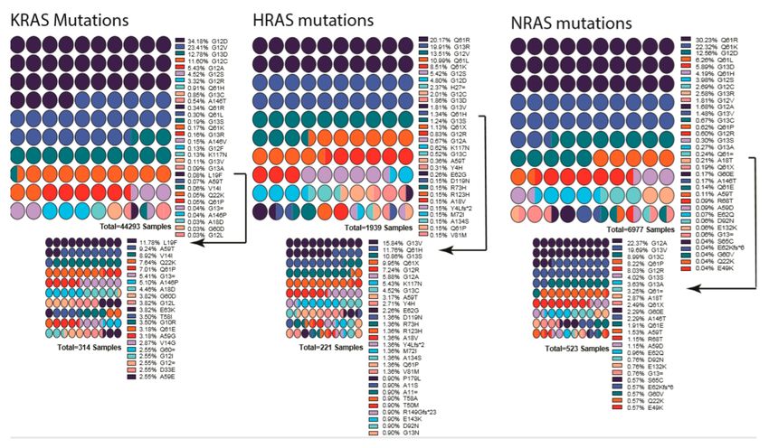
Figure 3. List of mutations detected in each RAS gene isoform. Percentages are indicated next to the
mutation, and colors indicate the mutation. Low-percentage mutations were shown as a smaller graph
underneath due to their being almost invisible in first graph. Arrows indicate from which point onward
graphs were cut into two. Color scheme repeats every ten mutations and should be interpreted in
combination of percentages and order. Graphs were drawn by using Prism 8.
10.2. RAF Mutations
Although V-RAF was discovered as a first oncogenic Ser/Thr protein kinase, its cellular prototype
CRAF is rarely mutated in cancers. The function of RAF as a prominent cancer driver was not
established until the discovery of BRAF(V600E) in 2002 [26,249]. In cancer genomes, BRAF is a major
target of oncogenic mutations and a single-point mutation, and V600E represents >90% of events,
while mutations in CRAF, ARAF, and KSRs are much less and have been detected in decreasing order
of frequency (Figure 4) [250].
Cancer-related mutations of RAF are enriched in special domains of the proteins and can be
categorized into multiple groups based on how they trigger the pathway. The first group (or Class I) of
RAF mutations activate RAF by mimicking phosphorylation of the activation loop. The second group
(or Class II) of RAF mutations turn on RAF activity by relieving the auto-inhibitory status. The third
group (or Class III) of RAF mutations have no, or impaired, kinase activity and agonist the pathway
through transactivating their wild-type counterparts.
Class I RAF mutations include V600E/D mutations in activation loop of BRAF and constitute
the largest group in the RAF mutation spectrum [251]. The V600E/D mutation stabilizes the active
conformation of BRAF by forming a salt bridge with K507 [252,253], thereby dramatically triggering
the kinase activity of BRAF independent of Ras [157,165,223,231]. Class II RAF mutations are mainly
located at the activation loop (K601 [254] and L597 [255,256]), or Gly-rich loop (P-loop, G464 [257]
and G469 [258]), and disrupt the inhibitory interaction of activation loop with Gly-rich loop and
thus destabilize the auto-inhibitory status [253]. This type of mutant has intermediate kinase activity
and increased dimer affinity, and it triggers ERK signaling with or without active RAS. Class III
RAF mutations are mostly found in the Gly-rich loop (G466 [257,259] and G469E [260]), the DFG
motif (D594 [254,261] and G596 [254,259]), the catalytic loop (N581 [252]), or the C-spine (V471F [91]).
These mutants have greatly reduced kinase activity compared with wild-type RAF and drive the
activation of ERK signaling by transactivating the wild-type RAF with their enhanced dimerizationCells 2020, 9, 198 13 of 33
affinity [160,252,262]. Unlike the Class I and Class II mutants, the Class III mutants require active Ras
to trigger signaling cascade [80,262].
Figure 4. List of mutations detected in each RAF gene isoform. Percentages are indicated next to the
mutation, and colors indicate the mutation. BRAF low percentage mutations are shown as a smaller
graph underneath due to their being almost invisible in the first graph. Arrow indicates from which
point onward graph was cut into two. Mutation data were obtained from COSMIC database. Color
scheme repeats every ten mutations and should be interpreted in combination of percentages and order.
Graphs were drawn by using Prism 8.
In addition to the highly prevalent mutations above, there are also some minor populations
of RAF mutations in cancer genomes. N-terminal truncations of CR1 and CR2 domains, or kinase
domain fusions with other proteins that relieve the N-terminal inhibition, have been identified in
RAF genes [167,169,263–265]. These N-terminal truncations or kinase domain fusions allow RAF to
dimerize and activate ERK signaling in a RAS-independent manner. Under physiological conditions,
RAF dimerization occurs on the plasma membrane. The mutations in the Cysteine-rich domain
and Ras-binding domain (RBD) of RAF can promote its plasma membrane recruitment, and thereby
dimerization, to trigger ERK signaling [250,266–269]. In addition, mutations that abolish the inhibitory
14-3-3 association with the N-terminus of RAF (S259 in CRAF and S365 in BRAF) were also found in a
small group of cancers [149,186,270,271].
10.3. MEK and ERK Mutations
In contrast to Ras and RAF mutations, MEK mutations are much less common in cancer genomes.
These mutations do not co-occur with Ras or RAF mutations, indicating that they function as cancer
drivers [272]. MEK mutations are classified into two groups according to their activation mechanism.
The first group of MEK mutations turns on the kinase activity of MEK by disrupting the inhibitory
intramolecular interaction mediated by the regulatory helix A, while the second group does so
through enhancing MEK homodimerization. These two types of MEK mutants also exhibit differential
sensitivities to MEK inhibitors in clinic or under clinic trials [222]. Like RAF, the elevated dimer
affinity may also result in the resistance to inhibitors. Finally, ERK mutations are very rare in cancer
genomes. However, a mutational hotspot has been identified on the site of D321/E322, which blocks
the Dusp6-mediated dephosphorylation of ERK and thereby extends the half-life of active ERK [273].Cells 2020, 9, 198 14 of 33
11. Targeted Therapies against Hyperactive Ras/RAF/MEK/ERK Signaling in Cancers:
The Present State and Perspectives
11.1. Is RAS Druggable?
Oncogenic Ras mutants have been considered “undruggable” over the decades due to their high
affinity with GTP and the lack of proper binding pocket for small molecule inhibitor binding [274].
Recently, the Shokat group at UCSF showed that KRAS G12C could be targeted by using a covalent
small molecule that docks in the switch II pocket and cross-links with Cys12 [275]. This discovery
spurred the race to develop KRAS-G12C-targeting drugs for clinical use. AMG510 and MRTX1257
were the first ones to be developed as drugs to target KRAS G12C mutant for non-small cell lung
cancer [274,276–278]. The results from phase I trials were promising and showed a 50% response rate
for patients with KRAS G12C mutations. However, these drugs still require further investigation;
besides validating efficacy, the impact of co-mutations and the development of drug resistance also
need to be evaluated [276]. Unfortunately, KRAS G12C is only represented in a small fraction of RAS
mutant cancers, and the challenge remains to drug the other RAS mutants.
Among alternatives to target mutant RAS isoforms, inhibiting their functionally relevant
post-translational modification represents a promising approach. The protein prenylation processing
pathway attracted significant attention in the early targeted therapeutic field [48,279]. Initial effort was
largely focused on protein farnesyltransferase, for good reason [48,279]. First, it was discovered as the
canonical prenylation enzyme for all RAS isoforms under normal cellular conditions [44,51]. Second, it
has been under careful genetic, biochemical, structural and functional studies [280–283]. However,
as the first targeted therapy development, inadequate understanding of the alternative prenylation
pathway of K- and N-RAS by geranylgeranyl transferase and corresponding inadequate stratification
method led to the dismal clinical results for farnesyltransferase inhibitors. In recent years, new trials
have been initiated that are designed to target the HRAS driven cancers, as H-RAS is not subject to
alternate prenylation [284]. It will be interesting to see the level of efficacy from these trials; early data
has shown promise (https://www.aacr.org/Newsroom/Pages/News-Release-Detail.aspx?ItemID=1350).
The earlier dismal trial results for farnesyltransferase inhibitors also sparked the interest in protein
geranylgeranyl transferase (GGTase I), which prenylates K- and NRAS when farnesyltransferase is
inhibited [284,285]. Targeting GGTase I, alone or in combination with FTIs, became an active area
of investigation following the FTI trials [286]. Indeed, there are major efforts in developing GGTase
I inhibitors for the treatment of certain cancers [286–288]. However, the toxicity of combination
therapy of GGTase I and FTase may have limited the pace of such development [289]. Among the
three steps of prenylation modifications, the last step of carboxymethylation by isoprenylcysteine
carboxylmethyltransferase (ICMT) has also attracted much attention. Genetic suppression of ICMT
in both cell and mouse models showed that ICMT inhibition led to the inhibition of tumorigenesis
in multiple cancer model systems [55,290]. Proof-of-concept ICMT inhibitors have largely replicated
the efficacy of genetic cancer models [291–293]. It will be quite interesting to follow the progress and
evolvement of the development.
For some RAS mutants, targeting their key effectors could be an effective alternative method.
The small molecule inhibitor Rigosertib has been used to block the interactions of active Ras with the
Ras-binding domain of effectors, and it has shown efficacy to target Ras-mutated cancers in phase III
clinic trials [294]. Sulindac and MCP110, two other Ras-effector interaction inhibitors, were also shown
to inhibit RAS driven tumorigenesis [295–297].
Like other oncogenes, Ras mutants need a set of cellular factors to facilitate their oncogenicity.
These synthetic lethal factors/networks could be targeted for cancer therapy. Two recent studies showed
that targeting RAF/MEK/ERK signaling together with autophagy signaling led to synthetic lethality
and achieved a promising efficacy on Ras-driven cancers [298,299], suggesting that synthetic lethal
factors/networks may be highly valuable for developing new drugs/methods against Ras-mutated
cancers. Indeed, prior evidence of manipulating autophagy by inhibiting ICMT has shown encouragingCells 2020, 9, 198 15 of 33
results [300–302], which supports that ICMT can be used to regulate RAS-driven signaling. For the
effort of identifying the targets downstream of ERK signaling in modulating autophagy, several screens
have been carried out with whole genome shRNA knockdown libraries, which yielded few druggable
factors [303]. The strong biology, however, appeals to future reliable screening approaches such as
Cas9-CRISPR-mediated knockout [304]. Nevertheless, exploring all synthetic lethal factors/networks
of Ras should provide better understanding how oncogenic Ras induces cancers and also accelerate
the development of drugs against Ras-mutated cancers.
11.2. RAF/MEK/ERK Inhibitors and Resistance
Among the members of the Ras/RAF/MEK/ERK signaling cascades, RAF is a key direct effector
of oncogenic Ras mutants and also a prominent target of oncogenic mutations. As the first kinase
in this pathway, RAF has been thought as an ideal target for drug development against cancers.
The first-generation RAF inhibitors, Vemurafenib [305,306], Dabrafenib [307], and Encorafenib [308],
were developed and applied to treatment of BRAF(V600E)-harboring cancers as single agents or
together with MEK inhibitors. These drugs achieved a promising efficacy at the initial therapeutic
phase, although this efficacy was abrogated by quick-rising drug resistance. Mechanistic studies
have shown that cancer cells reactivate this pathway upon drug treatment through two different
ways: (1) upregulating the cellular level of active Ras, which leads to paradoxical activation of ERK
signaling; and (2) alternative splicing of BRAF(V600E) to generate variants with truncated N-terminus,
which enhances BRAF(V600E) homodimerization and decreases drug affinity. Interestingly, these
drug-resistance cancer cells become addicted to RAF inhibitors, and drug withdrawal delays the growth
of resistant cancers (Figure 5). This phenomenon can be explained by the concept of a “sweet-spot”
for hyperactive Ras/RAF/MEK/ERK-signaling-driven cancer progression (Figure 5a). Specifically,
cancer cells need optimal ERK signaling for growth, and ERK signaling that is too high will induce
cell death or senescence and hence be toxic to cancer cells. Drug-resistant cancer cells have much
higher ERK signaling than drug-sensitive cancer cells. Drug treatment decreases the ERK signaling in
drug-resistance cancer cells to a level suitable for growth, while a drug withdrawal or “drug holiday”
will inhibit their growth [309,310] (Figure 5b,c).
The paradoxical effect of the first-generation RAF inhibitors not only abrogated their efficacy but
also induced secondary malignancies, a major side-effect of RAF inhibitors. To overcome the drug
resistance of the first-generation RAF inhibitors, second-generation of RAF inhibitors were developed
and underwent clinical trials; such inhibitors include pan-RAF inhibitors (such as LY3009120 [311],
TAK632 [312], TAK580 [313], CCT3833 [314], BGB283 [262], BAL3833 [315], LXH254 [316], and
RAF265 [317]), and paradox breakers (such as PLX8349 [318–320]). The pan-RAF inhibitors can inhibit
both protomers in RAF dimers with similar affinity, while the paradox breakers induce an αC helix-out
conformation upon loading and thereby prevent dimerization-driven transactivation.
To block hyperactive Ras/RAF/MEK/ERK signaling in cancers, MEK and ERK have also been used
as targets for drug designs. Two MEK inhibitors (trametinib and cobimetinib) have been developed
and approved for treating BRAF(V600E)-harboring cancers as single agents or together with RAF
inhibitors, while ERK inhibitors are still undergoing clinical trials. In contrast to RAF inhibitors, these
inhibitors have no paradoxical effect, but they do have a lower therapeutic index since they strongly
inhibit this signaling pathway in normal cells [321,322]. This also implies that targeting downstream
MEK/ERK may not be a good choice to treat Ras- or RAF-mutated cancers.You can also read