New Era in the Treatment of Iron Deficiency Anaemia Using Trimaltol Iron and Other Lipophilic Iron Chelator Complexes: Historical Perspectives of ...

Page created by Susan Larson
 
CONTINUE READING
New Era in the Treatment of Iron Deficiency Anaemia Using Trimaltol Iron and Other Lipophilic Iron Chelator Complexes: Historical Perspectives of ...
International Journal of
               Molecular Sciences

Review
New Era in the Treatment of Iron Deficiency Anaemia Using
Trimaltol Iron and Other Lipophilic Iron Chelator Complexes:
Historical Perspectives of Discovery and Future Applications
George J. Kontoghiorghes * , Annita Kolnagou, Theodora Demetriou, Marina Neocleous
and Christina N. Kontoghiorghe

                                            Postgraduate Research Institute of Science, Technology, Environment and Medicine, 3021 Limassol, Cyprus
                                            * Correspondence: kontoghiorghes.g.j@pri.ac.cy; Tel./Fax: +357-2627-2076

                                            Abstract: The trimaltol iron complex (International Non-proprietary Name: ferric maltol) was
                                            originally designed, synthesised, and screened in vitro and in vivo in 1980–1981 by Kontoghiorghes
                                            G.J. following his discovery of the novel alpha-ketohydroxyheteroaromatic (KHP) class of iron
                                            chelators (1978–1981), which were intended for clinical use, including the treatment of iron deficiency
                                            anaemia (IDA). Iron deficiency anaemia is a global health problem affecting about one-third of the
                                            world’s population. Many (and different) ferrous and ferric iron complex formulations are widely
                                            available and sold worldwide over the counter for the treatment of IDA. Almost all such complexes
         
                                     suffer from instability in the acidic environment of the stomach and competition from other dietary
                                            molecules or drugs. Natural and synthetic lipophilic KHP chelators, including maltol, have been
Citation: Kontoghiorghes, G.J.;
Kolnagou, A.; Demetriou, T.;
                                            shown in in vitro and in vivo studies to form stable iron complexes, to transfer iron across cell
Neocleous, M.; Kontoghiorghe, C.N.          membranes, and to increase iron absorption in animals. Trimaltol iron, sold as Feraccru or Accrufer,
New Era in the Treatment of Iron            was recently approved for clinical use in IDA patients in many countries, including the USA and in
Deficiency Anaemia Using Trimaltol          EU countries, and was shown to be effective and safe, with a better therapeutic index in comparison
Iron and Other Lipophilic Iron              to other iron formulations. Similar properties of increased iron absorption were also shown by
Chelator Complexes: Historical              lipophilic iron complexes of 8-hydroxyquinoline, tropolone, 2-hydroxy-4-methoxypyridine-1-oxide,
Perspectives of Discovery and Future        and related analogues. The interactions of the KHP iron complexes with natural chelators, drugs,
Applications. Int. J. Mol. Sci. 2021, 22,   metal ions, proteins, and other molecules appear to affect the pharmacological and metabolic effects
5546. https://doi.org/10.3390/ijms
                                            of both iron and the KHP chelators. A new era in the treatment of IDA and other possible clinical
22115546
                                            applications, such as theranostic and anticancer formulations and metal radiotracers in diagnostic
                                            medicine, are envisaged from the introduction of maltol, KHP, and similar lipophilic chelators.
Academic Editor: Oleg V. Mikhailov

                                            Keywords: maltol; ferric maltol; Feraccru; Accrufer; iron deficiency; iron deficiency anaemia;
Received: 13 April 2021
Accepted: 18 May 2021
                                            lipophilic chelators; alpha-ketohydroxyheteroaromatic chelators; pharmacology; clinical applications
Published: 24 May 2021

Publisher’s Note: MDPI stays neutral
with regard to jurisdictional claims in     1. Introduction
published maps and institutional affil-          Normal biological, metabolic, physiological activities, and bodily functions are main-
iations.                                    tained in life due to the supply of essential nutrients, including transition metal ions, such
                                            as iron, copper, and zinc [1–9]. Healthy living is ensured following the acquisition of
                                            daily dietary requirements and maintaining a concentration of iron and other metal ions
                                            in the tissues. In contrast, iron and other metal metabolic imbalances are associated with
Copyright: © 2021 by the authors.           serious clinical conditions [1–11]. In particular, one of these clinical conditions, namely iron
Licensee MDPI, Basel, Switzerland.          deficiency anaemia (IDA), affects about one-third to a quarter of the world’s population,
This article is an open access article      with outcomes including increased child and maternal mortality, pregnancy complica-
distributed under the terms and             tions, cardiac complications, fatigue, reduced physical and mental performance, paleness,
conditions of the Creative Commons          koilonychia, etc. [12–15]. In most of these cases, the symptoms of iron deficiency are cured
Attribution (CC BY) license (https://       using iron supplements, which are widely available.
creativecommons.org/licenses/by/
4.0/).

Int. J. Mol. Sci. 2021, 22, 5546. https://doi.org/10.3390/ijms22115546                                           https://www.mdpi.com/journal/ijms
Int. J. Mol. Sci. 2021, 22, 5546                                                                                            2 of 34

                                         Iron is found in all cells of the body. It is required for many essential bodily functions
                                   and physiological processes, including oxygen transport, storage, utilisation, and energy
                                   transduction [1–4,11].
                                         Different mechanisms, pathways, and proteins are involved in the uptake, distribution,
                                   utilisation, recycling, and excretion of iron and other essential metal ions in humans, as
                                   well as other organisms. In this context, each cell requires and utilises different amounts
                                   of these metal ions for different biological functions [1–4,11]. The transfer of iron to all
                                   cells of the body is accomplished by transferrin present in blood and is mediated through
                                   transferrin receptors present on the cell membrane [11,16–18]. Intracellular iron storage
                                   is accomplished by ferritin and haemosiderin. One molecule of ferritin can store up to
                                   4500 molecules of iron and haemosiderin is a cluster of ferritin molecules with a broken
                                   protein shell, which mainly predominates over ferritin in iron loaded conditions [11,19–22].
                                         In relation to the human body, about 4.5–5.0 g of iron is estimated to be present and
                                   distributed in blood and different organs of a 70–75 kg average adult man. Most of the
                                   iron is in the ferrous state in a complex form with a protoporphyrin ring (haem), in the
                                   protein haemoglobin (2.3–2.6 g) found in red blood cells (RBC), and myoglobin (0.32–0.40 g)
                                   found in the muscle. The remaining iron is distributed in the body mainly in the form of
                                   polynuclear ferric oxyhydroxide phosphate complexes in the iron storage proteins ferritin
                                   (0.7 g) and haemosiderin (0.3 g) found mainly in the liver, spleen, muscle, and bone marrow.
                                   Iron is also found in other proteins, such as mitochondrial cytochromes (17 mg), catalase
                                   (5 mg), transferrin (4 mg), and non-haem iron containing enzymes (0.1 g) [11].
                                         Under normal physiological conditions, iron is mainly found in the ferrous (Fe2+ ) or
                                   ferric (Fe3+ ) state forms, always bound to different ligands containing oxygen, nitrogen,
                                   and sulphur [11]. At physiological pH, ferrous iron in aqueous solutions is oxidised to
                                   ferric iron, which is only found in trace detectable levels, since it mostly precipitates by
                                   forming insoluble polymeric ferric oxyhydroxide complexes with a high stability constant
                                   (log K = 38) [11]. The solubility of iron and other metal ions increase at acidic pH including
                                   the acidic environment of the stomach.
                                         Many molecules, including food components and drugs, contain metal binding lig-
                                   ands, which can form complexes with iron. The complexes being formed can affect the
                                   solubility, interactions, and transfer properties of iron across cells in the gastrointestinal
                                   tract and in other parts of the body [11,23].
                                         Different pharmacological, toxicological, and therapeutic characteristics of iron com-
                                   plexes are observed in vivo, which depend on the size, solubility, lipophilicity, and stability
                                   of the complex, as well as other physicochemical parameters [11,24,25]. Furthermore, the
                                   characteristics of the iron complexes are also influenced by competing endogenous low
                                   molecular weight chelating molecules and iron chelating proteins, such as transferrin and
                                   lactoferrin [26,27]. Following these interactions and exchanges the chelator involved in the
                                   iron complex is, in most cases, dissociated, and the iron molecule enters the iron metabolic
                                   pathways. The chelator dissociated from the iron complex has different metabolic, toxico-
                                   logical, and pharmacological properties in comparison to the chelator iron complex [28].
                                         Iron and other metal metabolic imbalances are associated with serious medical condi-
                                   tions such as IDA, which affect billions of the people worldwide [12–15]. Iron deficiency
                                   anaemia could be caused by many genetic, nutritional, metabolic, and other factors, as well
                                   as diseases [12–15,29–33]. Iron supplementation is used in food products, such as cereals
                                   or prescribed by physicians in most cases for the treatment of IDA, including many and
                                   different oral ferrous and ferric iron complex formulations, which are widely available and
                                   sold worldwide at pharmacies [13].
                                         There are many safety concerns arising from the uncontrollable use of iron supple-
                                   ments, mainly because of the potential toxicity implications arising from the free radical
                                   catalytic and carcinogenic properties of iron. In general, only a small portion of orally
                                   administered iron is absorbed, and most of it is excreted in the faeces. The presence of
                                   excess iron is toxic to the cells of the gastrointestinal tract [34]. Furthermore, some iron
Int. J. Mol. Sci. 2021, 22, 5546                                                                                            3 of 34

                                   complexes, including those with nitrites and haem components found in processed and
                                   red meat, respectively, are suspected to be major causes of colorectal and other cancers [35].
                                         Despite the wide availability of oral ferrous and ferric iron formulations, and of intra-
                                   venous iron formulations in the treatment of IDA, there is scope for further improvement,
                                   especially in relation to safer and more effective targeting, with the ultimate aim to con-
                                   trol increased iron absorption and delivery to haematopoietic and other tissues. In this
                                   context, trimaltol iron (International Non-proprietary Name: ferric maltol, Feraccru or
                                   Accrufer), and other lipophilic chelator iron complexes appear to offer improved thera-
                                   peutic advantages in the treatment of IDA conditions in comparison to traditional oral
                                   iron formulations, which are generally linked to gastrointestinal toxic side effects, and in
                                   some cases, complications associated with exacerbation of other pre-existing underline
                                   diseases [11,23,34,36].

                                   2. Iron Absorption and Distribution Pathways
                                        Under normal conditions, the absorption of iron in the gastrointestinal tract and the
                                   distribution pathways to the haematopoietic and other tissues are regulated and controlled
                                   by a number of proteins and transcription factors, as well as other genetic, environmental,
                                   nutritional, and other factors [1–4,11,23,36].

                                   2.1. Iron Absorption in Humans
                                         Iron absorption in humans under normal conditions is thought to be controlled by
                                   a number of regulatory proteins of iron metabolism, which in conjunction with other
                                   regulatory pathways of iron utilization and excretion leads to body iron balance.
                                         In general, iron balance in the human body is maintained when the amount of iron
                                   absorbed is equivalent to the amount of iron lost through excretion and other routes [23].
                                   This dynamic balance is usually controlled and operated within limitations provided by
                                   the regulatory pathways and are mostly influenced by dietary iron and the haematopoietic
                                   activity [1–4]. Minor changes in iron balance can usually be restored at a different rate, de-
                                   pending on the level of change in the iron stores. For example, in blood donors or long dis-
                                   tance runners iron loss is gradually restored from increased dietary iron absorption [37,38].
                                   However, imminent blood transfusion is required following excessive bleeding, for exam-
                                   ple, after motor accidents or surgery, where there is substantial loss of blood and iron, as
                                   well as limited capacity in rapidly restoring these losses. Similarly, iron balance is restored
                                   when, for example, excess iron is gradually lost from patients who receive iron supplements
                                   long-term, or a small number of transfusions [23].
                                         In contrast, iron imbalance could also be caused and maintained in conditions where
                                   the rate of iron absorption may be greater than the rate of iron loss, as shown in African
                                   siderosis (Bantu siderosis), or where the rate of iron loss may be greater than iron absorption,
                                   as shown in vegetarian populations with IDA or in malnutrition, which is caused by
                                   insufficient intake of dietary iron [23]. In addition to genetic and regulatory factors, many
                                   nutritional, environmental, and other factors could also influence the rate of iron absorption
                                   and body iron load as shown in Table 1. In particular, the quantity and quality of iron
                                   entering the gastrointestinal tract could play a significant role in the overall absorption
                                   rate of body iron intake. Furthermore, dietary habits, such as the level of alcohol and
                                   water intake, as well as different food types and drugs, could also influence this process
                                   (Table 1) [23].
Int. J. Mol. Sci. 2021, 22, 5546                                                                                                      4 of 34

                                   Table 1. Non-Regulatory Factors Affecting Iron Absorption.

                                                                           Quantitative aspects
                                    The quantity of iron present in the diet, e.g., excess iron intake in African siderosis or insufficient
                                                                  iron intake in vegetarian populations
                                                                           Qualitative aspects
                                        The form of dietary iron present in food, e.g., haem, ferrous, ferric, ferritin, hemosiderin
                                                                        Role of reducing agents
                                     The presence of reducing agents, e.g., ascorbic acid converts Fe (III) to Fe (II) and increases iron
                                                                                absorption
                                                                     Effect of dietary molecules
                                    The presence of dietary molecules with chelating properties, e.g., phytates and tannins decrease
                                                                           iron absorption
                                                              Effect of drugs with chelating properties
                                     The presence of drugs with chelating properties, e.g., deferiprone, deferoxamine, tetracycline,
                                                                 hydroxyurea inhibit iron absorption
                                                                             Effect of fluids
                                       The quantity of water, alcohol, and other fluid intake can influence the dissolution of iron
                                                    supplements or other components in the gastrointestinal tract
                                                         Effect of molecules affecting cellular iron transport
                                       Dietary molecules and drugs affecting iron transport across the enterocyte (e.g., Nifedipine,
                                                             which is an L-type calcium channel blocker)
                                                               Dietary factors affecting iron solubilisation
                                     Dietary factors affecting the solubilization or precipitation of iron. Insoluble iron is not readily
                                                                                  absorbed
                                                                  Effect of pH on iron solubilisation
                                    pH of the stomach and intestine, e.g., the higher the pH the lower the solubility of iron; Antacids
                                                                        decrease iron absorption
                                                               Anatomical changes and iron absorption
                                     Gastrectomy and other surgical interventions, which can affect gastrointestinal iron absorption.
                                         Body weight, e.g., obese people absorb less iron than normal body weight individuals
                                                                  Effect of diseases on iron absorption
                                                     Malignancy, infectious and other diseases, haemoglobinopathies
                                                                    Changes in iron absorption levels
                                                                   Malnutrition, food poisoning, ageing

                                        In general, vegetarian meals low in dietary iron are likely to reduce the intake of
                                   iron and rate of body iron load in vegetarian populations [39–41]. In contrast, in meat
                                   eating populations, sufficient amounts of iron are absorbed because of the presence of
                                   high concentration of haem, which is the main form of iron to be found in the meat dishes.
                                   Haem is a lipophilic iron complex with a protoporphyrin ring, which is better absorbed
                                   from the gastrointestinal tract than other forms of dietary iron [11].

                                   2.2. The Role of Proteins of Iron Metabolism in the Regulation of Iron Absorption
                                         Iron uptake from the gastrointestinal tract is regulated by several mechanisms and
                                   specific proteins. Modifications or abnormalities in the regulatory mechanisms can cause
                                   changes in the rate of iron absorption and overall body iron load. For example, increased
                                   iron absorption is observed in genetic diseases, such as hereditary haemochromatosis, and
                                   thalassaemia intermedia leading to iron overload in both conditions [42].
                                         Under normal conditions there are several proteins and transcription factors that
                                   appear to play an important role in controlling the rate of iron transfer from the gastroin-
                                   testinal tract to the blood stream. The regulatory control of iron absorption and utilization
                                   in the enterocyte includes several steps and pathways, which appear to involve a number
Int. J. Mol. Sci. 2021, 22, 5546                                                                                                                                     5 of 34

Int. J. Mol. Sci. 2021, 22, x FOR PEER REVIEW                                                                                                                      5 of 35
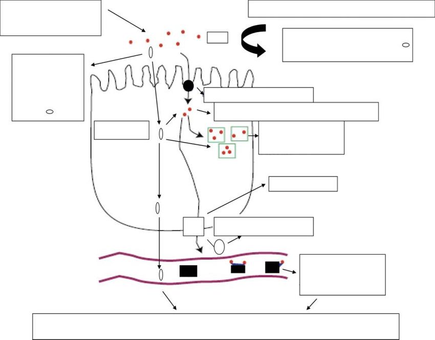
                                             of proteins such as ferrireductase(s), the apical divalent metal transported protein (DMT1),
                                        proteins such as ferrireductase(s), the apical divalent metal transported protein (DMT1),
                                             ferroportin, hepcidin, transferrin, and ferritin (Figure 1) [1–4,42–46].
                                        ferroportin, hepcidin, transferrin, and ferritin (Figure 1) [1–4,42–46].

                                                                                                           COMPETING HYDROPHILIC
                                                    LIPOPHILIC                                         CHELATORS AND OTHER METAL IONS
                                                    CHELATOR
                                                                                                 IRON               IRON DISPLACEMENT
                                                                                                                     AND INHIBITION OF
                                                  LIPOPHILIC                                                         IRON ABSORPTION
                                                  CHELATOR                                                  DMT1
                                                     IRON
                                                                                                      LOW MOLECULAR WEIGHT IRON POOL
                                                 COMPLEXES
                                                                                                                 FERRITIN AND
                                                                                                                 HAEMOSIDERIN

                                                                                                                   FERROPORTIN

                                                 ENTEROCYTE
                                                                                                          HEPCIDIN

                                                                                                                          TRANSFERRIN
                                                      BLOOD                                                               (CONTAINING
                                                                                                                            0,2,1, IRON
                                                                                                                           MOLECULES)

                                             Figure
                                        Figure        1. Mechanisms
                                               1. Mechanisms     of ironof  iron absorption
                                                                          absorption            at the enterocyte.
                                                                                       at the enterocyte.   AnimatedAnimated       image
                                                                                                                        image of the      of the regulatory
                                                                                                                                      regulatory
                                        pathways   involved
                                             pathways         in ironinabsorption.
                                                         involved                   Iron metabolic
                                                                        iron absorption.              pathways
                                                                                              Iron metabolic      involvinginvolving
                                                                                                                pathways      the apicalthe
                                                                                                                                         divalent
                                                                                                                                            apical divalent
                                        metalmetal
                                              transported    protein
                                                    transported       (DMT1),
                                                                   protein      ferritin,
                                                                           (DMT1),        hepcidin,
                                                                                      ferritin,      ferroportin,
                                                                                                 hepcidin,          and transferrin.
                                                                                                            ferroportin,              A parallel,
                                                                                                                          and transferrin.        non- non-
                                                                                                                                            A parallel,
                                        regulatory  iron  absorption   pathway   is also  shown   using  lipophilic  iron chelator  complexes.
                                             regulatory iron absorption pathway is also shown using lipophilic iron chelator complexes. In      In
                                        contrast to lipophilic
                                             contrast           chelators,
                                                       to lipophilic       hydrophilic
                                                                      chelators,           chelators
                                                                                 hydrophilic         appear
                                                                                                chelators     to inhibit
                                                                                                           appear        iron iron
                                                                                                                   to inhibit  absorption.
                                                                                                                                    absorption.

                                              The The initial   step step
                                                             initial    of theof mechanism
                                                                                  the mechanism    in thein regulatory
                                                                                                               the regulatory   pathwaypathway of iron     absorption
                                                                                                                                                      of iron    absorption is is
                                        thought
                                             thought to involve      the reduction
                                                            to involve     the reduction of dietary
                                                                                               of dietaryferricferric
                                                                                                                   iron iron
                                                                                                                          to ferrous
                                                                                                                                 to ferrousiron iron
                                                                                                                                                  by aby  ferrireductase
                                                                                                                                                             a ferrireductase
                                        at the
                                             atsurface
                                                 the surface of theofenterocyte
                                                                       the enterocyte before   its intracellular
                                                                                            before    its intracellular transport
                                                                                                                               transportby DMT1by DMT1 (Figure     1) [1–4].
                                                                                                                                                              (Figure     1) [1–4].
                                        Following
                                             Following  intracellular      entryentry
                                                               intracellular       in the inenterocyte,
                                                                                             the enterocyte,   iron iron
                                                                                                                     is partly       incorporated
                                                                                                                             is partly     incorporated  into into
                                                                                                                                                                ferritin    or or
                                                                                                                                                                       ferritin
                                        transferred
                                             transferred  intointo
                                                                 the the
                                                                       lowlow molecular
                                                                                  molecular  weight,
                                                                                                weight,  intracellular
                                                                                                             intracellulariron  ironpool.
                                                                                                                                        pool.In  In subsequent
                                                                                                                                                    subsequent steps,  steps, iron
                                        iron is
                                              is transported
                                                  transported to     to ferroportin
                                                                         ferroportin at    atthe
                                                                                              thebasolateral
                                                                                                    basolateralmembrane
                                                                                                                      membraneof          ofthe
                                                                                                                                              theenterocyte,
                                                                                                                                                    enterocyte,which  whichthen
                                        thenexports
                                              exportsititin    inplasma,
                                                                   plasma,where whereititisisbound
                                                                                                boundby    bytransferrin.
                                                                                                                 transferrin.Iron   Ironbound
                                                                                                                                           boundby    bytransferrin
                                                                                                                                                           transferrinisisthen
                                        thentransferred
                                               transferredtotoallallofof    thethe  cells
                                                                                 cells      of the
                                                                                        of the  body bodyvia an viauptake
                                                                                                                     an uptake         mechanism
                                                                                                                               mechanism                  by transferrin
                                                                                                                                                  by transferrin        receptors
                                        receptors
                                             present   present     at the
                                                          at the cell         cell surface,
                                                                         surface,    followed  followed        by intracellular
                                                                                                  by intracellular         dissociation dissociation
                                                                                                                                               of iron from of iron     from in
                                                                                                                                                                  transferrin
                                             an endosome
                                        transferrin                of acidic pH
                                                         in an endosome               [1–4]. pH [1–4].
                                                                                 of acidic
                                                      Hepcidin,
                                              Hepcidin,               a protein
                                                               a protein           hormone
                                                                              hormone             produced
                                                                                           produced        by the byliver,
                                                                                                                      the liver,      is believed
                                                                                                                               is believed             to play
                                                                                                                                                 to play    a majora major
                                                                                                                                                                         role role
                                        in iron absorption [43]. Hepcidin is a key regulator of iron metabolism, controlling the the
                                             in   iron    absorption       [43].   Hepcidin      is  a  key    regulator      of   iron    metabolism,        controlling
                                             release
                                        release    of ironof into
                                                              iron the
                                                                     intocirculation.
                                                                             the circulation.       In relation
                                                                                           In relation      to ironto     iron absorption
                                                                                                                      absorption         hepcidin   hepcidin
                                                                                                                                                       can prevent can prevent
                                                                                                                                                                          the
                                             theof
                                        release     release
                                                       iron intoof iron   into by
                                                                     plasma      plasma
                                                                                    binding by binding
                                                                                                 ferroportin  ferroportin       at the basolateral
                                                                                                                   at the basolateral           membrane   membrane
                                                                                                                                                                of the en-   of the
                                             enterocyte,
                                        terocyte,     causing   causing     its internalization
                                                                  its internalization                and degradation
                                                                                             and degradation            within  within      the enterocyte
                                                                                                                                     the enterocyte               [43–48].
                                                                                                                                                           [43–48].      As aAs a
                                             result,
                                        result,  iron iron
                                                         is notisreleased
                                                                  not released into into   plasma,
                                                                                     plasma,            but remains
                                                                                                 but remains        trappedtrappedin the inenterocytes
                                                                                                                                             the enterocytes and and      returns
                                                                                                                                                                    returns
                                        in the gut lumen since enterocytes are shed every 2–3 days. Several other pathways of iron of
                                             in   the   gut   lumen      since    enterocytes      are   shed     every     2–3    days.     Several    other      pathways
                                             iron metabolism
                                        metabolism         could becould        be affected
                                                                         affected               by hepcidin,
                                                                                      by hepcidin,        including including        the inhibition
                                                                                                                          the inhibition          of ironof release
                                                                                                                                                             iron releaseinto into
                                             plasma       from    macrophages        [1–4,43].    Furthermore,         these
                                        plasma from macrophages [1–4,43]. Furthermore, these effects and also other related ef- effects    and    also  other    related    effects
                                        fectscould
                                              couldinfluence
                                                         influencethe   theoverall
                                                                              overallrate
                                                                                        rateofofiron
                                                                                                  ironloading
                                                                                                         loadingand   anddistribution
                                                                                                                             distributionininthe     thebody,
                                                                                                                                                           body, including
                                                                                                                                                                    includ- the
                                             haematopoietic
                                        ing the    haematopoietic     tissues,   leading
                                                                            tissues,        among
                                                                                       leading         otherother
                                                                                                   among         thingsthings
                                                                                                                          to irontometabolic         imbalance
                                                                                                                                        iron metabolic                conditions,
                                                                                                                                                               imbalance
                                             such     as  IDA,    the   anaemia     of  chronic     disease,      and   iron
                                        conditions, such as IDA, the anaemia of chronic disease, and iron overload [1–4,42–48].overload        [1–4,42–48].
                                              Iron Iron      absorption
                                                      absorption       is alsois also  affected
                                                                                 affected          by changes
                                                                                             by changes              in other
                                                                                                                in other           regulatory
                                                                                                                            regulatory               molecules
                                                                                                                                              molecules              in addition
                                                                                                                                                              in addition
                                             to  hepcidin.       In  particular,    the  expression       of   ferroportin
                                        to hepcidin. In particular, the expression of ferroportin and DMT1 appear to be affectedand    DMT1       appear     to  be   affected by
                                        by several other factors and to be different in the duodenum, haematopoietic tissues, liver,liver,
                                             several      other    factors     and  to  be  different      in   the  duodenum,           haematopoietic           tissues,
                                             kidney,
                                        kidney,    and and also also
                                                                 otherother
                                                                          organsorgans    [43–49].
                                                                                    [43–49].    In thisIn context,
                                                                                                           this context,
                                                                                                                       several several     factors
                                                                                                                                     factors          are involved,
                                                                                                                                                are involved,       includ-includ-
                                        ing the signal to the intestine to increase iron absorption, which appear to be influenced
                                        by regulatory molecules, which sense the iron stores [1,49,50]. Similarly, different
Int. J. Mol. Sci. 2021, 22, 5546                                                                                             6 of 34

                                   ing the signal to the intestine to increase iron absorption, which appear to be influenced by
                                   regulatory molecules, which sense the iron stores [1,49,50]. Similarly, different regulatory
                                   mechanisms appear to control the production of haemoglobin in the haematopoietic tis-
                                   sues and to prevent anaemia. In this context, the regulatory peptide erythroferrone and
                                   the hormone erythropoietin play a key role in erythropoiesis and the regulation of iron
                                   absorption [1,2,4]. Overall, it appears that, in these cases, the influence of the regulatory
                                   molecules cause an increase in the expression of DMT1 and a corresponding increase in the
                                   uptake of iron [1,49,50].

                                   2.3. Differences among Individuals in Iron Absorption Requirements
                                         The daily requirements for iron differ among individuals and depend on several
                                   parameters including age, sex, life style, sport activity, health status, etc. [15,29–32]. Most
                                   of the iron present in the body, which is estimated to be about 4–5 g in normal adults, is
                                   conserved and recycled. Of particular importance to iron metabolism and especially the
                                   haematopoietic activity, is the recycling of iron present in haemoglobin, which amounts to
                                   more than 60% of the total body iron content [1,4,11,23]. In contrast, only a few milligrams
                                   of iron are excreted or lost and these are replaced from dietary iron, which is a small
                                   component of variable amounts in different foods. However, it is envisaged that small loses
                                   of iron and insufficient intake of dietary iron over long periods will result in iron deficiency.
                                         Under normal conditions, the absorption of iron in a western diet is generally esti-
                                   mated to be about 2 mg/day and equivalent losses allow the maintenance of body iron
                                   balance. However, there are many different dietary variations between individuals and
                                   also populations worldwide (Table 1) [11,23].
                                         Nutritional studies have suggested some general estimation for the daily iron require-
                                   ments in different categories or groups of people. In adult men and post–menopause
                                   women, the daily requirement is about 8 mg, which is generally considered to be low
                                   in comparison to other groups. The highest daily requirement for iron is for pregnant
                                   women, which is about 27 mg, whereas for adult women is 18 mg, and for breastfeeding
                                   women, 9–18 mg. In comparison, the daily requirements of iron for teenage boys and girls
                                   is estimated to be about 11 and 15 mg, respectively [29,30,51].
                                         Despite the differences in the daily iron requirements in each category, and each
                                   individual, the rate of body intake of iron could be affected by many other factors, and
                                   their combinations, including genetic predisposition, the erythropoietic activity of the bone
                                   marrow, the quantity and quality (haem or non-haem) of dietary iron, the presence of other
                                   dietary components, such as reducing agents, phytochelators, natural chelators, chelating
                                   drugs, and many others (Table 1) [29–31].

                                   3. Non-Regulatory Mechanisms of Iron Absorption
                                        Despite that major emphasis on the mechanisms of iron absorption is mainly associ-
                                   ated with the regulatory role of proteins of iron metabolism, the overall rate of iron absorp-
                                   tion is also governed by many dietary and other factors. Furthermore, the capacity of the
                                   protein regulatory pathway is limited and may not be effective under certain conditions.

                                   3.1. Dietary Molecules and Forms of Iron Affecting Its Absorption
                                        There are many dietary variations among individuals, groups, and populations, partic-
                                   ularly in relation to iron availability in the consumption of meat or vegetarian foods. Haem
                                   iron is much better absorbed than other forms of iron and it is the main source of dietary
                                   iron found in meat containing foods, which predominate in western diets (Figure 2) [41]. In
                                   contrast to western populations, the most severe pathological complications in relation to
                                   IDA are observed in the malnourished and vegetarian populations of developing countries.
                                   In the latter populations, haem iron in the diet is, by comparison, much lower, and overall
                                   iron absorption is not in significant amount from other dietary forms found in vegetarian
                                   meals [23,36–41].
In general ferrous iron is more soluble than ferric iron under the same conditions and
                                   is more readily absorbed. In general, ferrous iron forms or ferrous formulations, and the
                                   presence of reducing agents, such as ascorbic acid, will cause an increase in the solubility
                                   and overall absorption of iron from the enterocyte in comparison to most other ferric iron
Int. J. Mol. Sci. 2021, 22, 5546
                                   forms or formulations (Figure 2) [11,51,52]. In contrast, dietary molecules, such as tannins,
                                                                                                                           7 of 34
                                   phosphates, and other natural chelators or drugs, causing the precipitation of iron, will
                                   reduce the rate of iron absorption (Figure 2).

                                      A
                                                                                                                         B

                                                                                                                                    E

                                       C                                 D

                                                                                                                                    G
                                       F

                                   Figure 2. The chemical structure of molecules involved in iron absorption. Haem iron (A) is present
                                   Figure 2. The chemical structure of molecules involved in iron absorption. Haem iron (A) is pre-
                                   in meat
                                   sent     products
                                        in meat      and and
                                                 products  is better absorbed
                                                               is better        thanthan
                                                                         absorbed     non-haem
                                                                                         non-haemiron. Tannic
                                                                                                    iron.      acid
                                                                                                          Tannic     (B)(B)
                                                                                                                  acid    and
                                                                                                                            andphytic acid
                                                                                                                                 phytic acid(E)
                                   inhibit non-haem   iron  absorption.   Citric acid  (C) and ascorbic acid (D)   facilitate  non-haem
                                   (E) inhibit non-haem iron absorption. Citric acid (C) and ascorbic acid (D) facilitate non-haem iron   iron
                                   absorption. The chelating drugs deferiprone (F) and deferoxamine
                                                                                            deferoxamine (G)
                                                                                                           (G) inhibit
                                                                                                               inhibit iron
                                                                                                                        iron absorption.
                                                                                                                              absorption.

                                         Overall,   it appears
                                          The interactions   of that
                                                                 iron in  vegetarian
                                                                       with             and malnourished
                                                                             dietary molecules      or drugs populations
                                                                                                                and their effectsthe rate  of iron
                                                                                                                                     on iron    ab-
                                   loss
                                   sorption from the gastrointestinal tract could also be influenced by other essentialmeals,
                                         is higher   than  the rate   of iron absorption     from   the  iron  present   in  vegetarian      or xe-
                                   resulting   in a negative
                                   nobiotic metal              iron balance
                                                      ions competing      with and,
                                                                                iron in  the long-term, in iron deficiency [23].
                                                                                      [11,23,28,53].
                                         One of the major factors influencing the rate of iron absorption is the apparent solubil-
                                   ity
                                   3.2.of  iron
                                        Iron     at the enterocyte
                                              Chelating   Drugs and site   orDrugs
                                                                       Other  possibly    in other
                                                                                      Affecting  Ironsections   of the gastrointestinal tract,
                                                                                                       Absorption
                                   where iron may also be absorbed under different conditions (Table 1). The solubility of
                                          Many orally administered drugs, including iron chelating drugs, interact with iron
                                   ferric iron in aqueous solution at physiological pH is negligible (10−18 mol/L) and iron pre-
                                   in the   gastrointestinal
                                   cipitation   rapidly occurstract  and affectfluids
                                                                  in biological   iron absorption.    In this
                                                                                         in the absence        context,
                                                                                                           of low         themolecular
                                                                                                                    or high    hydrophilic     iron
                                                                                                                                           weight
                                   chelating occurring
                                   naturally   drugs, deferiprone
                                                           chelators, and
                                                                        suchdeferoxamine,      when
                                                                              as citric acid and       used orally
                                                                                                   transferrin,       (Figure 2F,G)
                                                                                                                  respectively          form
                                                                                                                                  (Figure       hy-
                                                                                                                                            2) [11].
                                   drophilic    chelator  iron  complexes,     which   do  not  facilitate  the  transport    of
                                         In general ferrous iron is more soluble than ferric iron under the same conditions and   iron in  the  en-
                                   terocyte   and   other cells, and   generally   inhibit  iron absorption     (Figure   1)
                                   is more readily absorbed. In general, ferrous iron forms or ferrous formulations, and the [53–56].   In  partic-
                                   ular, deferoxamine
                                   presence    of reducingisagents,
                                                              widely such
                                                                        usedasinascorbic
                                                                                  the prevention     of cause
                                                                                            acid, will  iron absorption
                                                                                                                an increaseininaccidental      iron
                                                                                                                                   the solubility
                                   poisoning     [55,56].
                                   and overall absorption of iron from the enterocyte in comparison to most other ferric iron
                                   forms or formulations (Figure 2) [11,51,52]. In contrast, dietary molecules, such as tannins,
                                   phosphates, and other natural chelators or drugs, causing the precipitation of iron, will
                                   reduce the rate of iron absorption (Figure 2).
                                         The interactions of iron with dietary molecules or drugs and their effects on iron
                                   absorption from the gastrointestinal tract could also be influenced by other essential or
                                   xenobiotic metal ions competing with iron [11,23,28,53].

                                   3.2. Iron Chelating Drugs and Other Drugs Affecting Iron Absorption
                                        Many orally administered drugs, including iron chelating drugs, interact with iron
                                   in the gastrointestinal tract and affect iron absorption. In this context, the hydrophilic
                                   iron chelating drugs, deferiprone and deferoxamine, when used orally (Figure 2F,G) form
                                   hydrophilic chelator iron complexes, which do not facilitate the transport of iron in the
                                   enterocyte and other cells, and generally inhibit iron absorption (Figure 1) [53–56]. In
                                   particular, deferoxamine is widely used in the prevention of iron absorption in accidental
                                   iron poisoning [55,56].
Int. J. Mol. Sci. 2021, 22, 5546                                                                                           8 of 34

                                         The iron chelating drugs are widely used for the treatment of iron overload by increas-
                                   ing iron excretion in iron overloaded patients [57,58]. It is envisaged that dietary molecules
                                   with properties similar to deferiprone and deferoxamine will not only cause a decrease in
                                   iron absorption when administered orally, but also an increase in iron excretion and an
                                   overall negative iron balance. The prolonged administration of iron chelating drugs in
                                   non-iron loaded individuals is expected to cause a reduction in the body iron stores and
                                   low availability of iron to the haematopoietic tissues, leading to IDA.
                                         There are many other orally administered drugs that can bind iron, but with lower
                                   affinity than the iron chelating drugs deferoxamine and deferiprone; in most of these,
                                   inhibition of iron absorption is expected including the cases of tetracycline, minocycline,
                                   and hydroxyurea. In addition, the absorption and bioavailability of these and other similar
                                   drugs, which form complexes with iron is also affected [59,60].
                                         The interaction of dietary molecules and drugs with chelating properties on the
                                   absorption of iron has not yet been fully investigated. However, in these cases, competition
                                   for iron between dietary molecules and drugs is suspected. Furthermore, the possibility of
                                   formation of ternary mixed iron complexes between these molecules will also likely affect
                                   the absorption of iron [61].

                                   3.3. Quantitative Aspects of Iron Affecting the Rate of Iron Absorption
                                        A major factor affecting the rate of iron absorption in humans is the quantity of soluble
                                   iron forms present in the gastrointestinal tract (Table 1). In general, the amount of iron
                                   absorbed is to some extent proportional to the concentration of iron in food. This effect
                                   can be highlighted from some unusual cases of consumption of excess amount of iron.
                                   For example, African siderosis in rural Africa, the use of iron cooking utensils for the
                                   preparation of traditional sorghum-based beer led to progressive iron overload in a large
                                   number of individuals [62]. The possibility of excess iron absorption been caused by the
                                   presence of a genetic component or the high iron content of the traditional sorghum-based
                                   beer has also been considered. Similar cases of acute iron overload and toxicity from
                                   increased iron absorption is observed mainly in children as a result of accidental iron
                                   poisoning. In these cases large amounts of iron are rapidly absorbed, causing serious toxic
                                   side-effects that are sometimes fatal [63,64].
                                        It appears that with the increased iron uptake observed both in African siderosis
                                   and accidental iron poisoning, the normal regulatory pathways involving hepcidin and
                                   ferroportin are overwhelmed and unable to influence or control the increased rate of
                                   gastrointestinal iron absorption and associated toxicity [62–64]. Similar questions have
                                   been raised for the role of hepcidin and ferroportin in the slow reduction of excess iron in
                                   iron loaded transplanted thalassaemia patients, who have not received any form of iron
                                   chelation therapy [52,65,66].

                                   4. Iron Formulations Used for the Treatment of Iron Deficiency Anaemia
                                        Many iron formulations are available and sold over the pharmaceutical counters
                                   worldwide for the treatment of IDA. The wide variety and selection of iron formulations is
                                   partly indicative of the commercial interest related to the large number of iron deficient
                                   patients worldwide, as well as the continuous efforts for the search and development of
                                   new, more effective, and less toxic iron complexes for IDA treatment. Usually the oral
                                   iron formulations are in a tablet, capsule, extended release tablet or capsule, or liquid
                                   preparations, which contain about 30–100 mg of elemental iron.
                                        The development of new and more effective iron formulations is also related to
                                   the increased and more selective requirements for more specific iron formulations by
                                   an increasing number of patients in addition to IDA, with other categories of anaemia
                                   not related to increased body iron requirements, such as pregnant women or vegetar-
                                   ian populations (Table 2) [52]. Many of these other categories of anaemic patients have
                                   a different pathophysiology and tolerance to iron formulations by comparison to IDA
                                   patients (Table 2) [13,67,68].
Int. J. Mol. Sci. 2021, 22, 5546                                                                                                9 of 34

                                   Table 2. Examples of anaemias treated with iron supplements.

                                                      Iron deficiency anaemia due to increased iron requirements
                                                       (e.g., pregnant and menstruating women, young children)
                                                        Iron deficiency anaemia due to insufficient dietary iron
                                                              (e.g., vegetarian populations, malnutrition)
                                          Anaemia of chronic disease or anaemia of inflammation, in neoplastic, infectious and
                                    inflammatory diseases, mainly in cases with concurrent iron deficiency or in cases of combination
                                     with erythropoietin treatment (e.g., Inflammatory bowel disease, cancer, rheumatoid arthritis)
                                                       Chronic kidney disease including haemodialysis patients
                                                                        Chronic cardiac failure

                                         One major category involving millions of patients are those suffering from the anaemia
                                   of chronic disease or anaemia of inflammation, including patients with inflammatory, in-
                                   fectious and neoplastic diseases, such as different cancer types and inflammatory bowel
                                   disease. In most of the anaemia of chronic disease cases, sufficient iron is stored in the
                                   body, but compartmentalised in the reticuloendothelial system and cannot become read-
                                   ily available to the haematopoietic tissues for the production of sufficient amounts of
                                   haemoglobin [67,68].
                                         There are many and different iron formulation products available worldwide for the
                                   treatment of IDA and other anaemias. Despite the large number and long term experience
                                   with most of the available formulations, there is no satisfactory treatment in many cases and
                                   always a scope for improvement in the treatment of different categories of iron deficient
                                   patients. In this context, new patented iron formulations appear in the pharmaceutical
                                   markets at regular intervals claiming improved response in patients.
                                         The risk/benefit assessment for the use of different iron formulations including ferrous,
                                   ferric, oral, intravenous, and slow release, in each disease category shown in Table 2 has
                                   not yet been fully examined or clarified. Furthermore, there is no general consensus among
                                   physicians in different countries for the use of any specific iron category of the available
                                   iron formulations.
                                         However, the large selection of iron formulations can benefit patients experiencing
                                   toxicity with one of them, such as in the cases of gastric irritation, or low efficacy in
                                   iron absorption. In these cases new iron formulations can be selected and prescribed for
                                   better tolerance or higher efficacy. Furthermore, the selection of iron formulations in most
                                   European and other countries is subject to budgetary controls in public health institutions
                                   and the cheaper available product is usually selected by comparison to new patented iron
                                   formulations, which are, in most cases, very expensive [69].
                                         Most of the ferric and ferrous iron formulation complexes used in IDA patients
                                   are based on naturally occurring sugar derivatives, which in general appear to partly
                                   increase the solubility of iron and facilitate its absorption from the gastrointestinal tract
                                   (Table 3) [70].
                                         Several other non-sugar iron derivatives have also been used successfully for many
                                   years for the treatment of IDA (Table 3). Ferrous sulphate is one of the classic, non-expensive
                                   iron formulations, which has been widely used worldwide with satisfactory results in
                                   many patients [71,72]. Many investigators are also proposing the use of ferrous ascorbate
                                   for the treatment of IDA because of its high efficacy and low toxicity [73–77].
                                         A new approach in the design and development of more effective and less toxic
                                   formulations containing iron complexes for IDA patients of different categories, is the use
                                   of iron trimaltol and other lipophilic iron chelator complexes, which were proposed many
                                   years ago, but only recently received approval for clinical use [25,78].
Int. J. Mol. Sci. 2021, 22, 5546                                                                                                   10 of 34

                                   Table 3. Examples of iron complexes used for the treatment of iron deficiency anaemia.

                                                                       Ferrous iron formulations
                                     (Ferrous sulphate, ferrous ascorbate, ferrous fumarate, ferrous gluconate, ferroglycine sulphate
                                                                        Ferric iron formulations
                                      (Ferric fumarate, ferric polymaltose, iron dextran, ferric gluconate, ferric iron sucrose, ferric
                                                                   saccharate, iron bis-glycinate chelate
                                                                  Ferric intravenous iron formulations
                                     (Iron sucrose, ferric carboxymaltose, ferric gluconate, ferumoxytol, iron isomaltoside-1000, iron
                                                                  dextran (low-molecular-weight forms)
                                                         Lipophilic (hetero)aromatic iron complex formulations
                                                     (Ferric maltol, ferric 8-hydroxyquinoline, ferric tropolone, ferric
                                                               2-hydroxy-4-methoxypyridine-1-oxide (L6)

                                   5. The In Vitro Properties of Iron Maltol and Other Lipophilic Iron Complexes
                                        Many ferrous and ferric formulations are widely used and sold at pharmacies for the
                                   treatment of IDA, but most of these have a low therapeutic index and are not satisfactory
                                   because of low efficacy, and are frequently non tolerated, leading to poor compliance
                                   (Table 3). The concept of the introduction of new, more specific iron formulations with a
                                   higher therapeutic index, including the use of iron maltol and other lipophilic iron chelator
                                   complexes for the treatment of IDA, was initiated about 40 years ago [25]. In particular,
                                   the trimaltol iron complex was originally designed, synthesised, and screened in vitro and
                                   in vivo in 1980–1981, by one of the authors (G.J.K.), following his discovery of the novel
                                   alpha-ketohydroxyheteroaromatic (KHP) class of iron chelators (1978–1981), which were
                                   intended for clinical use in iron overload and other iron metabolic disorders, including
                                   the treatment of IDA [25]. The slow progress in the development of iron maltol and other
                                   lipophilic iron chelator complexes appears to be related to commercial considerations [69,79].

                                   5.1. Physicochemical Properties of Lipophilic Chelators and Their Iron Complexes
                                        An original screening system for identifying investigational new drugs (IND) for the
                                   treatment of iron metabolic disorders, including iron overload and IDA, was previously
                                   tested using a large number of known and new chelators and chelator iron complexes [25].
                                   Many in vitro and in vivo procedures were involved in the screening process, including the
                                   physicochemical characterisation of the chelators and their iron complexes. In general, the
                                   physicochemical properties, such as the size, charge, and lipid/water partition of molecules
                                   affect their mode of action, including gastrointestinal absorption, membrane transport, and
                                   tissue distribution. In particular, IND of small size molecular mass, neutral charge, and
                                   high lipophilicity appear to facilitate passive transport across cell membranes.
                                        Within this context, several physicochemical parameters of IND and their metal
                                   complexes could be investigated and predict, to some extent, related pharmacological
                                   activity. These parameters include the affinity of the chelators for iron and other metals,
                                   the stability and stoichiometry of their complexes at acidic and physiological pH, the
                                   solubility, charge, the lipid/water partition coefficients of the chelators and their iron
                                   complexes, etc. [25,57]. Examples of some physicochemical properties of chelating drugs,
                                   phytochelators, and other synthetic chelators, as well as their iron complexes, are shown in
                                   Table 4.
                                        There is wide variation in the physicochemical parameters of both the chelators and
                                   their iron complexes. The differences between each chelator reflected in the parameters
                                   are the results of the structural features, metal binding ligands, and other side chains
                                   or substituents in the main structure of the chelator molecules. Similar differences in
                                   physicochemical parameters, including the charge, partition coefficient, and molecular
                                   mass are also observed among the chelator iron complexes (Table 4) [25,57].
                                        All the chelators listed in Table 4, including maltol and deferiprone, form a 3:1 sto-
                                   ichiometry chelator: iron complex at physiological pH, with exception of deferasirox
Int. J. Mol. Sci. 2021, 22, 5546                                                                                                                    11 of 34

                                   and deferoxamine, where 2:1 and 1:1 stoichiometry complexes are formed, respectively
                                   (Figure 2).

                                   Table 4. Physicochemical properties of chelators and their iron complexes.

                                                                                                                                               Charge
                                            Chelator                 Log β       MWt            Kpar         Charge         Kpar Iron
                                                                                                                                              Complex
                                              Maltol                  30           126          1.23         neutral            0.32           neutral
                                            Tropolone                 32           122          3.04         neutral            4.50           neutral
                                     8-Hydroxyquinoline               37           145          28.30        neutral           10.00           neutral
                                                L3                    30           127          0.09        zwitterionic        0.04           neutral
                                                L4                    NA           111          0.09        zwitterionic        0.95           neutral
                                                L6                    29           155          0.37        zwitterionic        4.85           neutral
                                            Omadine                   NA           127          0.04        zwitterionic        2.67           neutral
                                            Mimosine                  36           198          0.01        zwitterionic        0.01         zwitterionic
                                         Deferoxamine                 31           561          0.02         positive           0.02           positive
                                           Deferiprone                35           139          0.18         neutral            0.05           neutral
                                           Deferasirox                27           373          6.30         negative           NA            negative
                                   Iron complex stability constants (log β); molecular weight (MWt); n-octanol/water partition coefficients (Kpar);
                                   charge of chelator and chelator iron complex at physiological pH (charge); 2,4-dihydroxypyridine-1-oxide (L3);
                                   2-hydroxypyridine-1-oxide (L4); 2-hydroxy-4-methoxypyridine-1-oxide (L6). Not available (NA).

                                   5.2. In Vitro Properties of Maltol and the Maltol Iron Complex
                                                  Maltol is a naturally occurring alpha-keto hydroxy pyrone compound found in plants,
                                           e.g., in the bark of the larch tree and in pine needles [80]. It is also found in roasted malt and
                                           in bread, and is formed during caramelization. Maltol has been widely used and marketed
                                           for more than 50 years as a flavour enhancer and as a food additive. It is rapidly and
    Int. J. Mol. Sci. 2021, 22, x FOR PEER extensively
                                           REVIEW        absorbed from the gastrointestinal tract (similar to ethyl maltol—an  2 of 35analogue
                                           of maltol) and both are mostly metabolised to glucuronide conjugates [81] (Figure 3).

                                                         +      Fe                                          Fe

                                   Figure 3.
                                   Figure  3. The
                                              Theformation
                                                   formation  ofof
                                                                 thethe
                                                                      tris-maltol  ironiron
                                                                         tris-maltol     (III)(III)
                                                                                               complex  and maltol
                                                                                                    complex          glucuronide.
                                                                                                              and maltol            At physiolog-
                                                                                                                          glucuronide.      At physiological
                                   ical pH, maltol reacts with iron (III), forming the tris-maltol iron octahedral complex with iron in
                                   pH, maltol reacts with iron (III), forming the tris-maltol iron octahedral complex with iron in the
                                   the centre. During iron (III) binding, a proton is displaced from the hydroxyl group of each maltol
                                   centre.
                                   molecule,During
                                               formingiron  (III) binding,
                                                        a negatively   charged  a molecule,
                                                                                  proton iswhichdisplaced   from with
                                                                                                      coordinates  the hydroxyl
                                                                                                                        iron (III) ofgroup    of each maltol
                                                                                                                                      3+ charge,
                                   molecule,    forming
                                   forming a neutral      a negatively
                                                       trimaltol  iron (III)charged
                                                                             complex.molecule,
                                                                                         In humans,which
                                                                                                       maltol coordinates   with iron
                                                                                                              is mostly metabolised       (III) of 3+ charge,
                                                                                                                                       to the
                                   maltol glucuronide
                                   forming               conjugate,iron
                                             a neutral trimaltol      which   has
                                                                           (III)   no iron orInother
                                                                                 complex.             metalmaltol
                                                                                                  humans,    chelating capacity.metabolised to the maltol
                                                                                                                    is mostly
                                   glucuronide conjugate, which has no iron or other metal chelating capacity.
                                         The in vitro screening procedure of the chelators involved the study and characteri-
                                   sation of iron complex formation, the interaction with proteins of iron metabolism, and
                                   the iron transport effects in red blood cells (RBC), as well as the iron transport effects in
                                   rat jejunum permeation [25,57].
                                         In the iron binding studies, a 3 maltol:1 iron stoichiometry complex (trimaltol iron)
Int. J. Mol. Sci. 2021, 22, 5546                                                                                                                        12 of 34

                                                     Maltol was first identified as a potential iron chelator during the period of the four-
                                            Figure 3. The formation of the tris-maltol iron (III) complex and maltol glucuronide. At
                                              step synthesis of the iron chelating drug deferiprone, where it was used as the starting
                                            physiological pH, maltol reacts with iron (III), forming the tris-maltol iron octahedral complex
                                              material
                                            with  iron in [25]. It was
                                                          the centre.   included
                                                                      During          inbinding,
                                                                              iron (III) the list afor the isindisplaced
                                                                                                    proton      vitro and   inthe
                                                                                                                         from  vivo  screening
                                                                                                                                   hydroxyl groupprocedure
                                                                                                                                                    of       of
                                              natural
                                            each maltoland     synthetic
                                                          molecule, formingchelators     intended
                                                                              a negatively            for potential
                                                                                            charged molecule,    which clinical
                                                                                                                       coordinatesuse in iron
                                                                                                                                    with  iron(III)
                                                                                                                                                overload,
                                                                                                                                                    of    iron
                                            3+deficiency,
                                               charge, forming
                                                             anda other
                                                                   neutral diseases
                                                                           trimaltol iron
                                                                                        of (III)
                                                                                           ironcomplex.  In humans,
                                                                                                 metabolism            maltolThe
                                                                                                                   [25,57].   is mostly metabolised
                                                                                                                                   alpha-keto     hydroxy iron
                                            to the maltol glucuronide conjugate, which has no iron or other metal chelating capacity.
                                             binding site of maltol is identical to that of the chelating drug deferiprone (Figure 2F).
                                                    The in vitro screening procedure of the chelators involved the study and characterisa-
                                                  The in vitro screening procedure of the chelators involved the study and
                                             tion   of  iron complex
                                            characterisation        of ironformation,     the interaction
                                                                              complex formation,             with proteins
                                                                                                       the interaction     withof proteins
                                                                                                                                  iron metabolism,
                                                                                                                                             of iron and the
                                             iron   transport       effects  in red  blood   cells (RBC),    as well   as  the iron
                                            metabolism, and the iron transport effects in red blood cells (RBC), as well as the iron  transport   effects in rat
                                             jejunum      permeation       [25,57].
                                            transport effects in rat jejunum permeation [25,57].
                                                  InInthethe  iron
                                                           iron      binding
                                                                  binding       studies,
                                                                            studies,       a 3 maltol:1
                                                                                     a 3 maltol:1          iron stoichiometry
                                                                                                   iron stoichiometry     complexcomplex
                                                                                                                                     (trimaltol(trimaltol
                                                                                                                                                iron)      iron)
                                             wasformed
                                            was    formed      at physiological
                                                            at physiological         pH using
                                                                                pH using         the plot
                                                                                           the Job’s  Job’s  plot method
                                                                                                           method            as indicated
                                                                                                                    as indicated   in Figure in
                                                                                                                                             4B Figure
                                                                                                                                                 [25]. 4B [25].
                                            While
                                             Whilemaltol
                                                       maltol is aiswhite  solid,
                                                                      a white     forming
                                                                                solid,      a colourless
                                                                                        forming           solution,solution,
                                                                                                   a colourless     the iron complex
                                                                                                                               the ironofcomplex
                                                                                                                                           maltol is of maltol
                                            ofisdeep   red/orange      colour, and  appears   stable at a wide  pH
                                                 of deep red/orange colour, and appears stable at a wide pH range,   range,  including   the pHincluding
                                                                                                                                                  5–9        the
                                            range,   as  shown    in  Figure  4A  [25].
                                             pH 5–9 range, as shown in Figure 4A [25].

                                                OD

Int. J. Mol. Sci. 2021, 22, x FOR PEER REVIEW                                                                                            3 of 36

                                                                             (A)

                                                OD

                                                                       (B)
                                      Figure 4. Characterisation of the maltol iron complex. (A) Stability of the maltol iron complex over
                                             Figure 4. Characterisation of the maltol iron complex. (A) Stability of the maltol iron complex over a
                                      a wide pH range. Titration of a mixture of maltol (0.75 mM) and iron (0.25 mM) at a pH range 2–
                                             wide pH
                                      12, indicating  onerange.    Titration
                                                           iron complex       of aofmixture
                                                                          species            of maltol
                                                                                     red/orange          (0.75
                                                                                                 colour at  the mM)
                                                                                                                pH 6–10 and  iron The
                                                                                                                           range.   (0.25 mM) at a pH range 2–12,
                                                                                                                                        optical
                                      densityindicating     one iron
                                               (OD) monitoring     wascomplex    species
                                                                        carried out at theof red/orange
                                                                                          wavelengths    of colour
                                                                                                            320 nm at    the pH
                                                                                                                      (circles) and6–10
                                                                                                                                     280 range.
                                                                                                                                         nm      The optical density
                                      (squares)
                                             (OD)[25].monitoring
                                                       (B) Identification of the stoichiometry
                                                                    was carried                 of the tris-maltol
                                                                                   out at the wavelengths           ironnm
                                                                                                               of 320     complex    at and 280 nm (squares) [25].
                                                                                                                              (circles)
                                      physiological pH. Estimation of the stoichiometry of the maltol iron complex using the Job’s plot
                                             (B) Identification of the stoichiometry of the tris-maltol iron complex at physiological pH. Estimation
                                      method. Measurement of the optical density (OD) at 220 nm of different molar fraction mixtures of
                                             of mM)
                                      maltol (1  the stoichiometry
                                                       and iron (1 mM)ofatthepHmaltol
                                                                                7.0. Theiron  complex
                                                                                         horizontal  axisusing   thethe
                                                                                                          refers to    Job’s  plot
                                                                                                                          molar      method.
                                                                                                                                 fraction       Measurement of the
                                                                                                                                          of maltol.
                                              optical density (OD) at 220 nm of different molar fraction mixtures of maltol (1 mM) and iron (1 mM)
                                            An   octahedral
                                              at pH  7.0. Thestructure
                                                              horizontaliron
                                                                          axiscomplex
                                                                               refers to similar to fraction
                                                                                         the molar   deferiprone  and other bidentate
                                                                                                             of maltol.
                                      chelators is formed in the interaction of maltol with iron at physiological pH (Figures 3
                                      and 4). A proton is released from the coordinating hydroxyl group of maltol on binding
                                      ferric (3+) iron, forming three negatively charged maltol molecules (Figures 3 and 4) and
                                      an overall neutral trimaltol ferric iron complex. Both maltol and the maltol iron complex
                                      are lipophilic and have a neutral charge at physiological pH (Table 4).
Int. J. Mol. Sci. 2021, 22, 5546                                                                                                     13 of 34

                                         An octahedral structure iron complex similar to deferiprone and other bidentate chela-
                                   tors is formed in the interaction of maltol with iron at physiological pH (Figures 3 and 4).
                                   A proton is released from the coordinating hydroxyl group of maltol on binding ferric (3+)
                                   iron, forming three negatively charged maltol molecules (Figures 3 and 4) and an overall
                                   neutral trimaltol ferric iron complex. Both maltol and the maltol iron complex are lipophilic
                                   and have a neutral charge at physiological pH (Table 4).

                                   5.3. Interactions of Maltol and the Maltol Iron Complex with Proteins
                                            The pharmacological activity of maltol and the maltol iron complex is affected by a
                                       variety of proteins, including proteins involved in iron metabolism [28,57]. In particular, the
                                       interaction of maltol and the maltol iron complex with the iron transport protein transferrin
                                       and the iron storage proteins ferritin and haemosiderin is a major parameter characterising
                                       the mode of action of chelators, and is also a key part of the chelator evaluation for clinical
                                       use in diseases related to iron metabolism [19–22,24,25,28,57,82–85].
                                            Substantial differences were observed in the original studies of iron mobilisation
                                       from diferric transferrin using maltol and other KHP chelators in vitro [25,86]. The 59-Fe
                                       radioactive labelled iron transferrin equilibrium dialysis studies have indicated that maltol
                                       is not effective in the mobilisation of iron from diferric transferrin. For example, in four
                                       different experiments for up to six hours, iron release from 59-Fe radioactive labelled
                                       diferric transferrin using deferiprone (4.0 mM) was 77–90%, and using maltol (4.0 mM)
                                       was 4–23% at the same conditions [25,86]. More iron was released from transferrin
Int. J. Mol. Sci. 2021, 22, x FOR PEER REVIEW                                                                                  4 of by
                                                                                                                                    36
                                       maltol at 24 h incubations. However, UV-visible spectroscopic changes have also suggested
                                       that a ternary complex is formed between transferrin, iron, and maltol, but the maltol iron
                                       complex is not readily dissociated from transferrin (Figure 5) [25,86].

                                     OD

                                                 Fe - T - Fe

                                                                     Wavelength (nm)
                                    Figure 5. The interaction of maltol with iron saturated transferrin. Optical density (OD) spectral
                                    changes
                                   Figure  5.of theinteraction
                                              The   reaction ofof
                                                                maltol (1.0
                                                                  maltol    mM)
                                                                         with    with
                                                                              iron    diferric transferrin.
                                                                                   saturated   transferrin (Fe-T-Fe) (0.036 mM)
                                                                                                            Optical density (OD)asspectral
                                                                                                                                   a function
                                    of time from
                                   changes   of the1–220 minof
                                                    reaction  [25].
                                                                maltol (1.0 mM) with diferric transferrin (Fe-T-Fe) (0.036 mM) as a
                                   function of time from 1–220 min [25].
                                         Similar results were obtained in iron mobilisation studies from diferric lactoferrin. For
                                    example,  in equilibrium
                                        Similar    results weredialysis
                                                                  obtainedstudies using
                                                                             in iron    59-Fe radioactive
                                                                                     mobilisation  studieslabelled diferriclactoferrin.
                                                                                                            from diferric   lactoferrin
                                    ironexample,
                                   For   release at in
                                                     pHequilibrium
                                                         7.3 using deferiprone    (0.29 mM)
                                                                       dialysis studies      was59-Fe
                                                                                          using   87.4%,  and using maltol
                                                                                                        radioactive  labelledunder   the
                                                                                                                                diferric
                                    same conditions
                                   lactoferrin         was 9.4%
                                                iron release  at pH[87].
                                                                     7.3 using deferiprone (0.29 mM) was 87.4%, and using maltol
                                   underUnder     normal
                                           the same       conditions,
                                                      conditions   wastransferrin
                                                                         9.4% [87]. iron saturation in humans is only about 20–30%,
                                    withUnder
                                          apo-transferrin,    the  most   prominentiron
                                                  normal conditions, transferrin       species in blood
                                                                                           saturation  in by  comparison
                                                                                                          humans   is only to  the two
                                                                                                                            about   20–
                                   30%, with apo-transferrin, the most prominent species in blood by comparison to the two
                                   monoferric transferrins and diferric transferrin [17,18]. Apo-transferrin has high potency
                                   in mobilising mononuclear and low molecular weight iron, thus preventing microbial
                                   growth and the catalytic production of toxic free radicals, both requiring the presence of
Int. J. Mol. Sci. 2021, 22, 5546                                                                                            14 of 34

                                   monoferric transferrins and diferric transferrin [17,18]. Apo-transferrin has high potency in
                                   mobilising mononuclear and low molecular weight iron, thus preventing microbial growth
                                   and the catalytic production of toxic free radicals, both requiring the presence of iron [88].
                                         Apo-transferrin also has the capacity to mobilise iron from low molecular weight
                                   iron complexes, including iron from citrate, deferiprone and maltol iron complexes in
                                   plasma [88–90]. In this context, maltol or deferiprone iron complexes facilitate the transfer
                                   of iron to apo-transferrin, forming diferric and monoferric transferrin, which are then taken
                                   intracellularly through transferrin receptors in haematopoietic and other tissues [91–95].
                                         In the initial studies of the interaction of maltol with ferritin, it was shown that maltol
                                   (1–4 mM) was effective in the mobilisation of ferritin iron (0.6–1.8 mM) at a slow rate
                                   reaction, which reached about 3 days to completion. It was estimated that the amount
                                   of iron released from ferritin by maltol (1.0 mM) at the end of the reaction was 24% by
                                   comparison to deferiprone 46%, used under the same conditions [25,96]. In further studies
                                   with maltol and other chelators, iron mobilisation from ferritin was confirmed and was
                                   also extended and shown to take place from other polynuclear forms of iron, such as
                                   haemosiderin and freshly prepared iron precipitates [97]. The rate of iron solubilisation
                                   from polynuclear forms of iron was higher in freshly prepared iron precipitates by compar-
                                   ison to haemosiderin and less so from ferritin [97]. Furthermore, less iron has been shown
                                   to be mobilised by maltol and other chelators from ferritin and haemosiderin possessing
                                   lower amounts of iron [98].
                                         Similar results were observed in equilibrium dialysis 24 h studies using 59-Fe labelled
                                   horse spleen ferritin (0.15 mg/mL) containing 2200 molecules of iron per ferritin molecule
                                   and different chelators (1.2 mM). Under these conditions maltol caused the release of 9% of
                                   iron by comparison to deferoxamine 12% and deferiprone 21% [99].
                                         The interaction of maltol and the maltol iron complex has also been investigated
                                   in other proteins of iron metabolism. In particular, maltol in a comparative study with
                                   the iron chelating drugs deferoxamine and deferiprone, as well as other compounds has
                                   been shown to inhibit the iron containing proteins lipoxygenase and cyclooxygenase, and
                                   associated metabolic pathways, such as thromboxane A2 synthesis and the conversion of
                                   arachidonate to HETE and HPETE in platelets [100–102]. The inhibitory effect of maltol in
                                   both cases was concentration dependent, reversible in the addition of iron, and lower by
                                   comparison to deferiprone and deferoxamine [100–102]. In the case of the thromboxane A2
                                   inhibition, the median inhibitory concentration (IC50) was about 8 mM for maltol, 0.7 mM
                                   for deferoxamine, and 0.2 mM for deferiprone. Similarly, in the conversion of arachidonate
                                   to HETE and HPETE was 8 mM for maltol, 1.2 mM for deferoxamine, and 1.6 mM for
                                   deferiprone [101,102].
                                         Overall, it appears that the interactions and effects of maltol with proteins of iron
                                   metabolism are concentration dependent and involve several components. In particular,
                                   maltol has the ability to mobilise intracellular low molecular weight iron, ferritin, and
                                   haemosiderin iron forming trimaltol iron complexes. Furthermore, trimaltol iron complexes
                                   can interact and donate iron to apo-transferrin and form monoferric and diferric transferrin,
                                   which subsequently can release iron to cells for storage, haemopoiesis, or utilisation for
                                   other cellular functions.

                                   5.4. The Antioxidant Effects of Maltol and Other Iron Chelators
                                        Iron is the major catalyst of free radical reactions and oxidative stress toxicity in
                                   biological systems. Many factors influence the rate of free radical reactions catalysed
                                   by iron in biological systems, including the pH, the presence of reducing agents, and of
                                   chelating agents [103–106].
                                        The pro-oxidant and antioxidant effects of drugs are major parameters character-
                                   ising their mode of action, as well as their toxicological and pharmacological proper-
                                   ties. The modulation of free radical toxicity is more sensitive in the presence of iron
                                   and copper chelating drugs, which can strongly influence the catalytic activity of these
                                   metals [106–109].
You can also read