Mechanisms of Chemotherapy-Induced Peripheral Neuropathy - MDPI
←
→
Page content transcription
If your browser does not render page correctly, please read the page content below
International Journal of
           Molecular Sciences

Review
Mechanisms of Chemotherapy-Induced
Peripheral Neuropathy
Renata Zajaczkowska
           ˛                                 epska 2 , Wojciech Leppert 3 ,
                     1, * , Magdalena Kocot-K˛

Anna Wrzosek  1 , Joanna Mika  4  and Jerzy Wordliczek 1
 1    Department of Interdisciplinary Intensive Care, Jagiellonian University, Medical College,
      31-501 Krakow, Poland; surmiatko@interia.pl (A.W.); j.wordliczek@uj.edu.pl (J.W.)
 2    Department of Pain Research and Treatment, Jagiellonian University, Medical College,
      31-501 Krakow, Poland; makoco@wp.pl
 3    Chair and Department of Palliative Medicine Poznan University of Medical Sciences,
      61-245 Poznan, Poland; wojciechleppert@wp.pl
 4    Institute of Pharmacology, Polish Academy of Sciences, Department of Pain Pharmacology,
      31-343 Krakow, Poland; joasia272@onet.eu
 *    Correspondence: renia356@poczta.onet.pl

 Received: 31 January 2019; Accepted: 19 March 2019; Published: 22 March 2019

 Abstract: Chemotherapy-induced peripheral neuropathy (CIPN) is one of the most frequent side
 effects caused by antineoplastic agents, with a prevalence from 19% to over 85%. Clinically, CIPN is a
 mostly sensory neuropathy that may be accompanied by motor and autonomic changes of varying
 intensity and duration. Due to its high prevalence among cancer patients, CIPN constitutes a major
 problem for both cancer patients and survivors as well as for their health care providers, especially
 because, at the moment, there is no single effective method of preventing CIPN; moreover, the
 possibilities of treating this syndrome are very limited. There are six main substance groups that
 cause damage to peripheral sensory, motor and autonomic neurons, which result in the development
 of CIPN: platinum-based antineoplastic agents, vinca alkaloids, epothilones (ixabepilone), taxanes,
 proteasome inhibitors (bortezomib) and immunomodulatory drugs (thalidomide). Among them,
 the most neurotoxic are platinum-based agents, taxanes, ixabepilone and thalidomide; other less
 neurotoxic but also commonly used drugs are bortezomib and vinca alkaloids. This paper reviews
 the clinical picture of CIPN and the neurotoxicity mechanisms of the most common antineoplastic
 agents. A better understanding of the risk factors and underlying mechanisms of CIPN is needed to
 develop effective preventive and therapeutic strategies.

 Keywords: chemotherapy-induced neuropathy; cancer pain; drug neurotoxicity; pathophysiological
 mechanisms; anticancer drugs

1. Introduction
     Cancer is currently a leading cause of mortality worldwide [1]. However, thanks to advances in
medicine and modern technology, the availability of sensitive tests and diagnostic methods to detect
cancer at an early stage and the use of increasingly effective treatments, including chemotherapeutic
agents, the number of cancer survivors is rising: It is expected to increase by 35%, from 13.7 million
in 2012 to 18 million, by 2022 [2]. Although these survivors may have beaten cancer, many of them
have poor outcomes due to a number of syndromes that reduce the quality of life as a consequence of
cancer treatment, including pain, which they often experience for a long time after completing their
cancer treatment [3].
     Drugs used in cancer chemotherapy constitute an extremely effective tool in arresting the
progression of cancer since they have numerous targets and mechanisms of action aimed at eliminating

Int. J. Mol. Sci. 2019, 20, 1451; doi:10.3390/ijms20061451                         www.mdpi.com/journal/ijms
Int. J. Mol. Sci. 2019, 20, 1451                                                                     2 of 29

rapidly dividing cancer cells. Unfortunately, these drugs also affect normal cells and structures of the
body, causing various deleterious and sometimes even devastating side effects (e.g., anaemia, diarrhea,
nausea, vomiting, infections, neurological changes, fatigue, hair loss, infertility, pain and peripheral
neuropathy) [4], which may necessitate the tapering of chemotherapy regimens or even their cessation,
thereby limiting the efficacy of cancer treatment.
      Chemotherapeutic agents can damage nervous system structures and, depending on the
individual compound, can cause a variety of neuropathies: large and small fibre, sensory and/or
motor, demyelinating and axonal, cranial and autonomic [5]. The effects of chemotherapy on the
nervous system vary among the different classes of drugs, depending on the specific physical and
chemical properties of the drug used and its single or cumulative doses [6]. One of the most
common neuropathies caused by antineoplastic agents is a condition known as chemotherapy-induced
peripheral neuropathy (CIPN) [7]. The prevalence of CIPN is agent-dependent, with reported rates
varying from 19% to more than 85% [8] and is the highest in the case of platinum-based drugs
(70–100%), taxanes (11–87%), thalidomide and its analogues (20–60%), and ixabepilone (60–65%) [6].
Toxicity may occur either with a high single dose or after cumulative exposure. Observed symptoms
vary in intensity and duration and range from acute, transient thermal sensations to permanent
changes in peripheral nerves accompanied by chronic pain and irreversible nerve damage [9]. Recent
studies put the prevalence of CIPN at approximately 68.1% when measured in the first month after
chemotherapy, 60.0% at 3 months, and 30.0% at and after 6 months [9].
      CIPN is a predominantly sensory neuropathy that may be accompanied by motor and autonomic
changes [9]. Except for paclitaxel and oxaliplatin, which cause acute neuropathy during or immediately
after infusion [10], CIPN symptoms usually emerge late, that is, weeks or months after the completion
of chemotherapy, with their severity being usually proportional to the cumulative dose of the drug [11].
Some patients experience paradoxical worsening and/or intensification of symptoms after the cessation
of treatment [12], as well as a phenomenon known as “coasting”, where either mild neuropathy worsens
or new CIPN develops. This situation poses a challenge for oncologists since, during the chemotherapy
course, no signs or indications warrant a reduction in the dosage to mitigate CIPN symptoms [13]. Pain
and sensory abnormalities may persist for months or even years after the cessation of chemotherapy.
Therefore, patients may be cancer-free but may suffer from debilitating neuropathy induced by cancer
treatment [14].
      Clinically, CIPN manifests itself as deficits in sensory, motor and/or autonomic functions of
a varying intensity [15]. Sensory symptoms usually develop first, involve the feet and hands and
commonly present as a typical “glove and stocking” neuropathy with the most distal parts of the limbs
exhibiting the greatest deficits. The symptoms comprise numbness, tingling, altered touch sensation,
impaired vibration, paresthesias and dysesthesias induced by touch and warm or cool temperatures.
Moreover, painful sensations, including spontaneous burning, shooting or electric shock-like pain as
well as mechanical or thermal allodynia or hyperalgesia frequently occur [16]. In severe cases, these
symptoms can progress to a loss of sensory perception. Motor symptoms occur less frequently than
sensory symptoms and, as a rule, assume the form of distal weakness, gait and balance disturbances,
and impaired movements. These symptoms have a marked and often underappreciated impact on
quality of life and safety, e.g., cancer patients who develop CIPN are three times more likely to fall [17].
In severe cases, CIPN can lead to paresis, complete patient immobilization and severe disability [18].
Sensory disorders occur more frequently than autonomic symptoms, which usually involve orthostatic
hypotension, constipation and altered sexual or urinary function [18].
      In comparison to other peripheral neuropathies, for instance painful diabetic polyneuropathy,
patients with CIPN may present more fulminant symptoms, affecting at the same time the feet and
hands, with predominant pain, and symptoms have a faster progression as well. According to
findings coming from electrodiagnostic studies, CIPN may be characterized as an axonal sensorimotor
neuropathy, while painful diabetic neuropathy may be classified as a mixed neuropathy [19].
Int. J. Mol. Sci. 2019, 20, 1451                                                                     3 of 29

       CIPN is perceived by many clinicians as a side effect of life-saving or at least life-prolonging
therapy, which, due to its positive impact on a patient’s future fate, is deemed acceptable. However,
many patients judge it primarily from the perspective of extremely unpleasant complaints, which
cause suffering and hence significantly reduce the quality of life in the intervening years [20]. Given the
potential chronicity of chemotherapy-induced biochemical and cellular changes, oncologists involved
in chemotherapy should be aware of the magnitude and seriousness of the problem, should know the
factors that increase the risk of CIPN and should be aware of the fact that cancer survivors may require a
lifetime of medical monitoring and treatment of drug-induced health problems and comorbidities [21].
It is very important, especially in the case of platinum-based anticancer agents and taxanes; with these
drugs, CIPN may last several years after the completion of chemotherapy [22].
       A number of predisposing risk factors of CIPN have been identified, including patient age
(higher risk in older patients); the co-occurrence of neuropathy before the start of chemotherapy (e.g.,
diabetic neuropathy); a history of smoking; impaired renal function with reduced creatinine clearance;
exposure to other neurotoxic chemotherapeutic agents; paraneoplastic antibodies; and independent,
direct cancer-associated neuropathy. Genome-wide association studies (GWAS) identified some single
nucleotide polymorphisms (SNPs) associated with a higher risk of CIPN. The reported polymorphisms
are associated with a range of proteins, including voltage-gated sodium channels, Schwann cell
function–related proteins, receptors for cell surface collagen, receptors involved in neuronal apoptosis,
neuronal crest cell development and an enzyme involved in pyruvate metabolism [9]. The cumulative
dose of chemotherapeutic agents is another well-recognized major risk factor of CIPN [5,9,14].
       Chemotherapeutics exerting neurotoxic effects on the peripheral nervous system are used as
standard, routine medications against the most common types of cancer. Six main agent groups cause
damage to the peripheral sensory, motor and autonomic neurons, resulting in CIPN development:
platinum-based antineoplastics (particularly oxaliplatin and cisplatin), vinca alkaloids (particularly
vincristine and vinblastine), epothilones (ixabepilone), taxanes (paclitaxel, docetaxel), proteasome
inhibitors (bortezomib) and immunomodulatory drugs (thalidomide) [12]. Among them, the most
neurotoxic classes of anticancer drugs are platinum-based drugs, taxanes, ixabepilone and thalidomide
and its analogues; other, less neurotoxic but also commonly used drugs are bortezomib and
vinca alkaloids.
       The pathomechanism by which chemotherapeutics damage the nervous system structures and
cause CIPN is multifactorial and involves microtubule disruption, oxidative stress and mitochondrial
damage, altered ion channel activity, myelin sheath damage, DNA damage, immunological
processes and neuroinflammation [23]. In the subsequent part of this paper, we review the clinical
picture of CIPN and the exact neurotoxicity mechanisms associated with individual drugs most
commonly used in cancer chemotherapy, namely, platinum-based antineoplastics, immunomodulatory
drugs (thalidomide), taxanes, epothilones (ixabepilone), vinca alkaloids and proteasome inhibitors
(bortezomib).

2. Platinum-Based Antineoplastics (Oxaliplatin, Cisplatin and Carboplatin)
     Platinum-based chemotherapeutic agents (oxaliplatin, cisplatin and carboplatin) are widely
used in the treatment of several types of solid tumors. Oxaliplatin is indicated for the treatment
of digestive tract tumors (advanced colorectal, esophageal, stomach, liver and pancreatic cancers),
while cisplatin and carboplatin are indicated for the treatment of other types of tumors (small-cell
lung cancer, testicular, ovarian, brain, uterine and bladder cancers). Acute and chronic neurotoxicity
following platinum-based chemotherapy is a major limitation, contributing to prolonged infusion times,
dose reductions, treatment delays or even the cessation of treatment [24]. In addition to peripheral
neuropathy, cisplatin may also induce ototoxicity, myelotoxicity and nephrotoxicity. Cisplatin-induced
peripheral neuropathy (CisIPN) occurs in a time- and dose-dependent manner. The onset of neuropathy
may be variable, with some patients reporting onset of symptoms after the first dose and some reporting
onset after 12 cycles of therapy [25]. CisIPN develops after cumulative doses above 350 mg/m2 , and at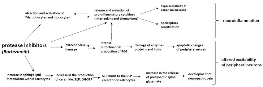
Int. J. Mol. Sci. 2019, 20, 1451                                                                    4 of 29

the cumulative dose of 500–600mg/m2 , CisIPN occurs in 92% of patients [26]. Epidemiological data
show that the incidence of neuropathic symptoms for cisplatin ranges from 49% to 100%, while chronic
CisIPN has been observed in 5–20% of patients at 12 months after therapy [15,27]. The severity of
CisIPN and the likelihood of chronicity increases with higher cumulative doses and longer exposure
times to cisplatin [28]. The development of CisIPN seems to be independent of pretreatment, age, sex,
tumor type and cotreatment with other chemotherapeutics [15,29]. Carboplatin seems to be less toxic,
with neuropathy observed in 13–42% of patients [30]. Therapy with oxaliplatin may induce side effects
such as myelotoxicity and enteric and peripheral neuropathy (OIPN, oxaliplatin-induced peripheral
neuropathy). Acute, transient OIPN develops in almost 65–98% of patients within hours of oxaliplatin
infusion at a dose ranging from 85 to 130 mg/m2 and may last up to 5–7 days. In patients receiving
12 cycles of chemotherapy, symptoms may persist up to 21 days or longer [31,32]. The symptoms of
acute OIPN include cold-related paresthesias of the hands and feet, pharyngolaryngeal dysesthesias,
jaw spasms, fasciculations and muscle cramps [33]. Cold-induced neuropathy after oxaliplatin is a
unique feature of OIPN, and this is the most important difference in the clinical presentation between
oxaliplatin and cisplatin-induced neuropathy [34]. Attal et al. have shown that the duration of cold-
(and touch-) evoked pain reported during the first three cycles was associated with the extent of the
chronic form of OIPN experienced one year later [35]. The chronic form, described as a pure sensory,
axonal neuropathy, with a classical stocking-and-glove distribution, is observed in 50–70% of patients,
but the incidence depends on the time point after oxaliplatin treatment and the intensity of symptoms
assessed [33,36–38]. According to data presented in a systematic review by Beijers et al. [39], OIPN
may be present in 26–46% of patients at the 12-month follow-up, in 24% of patients at the 15–18-month
follow-up or even in 84% of patients at the 24-month follow-up, which has been shown in the study of
Briani et al. [40].
     The most important risk factors of acute and chronic OIPN include the cumulative oxaliplatin
dose, the 2 h time of infusion, low body weight, younger age, a body surface area > 2,0, gene
variations (GSTP1, glutathione-S-transferase genes P1; GSTM1, glutathione-S-transferase genes M1;
and voltage-gated sodium channel genes SCN4A, SCN9A and SCN10A) and peripheral neuropathy
symptoms prior to chemotherapy [41–44]. High-grade chronic OIPN occurs in approximately 10% of
patients receiving cumulated doses ranging from 510 to 765 mg/m2 , while at doses higher than 1000
mg/m2 , this condition may be present in almost 50% of patients [45]. It is known that the acute form
of OIPN is also a risk factor for chronic OIPN; a higher intensity of the acute form results in a higher
incidence of chronic neuropathy [10].
     The antineoplastic mechanisms of platinum-based chemotherapeutic action include the
following [46–50]:

•     The binding to nuclear DNA (deoxyribonucleic acid) by cancer cells and the formation of
      DNA-platinum adducts, resulting in the inhibition of DNA replication and RNA (ribonucleic
      acid) transcription, followed by the arrest of cancer cell division, with the DNA adducts activating
      apoptotic pathways that induce cell death and tumor degradation;
•     The alteration of mitochondrial function followed by the disruption of the respiratory chain
      function and an increased production of reactive oxygen species (ROS);
•     The inhibition of mitochondrial DNA replication and transcription, leading to an altered
      mitochondrial function and the activation of apoptosis;
•     The activation of the immune system (macrophages, T-cells and monocytes) followed by the
      release of pro-inflammatory cytokines and the activation of apoptosis;
•     The influence on calcium signaling pathways and the function of protein kinase families (MAPK,
      mitogen activated protein kinases; JNK, c-Jun N-terminal kinase; PKC, protein kinase C; AKT,
      serine-threonine kinases), leading to tumor cell apoptosis.

     The exact mechanism of peripheral neuropathy induced by platinum-based chemotherapeutics
is not yet fully understood; however, it seems that their antitumor mechanisms are responsible for
Int. J. Mol. Sci. 2019, 20, 1451                                                                                       5 of 29

       Int. J.
      the      Mol. Sci. 2019,effect,
            neurotoxic         20, x FOR  PEER
                                       since   REVIEW
                                             chemotherapeutics                                                 5 of 29
                                                                 induce numerous changes either in the structure     or
      functioning of neuronal and glial cells [51]. Chemotherapeutic agents induce several alterations in
199    intracellular organelles
      intracellular       organelles (particularly
                                         (particularly mitochondria),
                                                       mitochondria), membrane
                                                                      membranereceptors
                                                                                receptorsand
                                                                                          andion
                                                                                               ionchannels,
                                                                                                   channels,followed
                                                                                                             followed
200   by alterations of the intracellular homeostasis, signaling and neurotransmission,all
       by   alterations     of   the  intracellular homeostasis, signaling and neurotransmission,   allofofwhich
                                                                                                            whichmay
                                                                                                                  may
201    resultin
      result    inneuroinflammation,
                    neuroinflammation, DNA     DNA damage
                                                     damage and
                                                             and axonal
                                                                  axonal degeneration
                                                                         degeneration (Figure
                                                                                      (Figure 1).
                                                                                              1).

202
            Figure 1. The mechanisms of chemotherapy-induced peripheral neuropathy (CIPN) induced by
203          Figure 1. The mechanisms of chemotherapy-induced peripheral neuropathy (CIPN) induced by
            platinum-based drugs: Platinum-based drugs induce the activation of glia cells, which leads to
204          platinum-based drugs: Platinum-based drugs induce the activation of glia cells, which leads to the
            the activation of the attraction and activation of immune cells and to the release and elevation of
205          activation of the attraction and activation of immune cells and to the release and elevation of
            pro-inflammatory cytokines (interleukins and chemokines), which results in nociceptor sensitization
206          pro-inflammatory cytokines (interleukins and chemokines), which results in nociceptor sensitization
            and hyperexcitability of peripheral neurons, and (together with ROS) damage the blood–brain barrier.
207          and hyperexcitability of peripheral neurons, and (together with ROS) damage the blood–brain
            These processes lead to the development of neuroinflammation. Mitochondrial damage caused by
208          barrier. These processes lead to the development of neuroinflammation. Mitochondrial damage
            platinum-based drugs leads to an increased production of reactive oxygen species (ROS), which leads to
209          caused by platinum-based drugs leads to an increased production of reactive oxygen species (ROS),
            enzyme, protein and lipid damage within neurons as well as the dysregulation of calcium homeostasis,
210          which leads to enzyme, protein and lipid damage within neurons as well as the dysregulation of
            which induces apoptotic changes in peripheral nerves and in DRG cells. Platinum-based drugs also
211          calcium homeostasis, which induces apoptotic changes in peripheral nerves and in DRG cells.
            alter the activity Na+, K+ and TRP ion channels, resulting in the hyperexcitability of peripheral neurons.
212          Platinum-based drugs also alter the activity Na+, K+ and TRP ion channels, resulting in the
            All of the above-described processes have the potential to alter the excitability of peripheral neurons.
213          hyperexcitability of peripheral neurons. All of the above-described processes have the potential to
214          alter the excitability of peripheral neurons.
            The unique feature of oxaliplatin is rapid nonenzymatic transformation to reactive platinum
      complexes and leaving-group oxalate. Oxalate has been proposed to contribute to acute cold-induced
215          The unique feature of oxaliplatin is rapid nonenzymatic transformation to reactive platinum
      neuropathy, which is most characteristic of oxaliplatin treatment. The study performed in laboratory
216    complexes and leaving-group oxalate. Oxalate has been proposed to contribute to acute
217   animals   by Pereira
       cold-induced         et al. showed
                        neuropathy,     whichthatiseither
                                                      mostoxaliplatin    or its oxalate-free
                                                              characteristic     of oxaliplatin  cytotoxic    analogue
                                                                                                      treatment.      The induced
                                                                                                                             study
218   peripheral    sensory  neuropathy,     although    oxalate   led  to  a  partial  and    later  decrease
       performed in laboratory animals by Pereira et al. showed that either oxaliplatin or its oxalate-free        in  mechanical
219   threshold
       cytotoxic in  comparison
                   analogue        withperipheral
                             induced     oxalate-free    analogue
                                                      sensory        [52].
                                                                 neuropathy,      although oxalate led to a partial and
220         Platinum   agent-induced      peripheral    neuropathy
       later decrease in mechanical threshold in comparison with        is initiated    by the
                                                                              oxalate-free        accumulation
                                                                                              analogue      [52].     of platinum
221   adducts    in dorsal
             Platinum       root ganglion
                        agent-induced          (DRG) neuropathy
                                          peripheral    and trigeminal       ganglion
                                                                       is initiated    by (TG)    neurons. This
                                                                                           the accumulation              process is
                                                                                                                    of platinum
222   probably    the major  mechanism      responsible    for  the  initiation   of  neurotoxicity
       adducts in dorsal root ganglion (DRG) and trigeminal ganglion (TG) neurons. This process           induced    by   this class
                                                                                                                                 is
223    probably the major mechanism responsible for the initiation of neurotoxicity induced by this class of
      of  chemotherapeutics      [53–55].    The   results   of Fujita   et al.   indicate    that    oxaliplatin    transporters
224    chemotherapeutics
      Octn1   and Mate1 are[53–55].
                               involved Theinresults
                                              platinum of Fujita  et al. indicate
                                                           accumulation       in DRG thatneurons,
                                                                                           oxaliplatin      transporters
                                                                                                      followed     by OIPN. Octn1
                                                                                                                                The
225    and Mate1 are involved in platinum accumulation in DRG neurons, followed by OIPN. The
      accumulation     of platinum    has  been   observed     in cells  overexpressing       Octn1     and   Mate1,     which   are
226    accumulation of platinum has been observed in cells overexpressing Octn1 and Mate1, which are
      correlated   proportionally   with  the  severity   of neuropathic     behavior    in  laboratory      animals    [56].
227    correlated   proportionally
            The exact  mechanism with      the severity ofagent
                                     of platinum-based       neuropathic     behavior
                                                                   neurotoxicity         in laboratory
                                                                                     in humans      is still animals
                                                                                                             discussed;[56].
                                                                                                                           however,
228          The exact mechanism of platinum-based agent neurotoxicity in humans is still discussed;in
      experimental    and  preclinical  studies   have  provided    insight    into the  processes     most    likely  involved
229    however, experimental
      neuropathy     pathogenesis.and   preclinical
                                     Some    of thesestudies   have provided
                                                       mechanisms                  insight
                                                                       are discussed      ininto
                                                                                             more  the  processes
                                                                                                      detail  below.most likely
230    involved in neuropathy pathogenesis. Some of these mechanisms are discussed in more detail
231    below.

232   2.1. Mitochondrial Dysfunction and Oxidative Stress
233       Mitochondrial dysfunction and oxidative stress have been highlighted as key players in the
234   pathophysiology of platinum-induced neuropathy. After entering neuronal and nonneuronal cells,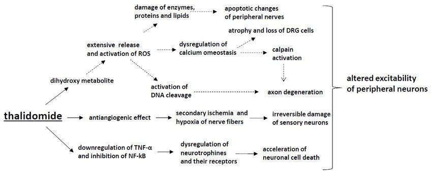
Int. J. Mol. Sci. 2019, 20, 1451                                                                      6 of 29

2.1. Mitochondrial Dysfunction and Oxidative Stress
      Mitochondrial dysfunction and oxidative stress have been highlighted as key players in the
pathophysiology of platinum-induced neuropathy. After entering neuronal and nonneuronal cells,
oxaliplatin and cisplatin bind to mitochondrial DNA (mDNA) and form mDNA adducts. These
pathological products cannot be repaired because there is no DNA repair system in mitochondria.
Platinum-mDNA adducts impair the physiological replication and transcription of mDNA, which
may lead to the synthesis of abnormal proteins, resulting in the impairment of the respiratory chain in
mitochondria [48,53,57–59]. The impairment of the mitochondrial physiological function leads, in turn,
to decreased cellular metabolism, to an increased production of ROS (reactive oxygen species) and to
oxidative stress [60–63]. Imai et al. showed that cisplatin and oxaliplatin may induce mitochondrial
dysfunction in cultured Schwann cells [64]. Oxaliplatin was shown to significantly increase superoxide
anion production and to induce lipid peroxidation, protein and DNA oxidation in both sciatic nerves
and the spinal cord in an in vitro study [65]. Further inhibition of cellular metabolism and pathological,
high intracellular levels of ROS may, in turn, lead to damage to enzymes, proteins and lipids, resulting
in structural changes of peripheral nerves [66]. Oxidative stress followed by apoptotic changes has
been observed in the sciatic nerves of cisplatin-treated mice [67].
      ROS can also activate the apoptotic pathway in neuronal cells through mitochondrial pathway
stimulation, including caspase activation and dysregulation of calcium homeostasis, resulting in
atrophy and loss of DRG cells [68,69]. The data from studies in vitro or in laboratory animals showing
a hypotrophy of the DRG along with neuronal atrophy seem to be contrary to the hypertrophy of
DRG found in MRI in patients treated with oxaliplatin [70]. However, the differences observed in the
structural abnormalities of patients’ DRG versus laboratory animals’ DRG may depend on the moment
of assessment and time after oxaliplatin treatment.

2.2. Intracellular Signaling
      Impairment in the physiological function of mitochondria may influence calcium signaling
pathways and promote further pathological functional and structural changes in neuronal and glial
cells. Mitochondrial and endoplasmic reticulum integrity, as intracellular stores of Ca2+ , are crucial
for Ca2+ homeostasis, since changes in the intracellular Ca2+ concentration may influence membrane
excitability, neurotransmitter release and gene expression of neuronal and glial cells [71]. An increase
in the intracellular Ca2+ concentration may result in calpain (potent protease) activation, which leads to
unregulated proteolysis, directly triggering axon degeneration [72]. The oxaliplatin metabolite oxalate
is a Ca2+ chelator and is likely involved in the pathogenesis of OIPN. The chelation of extracellular Ca2+
ions leads to an increase in Na+ conductance and a reduction in the threshold potential and membrane
resistance [34], resulting in the early phase of cold allodynia but not late mechanical allodynia. [73]. The
activation of protein kinases and caspases by chemotherapeutics may result in damage to intracellular
structures. Cisplatin and oxaliplatin can also produce MAPK-related apoptosis in DRG neurons, and
MAPK inhibitors may prevent DRG damage induced by platin-based agents in vitro [74]. The newly
discovered role of adenosine kinase in OIPN is described further in Section 2.4, Glial Cells.

2.3. Ion Channels
      Disturbances in the neuronal and glial functioning (membrane excitability and release of
neurotransmitters), resulting clinically in the development of peripheral neuropathy, may be partially
induced by the altered action of sodium channels (NaV), potassium channels (KV) and transient
receptor potential (TRP) channels. The voltage-gated sodium channels are necessary to facilitate the
initiation and propagation of action potentials in neurons and other excitable cells. Mutations in the
genes SCNA (sodium voltage-gated channel alpha subunit) encoding proteins forming NaV channels
lead to various diseases of the central and peripheral nervous systems, i.e., primary erythromelalgia,
small fiber neuropathy or insensitivity to pain [75]. Palugulla et al. confirmed that the presence of
Int. J. Mol. Sci. 2019, 20, 1451 7 of 29
polymorphisms in genes SCN4A (rs2302237), SCN9A (rs6746030) and SCN10A (rs12632942) predicts
either the development or the severity of chronic OIPN in cancer patients, while patients with mutations
in the SCN9A rs6754031 variant allele have a lower risk of severe chronic peripheral neuropathy
development. [44]. A study performed by Sittl et al. showed an increase in the Na+ current in rodent
peripheral axons and DRG neurons, with isoform NaV1.6 being involved in the development of
oxaliplatin-induced cold allodynia [76]. Altered NaV channel function induced by oxaliplatin has been
observed and confirmed further in numerous experimental in vivo and in vitro studies. [77,78].
Oxaliplatin injected directly into the rat hind paw leads to intense, short-duration mechanical
and cold allodynia, which has been suggested to be a direct action of oxaliplatin on NaV channels on
peripheral nerves but not on NaV channels in brain slices in vitro [79]. Experimental studies in vivo
confirmed the involvement of NaV1.7 channels [80] as well as NaV1.9 channels [81] in OIPN. In a
study by Heide et al., oxaliplatin induced the reversible slowing of sodium channel inactivation in
motor axons, and these changes were strictly related to reversible cold allodynia [82]. Acute OIPN
observed in clinical practice may be considered a cold-related acute channelopathy, mainly related to
NaV channels, as not only paresthesia but also muscle spasms and cramps are present, which could be
attributed to disturbances in NaV channel properties in both neurons and muscle cells [83].
Potassium (KV) channels are also involved in OIPN development, which has been shown in the
study of Descoeur et al. and subsequently confirmed in a study by Poupon et al. [84,85]. A single
administration of oxaliplatin to mice induced neuronal hyperexcitability, decreasing the expression
of potassium channels, TREK1 and TRAAK (members of the two-pore domain potassium channels
K2P subfamily) in DRG neurons. The oxaliplatin-induced downregulation of KV channels in cortical
and DRG neurons in vitro was shown in a study by Thibault et al., which might contribute to the
enhanced neuronal membrane excitability [86]. In a recent study by Viatchenko-Karpinski et al. [55],
it was shown for the first time that oxaliplatin leads to a significant downregulation of the KV4.3
channel expression in trigeminal V2 neurons. These changes in KV4.3 channels resulted in an increase
in membrane excitability, which could explain the cold allodynia in the orofacial region observed in
cancer patients.
Transient receptor potential (TRP) channels are nonselective cation channels that detect a vast
array of signals (thermal, mechanical and chemical). TRP channels play an important role in DRG
neurons and, thus, may be involved in the pathogenesis of OIPN. The TRPA1 (TRP ankyrin), TRPV1
(TRP vanilloid) and TRPM8 (TRP melastatin) channels are expressed in DRG neurons, and a few
preclinical studies have shown that they play a crucial role in cold and mechanical sensitivity induced
by oxaliplatin and cisplatin. Exposure to oxaliplatin and cisplatin results in the altered expression
and function of TRPV1, TRPA1 and TRPM8 channels in DRG neurons of laboratory animals [87,88].
TRPA1 channels can be activated by noxious cold (Int. J. Mol. Sci. 2019, 20, 1451 8 of 29
2.4. Glial Cells
Recent studies indicate that glial cells may also contribute to peripheral neuropathy induced by
platinum-based agents in animal models; however, it is not clear whether this mechanism may also be
involved in chemotherapy-induced neuropathy in cancer patients [97–99]. Oxaliplatin can activate
astrocytes, and the reduction of activation by minocycline decreases the intensity of neuropathic pain
behavior in rats [100].
Adenosine is a potent neuroprotective agent. Adenosine signaling at its adenosine receptors (ARs)
is driven by adenosine kinase (ADK) in astrocytes. In a study by Wahlman et al., oxaliplatin in rodents
caused ADK overexpression in activated astrocytes and reduced adenosine signaling at the A3AR
subtype (A3AR) within the spinal cord. The dysregulation of adenosine signaling was associated
with an increased proinflammatory and neuroexcitatory interleukin-1β expression [101]. These results
confirm the involvement of glial cells in the pathogenesis of OIPN.
Interestingly, oxaliplatin in an in vitro study did not activate microglia but surprisingly reduced
the number of microglial cells [102]. The results from experimental studies are still not consistent
because the activation of microglial cells has been observed in another study [103]. In a study by Imai
et al., it was shown that cisplatin and oxaliplatin may induce mitochondrial dysfunction in cultured
Schwann cells, followed by numerous disturbances in molecular function and cellular structure
contributing to peripheral neuropathy [64,104].
2.5. Inflammatory Mediators—Cytokines and Chemokines
The results from studies in human neuropathic pain and experimental animal models clearly
show that the activation of glial cells and the subsequent release and elevation of pro-inflammatory
cytokines (PIC): The IL-1b, IL-6 and TNF-a levels are common mechanisms of neuropathic pain
induced by chemotherapeutics [99,105–109]. The release of cytokines induced by chemotherapy may
be related to the ability of these agents to activate the Toll-like receptor (TLR) family, especially TLR4.
In knockout mice lacking that receptor, the pain behavior induced by cisplatin was decreased [110].
Pro-inflammatory cytokines can lead to the sensitization of nociceptors by the modulation of ion
channel properties, which has been confirmed in the study of Jin et al. [111].
Pro-inflammatory cytokines released from glial cells not only act in the peripheral nervous
system but also at the spinal and supraspinal levels. In the study of Xu et al., the administration of
oxaliplatin induced the activation of proinflammatory cytokines (PIC): IL-1b, IL-6, and TNF-α and
their receptors in periaqueductal gray matter (PAG). Blocking the PIC receptors decreased neuropathic
pain behavior induced by the administration of oxaliplatin. Additionally, PIC decreased the activity of
GABA (gamma-aminobutyric acid)-ergic-mediated descending inhibition, probably by the damage of
neuronal cells expressing GABA within periaqueductal gray matter (PAG) in the process of apoptosis.
GABA is one of the most potent antinociceptive neurotransmitters, and a correct GABA transmission
attenuated the mechanical and cold allodynia in this study. An enhanced release of PIC and subsequent
decrease of GABA transmission in PAG are likely to contribute to the development of mechanical and
cold hypersensitivity in oxaliplatin-treated animals [112].
Chemokines and their receptors are also involved in the pathogenesis of chemotherapy-induced
peripheral neuropathy [113]. CCLs (CC chemokine ligands) are responsible for the migration
and infiltration of monocytes/macrophages and other immune cells, thus contributing to
neuroinflammation and pain behavior in animal models [102]. Oxaliplatin can increase the level
of the CCL-2 chemokine, primarily released from astrocytes, and the level of CCL-2 is correlated with
the degree of hyperalgesia observed in rats [114]. The involvement of CCL-2 and its receptor CCR2
in neuropathy induced by oxaliplatin has been confirmed in the experimental study of Illias et al.
CCL2 and its receptor CCR2 were increased in the DRG after a single oxaliplatin administration and in
parallel with the development of mechanical hypersensitivity [115]. CCL2 increases the sensitivity of
neurons in other models of neuropathic pain, and probably the same mechanism can be observed in
neuropathy induced by chemotherapeutics [116]. Other studies have confirmed the role of chemokineInt. J. Mol. Sci. 2019, 20, 1451 9 of 29
CXCL12 [117] and chemokine CX3CL1 signaling in OIPN [118,119]. The hyperexcitability of DRG
neurons likely arises from the direct effect of CX3CL1 signaling on the function of ion channels in DRG
neurons [118].
2.6. Central Mechanisms
In recent years, many studies have shown that chemotherapy in cancer patients can influence their
cognitive and motor functioning. These data resulted in the description of a new neuropsychological
syndrome associated with cancer treatment/chemotherapy-induced cognitive impairment [120–123].
Whether the abnormalities in the central nervous system induced by chemotherapeutics may contribute
to peripheral neuropathy development and maintenance in humans is still unclear.
The blood–brain barrier (BBB) has been thought to prevent the access of oxaliplatin to the
brain [124]; however, a direct action of oxaliplatin on BBB endothelial cells (EC) has not been ruled
out. The possible mechanisms of BBB damage may include proinflammatory cytokines, ROS or other
neurotransmitters, all of which are involved in the peripheral nervous system toxicity induced by
chemotherapeutics [125,126]. In the study of Branca et al. [127], oxaliplatin administration in vitro
induced significant changes in the junctional and cytoskeletal apparatus of endothelial cells, and
these alterations of BBB may be associated with higher concentrations of oxaliplatin in the brain
and probably contribute to pain chronification. The study of Sanna et al. shows that oxaliplatin
administration induces changes in the levels of proteins in spinal and supraspinal levels in laboratory
animals and suggests a direct correlation between structural changes in the central nervous system
and chemotherapy-induced neurotoxicity [128].
3. Immunomodulatory Drugs (Thalidomide)
Thalidomide is a glutamic acid derivative and immunomodulatory drug that is approved by the US
Food and Drug Administration for the treatment of multiple myeloma [129]. The anticancer mechanism
of immunomodulatory drugs is poorly understood but may include the blocking of the production of
tumor necrosis factor alpha (TNF-α), the blocking of the activation of NF-kB (nuclear factor kappaB)
and the subsequent acceleration of neuronal cell death [130]. The second crucial anticancer mechanism
of thalidomide is its antiangiogenic effect by blocking angiogenesis through the inhibition of basic
fibroblast growth factor (b-FGF) and vascular endothelial growth factor (VEGF) [131]. Although the
thalidomide effectiveness in multiple myeloma patients improves the malignancy treatment outcome,
thalidomide-induced side effects may decrease the patients’ quality of life.
Thalidomide-induced peripheral neuropathy (TIPN) occurs in 25–75% of patients, with
dose-dependent prevalence and severity [132]. The risk of neurotoxicity increases in a dose-dependent
manner up to a cumulative dose of 20 g. In practice, thalidomide is administered at a maximum dose
of 200 mg daily and for a limited treatment duration [133].
In addition to the classical sensory symptoms and signs of peripheral neuropathy (stocking
and glove distribution), in severe cases of TIPN, thalidomide may induce motor impairment and
gastrointestinal and cardiovascular autonomic manifestations as well [132]. In approximately 15% of
patients, TIPN may lead to treatment discontinuation [134].
The data on TIPN prevalence also comes from studies on treatments with thalidomide in patients
with inflammatory diseases. In the pediatric population with inflammatory bowel disease, TIPN
was found in 72.5% of patients, but the prevalence depended on the time after treatment; thus, the
percentage of TIPN-free patients was 70.0% at 12 months and 35.6% at 24 months of treatment. The risk
of TIPN increased parallel to the mean daily dose, and TIPN was the cause of drug discontinuation in
41.8% of patients. These data confirm a dose- and time-dependent manner of TIPN development [135].
Risk factors for TIPN in multiple myeloma patients include advanced age, prior neuropathy
caused by myeloma by itself or other drugs [132]. The data on the role of genetics in predicting the
risk of TIPN are not consistent [136], although in the clinical study of Johnson et al., some genetic
susceptibility has been proposed: the ADME gene group (drug Absorption, Distribution, MetabolismInt. J. Mol. Sci. 2019, 20, x FOR PEER REVIEW 10 of 29
Int. J. Mol. Sci. 2019, 20, 1451 10 of 29
436 observed in a study by Badros et al. [138], where thalidomide was neuroprotective in patients
437 receiving it in combination with bortezomib. Since the anticancer mechanism of action of
438 thalidomide
and Excretion), is cytochromes,
an antiangiogenicsolute effect,
carrier this
familyprocess
genes,isand
also proposed
genes involvedto in
beneural
responsible for and
processes the
439 secondary
central ischemia
nervous system anddevelopment.
hypoxia of nerve It hasfibers, followed
also been by irreversible
suggested that the risk damage
of TIPN ofissensory neurons
associated with
440 [139,140].
single nucleotide polymorphisms (SNPs) in genes responsible for repair mechanisms and inflammation
441 in theItperipheral
has been proposed that the[137].
nervous system immunomodulatory effect can also contribute to TIPN (Figure 2).
442 Thalidomide may reduce neuronal
The pathophysiology of TIPN cell survival
remains by theunderstood,
not fully downregulation but it of TNF-α
has and the inhibition
been proposed that the
443 of NF-kB, resulting in the dysregulation of neurotrophins and their receptors
antiinflammatory effect of thalidomide may partially prevent neurotoxicity. This effect has been and the subsequent
444 acceleration
observed in aof neuronal
study cell death
by Badros [141].where
et al. [138], However, in experimental
thalidomide studies in in
was neuroprotective a patients
neuropathic pain
receiving
445 it in combination with bortezomib. Since the anticancer mechanism of action of thalidomide is the
model, the administration of thalidomide decreased mechanical hyperalgesia after injury of an
446 sciatic nerve in
antiangiogenic micethis
effect, byprocess
reducing TNF-α
is also levels
proposed in responsible
to be the sciatic for nerve, prefrontalischemia
the secondary cortex and
and
447 hippocampus;
hypoxia of nervethus, the
fibers, mechanism
followed of TIPN damage
by irreversible may beofrelated
sensory to other [139,140].
neurons processes induced by
448 thalidomide [142].
It has been proposed that the immunomodulatory effect can also contribute to TIPN (Figure 2).
449 In a study
Thalidomide may byreduce
Wani neuronal
et al., it was shown that
cell survival thedownregulation
by the dihydroxy metabolite
of TNF-α ofand
thalidomide was
the inhibition
450 capable
of NF-kB, of resulting
causing extensive redox-activated
in the dysregulation DNA cleavage,and
of neurotrophins andtheir
this mechanism
receptors and hasthebeen proposed
subsequent
451 to be crucial for thalidomide-induced teratogenesis. DNA cleavage was
acceleration of neuronal cell death [141]. However, in experimental studies in a neuropathic pain mediated through the
452 formation
model, the of ROS, as discussed
administration previously.
of thalidomide ROS-dependent
decreased mechanicalmechanisms
hyperalgesiahave been confirmed
after injury in
of the sciatic
453 platinum-based chemotherapeutic neurotoxicity development, but further
nerve in mice by reducing TNF-α levels in the sciatic nerve, prefrontal cortex and hippocampus; thus, preclinical and clinical
454 trials
the are neededoftoTIPN
mechanism confirm
maythis mechanism
be related in TIPN
to other [143].induced by thalidomide [142].
processes
455
456 Figure Themechanisms
2. The
Figure 2. mechanismsofofCIPNCIPN induced
induced byby thalidomide:
thalidomide: Thalidomide
Thalidomide downregulates
downregulates TNF-α
TNF-α and
457 and inhibits NF-κB, which leads to the dysregulation of neurotrophins and their
inhibits NF-κB, which leads to the dysregulation of neurotrophins and their receptors and,receptors and,
in
458 in consequence,
consequence, accelerates
accelerates neuronalcell
neuronal celldeath.
death.Moreover,
Moreover,the
theantiangiogenic
antiangiogenic effect
effect induced
induced by
by
thalidomide causes secondary ischemia and hypoxia of nerve fibres and, subsequently, irreversible
459 thalidomide causes secondary ischemia and hypoxia of nerve fibres and, subsequently, irreversible
damage of sensory neurons. The activation of the dihydroxy metabolite of thalidomide causes the
460 damage of sensory neurons. The activation of the dihydroxy metabolite of thalidomide causes the
extensive release and activation of ROS and activates DNA cleavage, though further preclinical
461 extensive release and activation of ROS and activates DNA cleavage, though further preclinical and
and clinical trials are needed to confirm the presence of such a mechanism in thalidomide-induced
462 clinical trials are needed to confirm the presence of such a mechanism in thalidomide-induced
peripheral neuropathy.
463 peripheral neuropathy.
In a study by Wani et al., it was shown that the dihydroxy metabolite of thalidomide was capable
464 4. Taxanes
of causing extensive redox-activated DNA cleavage, and this mechanism has been proposed to be
465 Taxanes
crucial constitute a class of
for thalidomide-induced antineoplastic
teratogenesis. DNA drugs actingwas
cleavage on mediated
microtubules, interfering
through with the
the formation of
466 normal
ROS, as cycling
discussedof microtubule depolymerization
previously. ROS-dependent and repolymerization,
mechanisms which causes
have been confirmed impairment of
in platinum-based
467 cancer cell divisionneurotoxicity
chemotherapeutic and consequently leads to cell
development, butdeath.
furtherThis class includes
preclinical paclitaxel,
and clinical trials docetaxel
are neededand
to
468 cabazitaxel.
confirm They have in
this mechanism been
TIPN approved
[143]. by the FDA for the treatment of various cancer types,
469 including ovarian cancer, breast cancer, non-small cell lung cancer and prostate cancer [144].
470 The incidence of CIPN from taxanes may be very high and ranges from 11 to 87%, with the
4. Taxanes
471 highest rates constitute
Taxanes reported for paclitaxel
a class [6,145]. Neuropathy
of antineoplastic caused
drugs acting by taxanes usually
on microtubules, presents
interfering withasthea
472 sensory dominant neuropathy, mostly affecting small diameter sensory fibers, manifesting
normal cycling of microtubule depolymerization and repolymerization, which causes impairment of usually
473 as paresthesias,
cancer dysesthesias,
cell division numbness,
and consequently altered
leads to cellproprioception and
death. This class loss of dexterity
includes paclitaxel,predominantly
docetaxel andInt. J. Mol. Sci. 2019, 20, 1451 11 of 29
cabazitaxel. They have been approved by the FDA for the treatment of various cancer types, including
ovarian cancer, breast cancer, non-small cell lung cancer and prostate cancer [144].
The incidence of CIPN from taxanes may be very high and ranges from 11 to 87%, with the
highest rates reported for paclitaxel [6,145]. Neuropathy caused by taxanes usually presents as a
sensory dominant neuropathy, mostly affecting small diameter sensory fibers, manifesting usually as
Int. J. Mol. Sci.dysesthesias,
paresthesias, 2019, 20, x FOR PEER REVIEW altered proprioception and loss of dexterity predominantly
numbness, 11 of 29 in
the toes and fingers (stocking-and-glove distribution); however, other localizations may appear. Motor
474 in the toes and fingers (stocking-and-glove distribution); however, other localizations may appear.
and autonomic involvement is less frequent but may also develop [146]. The symptoms may start
475 Motor and autonomic involvement is less frequent but may also develop [146]. The symptoms may
476 days after
start daystheafter
first the
dose. They
first areThey
dose. doseare
dependent and tend
dose dependent to tend
and improve after stopping
to improve the treatment.
after stopping the
477 In some patients, symptoms can continue up to 1–3 years after completion of
treatment. In some patients, symptoms can continue up to 1–3 years after completion of the therapy the therapy and can
478 sometimes last lifelong
and can sometimes [147].
last lifelong [147].
479 Such Such symptoms are mostintense
symptoms are most intensefor
for paclitaxel.
paclitaxel. The
Thedocetaxel
docetaxelintensity of of
intensity symptoms
symptoms is milder [5]. [5].
is milder
480 Protein bound paclitaxel (Nab-paclitaxel), developed to reduce overall toxicity, does not yield a
Protein bound paclitaxel (Nab-paclitaxel), developed to reduce overall toxicity, does not yield a reduced
481 reducedofincidence
incidence CIPN [148]. of The
CIPN [148]. The of
mechanisms mechanisms
neurotoxicity of neurotoxicity of taxanes
of taxanes (Figure (Figure 3) areand
3) are multifactorial
482 multifactorial
include and include
the following the following pathways:
pathways:
483
484 Figure
Figure 3. The
3. The mechanisms
mechanisms of CIPN
of CIPN induced
induced by taxanes:
by taxanes: Taxanes
Taxanes cause
cause microtubule
microtubule disruption,
disruption, which
485 whichaxonal
impairs impairstransport
axonal transport
and leadsandtoleads to Wallerian
Wallerian degeneration,
degeneration, altered
altered activity
activity of of
ionion channelsand
channels
486 and hyperexcitability
hyperexcitability of peripheral
of peripheral neurons.neurons.
Taxanes Taxanes also modify
also modify the expression
the expression and function
and function of K+
of Na+,
487 andNa+,
TRPK+ ionand TRP ion
channels, channels,
which resultswhich
in the results in the hyperexcitability
hyperexcitability of peripheral
of peripheral neurons. neurons.
Taxane-induced
488 Taxane-induced
mitochondrial mitochondrial
damage contributesdamage contributes
to the increased to the increased
production production
of reactive oxygenof reactive
species oxygen
(ROS), which
489 species (ROS), which leads to enzyme, protein and lipid damage as well as the dysregulation
leads to enzyme, protein and lipid damage as well as the dysregulation of calcium homeostasis within of
490 calcium homeostasis within neurons, which induces apoptotic changes and the demyelination
neurons, which induces apoptotic changes and the demyelination of peripheral nerves. These processes of
491 peripheral
alter nerves.ofThese
the excitability processes
peripheral alter the
neurons. Theexcitability
activation of peripheral and
of microglia neurons. The activation
astrocytes by taxanesofalso
492 microglia and astrocytes by taxanes also leads to the activation of attraction and activation of
leads to the activation of attraction and activation of immune cells and to the release and elevation of
493 immune cells and to the release and elevation of pro-inflammatory cytokines (interleukins and
pro-inflammatory cytokines (interleukins and chemokines), which results in the nociceptor sensitization
494 chemokines), which results in the nociceptor sensitization and hyperexcitability of peripheral
and hyperexcitability of peripheral neurons. These processes lead to nociceptor sensitization and the
495 neurons. These processes lead to nociceptor sensitization and the development of
development of neuroinflammation.
496 neuroinflammation.
4.1. Microtubule Disruption
497 4.1. Microtubule Disruption
Microtubule disruption is a principal mechanism of action of taxanes and is responsible for
498 Microtubule disruption is a principal mechanism of action of taxanes and is responsible for
their antineoplastic activity; however, it is also associated with the development of CIPN [149]. The
499 their antineoplastic activity; however, it is also associated with the development of CIPN [149]. The
aggregation and bundling of microtubules lead to changes in cell shape and cell stability but are also
500 aggregation and bundling of microtubules lead to changes in cell shape and cell stability but are also
501 responsible forfor
responsible the impairment
the impairmentofofaxonal
axonal transport ofsynaptic
transport of synapticvesicles
vesiclesloaded
loaded with
with essential
essential cellular
cellular
502 components, including lipids, proteins and ion channels [150–152].
components, including lipids, proteins and ion channels [150–152].
503 4.2. Mitochondrial Dysfunction
504 Damage to mitochondria, both in neuronal and nonneuronal cells, leads to oxidative stress and
505 the production of reactive oxygen species (ROS), such as hydroxyl radicals, peroxide, superoxide
506 and single oxygen. Impaired axonal transport of essential cellular components [151,153] and mRNAInt. J. Mol. Sci. 2019, 20, 1451 12 of 29
4.2. Mitochondrial Dysfunction
Damage to mitochondria, both in neuronal and nonneuronal cells, leads to oxidative stress and the
production of reactive oxygen species (ROS), such as hydroxyl radicals, peroxide, superoxide and single
oxygen. Impaired axonal transport of essential cellular components [151,153] and mRNA [154] to distal
neuronal parts due to microtubule disruption may have a significant impact on this process. Increased
levels of ROS have been detected in sensory neurons and the spinal cord [23,62,155–157]. Increased ROS
levels cause the activation of apoptotic processes, the disruption of cell structure and demyelinization.
These events lead to the impairment of signal transmission and the activation of immune processes,
including increased production of pro-inflammatory cytokines. The process is self-amplifying as the
above mechanisms can cause further mitochondrial damage [23,158–160]. Swelling, vacuolation and loss
of structure of mitochondria have been proven in a number of studies with paclitaxel [161,162].
4.3. Axon Degeneration
The direct damage of peripheral nerves, the loss of neuronal fibers and demyelinization caused
by paclitaxel have been reported in various studies [149,163–166]. Microtubule disruption and the
consequently impaired axonal transport of essential cellular components cause the degeneration of
distal nerve segments (Wallerian degeneration) and axonal membrane remodeling [167]. Boyette et al.
showed a decreased number of intraepidermal fibers in a rodent model of paclitaxel-induced CIPN [168].
Ferrari et al. described impaired corneal innervation in rats. Cytokine and chemokine signaling may
also play a role in axon degeneration. Zhang et al. showed that a decrease in the level of chemokine
MCP1/CCL-2 decreases nerve degeneration and CIPN behaviors in a rodent model [169].
4.4. Altered Calcium Homeostasis
The dysregulation of Ca2+ hemostasis has been shown to play a role in the pathogenesis of
CIPN [170]. The dysregulation of intracellular Ca2+ was observed in a paclitaxel neuropathy model
in both neuronal and nonneuronal cells [171–173]. Mitochondria and endoplasmic reticulum (ER) are
intracellular magazines of Ca2+ . Paclitaxel can cause the release of Ca2+ from mitochondria, and the
process is probably mediated by the activation of the mitochondrial permeability transition pore (mPTP),
leading to rapid mitochondria depolarization [172,173]. Paclitaxel can probably also stimulate the release
of Ca2+ from the ER; this process may be mediated by the 1,4,5-trisphosphate receptor (IP3R) [174,175],
leading to the increased expression of CaV3.2 channels in rats, with a suppression of these channels
reversing hyperalgesia [176].
4.5. Changes in Peripheral Nerve Excitability
The altered expression and function of ion channels (NaV, KV and TPR) is another mechanism
contributing to the development of CIPN. The decreased expression of K+ channels causing the
spontaneous activity of nociceptors was observed in the DRG in a paclitaxel-induced CIPN model [177].
The activation of cation channels TRPV1 and TRPA1, important components of pain signaling, was
detected in DRG neurons [178,179]. Antagonists of TRPA1 have been shown to relieve inflammation,
cold allodynia and hyperalgesia induced by paclitaxel [180]. Paclitaxel treatment increases the number of
NaV1.7 channels, which may be responsible for the development of CIPN [181,182]. Gheraldin et al. [80]
showed that blocking this channel attenuates hyperalgesia in rats.
4.6. Immune Processes and Neuroinflammation
Paclitaxel causes an increase in the production of pro-inflammatory cytokines (TNF alfa and
IL-1 beta) and a decrease in anti-inflammatory cytokines (IL-4 and IL-10) [23,155]. This process leads
to the attraction and activation of immune cells and the development of neuroinflammation [183].
Krukowski et al. [184] showed that IL-10 can attenuate paclitaxel-induced CIPN. Paclitaxel can also
lead to microglial and astrocyte activation [185,186] and an increase in macrophage number in DRGYou can also read