Central Locomotor Virtual Placements Student Guide 2020-2021 - QMplus
←
→
Page content transcription
If your browser does not render page correctly, please read the page content below
Central Locomotor Virtual
                 Placements
                         Student Guide
                             2020-2021

Lead Academics Dr. Dev Gadhvi (d.h.gadhvi@qmul.ac.uk) and Dr Rohini Sabherwal
(r.sabherwal@qmul.ac.uk )

Unit Moderator Mr. Jim Manzano (j.manzano@qmul.ac.uk )
Student Guide
During this extraordinary time we have been adapting our structured face-to-face teaching
to an online model. Our teaching sessions will of course run slightly differently. These
sessions are mandatory, attendance will be taken and remediation will be given if poor
attendance noted.
The aim of the module is to provide clinical knowledge of conditions commonly encountered
in primary care but also to give you the opportunity to develop and improve your
consultation skills.
Preparation for teaching session…
Please bring this student guide to each simulated surgery session, which you will
need to read in advance. The clinical notes and web links will provide you with
clinical information for the scenarios. You will also need to quick reference any
photos of rashes etc which are attached. You will find the consultations easier if you
have read up the clinical background beforehand.
Mondays ENT PM sessions are run slightly differently to the other days.
Pen and paper – to aid you with observing your colleagues and allow for more
constructive feedback
Ground rules and how the scenarios will work will be discussed in detail at the beginning of
the afternoon.
Please note during session –
Please switch mobiles off.
Please also switch off video/microphone if not speaking – this helps with overall
running of group but also with connectivity.
Please respect each others learning environment and follow good feedback practise
Group confidentiality and sensitivity – Any personal recording of the session is
prohibited. These sessions are also not recorded by QMUL.
If a scenario brings up particular sensitive/personal issues for you, please could you
let your GP tutor know at the beginning of the session to avoid being asked to role-
play that scenario. If any issues arise during or after the session, please inform your
GP tutor. You can use the chat function on the bottom right of the screen if you
require discretion. Click on your GP Tutors name and message them individually.
Please use chat function to ask questions or the raising hand function.
Remote Consulting - It is worth noting that the role-plays have not been changed to
reflect they are “video consultations”. This was to try and ensure learning in your
usual environment. It is worth discussing that there will still be possible examination
findings to discuss and possible rashes to describe.
These sessions provide a good opportunity to role-play with actors and receive constructive
feedback during your 4 year. We hope these will develop your consultation skills and also
th
enable you to start thinking about how you might investigate and manage patients. This
should help you in real-life situations with patients as an FY doctor and also in preparation
for OSCE examinations.
These sessions are facilitated by General Practitioners who are experienced in
communicating and consulting with patients and also have clinical knowledge and practical
wisdom to share regarding the reality of working as a doctor.
If your GP tutor has concerns about your consultation skills, they may address this with you
privately at the end of the session and inform the academic lead for the session. The
academic lead may contact you directly if any follow up is needed.
Please above all enjoy and GET INVOLVED!
Dermatology cases
Case 1 Dermatology – 3 year old boy with eczema
Student Information
Learning objectives
To understand the routine management of
eczema and to be able to explain this to patients and
their carers.
To understand the ways in which atopic
eczema may impact a patient and/or their family
psychosocially.
To manage parental anxiety and set realistic management goals to manage
moderate eczema
Student Instructions
You are a FY1 doctor on your GP attachment. During your morning surgery you are asked to
see Mrs/Mr Jones with her/his 3-year-old son. He has been irritable for the past 4 weeks,
constantly rubbing a rash on his arms and legs. He was seen by one of the partners 2 weeks
previously with the same complaint.
He was diagnosed with eczema and diprobase ointment, oilatum bath oil, aqueous cream
and hydrocortisone 1% ointment were prescribed. Mrs/Mr Jones feels that this has not
helped.
Take a history from the parent, ask to examine the rash (you will be shown a picture
of this)
Then explain your management plan. Please take into consideration how the
eczema could be better controlled as well as supporting Mrs/Mr Jones.
You may want to think about
Adequate use and application of prescribed medication
Is it necessary to increase medication, add in further medication?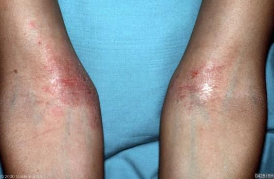
Looking for possible triggers (e.g. food allergy, atopic history).
Indications for referral
Patient quality of life
Key Points
Quality of life
Research on the impact of chronic skin disease on patient’s quality of life generally shows
that relative to the general population, patients report a lower level of psychological and
social well being. Consequently, when assessing a patient with a chronic skin disease it is
essential to explore the psychological and social impact of this condition on their life. It is
well documented that the quality of life of a parent with a child with atopic eczema can be
adversely impacted by the condition particularly when it is poorly controlled. NICE 2007
recommend asking about the effect of eczema on daily activities (school, work, and social
life), sleep, and mood and categorising the impact of eczema on quality of life and
psychosocial well-being in terms of
No impact on quality of life.
Mild: little impact on everyday activities, sleep, and psychosocial well being.
Moderate: moderate impact on everyday activities and psychosocial well-being, and
frequently disturbed sleep.
Severe: severe limitation of everyday activities and psychosocial functioning, and
loss of sleep every night.
Eczema – Atopic Eczema: NICE CKS 2007. Last revised in January 2018.
The stepped approach, recommended by the National Institute for Health and Care
Excellence (NICE), for the treatment of atopic eczema is shown in Table 1.
Treatment can be stepped up or down according to the severity of the condition. Treatment
of a flare will often require temporarily 'upping' the intensity of treatment (for example the
strength of corticosteroid).
Topical calcineurin inhibitors, phototherapy, and ciclosporin are less suitable for
the acute treatment of flares.
Bandaging and oral corticosteroids are unsuitable for maintenance treatment.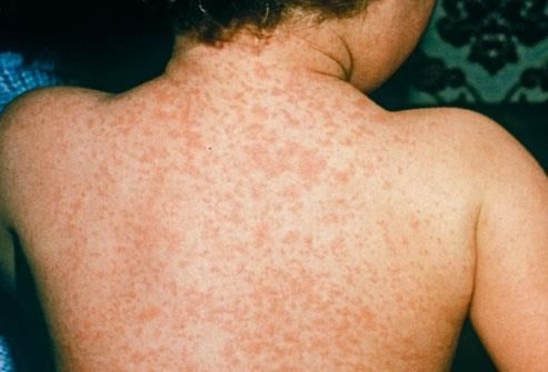
Table 1. Stepped treatment options for atopic eczema.

Mild atopic eczema               Moderate eczema                  Severe eczema
Emollients                       Emollients                       Emollients
Mild potency topical             Moderate potency topical         Potent topical
corticosteroids                  corticosteroids                  corticosteroids
                                 Topical calcineurin inhibitors   Topical calcineurin inhibitors
                                 (tacrolimus or                   (tacrolimus or
                                 pimecrolimus)*                   pimecrolimus)*
                                 Bandages*                        Bandages*

                                                                  Phototherapy†

                                                                  Oral corticosteroids‡

* Usually only prescribed by a specialist (for example a GP with a specialist interest in
dermatology, a dermatologist, or a paediatrician).

† Phototherapy is available in secondary care for the treatment of very severe eczema that
has proved resistant to standard treatment. Systemic immunosuppressants (for example
ciclosporin and azathioprine) are also available in secondary care for the same indication.

‡ Oral corticosteroids can be prescribed short-term in primary care for severe flares. Other
systemic treatments suitable for maintenance of severe eczema (for example ciclosporin or
azathioprine) require referral to secondary care.

With the use of potent immunosuppressive drugs initiated in Secondary Care and once
stabilised, there are often”Shared Care Guidance” between specialists and generalists.
Agreeing that the GP with take responsibility of prescribing the medication and any blood
monitoring etc. which is needed as a consequence.

Steroid Ladder

 Brand/Product Name            Generic Name                              Relative potency

 Dermovate                    Clobetasone Proprionate 0.05%              Very Potent

 Elocon                       Mometasone furorate 0.1%                   Potent

 Betnovate                    Beclomethasone valerate 0.1%

 Eumovate                     Clobetasone butyrate 0.05%                 Moderately Potent

 Hydrocortisone 1%            Hydrocortisone 1%                          Mild

Application of Emollients

Primary Care Dermatologist Society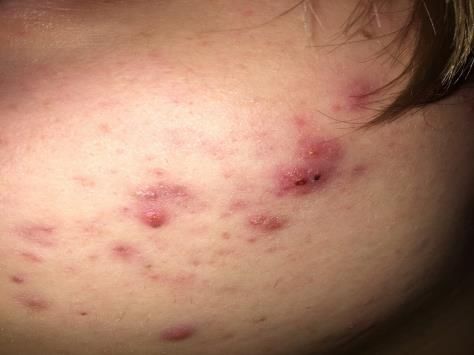
Step 1: general measures
As with other chronic skin conditions time is needed by the GP and / or practice
nurse to discuss the condition, advise on how best to use emollients and to provide
an individual management plan
Provide a patient information leaflet and a written management plan for the
patients/carers: e.g. British Association of Dermatologist (BAD)
Advise on a pre-payment certificate where appropriate
At each step it is essential to ensure patient compliance and to make sure that
copious amounts of emollients are being used
Step 2: initial management for patients presenting with a flare-up
In both children and adults it is more effective and safer to 'hit hard' using more
potent treatments for a few days than it is to use less potent treatments for longer
periods of time
Use a moderate to potent topical steroid.
For marked sleep disturbance consider the short-term use of sedating anti-
histamine. There is almost no role for non-sedating antihistamines in the
management of eczema, the only exception is patients needing treatment for co-
existent hay fever
Consider taking a skin swab if not settling
Review the patient in one to two weeks to discuss long-term management (see step
3 below)
Step 3: long-term management
Emollients - Complete Emollient Therapy
Emollients are the mainstay of therapy and without them it is not possible to
manage eczema effectively. Good evidence shows that the more emollients are
used, the less topical steroids are needed. Compliance is essential and so always
review patients to check they are happy with what has been prescribed - it may be
necessary to try a range of emollients before the patient settles on the best
combination.
Moisturisers
Most patients prefer creams and gels. The most important factor is to find one that
the patient likes and is happy to use
Ointments tend to be less well tolerated by patients, but they are less likely to cause
contact allergic dermatitis as they do not contain preservatives (this is for both
emollients and topical steroids)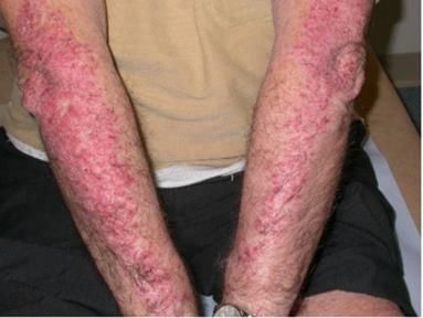
Encourage appropriate usage by prescribing generous amounts e.g. 500 grams of
moisturisers to use regularly (often QDS)
As with other topical treatments, moisturisers should be gently rubbed into the skin
until they are no longer visible. They should be applied downward in the direction of
the hairs to lessen the risk of folliculitis
Warn that they may sting for the first couple of days before soothing the skin
Ointments come in tubs and so can easily become cross infected with bacteria from
the skin - patients must not place hands into tubs but instead use a utensil to scoop
out the ointment
Order of application - if topical steroids are also being used, moisturisers can be
applied first and allowed to dry for 15-20 minutes before applying the topical
steroid
Application
Apply it liberally to the affected area of skin. Smooth it into the skin along the line of hair
growth, rather than rubbing them in. A full emollient regimen requires a bath oil, soap
substitute and regular emollient. They should all be used even when the rash resolves in
order to prevent relapses. Aim to apply the emollient liberally at least 2-4 times a day and
particularly after bathing. For optimum treatment it should be applied as often as the skin
looks dry. Advise patients should be using 200 -500g per week
Finger Tip Units (FTU)
In the UK most topical steroid tubes have a 5mm nozzle. Based on this, finger tip units (FTU)
can be calculated. An FTU is the amount of ointment/cream expressed from the distal skin
crease to the tip of the palmar aspect of the index finger. This is roughly the same in males
and females. One finger tip unit is enough to cover an area roughly equivalent to two hand
areas. If you have eczema covering the area of your palm, half a fingertip is needed of
topical steroid to cover this.
Useful references
Atopic eczema in children http://guidance.nice.org.uk/CG57
UK National Eczema Society www.eczema.org
Please note that a study in the BMJ 2018 showed not additional benefit in childhood
eczema with bath additives but soap free washes and leave on emollients continued
to show benefits. https://www.bmj.com/content/361/bmj.k1332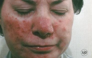
Case 2 Dermatology – 40 year old accountant with a longstanding rash on his/hers elbows
and knees
Student Information
Learning objectives
By the end of this station the student should
Have attempted to develop a shared management plan with a
simulated patient
Understand the basic principles of negotiating a shared management plan (See
Appendix 2)
Student Instructions
You are a FY1 Doctor on your GP attachment. Your next patient is a 40 year old man/women
with 2 year history of a rash that seems to be worsening. He/she has recently started
propranolol for stress at work and noticed that the rash got worse after starting this
medication. Sam Bailey.
Please speak to this patient in order to formulate a diagnosis and negotiate a management
plan.
You may want to consider general management:
Explanation of diagnosis to patient and patient given written information
Discuss treatment options, benefits and side effects and agree a management plan
Key points
Topical therapy: Try to keep the number of treatments per day to a minimum to improve
concordance
1st line treatments
Regular Emollient to reduce scale and itch
Topical steroids –short-term intermittent use of a potent topical steroid such as
beclomethasone 0.1% or in combination with calcipitriol e.g. dovobet
Vit D analoge eg. Dovonex
2nd line treatments
Coal tar (solution, cream or lotion)
Tazarotene gel
Short contact dithranol – for 30 minute exposures in patients with a few but
relatively large plaques
Useful references
Primary Care Dermatology Society: A –Z of Diagnosis: Psoriasis (including drugs
causing psoriasis) http://www.pcds.org.uk/p/a-z-of-clinical-guidance-how-to-use
http://www.patient.co.uk/doctor/Chronic-Plaque-Psoriasis.htm
Case 3 Dermatology – 50 year old landscape gardener with a
suspicious mole
Student Information
Learning objectives:
By the end of this tutorial the student should:
Be aware of two validated mole risk assessment tools (Glasgow seven point check
list and ABCDE system)
Have an attempt at discussing the implications of a suspicious mole and the next
steps in management.
This will involve breaking the news to the patient in a sensitive and tactful manner.
(See Breaking Bad News Framework in Appendix 3).
Student Instructions:
You are a FY1 doctor on your GP attachment. This 50 year old landscape gardener has come
to see you today because the mole on his/her back has become itchy and their partner told
them to.
Please take a history from this patient.
Describe the mole.
What is the likely diagnosis?
Please explain the next steps in management
Key Points
Malignant Melanoma of the Skin
Epidemiology
1. More common in women than men. 6th most common cancer in females and
males
2. Less common than non-melanoma skin cancer (BCC/SCC)
3. Incidence- women: 16.5/100,000. Men 15.9/100,000
4. Lifetime risk of developing melanoma in the UK 1in 61 for men and 1 in 60 for
women
5. Amongst white populations the incidence of malignant melanoma of the skin is
rising
6. Median age of diagnosis men 62 years of age, women 60 years of age
Distribution
Men: head and neck (22%), trunk (41%), arm (18%), leg (13%)
Women: head and neck (14%), trunk (19%), arm (23%), leg (40%)
Subtypes
Superficial spreading malignant melanoma – most common subtype (70% of all MM.
Most commonly presents on the trunks of men and legs of women)
Others: Nodular malignant melanoma, Lentigo malignant melanoma, Amelanotic
malignant melanoma, Acral lentiginous melanoma inc subungual – most common in
pigmented skins.
Risk Factors
PMH or FH of MM or DH of immunosuppresants e.g. Methotrexate, Ciclosporin etc. SH:
Higher socioeconomic class
Naevi are the most powerful predictor of melanoma.
A person with >100 has a 5-20 fold increased risk of MM
Sun exposure (short sharp burst of acute exposure in childhood, severe sunburn)
Occupation and leisure (gardeners, air crew, cricketers and those involved
with outdoor pursuits)
Past sunbed use particularly when 100 common moles (2mm in diameter) >two atypical naevi (5mm in diameter)
Naevi in unusual sites e.g. breast in females, buttocks, scalps, ears, dorsum
of feet and hands, irises
Useful references
M D Junction – Melanoma Support Group http://www.mdjunction.com/
Glasgow Seven point check list
Major features: Change in size, Irregular shape, Irregular colour
Minor features: Diameter >7mm, Inflammation, Oozing, Change in sensation
ABCDE system: Asymmetry Border irregularity Colour variation Diameter over 6 mm
Evolving (enlarging, changing)
Management in Primary care 2ww referral criteria for skin cancer.
Case 4 Dermatology – 32 year old with Acne
Student Information
Learning objectives
Being able to describe acne in dermatological terms and
have awareness of categories of severity.
Understand common steps to management.
Understand the psychological issues associated with acne.
Student Instructions
You are an FY1 in GP. This 31yr old women/man has come to see you today, she/he wishes
to discuss her/his acne. She/he saw your colleague 3-4 weeks ago and was prescribed a
topical treatment called DUAC (benzoyl peroxide/clindamycin).
Please take a history
Describe the rash
Discuss common management steps and follow up.
You will be shown a picture of her/his acne during the consultation and will be asked to
describe it by the tutor.
Key points
What are the clinical features of acne vulgaris? NICE CKSAcne affects areas of the body with a high density of pilosebaceous glands such as the face,
chest and back. Clinical features vary widely depending on severity and the person affected.
Comedones must be present for a diagnosis of acne to be made — if not present other
diagnoses should be considered.
Suspect acne in a person presenting with:
o Non-inflammatory lesions (comedones), which may be, open (blackheads)
or closed (whiteheads).
o Inflammatory lesions such as:
Papules and pustules
Nodules or cysts - In very severe acne nodules may track together
and form sinuses (acne conglobata).
Scarring
Pigmentation
Seborrhoea
There is no universally agreed scoring system for acne severity but categorising into
mild, moderate and severe can be helpful in selection of appropriate treatment and
monitoring of response:
Mild acne — predominantly non-inflamed lesions (open and closed
comedones) with few inflammatory lesions.
Moderate acne — more widespread with an increased number of
inflammatory papules and pustules.
Severe acne — widespread inflammatory papules, pustules and nodules or
cysts. Scarring may be present.
Images of acne and its clinical variants can be viewed at www.dermnet.org.nz.
How should I manage a person with acne vulgaris in primary care? NICE CKS
General advice
Cleaning — Acne is not caused by poor hygiene. Aggressive washing can
aggravate acne and should be avoided
Healthy diet — The role of diet in acne remains poorly understood — emerging
data suggests that high glycaemic index (GI) diets may exacerbate acne
Follow up
Several guidelines recommend follow up to determine the need for ongoing patient
education, escalation of treatment or maintenance therapy. Side-effects and lack of
knowledge about acne treatments are the two main reasons for non-adherence .
Maintenance therapy Maintenance therapy helps to prevent recurrence of acne by suppressing
development of microcomedones, which can be present in normal looking skin.
Explain the diagnosis and provide patient information:
Discuss treatment aims and advise the person:
To avoid over cleaning the skin (which may cause dryness and irritation).
If make-up, cleansers and/or emollients are used, non-comedogenic preparations
with a pH close to the skin are recommended.
To avoid picking and squeezing spots which may increase the risk of scarring.
That treatments are effective but take time to work (usually up to 8 weeks) and may
irritate the skin, especially at the start of treatment.
To maintain a healthy diet.
For people with mild-to-moderate acne:
Consider prescribing a single topical treatment such as:
A topical retinoid alone or in combination with benzoyl peroxide. Retinoids are
contraindicated in pregnancy and breastfeeding.
A topical antibiotic— antibiotics should always be prescribed in combination with
benzoyl peroxide to prevent development of bacterial resistance. Topical benzoyl
peroxide and topical erythromycin are usually considered safe in pregnancy if
treatment is felt to be necessary.
Azelaic acid 20%.
Creams or lotions may be preferable for people with dry or sensitive skin and less greasy gels
may be preferable for people with oily skin. Concentration or application frequency of
topical treatments may need to be reduced or lowered if skin irritation occurs.
Advise the person that frequency of application can be gradually increased from once or
twice a week to daily if tolerated.
For people with moderate acne not responding to topical treatment:
If response to topical preparations alone is inadequate consider adding an oral
antibiotic such as lymecycline or doxycycline (for a maximum of 3 months).
A topical retinoid (if not contraindicated) or benzoyl peroxide should always be co-
prescribed with oral antibiotics to reduce the risk of antibiotic resistance developing.
Macrolide antibiotics (such as erythromycin) should generally be avoided due to
high levels of P. acnes resistance but can be used if tetracyclines are contraindicated
(for example in pregnancy if treatment is felt to be necessary).Change to an alternative antibiotic if there is no improvement after 3 months, the
person is unable to tolerate side effects or acne worsens while on treatment.
If the person does not respond to two different courses of antibiotics, or if they are
starting to scar, refer to a dermatologist for consideration of treatment with
isotretinoin.
Combined oral contraceptives (if not contraindicated) in combination with topical
agents can be considered as an alternative to systemic antibiotics in women.
Refer the person to dermatology, with urgency depending on the clinical situation if:
They have a severe variant of acne such as acne conglobata or acne fulminans
(immediate referral is indicated).
They have severe acne associated with visible scarring or are at risk of scarring or
significant hyperpigmentation — primary care treatment should be initiated in the
interim.
Multiple treatments in primary care have failed.
Significant psychological distress is associated with acne regardless of severity —
primary care treatment should be initiated in the interim.
There is diagnostic uncertainty.
Arrange follow-up and review each treatment step at 8-12 weeks.
If there has been an adequate response continue treatment for at least 12 weeks.
If acne has cleared or almost cleared — consider maintenance therapy
If there has been no response consider adherence to treatment. Discuss a trial of an
alternative formulation or move on to the next step in treatment if appropriate.
Useful references:
GP notebook – indications, side effects and efficacy
https://gpnotebook.co.uk/simplepage.cfm?ID=1697972294
Informative patient leaflet
http://www.bad.org.uk/shared/get-file.ashx?id=2314&itemtype=documentCase 5 Dermatology – 2 year old girl presents with fever and rash
Student Information
Learning objectives
By the end of this station the student should have considered
the differential diagnoses of a viral rash that presents in
childhood in primary care.
Student Instructions
You are a FY1 doctor on your general practice attachment. Your next patient is a parent with
a 2 year old child who has been a bit unwell for two days with fever and a rash.
Please take a history from the child’s parent
Consider the differential diagnoses and management?
You may be asked to describe the rash.
Please focus on the diagnosis of the rash rather than the management of a sick child with a
fever since this will be covered during your human development placement
Key Points
Viral Exanthema (Rashes)
The term maculopapular is non-specific and used most commonly in reference to eruptions
that are caused by viral or bacterial infections. The term morbilliform means “measles-like
rash” and is used in reference to eruptions that resemble those caused by viruses.
Differential diagnosis for a maculopapular/morbilliform rash include
Viral exanthema e.g. parvovirus B19 (slapped cheek), HHV7 (roseola infantum),
rubella
Bacterial exanthema ( e.g. scarlet fever)
Drug eruptionBackground Information (from patient plus. http://www.patient.co.uk/doctor/Measles.htm) Measles Most cases are self limiting however approximately 10% of cases will require hospital admission and there is a 1:5000 fatality rate in the UK. Immunisation programmes in the UK and elsewhere had limited many modern clinicians' exposure to the disease. Falls in the uptake of immunisation following inappropriate concerns about the measles, mumps and rubella (MMR) vaccine safety have increased the susceptible population. The increasing numbers of measles since 2017 are thought to be partly due to teens/young adults migrating around the world whom have had poor vaccination rates. Lowering overall heard immunity. Transmission It is caused by a single-stranded RNA Morbillivirus from the paramyxovirus family. It is highly contagious. Transmission is airborne via respiratory droplets. These spread to surfaces and the virus can remain transmissible for up to two hours, removing the need for direct person- to- person contact. Incubation period: 14 days (range 6-19 days)
Duration of infectivity: 2-4 days before rash occurs up to 2-5 days after the rash has
resolved.
Epidemiology:
New Public Health England (PHE) stats – Jan to 10th Sept 2018 – 876 known
cases and 259 in 2017. Last death known was 2016.
Infection traditionally has occurred within 3 and 6 years of age on starting
education. Transmission is uncommon in vaccinated children of school age;
however, younger children and susceptible adults are at risk. Most were in
unvaccinated or incompletely vaccinated children. Good immunity needs at
least 2 doses
Presentation
Prodrome:
2-4 days with fever, runny nose, mild conjunctivitis and diarrhoea.
Koplik's spots (can be very small) are pathognomonic and appear on the
buccal mucosa - opposite the second molar teeth - as small, red spots, each
with a bluish-white speck (sometimes compared with a grain of rice) in the
centre. They occur in 60-70% of patients during the prodrome and for up to
2-3 days before the onset of the rash.
Rash (morbilliform = measles-like):
This is first seen on the forehead and neck, and spreads, involving the trunk
and finally the limbs over 3-4 days. It may become confluent in some areas.
The rash then fades after 3-4 days in the order of its appearance.
It leaves behind a brownish discoloration, sometimes accompanied by fine
desquamation.
Often, there is high fever (may be >40°C), a non-productive cough, and the
patient is clearly ill.
Also, swelling around eyes and photophobia can occur.
Clinical recovery in uncomplicated measles tends to occur soon after the appearance of the
rash.
Investigations
WHO-recommended clinical case definition:
Any person in whom a clinician suspects measles infection.
Any person with fever and maculopapular rash (i.e. non-vesicular) and cough, coryza
(i.e. runny nose) or conjunctivitis (i.e. red eyes).Laboratory confirmation is required.
Salivary swab or serum sample for measles-specific IgM taken within six weeks of
onset. Usually done by Public health England (themselves or by providing support to
the GP). RNA detection in salivary swabs or other samples.
Management
Measles is a notifiable disease in England and Wales
Uncomplicated measles is usually self-limiting and treatment is mainly symptomatic,
with paracetamol or ibuprofen and with plenty of fluids. Patients should remain at
home to limit disease spread.
Monitor patients carefully for signs of complications and consider hospitalisation if
these appear. Complications can be severe and life-threatening inc encephalitis, this
is why it is important to promote vaccination.
Useful references
E-learning module on BMJ –https://learning.bmj.com/learning/module-intro/quick-quiz-
rash-children.html?moduleId=10062910&locale=en_GB
http://www.patient.co.uk/doctor/Viral-Skin-Infections.htm
Case 6 Dermatology – 45-year-old woman/man with severe pustular facial rash
Student Information
Learning objectives
By the end of this scenario the student should
Be able to use a patient centred approach to history taking and effectively explore a
patient’s ideas, concerns and expectations during a consultation
Feel comfortable using dermatological terms to describe a rash
Make a diagnosis of the facial rash and draw up a differential diagnosis
Consider the psychological effect of the facial rash on the patient
Student Instructions
You are a FY1 doctor on your GP attachment. During the morning surgery you are asked to
take a history from a 46-year-old women/man with a longstanding severe facial rash, which
has worsened recently. Edward/Edwina Rourke.
Please take a history from the patient.
Explore the patient’s ideas, concerns and expectations regarding the effects of
treatment.Key Points
The onset of rosacea is often preceded by a history of episodic flushing. The features of the
rash include:
Erythema – initially intermittent but becomes more permanent
Telangiectasia
Papules and pustules
Absence of open comedones (blackheads), unlike acne vulgaris
Thickening of the skin can occur when chronic, for example, rhinophyma represents
marked thickening of the nasal skin and can cause serious disfigurement
Distribution – central face (forehead, nose, cheeks and chin with sparing of the peril-oral and
peril-orbital areas)
Eye involvement
Occurs in over 50% of patients
Gritty eyes, conjunctivitis, blepharitis, episcleritis. Keratitis is a more serious
complication
Epidemiology Age – adults and older patients. Bi-modal prevalence of 20 -30 years of age
with a larger peak at 40 -50 years. More common in women. More common in patients with
fair skin and blue eyes
Aggravating features
Anything that aggravates flushing including sunlight, caffeine, alcohol, spicy foods
Drugs that cause vasodilatation
Topical steroids
Differential Diagnosis
Acne – younger age group, blackheads, wider distribution and improvement with
sunlight
Seborrhoeic eczema – no pustules and eczematous changes present
Systemic Lupus Erythematosus – shows light sensitivity, erythema and scarring but
no pustules
Perioral dermatitis – occurs in women with pustules and erythema around the
mouth and on the chinManagement
Provide a patient information leaflet
Minimise factors that aggravate symptoms
Emollients
Papular/pustular lesions: Mild symptoms – topical agents e.g. metronidazole 0.75%
gel or cream bd or azaleic acid 15% cream bd as first-line treatment. More severe –
or where topical agents have failed, systemic treatment with a tetracycline
(doxycycline 100mg is the drug of first choice). Initial treatment should be for at
least three months. Severe symptoms that respond poorly to treatment or
psychological distress a referral to a dermatologist can be made.
Flushing/erythema/telangiectasia: If persistent, pulsed-dye laser treatment can be
effective though not permanent. Consider camouflage creams –refer to British Red
Cross clinics usually associated with hospital dermatology departments
Rhinophyma: Responds well to CO2 laser ablation). If present, referral should be initiated.
Useful References for Dermatology Scenarios
ABC of Dermatology 5th Edition BMJ Publications
Primary Care Dermatology Society A-Z of Diagnosis
http://www.pcds.org.uk/p/a-z-of-clinical-guidance-how-to-use
Musculoskeletal cases
Case 1 MSK – 74 year old woman with severe morning stiffness and pain across her
shoulders
Student Information
Learning objectives
To take a history from a patient with MSK symptoms
To manage a differential diagnosis of morning stiffness and consider treatment
Student instructions
You are a FY1 in General Practice you have been asked to take a history from Mrs Maguire, a
74 year old woman complaining of marked stiffness across the shoulders.
Please take a history
Consider a management plan for her.Key Points
PMR Polymyalgia Rheumatica (GP Notebook)
PMR is a chronic inflammatory disease of unknown aetiology, which presents with pain and
stiffness that is worse in the morning and particularly affects the shoulders and hips.
Features of PMR overlaps with those of Giant Cell Arteritis suggesting that they might
represent different types of the same disease process.
Although it's not a common condition, polymyalgia rheumatica isn't classed as rare. In
England, it is estimated that one in every 1,200 people will develop polymyalgia rheumatica
in any year.
Polymyalgia rheumatica is an age-related condition. It occasionally occurs in people in their
50s but is more common in people over 60, and especially in those in their 70s and 80s.
Polymyalgia rheumatica is two to three times more common in women than in men. It is
more widespread among white people, particularly those of Scandinavian descent, and it is
much less common in black people.
Managed almost exclusively in Primary Care. The British Society for Rheumatology
guidelines for the management of PMR recommends corticosteroids therapy in PMR should
commence only after a full assessment of the underlying cause is made.
A stepwise diagnostic approach has been proposed for the evaluation of polymyalgia
rheumatica:
assessment for core inclusion
o bilateral shoulder and/or pelvic girdle aching
o morning stiffness lasting more than 45 minutes.
o abrupt onset
o age over 50 years
o duration more than 2 weeks
o evidence of an acute phase response (increased ESR/CRP)
assessment of core exclusion features and mimicking conditions
o active infection
o active cancer
o evidence of active giant cell arteritis
abrupt-onset headache (usually temporal) and temporal tenderness visual disturbance, including diplopia
jaw or tongue claudication etc.
o other inflammatory conditions
o non inflammatory
o endocrine
o drug induced e.g. - statins
assessment of the response to a standardised dose of 15 mg prednisolone
o a patient-reported global improvement of 70% within a week of
commencing steroids is consistent with PMR, with normalization of
inflammatory markers in 4 weeks
o a lesser response should point towards an alternative condition
confirmation of diagnosis at early follow up
o during follow up (4-6 weeks) PMR should be confirmed (and should be
vigilant for mimicking conditions)
Useful references:
http://www.patient.co.uk/doctor/Polymyalgia-Rheumatica-(PMR).htm
www.patient.co.uk/doctor/Giant-cell-(cranial)-arteritis.htm
Case 2 MSK – 42 year old accountant with a painful left shoulder
Student Information
Learning objectives
To take a focused history from a patient with a frozen shoulder
To be able to discuss treatment options with someone with a frozen shoulder
Student instructions
You are a FY1 In General Practice. A 42yr old (Mr/Ms Du Vivier) attends with a painful
shoulder.
Please take a history and discuss the diagnosis and management with them.
If any other health issues arise during the consultation, please discuss these also if
there is time.You do not need to examine the patient, the tutor will tell you the findings at an
appropriate point during the consultation.
Key Points
This patient has probably developed a frozen shoulder (adhesive capsulitis)
Stage one: The "freezing" or painful stage, which may last from six weeks to nine
months, and in which the patient has a slow onset of pain. As the pain worsens, the
shoulder loses motion. The patient was at this stage.
Stage two: The "frozen" or adhesive stage is marked by a slow improvement in pain
but the stiffness remains. This stage generally lasts from four to nine months.
Stage three: The "thawing" or recovery stage is when shoulder motion slowly
returns toward normal. This generally lasts from 5 to 26 months.
If the student takes the opportunity for health promotion with regards to this patient’s
alcohol intake please encourage. If not it might be something you wish to explore with the
group.
Diagnosis: -
The diagnosis of frozen shoulder is based on clinical findings and generally further
investigations such as shoulder X-ray or US/MRI are not required – unless there is
uncertainty. Specialist surgical referral is only required in complex cases resistant to usual
management.
Aetiology
Thickening and contraction of the glenohumeral joint capsule and formation of adhesions
causes pain and loss of movement.
Localised trauma, Spontaneous, Conditions causing immobility
Epidemiology
Most commonly it affects ages 40-65 years; median age is 50-55 years.
It is more common in women.
It is more common in diabetics.
It is also associated with thyroid disease.
Treatment (patient.co.uk) and (https://cks.nice.org.uk/shoulder-pain#!scenario:1)
Aim to treat early. Ideally you want to prevent an episode of capsulitis becoming
frozen shoulder.
A holistic approach to treatment should be used considering psychological and
psychosocial factors. Encourage early activity
Provide a written patient information leaflet on shoulder pain.
Use analgesia - paracetamol as first-line with non- steroidal anti-inflammatory drugs
(NSAIDs) second-line provided there are no contra-indications. Use of a transcutaneous
electrical nerve stimulation (TENS) machine may also be helpful. Physiotherapy can be
helpful but may cause more pain. There is some evidence that intra-articular steroid
injections early in management WITH physio helps with pain and ROM, but likely only for 6
weeks. This can be done by specialist GPs in Primary care as well as Ortho/Rheum specialists
and Radiologists.
Acupuncture may be helpful in the short-term.
Some surgeons perform manipulation under anaesthetic and arthroscopic release of
the adhesions if conservative treatment fails.
Please see Appendix 2 for some useful points on “Negotiating skills”, especially in regards to
investigations/management and managing patient expectations.
Useful references
For RED FLAG symptoms for shoulder pain : http://www.patient.co.uk/doctor/Shoulder-
Pain.htm
examine the patient but the Tutor will discuss the findings at an appropriate point.
Case 3 MSK – 29 year old IT Consultant with a painful knee
Student Information
Learning Objectives
To be able to take a history of a non-traumatic painful knee
To consider the differential diagnosis of a non-traumatic painful knee
To understand the immediate management of a non-traumatic painful knee
Student Instructions
You are a FY1 in General Practice. This 29-year-old patient was booked as an emergency by
your colleague after a telephone call. He/she told your colleague he/she had a very painful
knee and really hoped to see someone today. Anthony/Antonia Rogers.
Please take a history from this 29-year-old patient.
Consider diagnosis and initial investigation and management for this patient.
You will not be expected to examine the patient but the Tutor will discuss the findings at an
appropriate point.Key Points
The differential diagnosis includes;
Septic Arthritis
Primary rheumatological disorders (e.g., rheumatoid arthritis, osteoarthritis), vasculitis,
gout and pseudogout (synovial fluid testing can help to distinguish)
Drug-induced arthritis.
Reactive arthritis, post-infectious diarrhoeal syndrome, post-meningococcal and post-
gonococcal arthritis, arthritis associated with intrinsic bowel disease.
Lyme disease.
Infective endocarditis.
Viral arthritis.
Useful references:
For RED FLAG symptoms for knee pain: http://www.patient.co.uk/doctor/Knee-Assessment-
(History-and-Examination).htm
https://cks.nice.org.uk/knee-pain-assessment
(Please note below new NICE Guidance on suspected Lyme Disease.)
ttps://www.nice.org.uk/guidance/qs186/resources/lyme-disease-pdf-75545724732613
Case 4 MSK – 35 year old postman/women with low back pain
Student Information
Learning Objectives
By the end of this tutorial the student should
Be able to discuss the indications and know how to complete of a “Fit note”
Know the red flag symptoms of back pain inc cauda equine syndrome.
Aware of the yellow flags for back pain and the impact this has on management
Student instructions
You are a FY1 doctor working in a busy inner city general practice that serves a deprived
area. A 35-year-old man/women presents to you complaining of back pain. Sam Tweed.
Please take a history from the patient
Suggest any further management for him.
Consider the use of a ‘Fit note’ in this case.You will not be asked to examine the patient but will be given findings at an appropriate
point by the Tutor.
Key Points
What’s new in the primary care management of low back pain
The STarT (Subgroups for Targeted Treatment) Back Trial (Lancet 2011; 378;1560)
The STarT Back Screening tool (a 9 or 6 point
questionnairehttp://www.keele.ac.uk/sbst/downloadthetool/) stratifies patients presenting
with low back pain in primary care into low, medium and high risk taking into consideration
the psychosocial dimension. Please see Appendix 4
Of first presentations in primary care:
55% low risk of poor outcomes – patients do well irrespective of treatment given
and many may be referred unnecessarily for further care
33% medium risk
12% high risk – includes patients not only emotionally distressed by their back pain
but also includes patients with complex pathology and social issues
In the research these three groups were matched to targeted treatment pathways. The
research found better outcomes for those in the high-risk group treated with CBT trained
physios. Using the STarT approach is the first evidence that taking a stratified approach
reduces costs both direct health costs and indirect costs through days lost not working but
also improves outcomes for patients. Low risk patients do not receive unnecessary
treatments and high-risk patients do not have treatments denied to them.
This tool is in part derived from the idea that there are yellow flags as well as red flags.
Please see...https://www.bmj.com/content/326/7388/535
It is very important to think about cauda equina syndrome and red flags. ANY concern in
regards to below would need emergency referral to Hospital Neurosurgeons or an AE
pathway
NICE March 2018
Red flag symptoms and signs
Serious conditions whose signs and symptoms may overlap with sciatica are listed below.
Cauda equina syndrome. Red flags include:
o Bilateral sciatica
o Severe or progressive bilateral neurological deficit of the legs, such as major
motor weakness with knee extension, ankle eversion, or foot dorsiflexion.
o Difficulty initiating micturition or impaired sensation of urinary flow, if
untreated this may lead to irreversible Urinary retention with overflow urinary incontinence
o Loss of sensation of rectal fullness, if untreated this may lead to irreversible
Faecal incontinence
o Perianal, perineal or genital sensory loss (saddle anaesthesia or
paraesthesia).
o Laxity of the anal sphincter.
Spinal fracture. Red flags include:
o Sudden onset of severe central spinal pain, which is relieved by lying down.
o There may be a history of major trauma (such as a road traffic collision or
fall from a height), minor trauma, or even just strenuous lifting in people
with osteoporosis or those who use corticosteroids.
o Structural deformity of the spine (such as a step from one vertebra to an
adjacent vertebra) may be present.
o There may be point tenderness over a vertebral body.
Cancer. Red flags include:
o The person being 50 years of age or more.
o Gradual onset of symptoms.
o Severe unremitting pain that remains when the person is supine, aching
night pain that prevents or disturbs sleep, pain aggravated by straining (for
example, at stool, or when coughing or sneezing), and thoracic pain.
o Localised spinal tenderness.
o No symptomatic improvement after four to six weeks of conservative low
back pain therapy.
o Unexplained weight loss.
o Past history of cancer — breast, lung, gastrointestinal, prostate, renal, and
thyroid cancers are more likely to metastasize to the spine.
Infection (such as discitis, vertebral osteomyelitis, or spinal epidural abscess). Red
flags include:
o Fever
o Tuberculosis, or recent urinary tract infection.
o Diabetes.o History of intravenous drug use.
o HIV infection, use of immunosuppressants, or the person is otherwise
immunocompromised.
Useful references
Statement of fitness to work: a guide for general practitioners and other doctors.
http://www.dwp.gov.uk/docs/fitnote-gp-guide.pdf
http://www.patient.co.uk/health/Back-Pain.htm.
Case 5 MSK – 30 year old shop assistant with aches and pains
Student Information
Learning objectives
To be able to take a history from a person with aches and pains
To learn how to broach the psychological aspects of disease
To be able to explore a diagnosis of fibromyalgia
Student instructions
You are a FY1 in General practice and have been asked to talk to this 30-year-old patient
about his/her pain.
Please try to discuss likely diagnoses
What tests you might like to organise.
An important aspect of this case is to think about how to manage this patient’s pain.
You will not be expected to examine this patient.
Key Points
What causes fibromyalgia?
Research shows that there is a direct relationship between the physical, mental and
psychological aspects of the illness. This means that the pain you feel is often affected by the
way you are feeling and vice versa. Feeling depressed or anxious can make the pain feel
worse, which in turn adds to the stress and anxiety, and so on...
Research has also shown that people with fibromyalgia are more sensitive to physical
pressure – this means that what would be a relatively minor knock for many people could be
extremely painful for someone with fibromyalgia. While this increased sensitivity is not fully
understood, we think this could be related to chemical changes in the nervous system. It's
also thought that sleep disturbance contributes to this increased sensitivity.
Guidelines for Primary Carehttps://www.guidelines.co.uk/musculoskeletal-and-joints-/fma-uk-fibromyalgia-
guideline/252644.article
Diagnosis
It is important to take a careful history and to acknowledge the individual's
experience and description of pain. Patients with fibromyalgia do not look ill and do
not appear clinically weak. Apart from restriction of movement due to pain and the
presence of the multiple tender points, physical examination tends to be
unremarkable. Blood tests, X-rays, and scans will typically yield a negative result
Red flags indicating other potential pathology could include:
involvement of the joints
systemic malaise, especially with weight loss
evidence of thyroid dysfunction
Classification criteria – 2010 American College of Rhuematology
The 31 point questionnaire was developed by ACR in 2010
it does not require a tender-point count
patients are assessed by
o the widespread pain index (WPI) - divides the body into 19 regions and
scores how many regions are reported as painfulo a symptom severity score (SS) - assesses severity of fatigue, unrefreshing
sleep, and cognitive symptoms
provides a severity scale for associated fibromyalgia symptoms.
A patient satisfies diagnostic criteria for fibromyalgia if the following 3 conditions are met:
widespread pain index (WPI) ≥7 and symptom severity (SS) scale score ≥5 or WPI 3 -
6 and SS scale score ≥9.
symptoms have been present at a similar level for at least 3 months.
the patient does not have a disorder that would otherwise explain the pain
Pharmacological management
Many patients may find available medications either insufficient to control their
symptoms, or difficult to tolerate due to a high incidence of adverse effects.
Therefore, all medications should be reviewed at regular intervals to monitor their
efficacy. Awareness by patients that some adverse effects may resolve in time can
encourage continuation with treatment
General intolerance to medication will dictate the treatment used. Individualised
programmes of pharmacological and non-pharmacological therapy may be more
effective than drug treatment alone
Management often involves the use of antidepressants and anticonvulsants. Low
dose tricyclic antidepressants (TCAs), such as amitriptyline, are used commonly to
reduce pain, and improve sleep and fatigue. However, tolerability and durability of
TCAs is poor
Selective serotonin reuptake inhibitors (SSRIs) can improve the symptoms of pain,
fatigue, and depression. SSRIs can cause insomnia and restlessness; therefore,
morning administration is recommended. Although better tolerated than TCAs,
beneficial effects of SSRIs can be less reliable
Serotonin and noradrenaline re-uptake inhibitors (SNRIs), e.g. duloxetine and
milnacipran, reduce pain and improve physical function and quality of life
The benefits due to any of the antidepressants are independent of their effect on
mood
Pregabalin and gabapentin also reduce pain and improve sleep quality, fatigue, and
quality of life
Alternatively, tramadol, a centrally acting analgesic with SNRI properties, will reduce
the pain; or pramipexole, a dopamine agonist, can improve pain, fatigue, function,
and global well-being Muscle relaxants, e.g. baclofen or tizanidine, can be helpful if muscle twitching or
cramps accompany the pain
Poor sleep quality is common in fibromyalgia and hypnotics such as zolpidem
improve sleep and fatigue, but do not modify pain
Benzodiazepines can be useful for initially re-establishing a sleep routine, but long-
term use may have associated risks
Irritable bowel syndrome is a common co-morbidity. Use of antispasmodics, e.g.
mebeverine or alverine, may reduce the spasm of hypersensitive bowels.
Intolerance to wheat and/or dairy products and excess fibre may exacerbate the
symptoms. A well-balanced diet is required, especially if complicated by medications
causing weight gain
Symptoms of depression can arise from the fear and isolation of living with chronic
pain. Coming to terms with living with fibromyalgia and adopting changes in attitude
and lifestyle is often sufficient to deal with depressive symptoms. It is important to
tackle any co-existing factors that may be contributing to the depression. In
persistent cases antidepressants can prove effective
Non-pharmacological management
Psychosocial factors play an important part in fibromyalgia and its successful
treatment
Cognitive behavioural therapy.
Exercise management: Graded therapy is gold standard and more evidence
emerging of the benefits.
Fatigue and poor sleep are common adjuncts to fibromyalgia, it can be helpful for
the patient to manage activity in a way that uses energy wisely. Prioritising,
planning, and pacing activity can make a significant impact on the amount people
can do in the long term
There is limited empirical research to substantiate the use of alternative therapies.
However, more focused on-going research is beginning to recognise some
physiological and emotional benefits of these interventions:
Case 6 MSK – 73-year-old man/women presents with a painful right hip and knee
Student Information
Learning Objectives
To be able to take a history from a person with joint pain.
To learn how to explain about incurable progressive conditions. To understand the principles of management of a person with OA hip. Student instructions You are a FY1 in General Practice. The next patient is a 73 year old with a painful right hip and knee. John/Joan Wise Please take a history Please consider initial management for this patient Note: When you examine the hip it is painful in all movements. The knee has no deformity/effusion and no crepitus. Key Points This case should enable you to discuss the issues that are involved with progressive conditions and how they can ultimately disable patients. Whilst NSAIDS are often prescribed in such cases please consider the risks associated with the use of NSAIDS and how to risk assess especially in the elderly. Also consider when to investigate and refer? It is generally accepted that this is dependent on how the patient’s ADLs (activities of daily living) are affected and how much pain they are in. The orthopaedic surgeon is not necessarily the first point of call, consider the merits of physiotherapy and analgesia. OA Osteoarthritis is a condition that affects the joints. It is the most common type of arthritis in the UK. Around 1 million people see their GP about it and the NHS in England and Wales performs over 140,000 hip and knee replacement operations every year. Pathology of OA (GP notebook) In osteoarthritis of the hip joint, there is a softening and fibrillation of the articular cartilage. Cyst formation and sclerosis occurs in the underlying bone. The changes seen in the joint are usually maximal at the point of maximum loading, i.e. the top of the joint. Characteristically there is osteophyte formation at the margins of the joint. Joint stiffness may be a result of synovial hypertrophy and capsular fibrosis. Treatment (GP notebook) Conservative treatment includes walking aids such as a stick or wheelchair. The stick is held in the hand opposite to the arthritic hip. Other options include the use of anti-inflammatory drugs, for example NSAIDS. Physiotherapy may help relieve pain. Obesity may accelerate progression and so obese patients should be advised to lose weight. Surgical treatment is employed if conservative treatment fails, or if there is nighttime pain - itself an indicator of significant disease. Surgical alternatives include: osteotomy, which allows redistribution of stress from a part where the joint is damaged to an undamaged part - rarely performed; arthrodesis, which is now rarely performed;
hip replacement, which is the most widely used operation. However this procedure
has a 10% revision rate after 10 years;
hip resurfacing arthroplasty.
Health care of the elderly cases
Case 1 HCOE – 67 year old woman/men presenting with tremor (Parkinson’s disease)
Student Information
Learning objectives for the case:
To be able to take a history from a patient presenting with a tremor
To be able to discuss possible diagnoses and advise on further management
To learn about the differential diagnosis for tremor
Student Instructions
You are a FY1 in General Practice and you have been asked to see Mr/Mrs Jones. He/ She
has a past history of asthma and is on no medication. On examination cranial nerves are
intact but he/she has some rigidity in his/her forearms.
Please take a history from this patient
Discuss the possible diagnosis, further investigations and possible treatment
Key Points
No clear cause why a person should develop Parkinson’s disease.
The disease results from the degeneration of dopaminergic neurones in the
substantia nigra. It becomes evident when approximately 80% of the dopaminergic
neurones in the nigrostriatal pathway have degenerated.
The main symptoms of Parkinson's are
hypokinesia ( poverty of movement);
bradykinesia (slowness of movement);
rigidity;
rest tremor;
However, there are many other symptoms of Parkinson’s, not all of which affect movement.
The different types of Parkinson's symptoms are often divided into 2 categories: motor
symptoms and non-motor symptoms.Motor symptoms are related to movement, while non- motor symptoms include problems
such as pain, depression, constipation and sweating.
Diagnosis
NICE has identified the recommendations below as priorities for implementation.
Referral to expert for accurate diagnosis
People with suspected Parkinson's should not be treated in Primary Care, but should be
referred quickly (within 6 weeks) to a specialist with expertise in the differential diagnosis of
the condition.
There is no specific test for Parkinson’s disease; often a trial of medication is used to
determine if the symptoms improve.
Treatment
Levodopa is one of the main drugs used to treat the symptoms of Parkinson's. It is a natural
amino acid that the brain converts into dopamine. It replaces the loss of the chemical
caused by Parkinson's.
Dopamine agonists are mostly used with levodopa to ease the control of symptoms in
people whose response to treatment is beginning to fluctuate.
Prognosis
The symptoms of Parkinson’s Disease tend to become gradually worse over time. However,
the speed of progression varies greatly from person to person. When symptoms first begin,
symptoms are relatively mild, treatment may not be needed.
Useful references
Tremor/PatientPlus (Professional) Article http://www.patient.co.uk/doctor/Tremor.htm
Case 2 HCOE - 68 year old for health review at the practice three months following a CVA
Student Information
Learning objectives for the case:
To be able to take a history from someone who has had a previous stroke
To understand and manage the risk factors for CVA
To address the issues around concordance and why patients do not take
medications
To practise the consultation skills e.g. FRAMES (behaviour change) and negotiation
skills needed for this consultation (See Appendix 2).
Student instructions:
You are a FY1 doctor in General Practice. You have been asked to review Mr/Mrs Dexter
following his/her visit to your practice nurse who found the patient’s blood pressure to beYou can also read