Jill C. Obrochta, RDH, BS - Continuing Education Units: 2 hours
←
→
Page content transcription
If your browser does not render page correctly, please read the page content below
Efficient & Effective Use of the Intraoral Camera
                                 Jill C. Obrochta, RDH, BS
                                  Continuing Education Units: 2 hours

This continuing education course is intended for the entire dental team: dentists, hygienists, dental
assistants, business employees, dental students, dental hygiene students, and dental assistant students.
This course will provide a broad range of techniques and parameters with which to best utilize an intraoral
camera within the dental practice. Intraoral cameras provide easy-to-use, high definition magnification and
are one of the most powerful diagnosis and teaching tools within dentistry. Knowing the advantages and
limitations of the intraoral camera will empower you to be an outstanding, cutting-edge clinician in this age
of innovation.

Conflict of Interest Disclosure Statement
•   The author reports no conflicts of interest associated with this work.

ADA CERP
The Procter & Gamble Company is an ADA CERP Recognized Provider.

ADA CERP is a service of the American Dental Association to assist dental professionals in identifying
quality providers of continuing dental education. ADA CERP does not approve or endorse individual courses
or instructors, nor does it imply acceptance of credit hours by boards of dentistry.

Concerns or complaints about a CE provider may be
directed to the provider or to ADA CERP at:
http://www.ada.org/prof/ed/ce/cerp/index.asp

                                                 Overview
Intraoral cameras (IOCs) had their debut in dentistry in 1987. Since then, their evolution has been
profound.  They have transformed from oversized mobile units to pocket-sized lightweight wands; from time
consuming to use, to time efficient sensations; from crude, analog to high-definition, digital images.  This
course will help you embrace and excel in the art of IOC usage by demonstrating techniques that will make
it easy to use, resulting in dramatically increased patient understanding and treatment acceptance.

                                                       1
                             ®
               Crest® Oral-B at dentalcare.com Continuing Education Course, March 4, 2011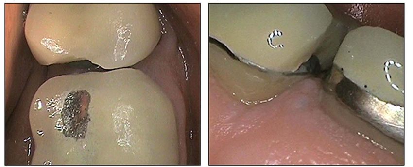
One of the biggest advancements of present day IOC technology is the ability to interface with most practice
management software, thus allowing storage and transmission of magnified, high-definition oral images and
video. As a result, this advancement has catapulted dentistry into a new information integration era.
While the dentist and hygienist will be the most apt to use the IOC routinely, the dental assistant can have
a significant role in using the IOC chairside. This is especially applicable when the dentist is performing a
hygiene exam. The dental assistant should utilize this time to review future potential work with her patient
using the IOC. Business team members must become “hands-on” with the IOC images themselves and
should know precisely how to use and share each custom image with patients, referring doctors and
insurance companies to ensure optimal understanding and care, in “larger-than-life” detail!
Learning Objectives
Upon the completion of this course, the dental professional should be able to:
• Understand the history of photography in relation to the practice of dentistry.
• Ascertain the evolution of the IOC.
• Be familiar with how to manipulate an IOC wand to focus and capture intraoral images.
• Understand how IOC images can be stored and referenced within dental practice software.
• Distinguish proper and improper disinfection techniques for IOC wands.
• Explain the advantages of color image magnification to the patient.
• Explain relevant dental conditions as they relate to each patient’s status.
• Describe various uses for IOC images (post-patient examination).
• Identify landmarks that may indicate various breakdown or disease conditions within a tooth.
• Understand how to use IOC images to monitor and track changes in oral soft tissues.
• Distinguish between fractures or craze lines within an IOC image and how each should be treated.
• Discuss with patient various conditions that may be present on intraoral images and optimal treatment
options.
• Explain at least 5 different educational applications for using the IOC.
• Understand how each dental team member can best utilize the IOC or IOC images on the patients’
behalf.
• Distinguish legal parameters for utilizing patient IOC images outside the scope of the dental practice (i.e.
internet, advertizing, etc.).
Course Contents Restorations within IOC Images
• History of the Intraoral Camera (IOC) Amalgams
• Types of Images Corrosion
• Quality & Illumination Landmarks
• Choosing an Intraoral Camera Leaks, Cracks, the "Black Halo Effect",
Key Feature Areas to Consider Patches & Percolation
Evolving Market Composites
• Handling Techniques for the Intraoral Camera Deep Stain vs. Caries on Occlusal Surfaces
(IOC) Caries vs. Stain Under Composite
• Ease of Use in Mouth Natural Dentition
• Creating a Dry Field Cracks vs. Craze lines
Steps to Create an Intraoral - Dry Field of Bruxism
View Abfractions, Cervical Erosion & Recession
• Proper Disinfection Enamel Hypocalcification, Hypoplasia &
• Introducing the IOC to Your Patients Fluorosis
Patient Learning Styles Occlusal Pit Defects
Patient Education Beyond Chairside Use Crowns, Bridges & Implants
• Detecting Defects & Landmarks on Soft Tissues
2
®
Crest® Oral-B at dentalcare.com Continuing Education Course, March 4, 2011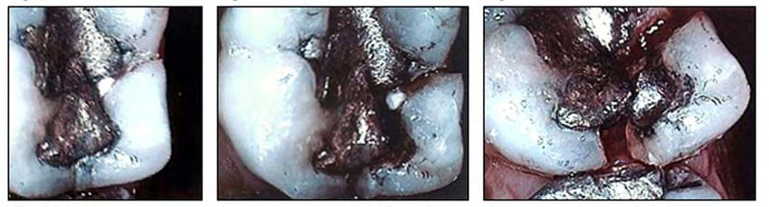
Soft Tissue Lesions photographs can be traced back to 1839. That
Periodontal Examinations was the year the first process of photography
Periodontal/Gingivitis Images and was presented to the world by Louis J. M.
Insurance Submission Daguerre at the Paris Academy of Sciences on
Tongue Health & Halitosis Education January 7th. Later that same year, Alexander
Oral Cancer Examinations S. Wolcott, a manufacturer of dental instruments
• Plaque Control & Plaque Disclosing Images from New York, designed and patented the first
IOC Images for Parent Education & camera from the Daguerre concept.1 These early
Specialist Referral photographs were called “daguerreotype” after
• IOC Photography at Various Patient their inventor and were a one-of-a-kind image
Examinations on a silver-coated copper plate.2 Until this time,
New Patient Examinations & Comprehensive all visual representations and descriptions of
Examinations dental conditions and procedures were subjective
Periodic Examination at Hygiene Visits interpretations expressed through drawings
During Treatment Evaluation & Discovery or models. The “photographic phenomena”
Before & After Comparison introduced a new era of objectively reproducing
• IOC Images & Each Professional’s Role and recording visual dental images. This new era
Dentist Use of IOC observed the inception of the world’s first dental
Hygienist Use of IOC journal, the American Journal of Dental Science,3
Dental Assistants Use of IOC and for the first time in literature preoperative and
Clerical Use of IOC postoperative photographs were published by
• Patient Motivation and the IOC Thompson and Ide.4
• Insurance Company Interface
• Sharing IOC Photos & Legal Confines Since that time Dentists have used extraoral
Doctor to Patient Sharing cameras with precision to capture images within
IOC Image Library the oral cavity. Photographs could capture and
Patient HIPAA / Patient Signature & Waivers monitor oral conditions to be used in ways that
• Conclusion allowed a dentist to predictably create the most
• Course Test aesthetic outcomes when fabricating restorative
• References and cosmetic cases (Figures 1 & 2).
• About the Author
The debut of the first true IOC that captured
History of the Intraoral Camera (IOC) images from inside the mouth came with the
If a picture is “worth a thousand words” then launch of the first Analog IOC System in the
modern dentistry has proven over the past several late 1980s. Fuji Optical Systems of Los Gatos,
decades that imagery conveys an understanding CA acquired the first registered trademark
of critical conditions. First accounts of intraoral of an intraoral camera on July 7, 1987. Fuji
Figure 1. Secondary reference planes for determining positioning
Figure 2. Teeth & Lip aperture cosmetic
measures for use in photographs
Photos courtesy of Dr. Davor Hribar
3
®
Crest® Oral-B at dentalcare.com Continuing Education Course, March 4, 2011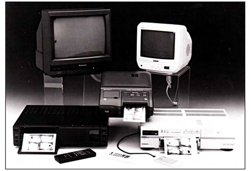
Figure 3. DentaCam™, original                                  Figure 4. Oral Video Scope
        intraoral camera system                                        (OVS) from 1989
        Photo courtesy of Patterson Dental

then released their IOC technology as the
DentaCam™ under Patterson Dental Supplies
later that same year (Figure 3).5

The Fuji DentaCam™ IOC was a derivative of
a medical grade unit and sold for $35,000 as a
cart system with a monitor and printer. About
that same time, Video Dental Concepts™ also
launched an intraoral camera (1989), using a
dental endoscopic handpiece. The design was
revolutionary and included components from
Panasonic Industrial Camera Division (NJ) and
ETS Groux Optical Corp (France). This was the
first component based IOC using a light source,
a remote head micro camera and a dental
endoscope. It inspired and set the standard for
over a decade. The Oral Video Scope (OVS),
cart and printer sold for approximately $12,000
(Figures 4 & 5).6

In different areas of medicine, particularly in
gastroenterology, endoscopes had been used for
many years. The potential of miniature intraoral               Figure 5. Original design dental endoscope
                                                               handpiece
cameras improved as manufacturers improved                     Photos courtesy of Video Dental Concepts.
them. Simultaneously, so called imaging systems
were being used in many areas of medicine and
industry with which digital photographs were

                                                        4
                                    ®
                    Crest® Oral-B at dentalcare.com Continuing Education Course, March 4, 2011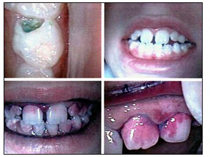
taken (home, clothing, face) and then enhanced              For example, the position of the hands of a clock
with the aid of software and computer. This                 is an analog representation of time.10 Original
re-imaging concept was introduced to dentistry              analog IOCs plugged into a monitor and printer.
in the late 1980s and was used to change                    The images could be viewed and printed. The
anatomical oral outlines to be used to aid in               early form of storage was a VCR. Movies of the
treatment planning and patient education.7                  patients mouth were recorded on VHS tape. In
                                                            1991, Panasonic introduced a video floppy recorder
According to the March 2009 Dental Products                 using a 2” disk that cost $10 and could only hold 50
Reports Technology Survey, 68% of practices                 pictures. Hence, storage of analog was costly and
have an intraoral camera, and 69% of practices              cumbersome at that time (Figure 6).
with computers in the operatory have an IOC
camera connected to their computers.8 Extraoral             Printing was through dye sublimation printers
and intraoral images are used for patient                   and printed copies would degrade over time.11
education and clinician erudition. While the                This limited the practical use of analog images to
images are not considered diagnostic, they                  real-time viewing and paper printing. As digital
play a role to augment and enhance diagnoses                imaging improved through the late 1990s, it slowly
when paired with other diagnostic measures like             replaced analog imaging as the optimal choice for
radiographs.9 Typically IOCs magnify teeth 40 to            most dental offices because digital representations
60 times their original size. This allows discovery         were smaller to store, could integrate with dental
of certain types of details and defects instantly in        practice management software and produced a
bigger, better format than ever before possible.            high quality picture.

Types of Images                                             Early digital imaging systems utilized video
Original IOCs were analog in nature. The word               capture cards to convert an analog image to a
analog means to measure or represent data by                computerized digital image. There are many
means of one or more physical properties that               ways to convert an analog image, but many dental
can express any value along a continuous scale.             offices will use a video capture card installed in the

          Figure 6. IOC video floppy disc recorder & printer (circa 1991)
          Photo courtesy of Video Dental Concepts

                                                        5
                                    ®
                   Crest® Oral-B at dentalcare.com Continuing Education Course, March 4, 2011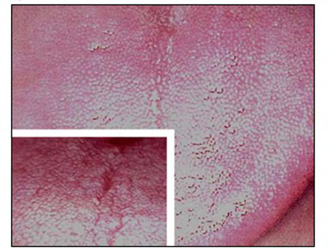
treatment room computer. To do the conversion,
the video output (either composite RCA or
S-Video) from an IOC is connected to the capture
card, which then converts the analog video to
a digital format. Once you freeze the video
image, computer electronics can capture, store,
enhance, or print that image. In other words,
the computer replaces the analog-printer which
was considered the memory of the old analog
IOC system. Still today, some IOCs with analog
composite output can be used with video capture
cards, others have direct USB output. For these,
it is not the camera that makes it a digital system;
it is what you plug it into.12

Today, IOCs can be either corded or cordless
direct USB to the PC or through a small docking
station. The cords of direct USB cameras house
both power and interface data transmission to
dental software. Cordless units are set on a                  Figure 7. First mobile IOC (circa early 1990s)
power docking station or may contain batteries                Photo courtesy of Video Dental Concepts

for power. Wireless receivers transmit the data
to the computer or video networks.  Cordless IOC           to operatory. Some of the original camera
units are very convenient to use and transport             systems were so heavy that a cart containing
form operatory to operatory. However, original             all components (camera, light source, monitor,
designs were known to pick up occasional                   and printer) was the best way to transport and
interferences with radiography emitting devices or         utilize the IOC system. This made fiber optic
other technology such as cell phones, landlines            IOC systems troublesome to use. These large,
and internet connections. Also, internal batteries         portable cart systems were very impractical
had difficulty staying charged. Newer cordless/            (Figure 7).
wireless IOC units use lithium ion rechargeable
batteries which eliminated this problem, and               Many dental clinicians found this inconvenient and
drastically increased the duration of the charge           the IOC units began to sit and collect dust instead
and life of the battery.13 Contemporary corded             of being actively used. One of the solutions
models are electric powered and have a quick               offered was to put a docking station in each
disconnection so that only the IOC handpiece               operatory. While this made carrying the camera
need be transported from room to room. They                much easier, it added on around $500 - $1500 per
are lightweight, easy to transport and do not rely         operatory to the overall cost.14
on batteries.
                                                           The inconvenience and bulkiness of the 1990s
Quality & Illumination                                     design of IOC systems motivated manufacturers
Visual illumination also plays an important role           to develop LED and USB camera systems. USB
in the quality of an IOC image. The rule is “the           stands for "Universal Serial Bus."  USB is the
more light, the more depth of field”. Original             most common type of computer “port” or “plug-
systems typically used very high-end optics to             in device” used in today's computers. USB-IOC
produce the best image quality possible. The               cameras are typically very lightweight, easy to
light source was in a separate “box” and there             carry from operatory to operatory and convenient
were fiber optic cables running from that box to           to use unlike their predecessors.  The USB light
the IOC handpiece. Until very recently, these              source is built into the IOC handpiece, usually
were the only types of IOC systems available.              as a ring of LED lights around the lens.  Like all
These camera and light systems were quite                  systems, there are pros and cons. Inferior quality
heavy and difficult to move from operatory                 LED lights can sometimes produce a reflection on

                                                       6
                              ®
                  Crest® Oral-B at dentalcare.com Continuing Education Course, March 4, 2011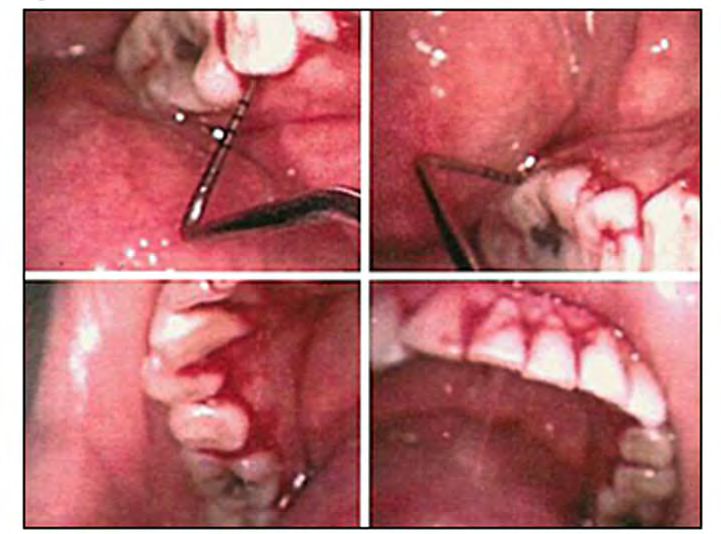
wet objects distorting a picture. Also, USB devices          •   Image Quality
require a small piece of software called “a driver”          •   IOC Features
be recognized for the USB camera to “bridge” and             •   Cost
work with specific software.  This again adds cost
(per-operatory) to operate a USB-IOC system.15               Researching various IOC options for your practice
                                                             can be done easily, using the internet or having
As of 2010, there are seamless software                      various manufacturers bring IOC units to your
solutions available from IOC manufacturers                   office for demonstration and discussion. During
that provide software drivers or "smart drivers"             your research you can create a list of “must have”
allowing the USB-IOC to directly integrate with              and “have not” options that will influence your
most dental imaging capture software. These                  final purchase. Experienced dental equipment
designs can be a less expensive software                     professionals can assist with ensuring that your
compatible solution for most dental offices. Most            IOC system integrates and performs for your
use a proprietary interface from the IOC to dental           intended use.
software (Figure 8).16
                                                             IOC images have many practical uses within the
Choosing an Intraoral Camera                                 clinical setting. While the images are primarily
There are many factors to consider when choosing             used for chairside patient education, they can
an IOC System. Present day IOC systems are                   also be stored within the patients records to
generally digital in nature. When choosing an IOC            be shared with insurance claims reports, other
for your clinical setting it is important to consider        dental specialists or even to be printed/disc
these factors:                                               copied for reference within the patient’s proposed
• Intent of Use                                              treatment plans. Image enhancement or video
• Compatibility & Integration with Practice                  capture capability, are also options for the dentist
    Software/Specifications                                  to consider. Deciding if storage of the images
• Space or Bulk of IOC Unit                                  is important and particular use of the images
• Ease in Handling                                           will help influence the type of IOC system you
                                                             purchase and what type of computer imaging
                                                             interface you will need. These specifications are
                                                             best discussed and decided with the help of a
                                                             dental technology equipment specialist since they
                                                             can vary from manufacturer to manufacturer. In
                                                             its simplest form, image management is like an
                                                             electronic photo album. It allows you to capture,
                                                             store, retrieve and display an image. However,
                                                             unlike a paper photo album, with digital image
                                                             management you can also transmit or enhance
                                                             an image,17 which is useful to present “before-and-
                                                             after” restorative treatment outcomes to patients.

                                                             An IOC will be most utilized within the clinical
                                                             dental setting if it is easy to use. If there are
                                                             clinicians in multiple treatment rooms, plan to use
                                                             the most convenient, portable IOC equipment
                                                             designs. Small, lightweight models are ideal
                                                             and less cumbersome to store and transport.
                                                             IOC handpieces and docking stations need to
                                                             have easy access to software “plug-in” ports.
                                                             This means reaching into an operatory without
                                                             disturbing another clinician and patient, needs
       Figure 8. DiscoveryULTRA with USB                     to be precisely thought out when designing your
       Photo courtesy of Videodental.com                     IOCs functionality.

                                                         7
                                     ®
                     Crest® Oral-B at dentalcare.com Continuing Education Course, March 4, 2011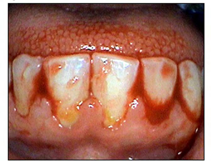
How easy your IOC is to handle is another                   The options are complex and seem endless!
important consideration. Contemporary focus                 Ted Takahashi, noted intraoral camera expert,
options are most commonly designed as “auto-                suggests following a more simplified approach
focus” or by a “twist or tap” setting within the            when choosing an IOC system.
IOC handpiece. Image capture options have
evolved from remote control (which required a               Key Feature Areas to Consider
second operator to assist with image capture) to            Quality of construction:  make sure the camera
foot pedal or handpiece button capture. The foot            docking station and power connection plugs can
pedal method can be challenging as it requires              withstand the punishing 2,000 insertions and
more body movement from the clinician and                   detachments it will have to endure per year. If
can cause distortion during the actual image                corded, inspect that the cord has “strain relief” or
acquisition. Handpiece buttons with sensitive tap           will it ultimately fray, leak and create "water spots"
controls seem to be the easiest to use.                     on image displays and printouts.

Image quality of an IOC depends on many                     Depth of field: with the IOC wand inside of the
factors: sharpness, noise (variations in                    mouth, note the amount of viewing area that is in
density), dynamic range of color capture, tone              focus. Superior IOCs require little or no focusing
reproduction, contrast, distortion, vignetting (light       inside of the mouth.
fall-off), exposure accuracy, color fringing (which
causes the lens to focus at varied distances),              Artifacts & Optics: compare what you see in
lens flare (glare of stray light), artificial color         the mouth to the monitor. Lighting will play a
banding and artifacts (low-contrast detail or               role in this, so it is important to have an in-office
over-sharpening that can occur during software              demonstration under typical operatory lighting
conversion). All of these factors affect digital            conditions. The optics distinguishes a good IOC
image quality.18                                            from a more inferior one. The best optic systems
                                                            are created by placement of the CCD chip at
Every digital image consists of a fundamental unit          the end of the wand next to the lens. This is
called “a pixel”. The pixel, invented by combining          more expensive than placing the CCD chip in
the words "PICture and ELement", represents                 the middle of the wand. When the CCD chip is
a single color dot, combined with millions of               in the middle of the wand an additional prism is
other color dots, to seemingly create a detailed,           used to direct the incoming image farther down
continuous image. Each pixel contains a series              the wand to the CCD chip. The addition of the
of numbers which describe its color or intensity            prism degrades image quality and produces more
to the computer’s software.  The terms pixels               artifacts (Figure 9).20
per inch (PPI) or dots per inch (DPI) were both
introduced to relate this theoretical pixel unit to         Artifacts can appear as either low-contrast detail
real-world visual resolution.19 IOC images are              or over-sharpening. They can occur during
measured by these terms in combination with the             software conversion resulting in an unreliable
above listed factors to indicate the quality of an          representation of present conditions. Avoid
IOC image.                                                  purchasing an IOC with high artifact quotient.

It is apparent that many features associated with           Evolving Market
IOCs play a role in selecting one for clinical use.         Through the years well over 150 companies
The options include:                                        have developed intraoral video cameras (IOVCs).
• Macro-to-infinity focusing                                IOVCs had component-based cameras that
• Anti-fog lens                                             required little engineering as they emerged
• USB interface                                             from 1988-1995.  IOVCs are considered Class I
• Corded / Cordless                                         devices which are not regulated by the FDA and
• Lithium battery powered / Direct power                    do not require FDA 510-k certification.  As LED
• Etc.                                                      and special optics were introduced and as the

                                                        8
                                ®
                  Crest® Oral-B at dentalcare.com Continuing Education Course, March 4, 2011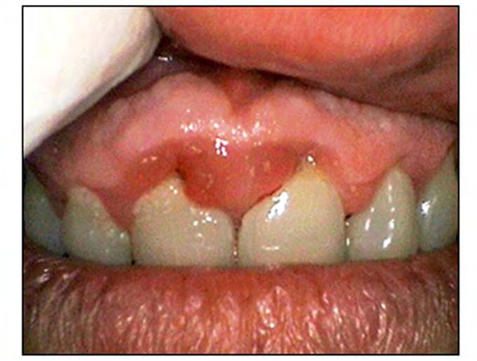
Figure 9. Mid-wand placement of CCD chip and prism degrades IOC image quality
 Photo courtesy of Videodental.com

market demanded better engineered handpieces,                low cost LED’s will only render average images.
over 80% of these companies went out of                      Loaner use and service is not available for
business between 1994 and 2004.21                            repairs on these low-cost dental cams”. Superior
                                                             image quality can only be achieved by the ideal
Presently IOC camera sales are dominated by                  combination of superior glass optical lenses
three major companies: Patterson & Henry                     paired with high resolution CCD chips. Durability,
Schein Dental Suppliers and Kodak Dental (a.k.a.             longevity and maintainability are derived from
CareStream Health of Canada) Kodak is selling                reliable manufacturers. While these newer low-
direct to dental practices and is a distributor of           cost cameras may be inexpensive to purchase,
imaging systems only. These companies offer                  they may have a life span of 3-6 months.  Most
practice management software that integrates                 quality cameras are made to last 10-20 years.
with imaging software but, the IOC capture                   To quote an old adage, “You get what you pay
is tied to a foot pedal operation. Secondary                 for”, still rings true with regards to IOC or IOVC
vendors offer button capture function that is an             investments. A clinician should carefully research
ease-of-use preference and typically less costly.            all aspects of current IOC technology, including
Integrating with practice imaging programs by use            compatibility with potential or existing dental
of bridging software may drive costs up further.             software and quality of images, before committing
                                                             to any IOC purchase.
While IOCs & IOVCs were first produced mainly
in the United States and later in Europe, Asia is            Handling Techniques for the Intraoral
now mass producing IOVCs and flooding the U.S.               Camera (IOC)
market.  The trend started in Japan (RF System               It is of fundamental importance to handle your
Lab, 1998), Korea (Sometech Corp 2005) and                   IOC with the utmost of care. The number one
now Taiwan and the Republic of China. These                  reason for repair is careless or mishandling of the
IOVCs are being mass produced using a new “on                IOC wand. If your IOC is portable from operatory
the chip technology” (CMOS) which originated for             to operatory, be sure you understand the proper
the web-cam industry. Dentists have commented                connection and disconnection of your IOC wand
on the DentalTown.com website of IOVC                        or unit, as it relates other ports or plug-in sources.
purchases as low as $149.00. E-bay lists IOVCs               When working with corded models, be sure to
as low as $49.00! Claude Berthoin, founder of                differentiate if disconnection is of “snap” or “twist”
Video Dental Concepts and co-inventor of the                 release. Forcing or jamming the connection will
IOVC technology comments, “On my recent flight               result in serious damage to the IOC wand. Read
to a dental tradeshow I was stunned to see one               the IOC Instruction Manual and ask for proper
of these Chinese IOVCs offered in the Sky Mall               guidance from experienced peers when learning
Magazine. Clearly more professional companies                to operate any IOC.
will go out of business as a result”. Berthoin
also added, “While purchasing a “cut-rate cam”               When not in use, be sure to store the IOC wand
will satisfy the need to show a tooth image, the             properly. Corded models should be stored within
combination of cheap plastic lenses and low                  docking cradles.  Portable-wand units are smaller,
quality imagers (640 x 480) like CMOS chips and              less stable and can have a tendency to drop or

                                                         9
                                     ®
                     Crest® Oral-B at dentalcare.com Continuing Education Course, March 4, 2011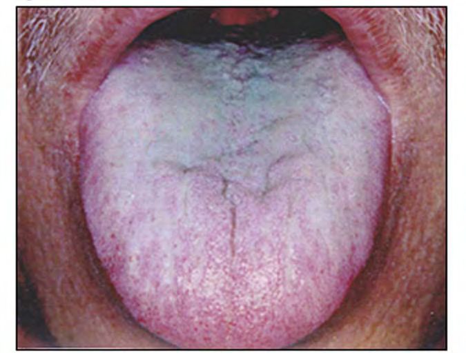
fall when not in use. Store cordless IOC wands                             Ease of Use in Mouth
in their docking cradle or securely in a storage                           The following video will demonstrate a very
case to avoid accidental damage.                                           efficient method that will enable you to “tour the
                                                                           mouth” and capture IOC images on patients
                                                                           proficiently. By practicing these techniques, your
                                                                           IOC skills will increase rapidly.

  Video 1. Handling Techniques for the Intraoral
  Camera
  To view this video, please go to www.dentalcare.com and find this
  course in the Continuing Education section.
                                                                             Video 2. Proper Use of the Intraoral Camera
                                                                             To view this video, please go to www.dentalcare.com and find
                                                                             this course in the Continuing Education section.
Navigation of your intraoral camera may take
some practice. Typically you will need to
maneuver your IOC wand while viewing your                                  1. Using the patient’s teeth as a fulcrum for the
navigation on a monitor.  Begin by making sure                                IOC wand, start at the distal most aspect of
your equipment is properly set up. Position                                   the lower arch.
a computer monitor within effortless view of                               2. Lay the camera wand “flat and parallel” on the
operator. Have the IOC wand inside of its                                     occlusal pane.
docking station engaged to the “on” position and                           3. Find orientation to the tooth above.
ready to use. Finally, ensure that the “capture                            4. Slide or drag the IOC wand forward to see the
method” of your IOC system is within reach. IOC                               consecutive teeth on the upper arch.
systems come with various methods to capture                               5. Have the patient “open & close” to enhance
images. A remote-controller, foot-pedal or a                                  focus.
button on the IOC wand are most common.                                    6. Use a “finger roll technique”, between thumb,
                                                                              index & middle fingers, to see lateral aspects
Set-Up & Capture                                                              of teeth.
Proficient navigation of the IOC wand, while                               7. Lift the wand from lower arch then extend
watching a computer monitor, may prove                                        wand towards left or right to gain full lateral
challenging at first. Maneuvering the IOC wand                                view, then capture the image.
inside the oral cavity without stability and purpose                       8. Return to original occlusal plane position IOC
can prove to be very frustrating. Follow these                                wand parallel and continue sliding the wand
“ease of use” recommendations to make IOC                                     forward to view consecutive teeth.
handling easy and efficient:                                               9. Repeat process for other side of mouth.
1. Pre-focus your IOC lens for the type of images                          10.Reverse it for upper arch to view lower arch.
    you will be capturing. Common Options
    include: full-face, smile, close-up, or super                          Remember, staying disciplined in practicing
    close-up. Setting the focus option before you                          these techniques will rapidly increase your IOC
    enter the oral cavity will ensure less fumbling.                       proficiency and skill.  You may want to take notes
    Auto-focus lenses will adjust automatically.                           on these videos and post a small “reminder” near
2. Remember to set your IOC to either “video” or                           your work station to promote exceptional IOC
    “still image” mode so that you can effectively                         handling techniques and habits.
    save and use your images.

                                                                      10
                                        ®
                       Crest® Oral-B at dentalcare.com Continuing Education Course, March 4, 2011
Creating a Dry Field the most proficient IOC photographer has to
Once you feel adept at moving the IOC wand consistently contend with managing excess
around the mouth while watching your monitor saliva flow.
and capturing images simultaneously, you can
then add an additional step that will enhance the Steps to Create an Intraoral - Dry Field of View
quality of your IOC images immensely. 1. Simultaneously hold IOC wand and air/water
syringe.
Creating a “dry field of view” just prior to capturing 2. Have patient hold saliva ejector in mouth.
your IOC images will dramatically increase what 3. Air dry each tooth just prior to capturing an
you and your patients can see. Saliva and a image.
“wet field” will hide or skew what actually exists 4. Help the patient to move the saliva ejector to
on the tooth’s surface. Look at the wet and dry the next appropriate place as you tour their
comparisons in these next examples (Figures 10 mouth capturing images.
& 11). Defects and diseased areas will seem to 5. Ask patient to “breath through their nose” to
“jump off the monitor” when you master how to avoid lens fogging.
keep your field-of-vision dry for IOC photography. 6. Some clinicians may use a dry angle or cotton
gauze 2 x 2 to aid in creating a dry field.
Establishing a dry field is much easier with two
clinicians. When only one clinician is taking IOC
images, add the following steps to the previous
“ease of use” suggestions for dramatically
improved images. You may need to retake
images that do get obscured with saliva. Even
Video 3. Creating a Dry Field for the Intraoral
Camera
To view this video, please go to www.dentalcare.com and find
this course in the Continuing Education section.
While a dry field IOC photo is most desirable for
educational purposes, be aware that managing
Figure 10. Wet view of amalgam filling
saliva to create a dry field can take up a
significant amount of time. Remain cognizant
to stay on schedule and diligent with the other
clinical duties you will have to perform. IOC
imaging can be rewarding but also frustrating
and initially very time-consuming. You may
even suggest rescheduling a patient for a more
comprehensive IOC and tactile examination if you
see the need for a more extensive evaluation.
Keep yourself on-schedule and advise the
patient if more time is required for all necessary
diagnostics.
Figure 11. Dry view of same amalgam filling
11
®
Crest® Oral-B at dentalcare.com Continuing Education Course, March 4, 2011Proper Disinfection
As with all dental equipment, proper and
consistent disinfection or sterilization is
mandatory. Most IOC wands cannot be sterilized
in a heat sterilizer because the fragile workings
of the internal lens will not hold up to the intense
heat. Delicate disinfection is in order with most
IOC wands. Immersion in sterilizing solutions
or spray disinfectants are also most always
contraindicated. Instead, gently wipe wand area
with either a sterile cloth moistened with water
and a gentle antibacterial soap (avoiding the
lens) or according to the manufacturer’s specific Video 4. Proper Disinfection of the Intraoral
disinfection guidelines. Camera
To view this video, please go to www.dentalcare.com and find
this course in the Continuing Education section.
Most IOC manufacturers advise against wiping
the delicate IOC lens directly, while others may
advise a light alcohol wipe. Dry-wiping can also the IOC to your patients will be to describe this
scratch and damage the delicate lens. Check as a patient education tool that will improve
manufacturer recommendations and do what both patient comfort and understanding of their
is best for your IOC’s longevity. To protect current dental conditions in a gentle, touch-
your IOC lens and adhere to universal infection less manner. Since the IOC wand is smooth,
control guidelines, use the IOC manufacturer’s slender and contained, it will be unobtrusive in
specified barrier sleeve. Typically an IOC barrier the oral cavity. A great introduction of the IOC
sleeve has an inner and an outer sheath. The could be worded like this: “Mrs. Smith, today
outer sheath ensures that the inner sheath included in your periodic dental examination, we
remains fresh, clean and ready for intra oral use. will be using a state-of-the-art dental technology
Single-layer sheaths are available but, provide called the intraoral camera. It is a comfortable
less infection control. To use, gently slide the miniature camera wand that will allow us to tour
IOC wand into the inner sheath and carefully and capture magnified pictures of your current
peal away the outer sheath. Avoid touching or dental conditions, then display them on a monitor.
handling the clean inner sheath. Your IOC wand The images will allow us to see fractures, leaks
and recommended sheath may demand specific in fillings and deteriorating areas close-up, so we
interface. For instance, you may have to insert can discuss potential problems together. We will
the camera wand “face-down” or “face-up” so that pair these visual findings with other traditional
the clearest side of the plastic interfaces with the diagnostic tools to plan any necessary treatment.”
lens. This will yield a clear field of view. Many
times the sheaths are marked with a stamp to
designate proper insertion for clearest viewing.
Improper sheath application may produce a
blurred lens and images.
Introducing the IOC to Your Patients
Two of the biggest obstacles for patients to
accept needed treatment are that they often do
not understand their condition and they just do
not enjoy the experience of dental treatment.
Even routine examinations can invoke within the
patient’s mind thoughts of pain, fear, buzzing
handpieces or pointed instruments. None of
these have pleasant association or make the Figure 12.
clinician’s tasks easy. A great way to introduce Photo courtesy of AbsoluteDentalImage.com
12
®
Crest® Oral-B at dentalcare.com Continuing Education Course, March 4, 2011It is important to point out to patients that patient can participate directly in the decision
IOC images do not replace conventional making process as far as preferred treatment is
diagnostic tools like radiographs, study models concerned.22 Some dentists even hand the IOC
or tactile examinations, but they will enhance wand directly to their patients and allow them to
understanding of the findings by providing tour their mouth themselves for inspection and
a magnified, clear, close-up look at current discovery prior to a clinical IOC exam. Clinicians
conditions. Point out too that these findings can should try varied approaches to ascertain what
be stored, shared with specialists, emailed with methodology works best to enroll patients in their
insurance claims, reprinted and cosmetically IOC image discovery and education.
enhanced. Patients will quickly understand the
far-reaching value and use that IOC images will Patient Learning Styles
lend to support their long-term oral health. Dental patients enter a dental setting with a
varied degree of understanding about their
Some patients may be resistant or squeamish to present oral conditions, need for treatment and
look at their IOC images. Reassure them that treatment options. This level of understanding
it is not compulsory that they view the monitors is said to be a patients “dental IQ”. As dental
but, it is necessary for the dentist to view them. professionals, we are constantly striving to
Most patients will reenroll themselves in the improve our communication skills, increase our
viewing process as the clinician is capturing and patients’ dental IQs and proceed with necessary
discussing the findings. dental care. Patient education has long been a
challenge for dental professionals to master and
Each dentist uses different methods to modify deliver chairside.
patient behavior and acceptance of treatment
proposed. The IOC allows patients to directly A common belief among teachers is that most
observe their intraoral conditions. Thus the people favor a particular method of information
processing/learning. Based on this concept,
teaching by the use of individualized "learning
styles" originated in the 1970s, and has been
used by educators ever since. One of the most
common and widely-used categorizations of the
various types of learning styles is Fleming’s VARK
model (sometimes VAK) which has its origins in
neuro-linguistic programming. The VARK model
is represented as follows:
1. Visual Learners
2. Auditory Learners
3. Reading/Writing=Preference Learners
4. Kinesthetic Learners or Tactile Learners23
In dentistry, Fleming’s VAK model, helps to
accelerate patient understanding when all
three styles (visual, auditory and kinesthetic)
are combined during the chairside examination
appointment. Since time is limited during a dental
appointment, combining the VAK model ensures
that all learning styles are addressed. The IOC
provides clear, magnified images in monitor-view
(visual learners) that can be easily discussed
and described (auditory learners). The clinician
Figure 13. Patient touring their own
mouth with IOC can also easily perform tactile examination, or
Photo courtesy of Osprey Dental, FL have the patient handle the camera wand for
13
®
Crest® Oral-B at dentalcare.com Continuing Education Course, March 4, 2011self-examination (kinesthetic learners). Greatest outside of the scope of patient/doctor care unless
impact of understanding results when IOC additional consent forms are signed by concerned
technology is utilized in this manner. patients. If you intend to use IOC images for
marketing or internet posting, you must get proper
High-tech guru Dr. John Jameson passes along clearance and with supporting legal consent
this information to us: "For doctors who capture documentation from your patients.
IOC or digital images of the patient, we have
seen an increase between 10% - 25% in case Detecting Defects & Landmarks on
acceptance."24 Restorations within IOC Images
It is certain that IOC imaging is a powerful
The IOC is an educational tool that will enlighten utility for finding, viewing and explaining dental
patients in a way that description alone cannot conditions to patients, as well as, supporting the
accomplish, confirming the old adage, “A picture dental clinician in diagnostic decision making.
is worth a thousand words.” In this case, However, IOC Systems are not regarded
a picture is worth acceptance of necessary as diagnostic means or measures. Most
treatment and years of optimum dental health! professionally recognized entities categorize IOCs
as educational devices. The American Dental
Patient Education Beyond Chairside Use Association does not take a hardened stance on
Contemporary digital IOC systems have improved the diagnostic and clinical use of IOC systems.
to allow better integration with dental software. ADA representatives encourage individuals to
This offers the ability to store and manipulate check with their respective State Dental Board
digital images with imaging software and provides or Department of Professional Regulations
a very broad scope of use for patient IOC concerning recommended practices as they
images. A dentist can choose which options best apply to IOCs and dental professionals.25 In
suit his/her needs. The following table illustrates checking several State Dental Practice Guidelines
common IOC image communications: it is consistently stated that the use of IOCs is
recommended for educational purposes. While
Use of IOC photographs and images may support diagnoses,
Communication with: IOC images are in no way a definitive means of
Images:
Printed Copy Patient – for at-home reference. analysis for disease prognosis or treatment.
Specialist Doctor – for referral / for
patient file That being said, a hybrid of an IOC and a light
Insurance Carrier – for visual claim scanning fluorescence device is in development
support for the detection of carious dentin. It is a novel
E-Mailed Patient – for at-home reference therapeutic concept using a light-induced
Transmission Specialist Doctor – for referral / for fluorescence evaluator for the detection and
patient file treatment of dental caries, referred to as LIFEDT.
Insurance Carrier – for visual claim LIFEDT uses variations of measured brightness
support of light to evaluate healthy vs. actively carious
Copy of Video Patient – for at-home reference dentin when illuminated. Active caries will appear
E-Mail Video Specialist Doctor – for referral / for red under the fluorescence. This device may be
patient file helpful in daily practice to discriminate between
Letters / Post Existing Patients or Potential Patients carious or healthy dentin.26 As technology
Cards advances, acceptable diagnostic practices will
E-Mail Blasts Existing Patients or Potential Patients follow suit.
For the remainder of this course we will be looking
While the use for IOC images multiplies, it at IOC images and explaining some commonly
is important to adhere to federally mandated recurring markers, anomalies and conditions that
restrictions. IOC images are considered are considered familiar “landmarks” within IOC
“personal health information” (PHI). Under dental photography. These are for educational use with
HIPAA mandates, IOC images cannot be utilized patients and should always be accompanied
14
®
Crest® Oral-B at dentalcare.com Continuing Education Course, March 4, 2011by a tactile examination and complimentary Corrosion in and of itself does not warrant filling
radiographs for formal diagnosis. replacement and can most times be polished back
to a high luster.
Amalgams
Landmarks
Corrosion
As dental professionals, we know that amalgam Leaks, Cracks, the “Black Halo Effect”,
fillings age and show signs of a breakdown Patches & Percolation
process. Fillings will begin to corrode, which is Amalgam fillings most often can begin to separate
the equivalent to an object becoming “rusty”. This from the tooth over time, creating a crevice that
becomes very obvious with the magnification of can harbor bacteria. This void can be referred
IOC images. Aging amalgam fillings appear dull- to as an open margin or a leak in the restoration.
gray or even black in color. Amalgams can also expand and contract in
response to the varying temperatures of food.
Cold foods will cause the amalgam to contract,
while hot foods will cause expansion. Continual
expansion and contraction may place undue
pressure against the surrounding tooth structure
and create cracks in the walls of the tooth. These
cracks can also allow bacteria and acids to
penetrate the tooth causing damage (Figures 16
& 16A).
Figure 14. Shiny amalgam filling with no signs of
corrosion
Figure 16. IOC image
Figure 16A. IOC image with corresponding landmarks
Figure 15. Corroding amalgam filling
15
®
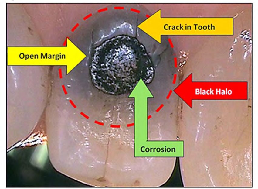
Crest® Oral-B at dentalcare.com Continuing Education Course, March 4, 2011Changes in temperature can also lead to on the IOC image, look for dark corrosion areas,
“amalgam percolation”, a separation and popping actual separation of the filling from the tooth
effect of the amalgam from its original position. (leaks/open margins), cracks in the tooth, cracks
This can occur due to the micro-gap between in the amalgam and “black halos” surrounding the
tooth structure and the filling material.27 Within amalgam itself (Figure 18).
this micro-gap oral fluids enter, are effected by
body heat and the result is percolation which A “black halo” is the appearance of a blackened
allows the filling to move, rise or separate from area emanating from the amalgam and spreading
its original position. While the amalgam filling through the tooth’s image. This will typically merit
may stay secure within the prep, the separation further diagnostic inspection with radiographs
creates continual micro leakage and should be of the corresponding tooth to determine the
evaluated with other diagnostic measures such as presence and extent of decay. The “black halo”
tactile exam and radiographs. appearance is a landmark for cavity detection.
It is important to look for certain IOC landmarks Amalgam fillings can also crack, which indicates
around the margins of all amalgam fillings. As that the filling is weak or defective. Another
you inspect the circumference of an amalgam common landmark visible on IOC images is
the presence of “patched amalgams”. In an
effort to restore cracks within amalgams, some
dentists have added additional amalgam to
patch and repair the restoration. It is important
to look carefully and closely at amalgam fillings
to differentiate between cracks and patched
outlines.
Typically it is difficult for a clinician to see these
defects with the naked eye and patients may not
see or feel discomfort from this breakdown at all.
IOC photos bring this erosion into magnified view.
Figure 17. Percolation of an amalgam filling
(Notice the edges separated and “popped-out” from its Remember the importance of air-drying each
original position in the prep.) tooth, as saliva will coat over and “camouflage”
Figure 18. Landmarks on IOC photo
16
®
Crest® Oral-B at dentalcare.com Continuing Education Course, March 4, 2011fine defects especially at the margins of fillings.
Dry teeth will yield enhanced IOC images.
Composites
As composite fillings improve in compressive
strength, bond strength, luster and color
compatibility, they gain popularity within the
professional dental community. Many dental
offices only offer non-mercury fillings made of
resin or porcelain. Studying composite landmarks
and potential defects is an essential skill in
mastering the art of IOC photography. Let’s take
a look at some composites conditions under IOC
Figure 19. Look closely at the
magnification:
center of this amalgam to detect
small circular patch.
Deep Stain vs. Caries on Occlusal Surfaces
IOC capture of stain in occlusal pits is a very
powerful visual for the patient, but do not let them
be fooled. In this example, the more significantly
stained margins proved to be decay-free, while
the lesser stained pits had incipient caries.
This could only be determined by following up
the IOC exam with a traditional mirror-and-
explorer tactile examination. IOC photography
never replaces dependable diagnostics, but is
maximized when the clinician knows when and
how to combine diagnostics with educational tools
like IOC images.
Figure 19A. Two separate amalgam
fillings (MO +DO). Interface of filling
creates an open margin.
Figure 19B. Close tactile
examination reveals these to be just Figure 20. Upon tactile and radiographic examination
scratches in the amalgam. most stained areas were sound. Distal pit was found to
be carious.
17
®
Crest® Oral-B at dentalcare.com Continuing Education Course, March 4, 2011Caries vs. Stain Under Composite
It takes some training of the eye to distinguish
subtle color differences in IOC photography. The
“black halo” landmark is a very helpful tool but
can be tricky when seen around a composite
filling. Black halos under composite fillings can
merely represent stain left behind from a previous
amalgam that takes on a dark translucent value
through the composite material. How do you tell
the difference? Always cross check for caries
with a tactile exam and radiographs (Figure 21).
Natural Dentition
Cracks vs. Craze lines
Visible crack lines on teeth (within IOC images) Figure 21. Composite filling with Black Halo
can be misleading. A savvy clinician must
become very aware of the differences in cracks/
fractures vs. craze lines. Use of the IOCs fiber
optic light will help you make the distinction. If a
tooth is truly cracked, light will not transilluminate
through the tooth. It will instead bury itself in
the floor of the crack. Conversely, if the light
source does transilluminate through the tooth,
the apparent crack is merely a surface craze
line. In illustrations (22 & 22A), you can see the
difference.
Craze lines are for the most part inconsequential
and do not need to be corrected with dental
treatment except for cosmetic improvement. True
cracks on the other hand, can be very serious!
Vertical cracks that travel to the gumline may Figure 22. Illuminated light gets
require a full-coverage crown restoration. If the buried into a tooth fracture
vertical crack travels below the gumline, the tooth
may require endodontic treatment with crown
lengthening or extraction. Additional diagnostic
measures would be needed to determine
appropriate treatment options. It is important to
understand the distinctions between cracks and
craze lines.
Bruxism
Bruxism, clenching or grinding teeth can have
a profound mix of effects on the dentition,
temporomandibular joint, head and neck
musculature and even sleep patterns. The cause
of bruxism is not completely agreed upon, but
daily stress may be the trigger in many people.
Bruxism can cause severe pain to none at all.
Many patients will be unaware or asymptomatic Figure 22A. Illumination travels
through a craze line
to their bruxing habit. IOC images will capture
18
®
Crest® Oral-B at dentalcare.com Continuing Education Course, March 4, 2011Figure 23. Craze lines in central incisor Figure 24.
Figure 23A. True fracture in tooth at upper third of
incisor
Figure 24A.
surface wear facets on occlusal and incisal this type of damage abfraction in a paper
edges of teeth (Figures 24 & 24A). These visual published in 1991 (Figures 25 & 26).29
landmarks should alert the dental clinician to
examine the patient more thoroughly for bruxism While there are varied opinions on whether or
symptoms and measures to arrest its ill effects not to treat cervical tooth wear with composite
over time. restoration or prosthetic mouth guards, the
incident of such wear is prevalent. Dental
Abfractions, Cervical Erosion & Recession clinicians can view and capture cervical wear
Through the years the dental profession has on teeth with the IOC and review treatment
held a variety of theories about the causes of recommendations with each patient.
abfractions, including the chemical wasting of
teeth, the effects of tooth brushing or tooth pastes Gingival recession is characterized by the
and lateral forces.28 displacement of the gingival margin apically from
the cementoenamel junction, or CEJ, or from the
In the early 1990's, Dentist, J. O. Grippo former location of the CEJ in which restorations
concluded that cervical erosions were the result have distorted the location or appearance of the
of flexing of the teeth at the gum line due to CEJ. Many people may exhibit gingival recession
heavy bruxing (grinding). This flexure produced without having any awareness of the condition.30
damage to the enamel rods at the gum line Using IOC images to record and discuss the
resulting in their loosening and consequently implication of gingival recession with patients can
flaking away of the tooth structure. He named be critical, especially when such conditions are
19
®
Crest® Oral-B at dentalcare.com Continuing Education Course, March 4, 2011advancing asymptomatically. Images are also
valuable to share with referring dental specialists
or within insurance documentation (Figure 27).
Enamel Hypocalcification, Hypoplasia &
Fluorosis
The appearance of white spot defects on IOC
images is quite common. White spot lesions may
occur as a result of an injury to, or a large cavity
in the primary tooth. When the permanent tooth
erupts it may have a roughened, pitted area in
the enamel, corresponding to the defect. This
is sometimes referred to as a "Turner's tooth",
or "Turner's hypoplasia". It is most common in
Figure 25.
(permanent) bicuspid teeth (secondary to infected
primary molars) and permanent central incisors
(secondary to injury to the primary incisors).
An event such as fever, malnutrition, or
hypocalcemia during teeth formation can occur
during fetal development or early childhood. This
is a related condition in which the ameloblast cells
affect the tooth enamel. When the front teeth
and six-year molars are affected, the event most
likely occurred in the first year of life. When the
bicuspids and second molars are affected, the
event likely occurred around age three. Disruption
can appear as white or brown spots on the teeth
Figure 26. (Figure 28).31
Images courtesy of Dr. Brian Palmer
Mild fluorosis is the appearance of small white
opaque flecks, which can be magnified with the
IOC and are more visible near the incisal edges.
The pattern becomes more obvious when the
teeth are dried prior to image capture. This
Figure 28. Enamel hypoplasia exhibiting both white
Figure 27. Gingival Recession and brown lesions in enamel
20
®
Crest® Oral-B at dentalcare.com Continuing Education Course, March 4, 2011Figure 29.
Photo courtesy of Cosmetic Dentistry Center
Figure 30.
condition has been linked to over use of fluoride.
It can be corrected with a course of professional
treatments that combine in-office etchants, tri-
calcium phosphate paste infusion and whitening
products (Figure 29).
Occlusal Pit Defects
While IOC images alone cannot diagnostically
determine if a tooth lesion is carious, the
magnification can provide clinicians and patients
dramatically close-up views which can aid in the
early detection and treatment when combined
with other diagnostic measures. Figures 30 & 31
are ideal examples. Figure 31.
Crowns, Bridges & Implants
IOC photography can be useful when inspecting
margins of existing crown and bridgework. Many
times difficult to see or reach areas come to full
view with the IOC wand and capture. Defects
not discernable by plain view, tactile or even
radiographic inspection can be revealed with IOC
magnification. The dentist can then discuss with
patients possible treatment options to correct
such defects. Figures (32 - 34C) are some
examples.
Soft Tissues
Figure 32. Mesial fracture in this filling prep reveals
Soft Tissue Lesions
need to discuss benefits of crowning this tooth with
IOC images can be helpful in detecting and patient.
monitoring soft tissue abnormalities as well
as tracking them for changes. Since capture,
21
®
Crest® Oral-B at dentalcare.com Continuing Education Course, March 4, 2011You can also read