On the Systematics and Biodiversity of the Opheliidae and Scalibregmatidae - MDPI
←
→
Page content transcription
If your browser does not render page correctly, please read the page content below
Review

On the Systematics and Biodiversity of the Opheliidae and
Scalibregmatidae
Julio Parapar 1,*, Alejandro Martínez 2 and Juan Moreira 3

                                            1 Departamento de Bioloxía, Facultade de Ciencias, Universidade da Coruña, 15008 A Coruña, Spain
                                            2 Molecular Ecology Group (MEG), Water Research Institute (IRSA), National Research Council of Italy
                                              (CNR), Largo Tonolli 5, 28922 Pallanza, Italy; alejandro.martinezgarcia@cnr.it
                                            3 Departamento de Biología (Zoología) & Centro de Investigación en Biodiversidad y Cambio Global

                                              (CIBC‐UAM), Facultad de Ciencias, Universidad Autónoma de Madrid, 28049 Madrid, Spain;
                                              juan.moreira@uam.es
                                            * Correspondence: julio.parapar@udc.es

                                            Abstract: In this paper we review the systematics, diversity, and ecology of two related annelid
                                            families: Opheliidae Malmgren, 1867 and Scalibregmatidae Malmgren, 1867. Opheliids are deposit‐
                                            feeders and that are mainly found as burrowers in sandy sediments. Morphologically, opheliids are
                                            characterized by the smooth cuticle, as well as the presence of a conspicuous ventral groove, re‐
                                            duced parapodia, and a tubular‐shaped structure often projecting from the posterior end. Scalibreg‐
                                            matids are also deposit‐feeders, but compared to opheliids, they have a characteristic arenicoliform
                                            body, a T‐shaped anterior end and a glandular, reticulated epidermis. For each family, we summa‐
                                            rize the available information about the evolutionary relationships, taxonomic history, geographical
                                            distribution, ecological preferences and diversity of life strategies along with the techniques most
                                            commonly used for their study. By highlighting the main gaps in knowledge on each of these topics,
                                            this review ultimately aims at stimulating further research into members of these two families in
                                            the future.

Citation: Parapar, J.; Martínez, A.;        Keywords: Opheliidae; Scalibregmatidae; diversity; taxonomy; anatomy; biology
Moreira, J. On the Systematics and
Biodiversity of the Opheliidae and
Scalibregmatidae. Diversity 2021, 13, 87.
https://doi.org/10.3390/d13020087
                                            1. Introduction

Academic Editor: Luc Legal
                                                  Opheliidae Malmgren, 1867 is a well‐known family of annelids distributed through‐
Received: 30 December 2020
                                            out the world mostly in sandy sediments [1–3]. Most of the described five to six genera
Accepted: 12 February 2021                  and ca. 160 species of opheliids include elongate, deposit‐feeding burrowing worms,
Published: 18 February 2021                 which are easily recognized by the smooth cuticle and the presence of a conspicuous ven‐
                                            tral groove along at least the posterior half of the body (Figure 1). Opheliids usually have
Publisher’s Note: MDPI stays neu‐           a conical to pointed prostomium that lacks lateral antennae, whereas their pygidium often
tral with regard to jurisdictional          develops a tubular‐shaped prolongation that may bear cirri and marginal papillae. Alt‐
claims in published maps and insti‐         hough some species may reach 100 mm in length, most opheliids range between 5–70 mm
tutional affiliations.                      and their trunk comprise about 30–60 segments [4].
                                                  The knowledge on opheliid taxonomy and systematics has been substantially im‐
                                            proved in the last two decades, including the delineation of subfamilies and phylogenetic
                                            affinities [1]. However, further work is still needed in order to assess the validity of the
Copyright: © 2021 by the authors.
                                            genus Ammotrypanella McIntosh, 1879 and some species of Ophelia Savigny, 1822 and Ophe‐
Licensee MDPI, Basel, Switzerland.
                                            lina Örsted, 1843, as well as the status of the many synonymies attributed to the presum‐
This article is an open access article
                                            ably cosmopolitan Polyophthalmus pictus (Dujardin, 1839). The opheliid fauna of some ge‐
distributed under the terms and
                                            ographic areas is well known (e.g., North Atlantic, California) whereas other regions re‐
conditions of the Creative Commons
                                            main clearly understudied and may potentially hold many undescribed species (e.g.,
Attribution (CC BY) license
(http://creativecommons.org/licenses
                                            Tropical Atlantic, Indo‐Pacific and Australasia). The biology, ecology, and burrowing be‐
/by/4.0/).                                  havior of some species were studied in detail due to their ecological importance in the

Diversity 2021, 13, 87. https://doi.org/10.3390/d13020087                                                            www.mdpi.com/journal/diversity
Diversity 2021, 13, 87                                                                                                  2 of 36

                         intertidal and shallow subtidal of sandy beaches at temperate and tropical latitudes (e.g., [5–
                         10]). Some of these shallow water opheliids represent promising bioindicator species and have
                         been even the target of experimental toxicological studies [11,12]. In contrast, we know virtu‐
                         ally nothing on the biology of the opheliid species found at greater depths, despite their nu‐
                         merical importance in many macrofaunal assemblages in the deep‐sea [13].
                               Traditionally, opheliid taxonomy has been based on conspicuous morphological charac‐
                         ters, such as the number of branchiate chaetigers and different features associated to the anal
                         tube. However, the branchiae and the anal tube are easily detached or damaged, leading to
                         the wrong assessment of their absence or presence during species descriptions and identifica‐
                         tion and producing too much taxonomic confusion in the past (e.g., [1,2,14,15]). On the other
                         hand, recent studies based on scanning electron microscopy (SEM) have revealed that the ex‐
                         tended presence of lateral organs as well as a variety of nuchal organs features [1,15] may
                         represent reliable taxonomic characters in those animals with simple bodies, reduced parapo‐
                         dia, and apparently similar simple chaetae. The internal anatomy of several opheliids has been
                         studied in detail during the first half of the 20th century [16,17], when much attention was
                         paid, for instance, to the structure of the sensory organs (e.g., [18,19]) and the arrangement of
                         the body musculature (e.g., [20,21]). Methodological approaches such as the use of microcom‐
                         puted X‐ray tomography (Micro‐CT) may update some of the results from these studies and
                         provide further morphological support for the described genera (e.g., features of the digestive
                         tract) by revealing new phylogenetically informative characters.

                         Figure 1. Stylized drawings of opheliids of the subfamily Opheliinae (B,C) and Ophelininae (A,D,E).
                         (A) Polyophthalmus pictus in latero‐ventral view; (B) Thoracophelia japonica in lateral view (chaetiger
Diversity 2021, 13, 87                                                                                                 3 of 36

                         numbers mark limit between body regions); (C) Ophelia bicornis in lateral view; (D) Ophelina abran‐
                         chiata in lateral view; (E) Armandia cirrhosa in lateral view. (A,C–E) redrawn after Parapar [4]; (B)
                         modified after Misaka and Sato [22]. Abbreviations: Ab—abranchiate chaetigers; Ar—abdominal
                         region; At—anal tube; Br—branchia; Ch—chaetiger; Cr—cephalic region; Le—lateral eye; Mo—
                         mouth; Pd—pygidium dorsal papillae; Pe—prostomial subdermal eye; Pm—pygidium marginal
                         papillae; Pr—prostomium; Tr—thoracic region; Uc—unpaired anal cirrus; Vg—ventral groove.

                               Scalibregmatidae is a worldwide distributed family of sedentary annelids currently
                         including ca. 70 described species classified in 14 genera (see below) [3,23]. Most species
                         are subsurface deposit‐feeders and prefer muddy bottoms at considerable depths or in
                         high latitudes. Typically, they range between 5–70 mm in body length, exhibiting a vividly
                         red pigmentation and a relatively simple external morphology [24]. Traditionally, the
                         body shape has been categorized either as arenicoliform, i.e., more or less elongated and
                         tapering towards the posterior end, or as maggotlike, i.e., relatively short and stout [25].
                         The epidermis is thick and glandular, and each trunk segment is often divided in one to
                         six annulated rows of elevated pads that give the body a characteristic tesselate appear‐
                         ance. The prostomium is usually small and forms a pair of lateral or frontal prostomial
                         appendages, which give the anterior end a characteristic T‐shaped appearance. The py‐
                         gidium is typically simple and possesses a variable number of cirri. However, there are
                         several exceptions to this body plan within morphologically divergent species classified
                         in the genera Axiokebuita, Speleobregma, and Scalibregmella [26,27].
                               Scalibregmatids have been known for a relatively long period of time, and indeed,
                         quite extensive monographs on the group were already published during the 19th and the
                         early 20th centuries [28–30]. However, despite this early interest, the phylogenetic position
                         of the family as well as the relationships amongst its genera remain poorly understood.
                         This is despite the several taxonomic revisions that the family has undergone during the
                         last few decades, notably involving the rearrangement of several genera [25,31,32] and the
                         transference of the genus Travisia to the newly erected family Travisiidae [33]. While most
                         Scalibregmatidae has been described from the Northern Atlantic [34,35], the family is un‐
                         usually diverse in the Antarctic Ocean, from where 16 species have been described so far
                         [31,36,37]. Most of those Northern Atlantic and Antarctic species have been recorded from
                         muddy bottoms, where they might become very abundant and even locally dominate the
                         benthic community. Records of scalibregmatids in lower latitudes are scarcer but often
                         come from a wider range of environments, including sandy bottoms, Posidonia and Zostera
                         seagrass meadows [38], corals and sponges [25,39], mussel beds [40], or even marine and
                         anchialine cave systems [26].
                               Despite that the internal anatomy of Scalibregmatidae has long been known [28,41],
                         no recent studies have revisited these early anatomical studies using modern imaging
                         techniques. This has hampered our understanding of both the phylogenetic position of
                         the family as well as its internal relationships insofar as the homology of many scalibreg‐
                         matid characters in relation to other annelids [36,42,43], as well as the character evolution
                         within the group remain obscure. Consequently, both the family Scalibregmatidae as well
                         as many of its genera are diagnosed without any synapomorphies [24,44], but rather based
                         on combinations of few external morphological characters [23,24,30,44] whose inter‐ and
                         intraspecific variability remain, in general, poorly understood. The fact that many scali‐
                         bregmatids have been described from limited or fragmented material has aggravate this
                         situation [27,40], also because many traditional characters vary substantially across life
                         stages of the same species [26,36]. This situation can be improved integrating different
                         microscopical techniques in future taxonomic descriptions. This approach has already
                         been followed by recent studies, which have successfully included previously overlooked
                         characters, such as arrangement of ciliary bands, glands, or patterns of the epidermal or‐
                         namentation, in the diagnoses of several new species [26,36].
                               In this contribution, an updated revision of the current biodiversity knowledge of the
                         families Opheliidae and Scalibregmatidae is provided, and an update in taxonomy,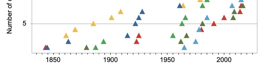
Diversity 2021, 13, 87                                                                                           4 of 36

                         classification, and systematics of the members of both taxa, highlighting where major gaps
                         in knowledge lie and where future efforts could be made.

                         2. Methods
                               Published literature on opheliids and scalibregmatids was reviewed thoroughly aim‐
                         ing for information on diversity, ecology, and distribution. The World Register of Marine
                         Species [3] database was mostly used as the basis for systematic arrangement, synonymies
                         and valid genera and species, as well as Blake and Maciolek [1] for Opheliidae and Blake
                         [23] for Scalibregmatidae. Furthermore, brief accounts on systematics and general morphol‐
                         ogy of these families are also provided as well as tables with valid nominal species includ‐
                         ing type locality, depth (from original description) and marine realms (sensu [45]) (Tables
                         A1 and A2 in Appendix A).

                         3. Results
                         3.1. Opheliidae Malmgren, 1867
                         3.1.1. Systematics
                               Until recently, the Opheliidae comprised three subfamilies: Opheliinae Hartman‐
                         Schröder, 1971, Ophelininae Hartman‐Schröder, 1971 and Travisiinae Hartmann‐Schröder,
                         1971. The latter only included the genus Travisia Johnston, 1840 that differed from other
                         opheliids in having a grublike appearance and a papillated cuticle. Indeed, recent molecular
                         phylogenetic analyses have demonstrated the monophyly of opheliids if the Travisiinae
                         are excluded [46], subsequently motivating the establishment of Travisiinae as a family
                         by Blake and Maciolek [33]. In fact, this possibility had been already proposed by Blake
                         [47], Bleidorn et al. [48] and Hall et al. [49]. The morphological differences between Travisi‐
                         inae and the other two subfamilies were further supported by Belova and Zhadan [50].
                         These authors suggested that the presence of several shared anatomical and ultrastruc‐
                         tural features of the gills amongst several opheliid genera but absent in Travisia, might
                         support the exclusion of the latter from Opheliidae and would constitute synapomorphies
                         of the Opheliinae and Ophelininae. The Travisiidae is now considered the sister group to
                         the Scalibregmatidae, while molecular analyses have highlighted the affinities of opheliids
                         to capitellids and echiuroids [51,52] and to other “sedentary” families as well (e.g., Are‐
                         nicolidae). Therefore, today Opheliidae includes only the subfamilies Opheliinae and
                         Ophelininae [1,46]. The two subfamilies are represented only by species with elongated
                         bodies and smooth cuticle, all sharing the presence of a conspicuous ventral groove [1].
                               According to Blake and Maciolek [1], Opheliidae comprises five genera distributed in
                         the subfamilies Opheliinae (Ophelia and Thoracophelia Ehlers, 1897) and Ophelininae (Arman‐
                         dia Filippi, 1861, Ophelina and Polyophthalmus Quatrefages, 1850). However, there has been
                         much confusion with the generic arrangement within the Opheliidae (e.g., synonymies and
                         changes in diagnosis of genera). Sene‐Silva [53] performed a cladistic morphological analy‐
                         sis of the family that have led to a redefinition of the previously established genera. In this
                         context, Lobochesis Hutchings and Murray, 1984 was synonymized with Thoracophelia, which
                         subsequently replaced Euzonus Grube, 1866 (a homonym of the diplopod myriapod Euzonus
                         Menge, 1854 [54]). According to Blake and Maciolek [1], the genera Tachytrypane McIntosh,
                         1879 and Ammotrypanella would fall within the current diagnosis of Ophelina because the
                         presence and distribution of branchiae seemed much variable within the latter. However,
                         Ammotrypanella was retained by Wiklund et al. [2] who also amended the redefinition of the
                         genus as given by Schüller [55]. Furthermore, the abranchiate Antiobactrum Chamberlin,
                         1919 is regarded as a valid genus in the World Register of Marine Species [3] but considered,
                         in turn, as a synonym of Ophelina by Blake and Maciolek [1]. In this context, Paul et al. [46]
                         have provided a phylogenetic analysis of the family but considering only a limited number
                         of species; therefore, an analysis based on molecular and morphological characters includ‐
                         ing a greater taxa sampling would be desirable to assess the actual definition of genera.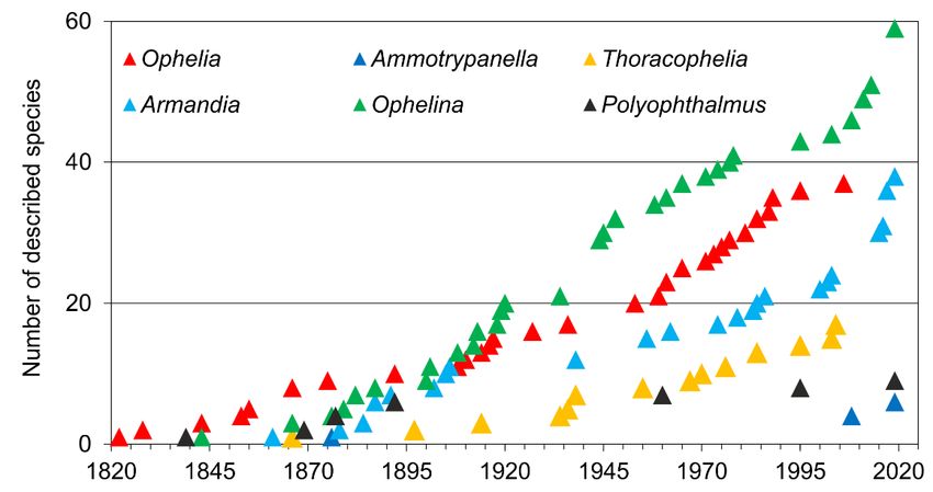
Diversity 2021, 13, 87                                                                                               5 of 36

                         3.1.2. Taxonomic History
                               The first described species was Ophelia bicornis Savigny, 1822. The number of newly
                         described taxa increased gradually during the second half of the 19th century and along
                         the first two decades of the 20th century. After WWII new species were described at a rate
                         of about 10 per decade, whereas in the last decade (2010–2019) 30 new species were added
                         to the family from all around the globe (Figure 2A). This overall tendency closely resem‐
                         bles those exhibited by each of the most speciose genera (Figure 3).

                         Figure 2. (A) number of opheliid species described per decade; (B) number of valid opheliid species
                         listed under the bioregion (sensu Spalding et al. [45]) according to type locality. Abbreviations:
                         ARC—Arctic; TNP—Temperate North Pacific; EIP—Eastern Indo‐Pacific; TEP—Tropical Eastern
                         Pacific; CIP—Central Indo‐Pacific; WIP—Western Indo‐Pacific; TAU—Temperate Australasia;
                         TNA—Temperate Northern Atlantic; TSM—Temperate South America; TSA—Temperate South Af‐
                         rica; TRA—Tropical Atlantic; SOC—Southern Ocean.

                              There are a number of identification keys for Opheliidae from regions such as South
                         Africa [56], California [47], the United Kingdom [54], and the Iberian Peninsula [4]. Some
                         papers also provided tables that compile morphological features for species of the genus
                         Thoracophelia (as Euzonus and Lobochesis; [57]), Ophelina from Australia [58], Armandia [59]
                         and Polyophthalmus [60], and identification keys for Ophelina from NE Atlantic [61] and
                         Armandia from Australasia and Central Indo‐Pacific [62,63].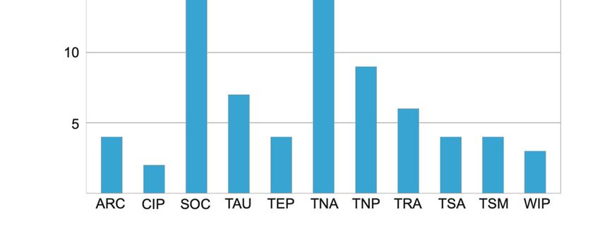
Diversity 2021, 13, 87                                                                                         6 of 36

                         Figure 3. Number of described species (accumulated) of each opheliid genus (including Ammo‐
                         trypanella) from 1820 to 2020.

                         3.1.3. Taxonomic Characters and External Morphology
                               The opheliid body is usually elongated and divided into a defined number of seg‐
                         ments, usually ranging between 30–70. The anterior end is inflated in the Opheliinae, but
                         typically sleek and more elongate in the Ophelininae [1,4]. The trunk may be entire, as in
                         Ophelininae (Figure 1A,D,E), or divided in two (e.g. Ophelia) (Figure 1C) or three regions
                         (e.g. Thoracophelia) (Figure 1B). In Thoracophelia, the modified chaetiger 10 marks the limit
                         between the thoracic and the abdominal region (Figure 1B). A conspicuous ventral groove
                         is always present, but it may extend continuously throughout the trunk, as in Ophelininae
                         (Figure 1A,D and Figure 4B,H,I), or be restricted to its posterior half, as in Opheliinae
                         (Figure 1C). Some species present two additional longitudinal lateral grooves, one on each
                         side of the body (Figure 4H). Paired lateral branchiae attached dorsally to the parapodia
                         are present in many species, either along the entire trunk or limited to its posterior 1/2–
                         2/3 portion. Branchiae are always absent in last few chaetigers (Figure 1B,E, Figure 4I,J
                         and Figure 5A,C,H). Branchiae are bifurcate or pectinate in some Thoracophelia, but simple
                         and cirriform in the remaining genera (Figure 1B).
                               The prostomium is elongated, tapered, or conical in most species (Figure 4A,C), but
                         rounded in Polyophthalmus (Figure 4B). It lacks lateral appendages, but a terminal palpode
                         (sometimes biarticulated) is present in Armandia and several Ophelina species (Figure
                         4A,C). The proboscis is often an axial, nonmuscular eversible structure (Figure 4A,D and
                         Figure 6A,B), but it might consist of several retractable ciliated tentacles in some species
                         of Armandia [63,64]. Nuchal organs are eversible and represented by one pair of conspic‐
                         uous ciliated pits/slits of various shapes depending on the species [65] (Figure 4A,C–G).
                         Exceptionally, two pairs of nuchal organs are present in Polyophthalmus spp. and Armandia
                         polyophthalma Kükenthal, 1887 (see [65]), often slightly pigmented [2]. Subdermal pig‐
                         mented eyes (two to three) are present in several species [66] (Figure 1E); these simple
                         eyes are present in larvae and may be retained in the adult [67]. Additional pairs of seg‐
                         mentally arranged pigmented eyes are present in Armandia and Polyophthalmus, at least
                         on several midbody segments (Figure 1E).
                               Parapodia are biramous and consist of small lobes or tori provided with simple ca‐
                         pillary chaetae (Figure 4I,J and Figure 5A–F). A ventral cirrus is present and a small spher‐
                         ical projection may be also found dorsally on the prechaetal lobe in Armandia (termed as
                         “dorsal cirrus”: [15,62]; Figure 5C). Parapodial ciliated sensory organs were reported on
                         the prechaetal lobe in several species of Armandia [62]. Lateral organs are usually present
                         as ciliated pits in between noto‐ and neuropodia and may also occur in the anterior achae‐
                         tous segments [15] (Figure 5E).
Diversity 2021, 13, 87 7 of 36
The last segments may be achaetous and are often retractile. The pygidium typically
prolongs into a tubular funnellike structure (termed anal cone, funnel, or tube) that may
be quite long in comparison to body length in some species of Ophelininae (Figure 1D and
Figure 5G–K). The shape of the funnel, as well as the presence of accessory structures,
such as dorsal/marginal papillae and unpaired/paired cirri, diagnoses few genera and spe‐
cies (Figure 1C–E and Figure 5G–K).
Figure 4. SEM micrographs of several Opheliidae showing main diagnostic characters. (A) Ophelina breviata, anterior end
in lateral view, showing pointed prostomium; (B) Polyophthalmus pictus, anterior end in ventral view, showing the distally
rounded prostomium; (C) Ophelina helgolandiae, anterior end in lateral view; (D) O. breviata, anterior end in dorsal view;
(E) O. helgolandiae, nuchal organ; (F) Armandia buccina, nuchal organ; (G) Ophelina abranchiata, nuchal organ; (H) O. abran‐
chiata in lateral view; (I) A. buccina, anterior chaetigers in lateral view; (J) Armandia opisthoculata, mid‐body chaetigers in
lateral view. Abbreviations: Br—branchia; Lg—lateral groove; Mo—mouth; No—nuchal organ; Pa—palpode; Pb—pro‐
boscis; Pc—prechaetal lobe; Vg—ventral groove.
Most opheliids have a relatively simple body, reduced parapodia, and simple chae‐
tae. Therefore, the taxonomy of the family has traditionally relied on the limited number
of available external characters. This is particularly evident among Polyophthalmus, a ge‐
nus in which most described species are nearly identical morphologically (e.g., [19]).
Opheliid genera are defined according to whether the body is divided in distinct regions
or not, the extension of the ventral groove, as well as the presence of branchiae and lateral
eyes. Species are instead diagnosed based on several parapodial features (e.g., shape of
prechaetal lobe, ventral cirrus and presence of “dorsal cirrus”, relative length of chaetae
across body), the number of lateral eyes (if present), the length of the branchiae, as well as
the number of branchiate segments and pygidial cirri. Features of the anal tube are mainly
relevant to identify species of Ophelininae, and include its shape, length relative to last
chaetigers, number and shape of marginal anal cirri/papillae, as well as presence, shape,
and position of the unpaired ventral cirrus and the paired basal cirri [58]. Unfortunately,Diversity 2021, 13, 87 8 of 36
the fact that branchiae and anal tube are easily detached has generated much confusion
regarding the taxonomic status and identifications of some opheliid taxa (e.g., [2]). The
many species described based on single/damaged specimens have contributed to worsen
the situation [58], together with the lack of information on the intraspecific variation ex‐
hibited by some characters, such as number and presence of lateral eyes and anal tube
papillae, which may change through different ontogenetic stages in the same species [66].
Examination of a sufficient number of specimens of several sizes is therefore crucial to
alleviate this situation in the future [2,58,66].
Figure 5. SEM micrographs of several Opheliidae showing main diagnostic characters. (A) Ophelina basicirra, parapodium
and branchia; (B) Armandia laminosa, parapodium; (C) Ophelina helgolandiae, parapodium and branchia; (D) Ophelina abran‐
chiata, mid‐body parapodia; (E) Polyophthalmus pictus, lateral organs; (F) Armandia paraintermedia, parapodium and bran‐
chia; (G) O. abranchiata, anal tube in lateral view; (H) Armandia parva, posterior end in lateral view; (I) Armandia tubulata,
anal tube in lateral view; (J) Ophelina bowitzi, anal tube in lateral view; (K) Ophelina cylindricaudata, anal tube in lateral
view. Abbreviations: Br—branchia; Dc—ʺdorsal cirrusʺ; Lo—lateral organs; Nc—notochaetae; Ne—neurochaetae; Pc—
prechaetal lobe; Pm—pygidium marginal papillae.Diversity 2021, 13, 87 9 of 36
Parapar et al. [15] suggested that features of nuchal and lateral organs might repre‐
sent useful characters to diagnose species in the future, in spite that the latter, for instance,
can be easily overlooked or is difficult to examine due to state of preservation [2]. In this
context, the use of SEM for examination of properly fixed specimens seems mandatory to
fully assess features of parapodia, as well as nuchal and lateral organs (e.g.,
[2,58,60,62,63,68]).
3.1.4. Internal Morphology
The internal anatomy of opheliids has been studied mostly in several intertidal spe‐
cies [16,17,69], including later detailed accounts on the structure of the proboscis [64],
body musculature [20,21], respiratory system [50], and sensory organs (see below).
Opheliids lack circular muscle fibers, but they possess bands of longitudinal muscles
protruding along the body surface [20], as well as oblique muscles that insert into the
midventral line thereby contributing to shape the typical opheliid ventral groove. The
structure of the proboscis varies greatly among taxa, corresponding to several of the types
described by Tzetlin and Zhadan [64]: type 1, symmetrical, bubblelike, and ciliated as
found in the Opheliinae; type 3, asymmetrical, dorsal‐lobed (e.g., Ophelina, Polyophthal‐
mus); type 4, formed instead by several retractable ciliated tentacles (some species of Ar‐
mandia). Exceptionally, the proboscis of Armandia amukusaensis Saito, Tamaki and
Imajima, 2000 has been reported as flanked by several “filaments” [66]. The digestive tract,
and particularly the intestine, might be regionalized in certain species [70] (Figure 6).
Figure 6. Microcomputed tomography (CT) sections of Ophelina acuminata from Iceland. (A)
frontal, (B) right sagittal and (C,D) transversal body sections showing internal anatomy. White dis‐
continuous lines in (A,B) marking regions showed in (C,D). Abbreviations. Br—branchia; Bw—Diversity 2021, 13, 87 10 of 36
body wall musculature; Cl—coelomic space; Fg—foregut; Hg—hindgut; Mg—midgut; Pb—probos‐
cis.
The circulatory system is closed [17]. Gills appear as body wall protrusions contain‐
ing coelom or vessels connected to blood sinuses [50]. Metanephridia are present in sev‐
eral species [71], although protonephridia have been reported in Thoracophelia mucronata
(Treadwell, 1914) by McConnaughey and Fox [17]. The ultrastructure of sensory organs
has been described thoroughly in several opheliids, including the nuchal organs in Ophelia
bicornis [72] and Ophelia rathkei McIntosh, 1908 [65]), the subdermal eyes in Armandia brevis
(Moore, 1906) [18] and the juveniles of O. rathkei [67], as well as the lateral eyes in A. brevis
[73], P. pictus, and Polyophthalmus qingdaoensis Purschke, Ding and Müller, 1995 [19].
In this sense, the consistent differences in the ultrastructure of lateral eyes in Polyoph‐
thalmus (e.g., size and number of cells, number, and dimensions of cellular elements) seem
also useful to distinguish species [19]. Thus, future ultrastructural studies might provide
phylogenetically informative morphological characters, perhaps further illuminating the
delineation of genera. In the same line, the use of micro‐CT seems a promising source for
phylogenetically informative characters insofar as it offers a comparatively easy overview
of the internal anatomy and produces a minimum damage to the examined specimen (e.g.,
[74]) (Figure 6). It therefore represents a useful tool to compare, for instance, the regional‐
ization of the digestive tract as well as the organization of the circulatory system across
genera and/or species.
3.1.5. Species Diversity and Distribution
The most speciose genera are Ophelina (about 59 species, excluding Ammotrypanella),
Armandia (38) and Ophelia (37); Thoracophelia comprises 17 species. Depending on the
sources, Polyophthalmus is composed of four [3] to nine [60] species, highlighting the need
for further morphological and molecular work in order to assess its actual diversity as
well as a fully review the synonyms and material attributed worldwide to Polyophthalmus
pictus [4,60]. Finally, six species are classified into Ammotrypanella by those authors who
consider the genus as valid [2,15,55].
Opheliids have been reported or described from the poles to the equator across all
the 12 marine ecoregion realms defined by Spalding et al. [45] (Figure 2B). Similar distri‐
bution patterns are found in the genera Armandia, Ophelia, and Ophelina; whereas Thoraco‐
phelia is mostly restricted to the temperate realms (14 out of 17 species). Many opheliid
species have been described from Temperate Northern Atlantic and Central Indo‐Pacific
(33 and 34, respectively) in comparison to other regions (ranging from 4 to 17). The type
localities of half of the known species of Ophelia (16) are in the Temperate Northern At‐
lantic and about one third of each Armandia and Ophelina are found in the Central Indo‐
Pacific. These numbers, however, may be explained by the greater sampling effort histor‐
ically performed in those areas and the subsequent more detailed knowledge that we have
on their annelid faunas of the NW and NE Atlantic, California, and some areas of the
Pacific Ocean [47,75]. Indeed, recent work done in unexplored Pacific areas has yielded
many new taxa. For instance, Magalhães et al. [60] have described five new species from
several western Pacific islands and Wiklund et al. [2] eight new species of Ammotrypanella
and Ophelina plus other still formally undescribed taxa from the eastern Clarion‐Clipper‐
ton Zone (central Pacific). Furthermore, Parapar and Moreira [62] and Moreira and Para‐
par [63] have described eleven new species of Armandia from Lizard Island (Great Barrier
Reef) whereas only two valid species of this genus are present in the comparatively better‐
known Western Europe. These findings suggest that the actual diversity in other temper‐
ate and tropical regions may be greater, including other Pacific areas as well as Temperate
Australia (only 13 species described so far) and Tropical Atlantic (nine species).
A wide geographic distribution has been reported for species such as P. pictus, Ar‐
mandia intermedia Fauvel, 1902, Ophelina acuminata Örsted, 1843 and O. abranchiata Støp‐
Bowitz, 1948. However, these taxa might represent complex of cryptic species asDiversity 2021, 13, 87 11 of 36
suggested by recent molecular analyses of several populations previously attributed to O.
abranchiata [2,76]. On the contrary, many taxa have not been reported after original de‐
scription thus making it difficult to assess their distribution patterns. Finally, reports of
species far away from their type locality should be considered with caution because of the
lack of knowledge of local faunas (see [60]).
3.1.6. Biology and Ecology
Most Ophelia species inhabit clean sandy sediments from the intertidal fringe to the
shallow subtidal down to depths of about 100 m [75]. The exception is Ophelia profunda
Hartman, 1965 and Ophelia pulchella Tebble, 1953 that prefer, in turn, muddy bottoms; the
former being reported down to 1700 m depth. Species of Armandia, Polyophthalmus, and
Thoracophelia prefer coastal areas, the only remarkable exception being Thoracophelia pro‐
funda (Hartman, 1967) (4000 m). Polyophthalmus translucens Hartman, 1960 has been re‐
ported at depths of 900 m but Sene‐Silva [53] suggested that this species may correspond
to the genus Ophelina. Indeed, Ophelina shows a wider range of ecological preferences,
with some species restricted to intertidal‐shallow depths while others show wide bathy‐
metric ranges (subtidal/shelf depths down to 2000–3000 m), or, alternatively, are limited
to the deep‐sea (at depths below 1000 m). Ammotrypanella species are distributed at depths
below 400 m, more than reaching the abyssal realm.
The majority of opheliids burrow in coarse to fine sand or in muddy sediments. Ecol‐
ogy of several intertidal species of Armandia, Ophelia, and Thoracophelia have been exten‐
sively studied when compared to deep‐sea species [13]. Some opheliids such as Thoraco‐
phelia furcifera Ehlers, 1897 and T. mucronata may reach high abundances in the intertidal
of sandy beaches (2000–40,000 individuals per m2 [8,17]). Spatial variations in abundance
have been related to beach morphodynamics, granulometry, and organic content (e.g.,
[10]). Experimental work has suggested that the abundance of A. brevis is correlated neg‐
atively with proliferation of tube‐building infaunal species [77]. In general, opheliids are
found within well‐oxygenated sediments but some Ophelina species thrive in muddy sed‐
iments with low oxygen content [50] or a high concentration of heavy metals [58]. On the
other hand, P. pictus usually dwells among intertidal algae, reaching densities that surpass
5000 ind. per m2 in Cystoseira mats where is also present all the year round [78]; Polyoph‐
thalmus is also found among fouling communities in artificial habitats [79].
Opheliids show two strategies to burrow into the sediment, i.e., peristalsis based on
oblique muscular fibers acting in conjunction with cuticular fibers (e.g., Thoracophelia) re‐
sulting in a dual anchor burrowing mechanism [9,21] or, rather, by undulatory move‐
ments (e.g., Armandia). Regarding the latter, A. brevis lacks circular musculature and there‐
fore relies on bands of oblique muscles that act antagonistically to longitudinal muscles.
This muscular arrangement allows for lateral bending and undulating movements that
rearrange the sediment grains around by creating a burrow [80]. Armandia brevis and other
Ophelininae species display a similar pattern of movement when swimming in water;
while there is no report of such behavior in Opheliinae.
These burrowing abilities facilitate the migration of intertidal species of Thoracophelia
downwards or upwards into the sediment to cope with wave turbulence or avoid of low
levels of oxygen in the interstitial water [81]. Thoracophelia is also capable to migrate hori‐
zontally seaward or landward into the sediment in response to changing beach morpho‐
dynamics in high‐energy environments [7]. Vertical migration in Ophelia has also been
related to the release of gametes/eggs near the sediment surface [82] or to the avoidance
of interspecific competition [83]. Tamaki [84] reports that specimens of Armandia sp. mi‐
grate in offshore direction as they grow. Giangrande et al. [10] has suggested that the spa‐
tial migration in Ophelia barquii Fauvel, 1927, from the upper intertidal to upper infralitto‐
ral zones, may occur as a response to seasonal changes in hydrodynamics. Because of their
burrowing activity, opheliids are important agents in sediment bioturbation [6].
Opheliids are nonselective deposit‐feeders by swallowing sediment with the everted
proboscis [85]. Feeding behavior has been studied in several species of Ophelia, Ophelina,Diversity 2021, 13, 87 12 of 36
and Thoracophelia; intertidal and shallow‐water species show high ingestion rates [86]. On
the contrary, P. pictus has been suggested to be a selective feeder [87].
In general, opheliids are mostly dioecious and synchronously release large amounts
of gametes or eggs [88]. Life cycle and reproduction of several Ophelia species has been
studied in North Atlantic and the Mediterranean. Life span extends from one to six years
and reproduction occurs from spring to autumn. In general, species breed once a year. On
the other hand, adults of A. brevis and P. pictus experience an epitokous planktonic phase
in which they swimming into water to release their gametes [89,90]. Presence of similar
epitokous phases has also been suggested for A. polyophthalma at least in aquarium condi‐
tions. Epitokous specimens show longer chaetae on the posterior five chaetigers that are
in turn slightly compressed laterally [90].
Larvae might go through a short lecithotrophic planktonic stage of 4–12 days
[5,10,82] or a longer planktonic life thus allowing for a greater dispersal ability (e.g., A.
brevis; [89,91]). Larvae of Ophelia, Thoracophelia, Armandia cirrhosa Filippi, 1861, and A. poly‐
ophthalma consist only of two to five chaetigers right before settlement whereas those of
A. brevis may have up to 20 segments [87,89,90,92,93]. Miner et al. [91] described the feed‐
ing mechanisms of the larvae of A. brevis that includes action by ciliary bands and direct
ingestion with the mouth. After this pelagic phase, the larva settles on the substrate and
the body enlarges to become a juvenile worm. Wilson [94] has demonstrated, after several
experiments that the settlement of O. bicornis larvae is conditioned by the presence of bac‐
teria in the sand grains rather than by the grain size itself.
Polyophthalmus pictus is among the few polychaetes unable to regenerate body seg‐
ments although it may show wound healing of posterior segments [95].
Deep‐sea opheliids are known to be the hosts of two parasitic cyclopoid copepod
species of the genus Ophelicola [96]. Opheliids are also consumed by several fishes and
crabs; for example, Ophelia limacina (Rathke, 1843) has been found in the digestive tract of
demersal fishes [97] and P. pictus in Trachurus mediterraneus (Steindachner, 1868) [98].
Kicklighter and Hay [99] also suggested that A. agilis may have some chemical deterrents
that make it unpalatable for some fishes.
Some opheliids have been the subject of a number of ecotoxicological studies by ex‐
posing them in experimental conditions to contaminants (e.g., heavy metals) or antifoul‐
ing compounds (e.g., tributyltin) in sediments. For instance, O. bicornis has been demon‐
strated to be sensitive to cadmium [12] whereas the exposition of A. brevis to TBT resulted
in changes in body growth rates [11]. Armandia agilis (Andrews, 1891) has been suggested
as an appropriate target species to discriminate between clean and contaminated sedi‐
ments [100] and Armandia cyprophilia Neave and Glasby, 2013 is abundant in sediments
with high concentrations of copper in otherwise depauperated polychaete assemblages
[58]. Therefore, the use of opheliids as indicators of marine pollution seems a promising
field of study.
3.2. Scalibregmatidae Malmgren, 1867
3.2.1. Systematics
The first described scalibregmatid was Scalibregma inflatum Rathke, 1843 [30], origi‐
nally classified as an allied to the genus Arenicola Lamarck, 1801 [24], until Malmgren [101]
established the family Scalibregmatidae in 1867. Later classifications considered Scalibreg‐
matidae as part of the suborder Opheliida [97,102]. This placement was congruent with
the results of subsequent morphological analyses, which nested Scalibregmatidae within
the clade Scolecida as sister group of Opheliidae, although without any synapomorphy
[44]. In contrast, molecular data have more frequently favored a sister‐group relationship
between Scalibregmatidae and Arenicolidae, often including the genus Travisia Johnston,
1840 [46,103], nowadays classified as a separated family [23]. However, the placement of
Scalibregmatidae must be considered unresolved, as those analyses were limited to few
molecular markers and did not include morphological information. DespiteDiversity 2021, 13, 87 13 of 36
phylogenomic information is available for at least one species in the family [104], Scali‐
bregmatidae has never been included in broad phylogenomic analyses [105].
Scalibregmatidae comprises about 68 described species and 14 valid genera [23]. How‐
ever, there has been much confusion regarding the species composition of several of them,
hampered by the fact that many scalibregmatid species have been described based on in‐
complete specimens or limited material [27,40]. Scalibregmatids have been traditionally
categorized as arenicoliform or maggotlike depending on their overall body shape, alt‐
hough without assigning to these groups any systematic value. Arenicoliform species are
typically elongated, inflated in the anterior end, and tapering towards the pygidium;
whereas maggotlike species are shorter and stouter [31]. This distinction has been pro‐
gressively abandoned partly because we know that these differences often rely on preser‐
vation artefacts and post mortem contraction; but mostly because intermediate forms also
exist and this character even changes during the development of certain species [26,36].
There have been no attempts to resolve the internal relationships of Scalibregmatidae
apart from few studies aiming at placing a few specific taxa [2,26,46,103], so the character
evolution within the group remains unknown [43].
3.2.2. Taxonomic History
The study of Scalibregmatidae received a notable attention during the 19th century.
By the beginning of the 20th century, many common European species were already de‐
scribed [30,106–110] including also a few species from Australia [111], New Zealand [112],
Cuba [113], and South Africa [111] (Table A2). This level of attention did not decline dur‐
ing the 20th century, when new species of Scalibregmatidae were described nearly every
decade (Figure 7A and Figure 8).Diversity 2021, 13, 87 14 of 36
Figure 7. (A) number of scalibregmatid species described per decade; (B) number of valid scalibreg‐
matid species listed under the bioregion (sensu Spalding et al. [45]) according to type locality. Ab‐
breviations: ARC—Arctic; CIP—Central Indo‐Pacific; SOC—Southern Ocean; TAU—Temperate
Australasia; TEP—Tropical Eastern Pacific; TNA—Temperate Northern Atlantic; TNP—Temperate
North Pacific; TRA—Tropical Atlantic; TSA—Temperate South Africa; TSM—Temperate South
America; WIP—Western Indo‐Pacific.
The first major revision for the family was published in 1925 [114], followed by the
work by Kudenov and Blake [38], Kudenov [25] and Blake [31,36,47]. There have also been
important works focused on individual genera, such as Axiokebuita Pocklington and Four‐
nier, 1987 [26,61], Oligobregma Kudenov and Blake, 1978 [37], and Scalibregma [35,36]. The
status of the systematics of the family has been recently reviewed by Blake [23], who has
summarized and amended the diagnoses of all the currently valid genera.Diversity 2021, 13, 87 15 of 36
Figure 8. Number of described species (accumulated) of the main scalibregmatid genera from 1820 Table 2020. Genera
with less than five described species have been grouped under “Other genera”.
3.2.3. Taxonomic Characters and External Morphology
Members of Scalibregmatidae are relatively large annelids with few taxonomically
informative characters. The prostomium is generally rounded or triangular, lacking an‐
tennae but often bearing a pair of lateral or frontal extensions, whose homology with palps
remains unclear [43] (Figure 9 and Figure 10B–E). Due to the presence of these structures,
the prostomium has been often described as T‐shaped [see 23]. Prostomial extensions are
well developed in the species of Axiokebuita and Speleobregma Bertelsen, 1983, where they
are separated from the prostomium by a basal furrow and bear longitudinal bands of mo‐
tile ciliary bands capable of producing water currents (Figure 10D,E) [26]. Prostomial ap‐
pendages are also long in the enigmatic Scalibregmella antennata Hartman and Fauchald,
1971, only known from its original collection off New England at 4800–5000 m depth [27],
although the presence and arrangement of ciliary bands remain unknown (Figure 9G). In
contrast, in the species of the genera Asclerocheilus Ashworth, 1901, Oligobregma, Scali‐
bregma, and Sclerobregma Hartman, 1965 prostomial appendages consist of stiff hornlike
prolongations, lacking ciliation and a basal furrow (Figure 9A,B) [31]. Despite these mor‐
phological differences, the fact that prostomial appendages follow a similar development
in all investigated scalibregmatids suggests their homology across the family [26,36]. Ep‐
idermal eyes are sometimes present as simple ocelli (Figure 9F) or more complex struc‐
tures composed of multiple ocelli (Figure 9B). Nuchal organs are usually small and often
found retracted into grooves that extend transversally between the prostomium and the
peristomium (Figure 10B,E). When they are everted, they resemble expanded bulbous
vesicles [23]. Nuchal organs are associated with additional transverse bands of motile cilia
in Axiokebuita cavernicola Martínez, Di Domenico and Worsaae, 2013 and Speleobregma
lanzaroteum Bertelsen, 1983 [26] (Figure 9G, Figure 10D,E and Figure 11D).
The peristomium typically consist of one dorsal and one to three ventral rings, merg‐
ing into the upper and lower lips of the mouth. Unfortunately, detailed morphological
descriptions of the peristomium have only been provided for a few species [23,36] (Figure
10C–E). The ventral mouth is connected to an axial proboscis, which is multilobed when
everted and divided into proximal unciliated and distal ciliated zones [64]. The posterior
part of peristomium possesses a pair of rounded ciliated areas of unknown function in
Axiokebuita and Speleobregma, with potential taxonomic value (Figure 11F) [26].Diversity 2021, 13, 87 16 of 36
Figure 9. Stylized drawings showing the main taxonomic characters of different genera in Scalibregmatidae, artificially
grouped according to the most conspicuous external traits. (A) Arenicoliform scalibregmatid genera with branchiae; genus
Scalibregma, (1) S. hanseni, dorsal view, (2) S. inflatum, anterior end in dorsal view, (3) S. hanseni, left parapodium of chae‐
tiger 21 in posterior view; genus Scheme 4. S. branchiatum, dorsal view, (5) chaetiger 3 in posterior view and (6) abdominal
parapodium in anterior view; genus Cryptosclerocheilus, (7) C. baffinensis, dorsal view, (8) anterior end in ventral view;
genus Parasclerocheilus, (9) P. capensis, chaetiger 40 and (10) anterior end in lateral view. (B) Arenicoliform scalibregmatids
genera without branchiae and with spines; genus Asclerocheilus, (11) A. tasmanius, dorsal view, (12) A. kudenovi, anterior
end dorsal view, (13) A. beringianus, chaetiger 15 in anterior view; genus Oligobregma (14) O. quadrispinosa, anterior view,Diversity 2021, 13, 87 17 of 36
(15) O. mucronata, anterior end in ventral view, (16) and posterior parapodium in anterior view; genus Sclerocheilus, (17) S.
unoculus, anterior end in dorsal view, (18) chaetiger 16 in posterior view, and (19) chaetiger 29 in posterior view. (C) Are‐
nicoliform scalibregmatid genera without branchiae and spines; genus Hyboscolex, (20) H. quadricincta, anterior end in
dorsal view, (21) H. pacificus, median parapodium in anterior view; genus Pseudoscalibregma, (22) P. papilia, dorsal view,
(23) P. usarpium, anterior end in dorsal view, (24) P. hartmanae, posterior chaetigers in anterior view. (D) Maggotlike scal‐
ibregmatids genera; genus Polyphysia, (25) P. crassa, lateral view and (26) anterior end in anterior view; genus Lipobranchius,
(27), L. jeffreysi, frontal view. (E) Morphologically divergent genera; genus Scalibregmides, (28) S. peruanus, anterior end in
dorsal view, (29) S. chilensis; genus Scalibregmella, (30) S. antennata, anterior end in dorsal view; genus Speleobregma, (31) S.
lanzaroteum, anterior end in dorsal view; genus Axiokebuita, (32) A. minuta, anterior end in ventral view. Abbreviations:
Br—branchiae; Dc—dorsal cirri; Ip—interramal papillae or ciliation; Pa—prostomial appendages; Pl—parapodial lobe;
Sp—spines; Vc—ventral cirri. Modified from (1,3) Bakken et al. [35]; (2) Mackie [34], (4–6) Hartman [115], (7–8) Blake [116],
(9–10) Day, [117], (11) Kirkegaard [118], (12) Blake [119], (13) Imajima [120], (14) Schüller and Hilbig [37], (15–16) Blake
[36], (17‐20) Kudenov [25], (21) Imajima [121], (22) Schüller [55], (23–24, 28–29) Blake [31], (25) Støp‐Bowitz, [122], (26)
Hartmann‐Schröder [97], (27) Wesenberg‐Lund [123], (30) Blake [23], (31) Bertelsen [124], (32) Parapar et al. [61].
The trunk includes up to 60 segments, each of them typically bearing one to six rows
of elevated pads giving the worms an areolate appearance (Figure 9C and Figure 10A–C).
The number of these rows, as well as the number and size of the pads that form each of
them, varies across different species and body regions. The pattern formed by the pads
has been used to diagnose certain species, suggesting that these patterns might be species‐
specific in some genera [36]. Epidermal papillae are absent in Scalibregmatidae. A
midventral groove is present in most genera, extending from the mouth towards the py‐
gidium along the longitudinal body axis (Figure 10A). It is not clear, though, whether this
structure bears systematic information or if its appearance depends on the post mortem
contraction of the trunk musculature [23]. Transverse bands of presumably motile cilia
have been described on S. lanzaroteum and A. cavernicola [26] (Figure 10D,E).
Branchiae have been considered as an important taxonomic character. The presence
of branchiae in the anterior segment characterizes the genera Scalibregma, Sclerobregma,
Cryptosclerocheilus Blake, 1972, and Parasclerocheilus Fauvel, 1928 (Figure 9A), in which
they are attached to the notopodium from segment 2 up to segment 5–7. Branchiae are
arborescent in most species, branching dichotomously a variable number of times; but can
also be pectinate, with individual branchial filaments arising from an elongate flattened
lamella, as in Sclerobregma branchiatum Hartman, 1965 (Figure 9A) [23]. However, recent
studies suggest that their number and arrangement might vary ontogenetically within the
same species [23,36]. This has raised concerns about the validity of certain species identi‐
fication, particularly when few small individuals have been studied, and growth series
are not incorporated into species descriptions. More information on the ontogeny of other
species of Scalibregmatidae can be found elsewhere [23,26,36].Diversity 2021, 13, 87 18 of 36
Figure 10. SEM micrographs of several Scalibregmatidae showing main prostomial diagnostic characters. (A) Pseudoscali‐
bregma sp., Canary Islands, anterior end in ventral view, showing the pattern formed by the pads as well as the structure
of the parapodia; (B) Asclerocheilus sp., Canary Islands, anterior end in dorsal view; (C) Asclerocheilus sp., northwestern
Spain, anterior end in dorsal view; (D) S. lanzaroteum, anterior end in dorsal view; (E) A. cavernicola, anterior end in dorsal
view. Notice the difference in the prostomial shape and appendages amongst (B–E), as well the presence of different
development of the peristomium, and the presence of different types of chaetae. Abbreviation: Pa—prostomial append‐
ages.
Parapodia are biramous in all scalibregmatids. The development of each ramus
largely varies across species and body regions, but they are typically smaller anteriorly
and more elongated towards the posterior body end. Parapodial structures, such as inter‐
ramal papillae and parapodial cirri have been described in some species, holding useful
taxonomic information. Interramal papillae are retractile and ciliated in S. inflatum [28]Diversity 2021, 13, 87 19 of 36
and Asclerocheilus (Figure 12B); whereas species of Oligobregma present interramal cili‐
ated areas (Figure 12C). Interramal papillae in A. cavernicola and S. lanzaroteum project
from the body wall and bear terminal ciliation [26,61] (Figure 12A,D). Nonciliated glan‐
dular papillae have been observed in S. minutus Grube, 1863 [41], and P. palmeri Blake,
2015 [36]. Parapodial dorsal and ventral cirri may help discriminating amongst species.
Cirri are filiform in Axiokebuita and Speleobregma (Figure 9G), and leaf‐shaped in Oli‐
gobregma, Pseudoscalibregma Ashworth, 1901, Scalibregma, and Sclerobregma. Cirri of‐
ten exhibit glands, which are tubular in some species of Scalibregma, Oligobregma, and
Pseudoscalibregma; but circular in Axiokebuita and Speleobregma. Parapodial lobes or
lamellae are described in Asclerocheilus californicum and in the two species of the genus
Scalibregmides (Figure 9F) [31,40].
Figure 11. SEM micrographs of several Scalibregmatidae showing main diagnostic characters. (A)
Pseudoscalibregma sp., Canary Islands, anterior end in lateral view; (B) Asclerocheilus sp., northwest‐
ern Spain, anterior end in lateral view; (C) Asclerocheilus sp., Canary Islands, anterior end in lateral
view; notice the different morphology and epidermal pattern found on the anterior end on (A–C).
(D) A. cavernicola, anterior end in lateral view; (E) Asclerocheilus sp., Canary Islands, anterior end inDiversity 2021, 13, 87 20 of 36
frontal view, compare the arrangement of the ciliary patterns between (D,E); (F) A. cavernicola, an‐
terior end in ventral view, showing the ventral ciliary pads pn the peristomium; (G) Asclerocheilus
sp., northwestern Spain, posterior end in dorsal view, showing a typical shape and arrangement of
the pygidium in Scalibregmatidae; (H) A. cavernicola, posterior end in dorsal view, showing the ad‐
hesive pygidium typical of the genera Axiokebuita and Speleobregma.
The arrangement of chaetae is a very important taxonomic characteristic in Scalibreg‐
matidae. Chaetae are always simple and might include long capillaries (Figure 10A), ge‐
niculated (Figure 12I), lyrate (Figure 12E,G), short spinous (Figure 12H), and acicular (Fig‐
ure 12F,H). Simple capillary chaetae are present in all described species, while the pres‐
ence or absence of other types of chaetae is an important character to diagnose genera.
The absence of lyrate chaetae characterizes the genera Speleobregma and Axiokebuita,
whereas the morphology of these chaetae is useful to diagnose species in genera such as
Hyboscolex and Asclerocheilus, amongst others. Spinous chaetae are small and typically
arranged as a single row restricted to the anterior most body segments. Since they occupy
similar position to the lyrate chaetae, they are presumed as homologous to the former and
rarely used in taxonomy. The presence of acicular chaetae, in contrast, is very useful and
characterizes the genera Sclerobregma, Parasclerocheilus, Asclerocheilus, Sclerocheilus,
and Oligobregma. Acicular chaetae are large and conspicuous, typically sickle‐shaped or
curved, and covered with fibrils visible in the scanning electron microscope. They are re‐
stricted to the anterior most segments and their arrangement is useful for species diagno‐
ses. They can extend through a variable number of segments either on the notopodia or
in both rami. Finally, geniculate chaetae are only found in S. lanzaroteum [124].
Figure 12. SEM micrographs of several Scalibregmatidae showing main diagnostic characters. (A)
Speleobregma lanzaroteum, parapodia on the anterior segments in dorsal view, showing the presenceDiversity 2021, 13, 87 21 of 36
of cirri; (B) Asclerocheilus sp., Canary Islands, parapodia on the anterior segments in lateral view; (C)
Asclerocheilus sp., northwestern Spain, mid‐body parapodium in frontal view; (D) Axiokebuita caver‐
nicola, mid‐body parapodium in frontal view; (E), Asclerocheilus sp., Canary Islands, lyrate chaetae
on anterior segments in lateral view; (F) Asclerocheilus sp., northwestern Spain, spines on segment
1; (G) Asclerocheilus sp., Canary Islands, lyrate chaetae on mid‐body segment; (H) Asclerocheilus sp.,
Canary Islands, spines; (I) S. lanzaroteum, geniculate chaetae.
The pygidium is quite variable across different scalibregmatids. However, since scal‐
ibregmatids are found lacking the posterior end in most samples, the usefulness of this
character is limited. In most species, the pygidium is simple and bears a typically terminal
anus and surrounded by a variable number of cirri (Figure 11G) whose arrangement,
length, and number are potentially useful to identify species. Species of Axiokebuita and
Speleobregma possess two enlarged rounded pygidial lobes covered with adhesive papil‐
lae (Figure 11H).
3.2.4. Internal Morphology
The internal morphology of Scalibregmatidae was thoroughly investigated during
the early 20th century, particularly in the species S. inflatum [28] and S. minutus [41] mostly
based on histological sections. Unfortunately, after these early works, very few studies
have been undertaken using more modern microscopical techniques.
The body wall consists of the epidermis, which comprises elongated columnar cells
and mucous secreting cells, as well as a muscular layer of circular muscles surrounding
dorsal and ventral longitudinal muscular bundles [28]. Narrow oblique muscles are also
present, arising ventrally from each side of the nerve cord and inserting into the body wall
near the notopodial chaetal sacs. Parapodial musculature is limited to the chaetal sacs as
well as the parapodial retractor muscles [28]. There is also a relatively strong mouth and
pharyngeal musculature, with retractor muscles attached to the proboscis and two short
muscles supplying the nuchal organs [28].
A thin epithelium delineates the coelomic cavity, which is well developed and spa‐
cious. As an adaptation for burrowing, septa are reduced along most of the body [28,41].
The gut is linear and attaches to the body cavity by few strands of muscular tissue in S.
inflatum. The esophagus is straight and covered with secretory glands; whereas the mid‐
gut is wider and curled, and the hindgut is short, linear, and opens directly into the anus.
Several blood sinuses are associated with the stomach in S. inflatum and S. minutus [28,41].
There is also a well‐developed vascular system [28,41], consisting of dorsal and ven‐
tral vessels and their derivatives. The dorsal vessel extends along the alimentary canal
supplying it with capillary vessels. It forms a blood reservoir near the anterior end of the
stomach and a conical heart‐like bulb before branching off to supply the pharynx, the per‐
istomium, and the brain. The ventral blood vessel originates near the mouth and continues
posteriorly, extending dorsally along the nerve cord. In S. inflatum, it supplies the bran‐
chiae, the stomach, and nephridia, as well as the chaetal sacs and their adjacent tissues.
A pair of metanephridia occurs in each chaetigerous segments, except for those most
anterior. Gonads are associated with each metanephridium and are formed by the prolif‐
eration of cells covering the septum by which the nephrostome is attached to the body
wall [28]. The gametes are released from the gonad at an early stage and complete their
maturation in the coelom. Male gonads form sperm platelets bearing spermatids in S. aus‐
tralis and O. mucronata, and they mature into ect‐aquasperm [36].
The brain has an anterior lobe associated with the prostomium and two posterior
lobes associated with the nuchal organs. The prostomial appendages are innervated by a
pair of nerves originating from the anterior lobe of the brain, whereas the esophageal con‐
nectives and the nerves innervating the nuchal organs arise from the middle and posterior
lobes, respectively. The palps are innervated by one ventral and one dorsal nerve, corre‐
sponding to the fourth and ninth pairs respectively [42].You can also read