Advances in 3D Printing for Tissue Engineering - IPPT PAN
←
→
Page content transcription
If your browser does not render page correctly, please read the page content below
materials
Review
Advances in 3D Printing for Tissue Engineering
Angelika Zaszczyńska                , Maryla Moczulska-Heljak, Arkadiusz Gradys and Paweł Sajkiewicz *

                                          Institute of Fundamental Technological Research, Polish Academy of Sciences, Pawinskiego 5b St.,
                                          02-106 Warsaw, Poland; azasz@ippt.pan.pl (A.Z.); mheljak@ippt.pan.pl (M.M.-H.); argrad@ippt.pan.pl (A.G.)
                                          * Correspondence: psajk@ippt.pan.pl

                                          Abstract: Tissue engineering (TE) scaffolds have enormous significance for the possibility of regener-
                                          ation of complex tissue structures or even whole organs. Three-dimensional (3D) printing techniques
                                          allow fabricating TE scaffolds, having an extremely complex structure, in a repeatable and precise
                                          manner. Moreover, they enable the easy application of computer-assisted methods to TE scaffold
                                          design. The latest additive manufacturing techniques open up opportunities not otherwise available.
                                          This study aimed to summarize the state-of-art field of 3D printing techniques in applications for
                                          tissue engineering with a focus on the latest advancements. The following topics are discussed:
                                          systematics of the available 3D printing techniques applied for TE scaffold fabrication; overview of
                                          3D printable biomaterials and advancements in 3D-printing-assisted tissue engineering.

                                          Keywords: tissue engineering; 3D printing; biomaterials

                                          1. Introduction

                                        Recent progress in the 3D printing method stems from the regenerative ability of the
Citation: Zaszczyńska, A.;               human body. It was reported that there were about 31 million Americans who suffered
Moczulska-Heljak, M.; Gradys, A.;         from body defects [1]. Every year, there is globally an increasing number of patients
Sajkiewicz, P. Advances in 3D             suffering from various types of body defects caused by injuries and degenerative processes
Printing for Tissue Engineering.          of various origin [2,3]. Critical defects require support for the growth of the cells [4]. Native
Materials 2021, 14, 3149. https://        regeneration of the human body is limited by multiple elements such as availability of the
doi.org/10.3390/ma14123149                growth hormones or by functionality of the defected tissue. For many years, the standard
                                          medical treatment in such cases has been autologous transplantation (less frequently,
Academic Editor: Brunella Grigolo         allologous) or implantation of an endoprosthesis imitating the lost organ. The above-
                                          mentioned methods allow to restore the full or partial function of the lost organ (tissue
Received: 15 May 2021
                                          defect); however, it should be noted that they are characterized by many disadvantages
Accepted: 4 June 2021
                                          affecting the comfort of the patient’s life. Hence, the idea of developing methods supporting
Published: 8 June 2021
                                          the full regeneration of tissue defects was born, which are based on laboratory cell cultures
                                          collectively referred to as tissue engineering (TE).
Publisher’s Note: MDPI stays neutral
                                               Tissue engineering belongs to a group of relatively new fields of human activity. It
with regard to jurisdictional claims in
                                          combines elements of biology, medicine, material engineering, and mechanics. The basic
published maps and institutional affil-
                                          aim of tissue engineering is to develop methods supporting the regeneration of damaged
iations.
                                          tissues and organs, especially those so far considered to be non-regenerative. Examples
                                          of such tissue and organ damage are provided by everyday clinical practice. These are
                                          usually critical defects of bone, skin, or nerve tissue. The most common cause of such
                                          defects is various types of trauma, with the second most common being those resulting
Copyright: © 2021 by the authors.
                                          directly from tumor activity or those resulting from resection of tumor sites. Typically, the
Licensee MDPI, Basel, Switzerland.
                                          regenerated tissue (cell culture) is initially cultured in vitro (in a bioreactor), and then the
This article is an open access article
                                          partially regenerated tissue is implanted in situ at the site of the defect. To ensure an even
distributed under the terms and
                                          distribution of the cells in the defect space, so-called TE scaffolds are used, which are porous
conditions of the Creative Commons
Attribution (CC BY) license (https://
                                          structures that provide an appropriate substrate for the cultured cells and, at the same
creativecommons.org/licenses/by/
                                          time, allow free access to nutrients and drainage of cell metabolism products. An equally
4.0/).
                                          important task of tissue scaffolds is to take over the mechanical function of the damaged

Materials 2021, 14, 3149. https://doi.org/10.3390/ma14123149                                              https://www.mdpi.com/journal/materials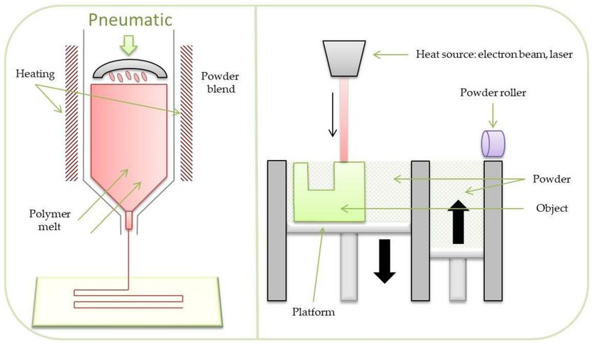
Materials 2021, 14, 3149                                                                                            2 of 28

                           tissue (organ). For this reason, they should be characterized by appropriate stiffness. It
                           is also expected that the implanted scaffold will be fully resorbed by the time the tissue
                           defect is fully regenerated. To meet this requirement, scaffolds are most often fabricated
                           from biodegradable polymers, either natural, such as chitosan or cellulose, or synthetic
                           (polycaprolactone (PCL), polylactide (PLA), etc.). It is not uncommon to use ceramic
                           materials (β-TCP, hydroxyapatite) in a polymeric matrix to improve the biocompatibility
                           of the material used. The designed TE scaffolds must meet many different requirements (in
                           practice, often contradictory). It also turns out that how the scaffold performs its function
                           is determined by factors of various nature, ranging from purely biological to mechanical.
                                 There are numerous methods of TE scaffolds’ fabrication. Amongst them, one can
                           mention a few conventional methods, such as the solvent casting method, phase separation,
                           or electrospinning, which enable limited control over the scaffold geometry. Additionally,
                           they are characterized by poor repeatability. The above limitations do not apply to the
                           additive manufacturing (AM) methods, commonly known as 3D printing methods. Ad-
                           ditionally, 3D printing methods enable easy application of computer-assisted methods of
                           TE scaffold design. Presently, there are a multitude of 3D printing techniques applied for
                           TE purposes. They enable fabrication of TE scaffolds made of different types of materials
                           including polyesters, ceramics, metals, or hydrogels.
                                 Generally, an incredible advantage of 3D printing is the possibility of the fabrication
                           of complex structures, unprofitable to manufacture using injection molding methods [5].
                           Furthermore, 3D printers have been improved for extremely high resolution, which fosters
                           their use in tissue engineering. There are documented attempts of the adaptation of indus-
                           trial printers to make them usable for printing scaffolds for tissue engineering. Nowadays,
                           3D printing methods enable fabrication of TE constructs used for the regeneration of dif-
                           ferent types of tissues, such as skin [6], cartilage [7], and vascular networks [8], as well as
                           whole organs [9].
                                 This review summarizes limitations and general principles of the most extensively
                           used additive manufacturing technologies, including extrusion-based as well as jetting
                           systems. Thus, current methods of printing and printable materials will be discussed.
                           Additionally, the article highlights advanced scaffold fabrication methods for tissue engi-
                           neering applications.

                           2. Scaffolds for Tissue Engineering
                                 Daily, by average, 13 people die due to a long waiting time for organ transplanta-
                           tion [10]. There exists also a problematic issue related to tissue compatibility. In such
                           a situation, tissue engineering may offer various unique methods of scaffold formation,
                           where the tissue compatibility issue may be easily overcome. The idea and the goal is
                           to deliver a functional compatible organ using the patient’s own cells. However, such
                           a process may be a highly complex task as there exist numerous factors related to the
                           organism’s physiology, such as culturing many cell types [11]. In general, scaffolds are
                           essential for the creation of graft structures. TE scaffolds are a substratum for cells’ migra-
                           tion/differentiation and the creation of new regenerated tissue. Thus, properties of the
                           materials, especially chemical and physical, as well as the architecture and morphology, are
                           crucial for cell proliferation and viability [12,13]. Moreover, successful repair of the defects
                           sometimes requires reconstruction of different types of coexisting tissues, such as bones,
                           glands, muscles, vessels, ligaments, nerves, and cartilage. The scaffolds’ morphology and
                           architecture are crucial at various levels: macro, micro, and nano. At the macro level, the
                           architecture is related to the scaffold size and shape from the perspective of the size and
                           shape of the defect, which are essential for the contact and interactions between the scaffold
                           and the native tissues, matrix-cell interactions, and nutrients’ transport [14]. At the micro
                           level, it is characterized by scaffold porosity, pore shape, or pore spatial distribution, each
                           of which is responsible for general scaffold permeability. At the nano level, the morphology
                           is related to the fiber surface characteristics, which are supposed to be responsible for cells’
                           differentiation and proliferation [15].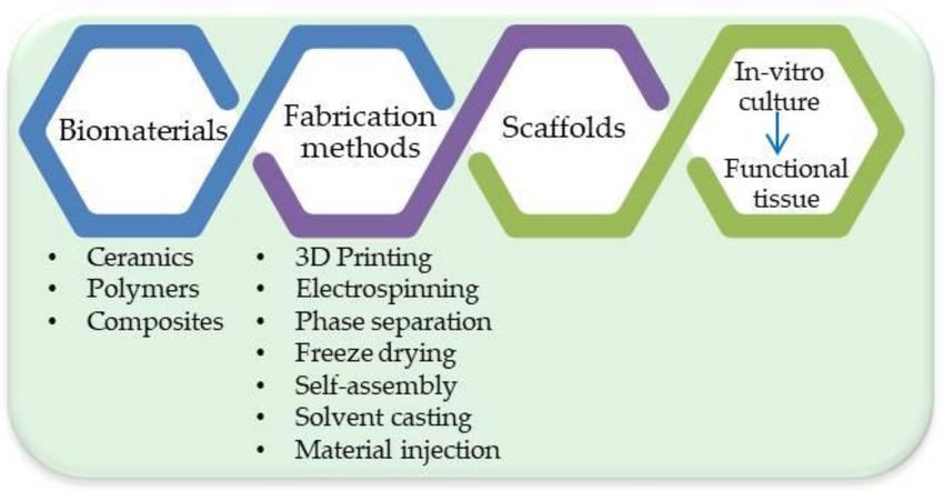
Materials 2021, 14, 3149                                                                                            3 of 28

                                The most critical factors in 3D printing scaffolds are the type of fabrication method
                           and the choice of a biomaterial. Biomaterials interact with biological systems and can be
                           classified by various criteria such as biodegradability, physical and chemical composition,
                           or the application of certain modifications [16]. The choice of a biomaterial is connected
                           with the character of the damaged tissue. Favored materials are usually biodegradable
                           and piezoelectric biomaterials. The main groups of these materials consist of polymers
                           (synthetic and natural), ceramics, and composites. Ceramic scaffolds are preferred in or-
                           thodontic applications; composite scaffolds have applications in dental tissue engineering,
                           whereas polymers are used in soft tissue engineering [17].

                           2.1. Different TE Strategies
                                 Generally, two distinct strategies are used in TE to treat tissue defects using tissue
                           scaffolds [18]. In each, the fabricated scaffold is seeded with cells (sometimes cells are
                           embedded in a scaffold matrix), followed by cell culture in a bioreactor, after which the
                           scaffold filled with the newly formed tissue is implanted into the defect site. The difference
                           lies in the choice of the moment of implantation. In the first of the strategies, fully matured
                           and remodeled tissue is implanted in the defect site. In this case, the scaffold should be
                           completely degraded and metabolized before the moment of implantation. In the second
                           strategy, a scaffold filled with not fully matured tissue is implanted. Depending on the
                           strategy chosen, the implanted scaffold should be characterized by different degradation
                           (erosion) kinetics.
                                 TE scaffolds’ fabrication is followed usually by adequate surface modifications in
                           order to achieve the desired structure/properties from the cells’ perspective. Various
                           hormones or growth factors are usually added during the cell culture. Figure 1 shows the
                           process of creating the tissue engineering product.

                           Figure 1. Tissue Engineering process.

                           2.2. Conventional TE Scaffold Fabrication Techniques vs. 3D Printing Techniques
                                There are various methods of scaffold formation allowing them to meet the require-
                           ments in various specific applications. In addition, many biomaterials are constantly
                           improved for more effective use in tissue engineering. A schematic illustration is shown
                           in Figure 2.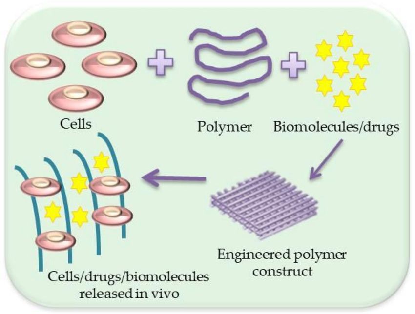
Materials 2021, 14, 3149                                                                                               4 of 28

                           Figure 2. Schematic illustration of scaffold with cells/drugs or biomolecules’ formation.

                                The mostly used scaffold fabrication methods include: electrospinning, additive man-
                           ufacturing, phase separation, solution casting, foaming, extrusion, and self assembly [19].
                           In order to limit some disadvantages of the methods, a combination of them is often
                           used, which sometimes leads to very interesting and promising effects [20]. Figure 3
                           shows various techniques to fabricate three-dimensional scaffolds while some of them are
                           described further.

                           Figure 3. Scaffolds’ fabrication techniques.

                                 One of the most popular processes for scaffold formation is the electrospinning tech-
                           nique (Figure 4). The spinneret filled with electroconductive polymer, usually a solution, is
                           connected to a high electric potential (several to tens kV) at low current. The polymer is
                           spun in the form of fibers, while solvent is evaporating on the way between a spinneret and
                           a collector. A collector is electrically grounded or at a low counter potential and may be sta-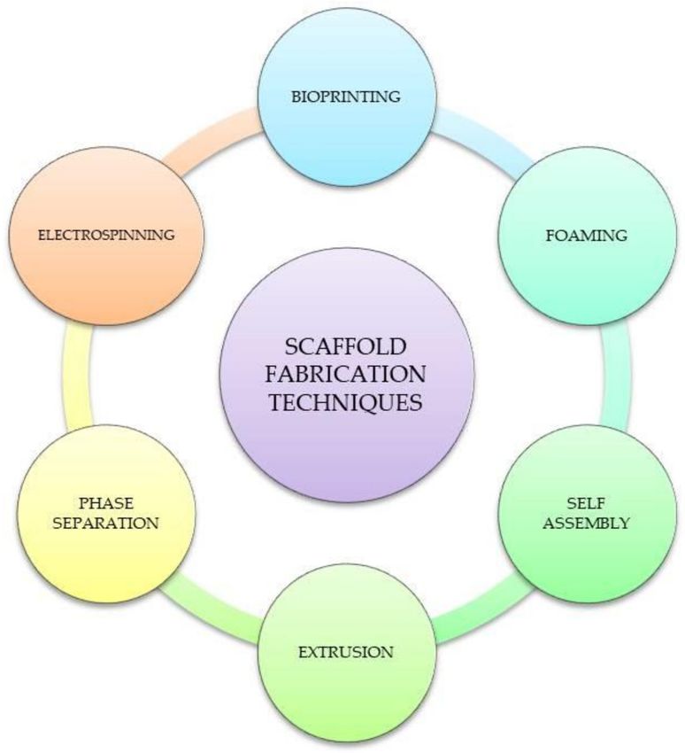
Materials 2021, 14, 3149                                                                                            5 of 28

                           tionary or rotating. The resulting scaffold consists of a micron, submicron, or nanofibrous
                           architecture, either random or aligned, depending on the collector type and mode used.
                           This method of scaffold fabrication allows the formation of fibrous nonwovens with mor-
                           phology and architecture mimicking the fibrous structure of the extracellular matrix (ECM)
                           which is crucial from the perspective of cells. In this process, a large number of various
                           polymers and solvents can be used, both natural, such as gelatin, chitosan, collagen, etc.,
                           and synthetic, such as polycaprolactone (PCL) [21], polyvinylidene fluoride (PVDF) [22],
                           poly(3-hydroxybutyrate-co-3-hydroxyvalerate) (PHBV) [23], poly(methyl methacrylate)
                           (PMMA) [24], etc. By connecting different types of materials, hybrid materials can be
                           developed, particularly as a mixture of synthetic and natural polymers. Although elec-
                           trospinning is a relatively simple process from the instrumental perspective, it is quite
                           complex when analyzing physical phenomena on the process between the moment of jet
                           formation and collection of fibers on the collector. The electrostatic field between the liquid
                           and collector results in a cone-shaped polymer solution to flow out (s.c. Taylor cone). Then,
                           the polymer jet is ejected from the Taylor cone when the electric field exceeds the polymer
                           liquid’s surface tension, followed by various instabilities including bending instability
                           of the jet due to repulsion of the electric charges on the jet surface. By modifying some
                           parameters of the electrospinning process, of the materials, and of external conditions, such
                           as the solution flow rate, spinneret-collector distance, the rotational speed of the collector,
                           voltage, polymer concentration, polymer molecular weight, humidity, and temperature,
                           the morphology and architecture of the scaffold can be dramatically changed according to
                           the desired application [25].

                           Figure 4. Scheme illustration of electrospinning technique.

                                 Phase separation is another traditional method to produce complex and high-porosity
                           three-dimensional scaffolds. There are various modifications of this method, which are
                           generally based on two processes, namely, liquid-solid and liquid-liquid phase separation.
                           They are technically implemented by using either thermally or non-solvent-induced pro-
                           cesses. In the first case, separation is obtained by reducing polymer solubility through
                           a change in the solution temperature resulting in polymer precipitation. In the second
                           case, phase separation is obtained by immersion of a polymer solution in a non-solvent
                           (for the polymer) bath in order to leach away the polymer solvent (wet phase inversion
                           method). There are additional techniques of porous scaffolds’ formation by phase sep-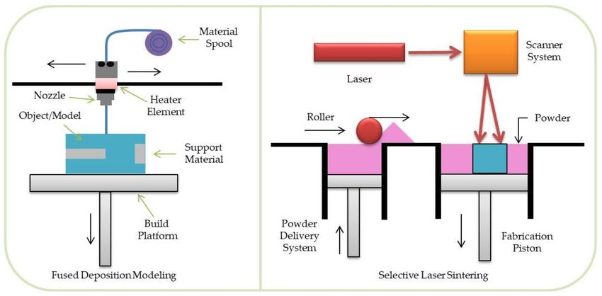
Materials 2021, 14, 3149                                                                                                    6 of 28

                                  aration techniques based on highly compressed gases or supercritical fluids. The phase
                                  separation method has been developed and improved over the years and used as a man-
                                  ufacturing method to form porous polymer membranes. The resulting morphology of
                                  phase-separated scaffolds is extremely sensitive to the process parameters, so desired
                                  parameters of three-dimensional scaffolds can be achieved by adjusting various process
                                  and materials’ parameters [26].
                                       Mixing an organic solvent with polymers, adding granules or spheres as porogens,
                                  and casting this solution to the mold, followed by the extraction of porogens, is known as
                                  the solvent casting/particulate leaching method (SCPL). It is possible to control the final
                                  pore size by the size, content, and distribution of porogens. The solvent evaporates, and
                                  the porogen is removed by dissolving, leaving behind a porous structure. Solvent scaffolds
                                  can be used, for instance, for cardiac tissue engineering applications due to the uniform
                                  distribution of endothelial cells [27]. This technique allows preparation of structures with
                                  regular porosity, but with a quite limited thickness. A summary of these methods is given
                                  in Table 1.

                 Table 1. Selected scaffolds’ formation techniques—main applications and advantages/disadvantages.

         Method                    Applications                        Advantages                        Disadvantages
                           Bone, nerve, skin, and cardiac   High surface area to volume ratio,
    Electrospinning                                                                                 Limited range of polymers
                              tissue engineering [28]          high porosity, easy process
                                                                 Bioactive agents can be
                           Protein delivery applications                                           Limited ranges of pore size,
    Phase separation                                         incorporated into the structure,
                             and/or drug release [29]                                            problems with residual solvents
                                                                      high porosity
                            Vascular tissue engineering                                          Low mechanical strength, limited
     Solvent casting                                        Simple method, controlled porosity
                                 applications [30]                                                  thickness, small pore size

                                       Tissue engineering requires fundamental systemic understanding of the human or-
                                  ganism including cellular differentiation and proliferation [31–34]. To summarize, the
                                  prerequisites of TE scaffolds (not only those 3D printed) are extremely challenging and
                                  manifold. They include: he material for TE scaffold fabrication should be biocompatible
                                  (that is, scaffolds cannot cause any cytotoxicity or immune response); scaffolds should be
                                  easy to sterilize to prevent infections. Moreover, mechanical properties should be enough
                                  for patients’ regular life and activity [18].

                                  3. 3D Printing of Tissue Engineering Scaffolds
                                  3.1. Overall Characteristics of 3D Printing Techniques
                                       Since the emergence of the concept of using tissue engineering products in reconstruc-
                                  tive medicine, many methods of producing TE scaffolds have been developed, starting
                                  from the simplest ones, such as the method of sugar- or salt-crystal-leaching from a solid
                                  structure, to the most advanced ones, which include rapid prototyping (RP) and rapid
                                  manufacturing (RM) methods. The methods of rapid manufacturing are currently a very
                                  dynamically developing field. Practically on an ongoing basis, modifications are made
                                  to existing methods; new methods and devices are created, and the RM industry is now
                                  created both by scientific institutions and commercial manufacturers of hardware and
                                  software. Unfortunately, the dynamism of industry development makes it difficult to
                                  systematize existing methods. Many of the common names of RM methods are registered
                                  trademarks, which means that often even several manufacturers produce very similar
                                  devices using different names for virtually the same manufacturing method used by the
                                  devices. These names come into common use at the same time, which creates a lot of
                                  confusion. One needs to be aware of the fact that terms such as additive manufacturing,
                                  rapid prototyping/manufacturing, solid free-form fabrication, as well as 3D printing, are
                                  essentially synonymous. In the remainder of this paper, we have chosen to use the term 3D
                                  printing. It is a relatively new method of the fabrication of TE scaffolds with controlled
                                  architecture. Despite the fact that there are many various 3D printing techniques, including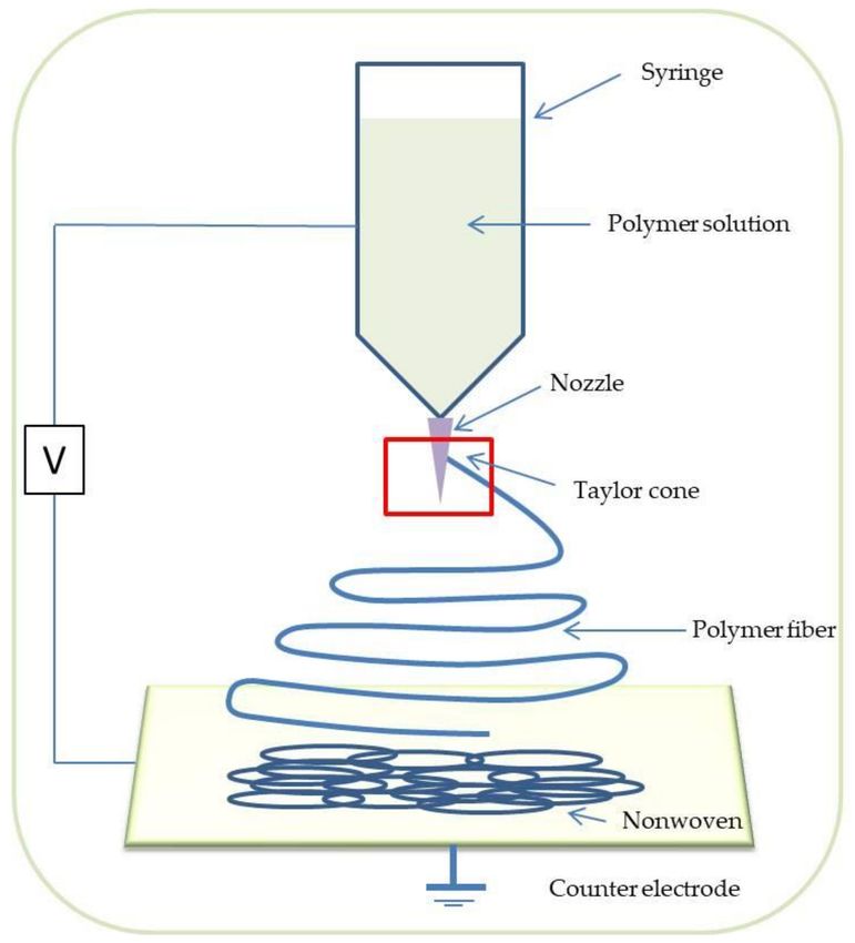
Materials 2021, 14, 3149                                                                                           7 of 28

                           stereolithography, bioprinting, inkjet printing, fused deposition modelling (FDM), PED
                           (Precision Extruding Deposition), laser beam melting, polyjet, electron beam melting, digi-
                           tal laser printing (DLP), and selective laser sintering (SLS) [35], the common feature of all
                           mentioned methods is the general principle of material deposition layer-by-layer until the
                           final product is created [36].
                                 Thus, the 3D TE scaffold is fabricated by the successive addition of consecutive 2D
                           layers of a material. Additive manufacturing has numerous advantages, such as the ability
                           to create complex structures and the possibility of the application of the Computer-Aided
                           Design (CAD) methods. It enables the use of various types of biomaterials [37]. Using
                           living cells and biodegradable polymers allows for the development of methods and novel
                           strategies to create complex tissues and, possibly in the future, whole organs [38]. A
                           3D-printed TE scaffold can be designed using patient-specific data. The CAD method
                           allows for the precise designing of the 3D organ or its missing part. Selected features of
                           living organs, such as porosity or vasculature, may be taken into account in the CAD 3D
                           model. Due to these remarkable advantages, 3D printing is gaining significant interest in
                           regenerative medicine and tissue engineering [39].
                                 In 3D printing, techniques may be distinguished into two categories—binder 3D
                           printing and direct 3D printing.
                                 The former is also called the “drop on powder technique” (Figure 5) [40]. Objects are
                           made by an inkjet liquid printing binder solution on a powder base [41–43]. The process
                           starts by spreading the powder layer on the building platform. Positioning software
                           prints the pattern using a deposition of droplets on the layer with powder. Next, the
                           building platform, powder, and part are lowered, and the next layer can be applied. Then,
                           the powder is removed, and one can observe the printed part. The disadvantages of
                           the method include relatively low resolution and problems with printhead reliability. A
                           small nozzle can have better quality but is more prone to clogging. As an advantage,
                           the fabrication of complicated scaffolds with internal channels is feasible because the
                           surrounding powder supports objects.

                           Figure 5. Scheme illustration of direct 3D printing technique (left) and “drop on powder tech-
                           nique” (right).

                                In the case of direct 3D printing, which is shown in Figure 5, the nozzle of a 3D
                           printer moves back and forth dispensing waxes or plastic polymers, which solidify to form
                           consecutive layers of the fabricated 3D object.

                           3.2. 3D Printing Techniques Applicable to TE
                                Below, the most-known 3D printing techniques, which are applicable to TE, are listed.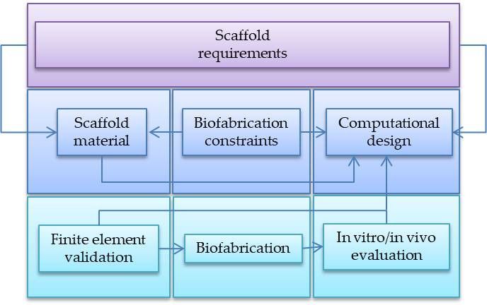
Materials 2021, 14, 3149 8 of 28
3.2.1. Bioprinting
This method allows for the fabrication of soft 3D tissue scaffolds combining biomateri-
als, living cells, as well as growth factors. It enables the fabrication of biomedical parts that
maximally imitate natural tissue characteristics. Generally, 3D bioprinting utilizes the layer-
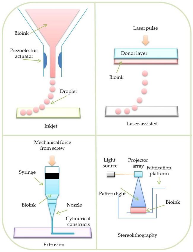
by-layer deposition of materials known as bioinks to create tissue-like structures. There
are four main categories of 3D bioprinting: inkjet bioprinting, laser-assisted bioprinting,
extrusion bioprinting, and stereolithography [44].
3.2.2. Inkjet Bioprinting
In this type of bioprinting method, a mixture of living cells and a bioink is stored in a
chamber joined with the printhead [45]. During the process, the piezoelectric transducer
deforms the printhead. Spatially defined droplets establish tissue constructs (Figure 6).
The main advantage of the method is its low cost and high cell viability [46]. Nevertheless,
this method is limited by numerous problems, such as printhead clogging, uneven distri-
bution of the cells, and inability to print viscous materials. Due to these problems, inkjet
bioprinting has received less consideration by researchers in recent years [47].
Figure 6. Four main categories of bioprinting.Materials 2021, 14, 3149 9 of 28
3.2.3. Laser-Assisted Bioprinting
Typical laser-assisted bioprinting (LAB) involves specialized layers, such as a bioink
layer, an energy-absorbing layer, a donor (quartz/glass), and a collecting layer, to form
structures [48]. During the process, a laser beam is focused on the energy-absorbing layer.
Next, this layer vaporizes and creates an air bubble between the bioink and donor layers.
The formation of a bubble ejects the desired amount of the bioink on the collecting layer. A
tissue structure is created in a droplet-by-droplet manner (Figure 6) [49]. LAB is feasible
for use with high cell density and viscous materials. Additionally, it has been reported
that the method is characterized by high cell viability (95%) and resolves the clogging
issues. Nevertheless, LAB is an expensive process, which generates a very high cost with
large-scale projects. Therefore, only a few printer prototypes were created. [50,51].
3.2.4. Extrusion Bioprinting
The extrusion bioprinting technique is based on liquid extrusion (paste, solution)
from a pressurized syringe through a needle to a solution with controlled density. The
materials are extruded in a form of long strands or dots to create complex structures [52].
The printing process can be conducted at room temperature and used to print natural
biomaterials, especially hydrogels (Figure 6) [53].
3.2.5. Stereolithography
Stereolithography (SLA) is the first developed method of rapid prototyping expanded
in the late 1980s [54]. Stereolithography rasters use a laser beam to control the polymeriza-
tion process of bioinks in a 2D layer. After the deposition of each layer of a material, curing
follows. During the curing process, a photosensitive hydrogel is subjected to UV or visible
light. When a given layer is polymerized, the process is repeated, overlapping the previous
layer, up to the moment when the whole scaffold is completed. This method allows the
use of the following hydrogel materials (Figure 6) [55]: Polyethylene glycol diacrylate
(PEGDA) and gelatin methacryloyl (GelMA) [56]; photo-initiators can be also added [57,58].
The adjustment of various polymerization process parameters, including light energy and
intensity, speed of printing, layer thickness, and exposure time, enables the achievement of
a high-quality (including resolution) product [59–64]. Nevertheless, compared to the other
methods, the SLA process is relatively time consuming, which makes the process feasible
for small-detailed objects.
3.3. Fused Deposition Modeling and the Other Microfiber Extrusion Methods
In the fused deposition modelling (FDM) (Figure 7, left) technique, a coiled polymer
filament is heated up and extruded through a nozzle on the platform. When the polymer
contacts with the platform, it solidifies [65]. The main limitations of using FDM printers in
TE include spatial resolution and possible thermal degradation of the polymeric material.
FDM enables the use of thermoresponsive polymers such as polycaprolactone (PCL), poly-
lactide (PLA), or polyglycolide (PGA). One of the criteria for selecting a material suitable
for FDM is its high thermal stability, which many aliphatic polyesters, unfortunately, do
not have. Thermal degradation of plastics is a particularly noticeable problem in the case
of devices processing polymer granules (the original FDM method is less exposed to the
negative effects of this phenomenon). The polymer heated for a long time loses its viscosity
suitable for the proper course of the manufacturing process. In other 3D printing tech-
niques belonging to the polymer microfiber extrusion group, the method of the material
supplying may be different. In the case of the precision extruding deposition (PED), the
material is supplied in the form of polymer granules, which are thermally plasticized and
extruded under pressure through a nozzle. The described group of methods has been
successfully used in the fabrication of TE scaffolds for many years. Thanks to the methods
based on microfiber extrusion, tissue scaffolds with a strictly planned fibrous structure
can be obtained. The disadvantages of the method include the fact that, due to a too-highMaterials 2021, 14, 3149 10 of 28
polymer processing temperature, it is not possible to produce scaffolds with biomolecules
or living cells incorporated into the fiber structure.
Figure 7. Scheme illustration of FDM (left) and SLS (right) process.
3.4. Selective Laser Sintering
In the method, the polymeric powder particles are heated up slightly above the
polymer glass transition temperature by a laser beam [66]. This leads to partial melting of
the particles [67], during which molecular diffusion on the particles’ surface takes place,
which leads to particles’ fusion. After fabrication of each object layer, the building platform
is lowered, a new layer of powder particles is spread on the top and connected with the
previous layer (Figure 7, right).
3.5. Melt-Spinning
Melt electrospinning (MES) is a relatively new 3D TE scaffold fabrication technique,
being the alternative to conventional solution electrospinning (SES) known for disadvan-
tages related to toxic polymeric solutions [68]. Residues of solvents, e.g., chloroform,
DMSO (dimethyl sulfoxide), DMF (dimethyloformamid), that can be used by SES may
be harmful to living cells seeded on the scaffold. SES limitations were overcome by the
use of the molten polymer instead of the polymer solution. To be jetted in an electric
field, the molten polymer should be characterized by a suitable viscosity. The molten
polymer would be collected by a rotating drum; however, implementation of the numerical
control (NC) enables the precise deposition of fiber in X, Y axes. The mentioned approach
makes the MES another class of 3D printing techniques [69]. Recent works on the melt
electrospinning report that this technique allows for depositing continuous fibers charac-
terized by a diameter less than 1 micrometer, which is comparable to the classic solution of
electrospinning [70].
Summarizing information about 3D printing methods are listed in Table 2.Materials 2021, 14, 3149 11 of 28
Table 2. The most popular 3D TE Scaffolds fabrication techniques—applications, advantages, and disadvantages.
Method Applications Advantages Disadvantages
- requires support
- scaffolds manufacturing [71] structures
- hydrogels [72] - prints viable cells
Bioprinting - nozzle limitations
- tissue engineering [73,74] - soft tissue applications
- must be not cytotoxic
- cell growth [75] during process
- only thermoplastics
• pharmaceuticals [76,77]
materials [82]
• scaffold manufacturing [78] - low cytotoxicity
Extrusion-based - low resolution [81]
• bone tissue engineering [79] - low cost [81]
methods - non-biodegradable
• cardiovascular medical - inexpensive printers
materials can be used
devices [80]
- post-processing
- pharmaceutical [83] - high mechanical
- photo-sensitive
- biomedical manufacturing properties
Indirect methods materials
[84,85] - SLS: powder supporting
(Selective Laser - expensive
- bone tissue engineering the structure
Sintering; - support systems in case
[86,87] - high resolution
Stereolitography) of very complicated
- pharmaceutical [76] - smooth surface
structures
- drug delivery [88] - short time of the process
4. Design Strategies of 3D Printed Scaffolds
4.1. Idea of Computer-Aided Tissue Engineering
Modern tissue engineering probably could not exist without the use of various types
of computer-aided methods; however, it was not until numerically controlled 3D printers
were introduced in TE that all the advantages of computer-aided TE scaffold design became
fully available. They are present at almost all possible stages of creating the so-called tissue
engineering product. This chapter aims to characterize selected computer-aided design
methods and determine the role they play in the process of tissue scaffold design and
fabrication by 3D printing techniques. Generally, the role of computer-aided design in tissue
engineering is so important that the term CATE (Computer-Aided Tissue Engineering) has
emerged and been used in the literature for some time now [89,90].
Figure 8 shows a block diagram that, in simplified terms, describes the operation of the
CATE system. The blocks in the diagram symbolize the individual modules of the system.
In brief, the task of the CATE system is to generate (based on the defect geometry and a
set of appropriately selected criteria) a tissue scaffold design in a form comprehensible for
numerically controlled manufacturing devices such as 3D printers.
Figure 8. General idea of CATE (based on [90]).Materials 2021, 14, 3149 12 of 28
4.2. TE Scaffold CAD Geometry Development
Scaffold geometry can be generated from the start to the finish using CAD software.
Such a model is usually described by a set of solid virtual objects with surfaces that precisely
define its shape. However, it should be kept in mind that the geometry of the scaffold is a
representation of the tissue defect, which usually has an irregular shape. In such a case,
a more adequate way to acquire the geometry of the designed scaffold would be to use
the reverse engineering techniques enabling one to define precisely the defect shape based
on the results of medical imaging by means of computed tomography (CT) or magnetic
resonance (MRI). The result of the CT examination is a series of cross-sectional images
of the examined object called tomograms. The tomograms usually require filtering of all
kinds of noise and artifacts typical for this method. The next stage is the binarization
of grayscale tomograms. Based on the series of binarized tomograms, one can create
a CAD model of the designed scaffold using commercial or free software (Materialise
MIMICS, 3DSlicer, InVesalius). The CAD model is usually saved in one of the neutral
formats used by additive manufacturing systems. Probably the most common format
used in additive manufacturing systems is the Standard Tessellation Language (STL). It
was originally developed for stereolithography but later became popular in other additive
manufacturing methods. In the STL format, the shape of an object is approximated by a
mesh of triangles; hence, the contents of an STL file are the x, y, z coordinates of each vertex
and a vector normal to the triangle plane. In addition to STL, there are other less common
geometry storage formats such as SLC (a format containing consecutive sections described
by polylines), HGPL (HP Graphical Language), and CLI (Common Layer Interface).
4.3. Computer-Assisted Optimization of TE Scaffolds
The optimal tissue scaffold should be characterized by many, often contradictory,
features. In turn, the number of design variables describing the tissue scaffold structure is
so large that a trial-and-error design usually becomes tedious and inefficient, given that
experimental evaluation of the design variants involves lengthy and expensive in vitro and
in vivo testing. Design variables that directly affect the quality of the designed scaffold
include the mechanical properties of the material used, porosity, scaffold stiffness (depen-
dent on the material and scaffold structure), biological activity, and chemical activity of
the chosen material. Several theories have emerged as to what the optimal scaffold should
be, but for a long time, there was a lack of proven methods for assisting the design of
tissue scaffolds. It was not until the mid-1990s that the first attempts to use computer-
aided design methods appeared. Until recently, the use of computer methods in tissue
engineering was usually limited to the computer-aided design of TE scaffold geometry
or the use of in silico models (mainly based on the finite element method (FEM)) at the
stage of evaluation of the designed structure. The end of the first decade of this century has
brought a significant change in the approach to the problem of tissue scaffold design [91].
At that time, the first attempts were made to use optimization algorithms, both classical
ones and those based on artificial intelligence methods [92–94].
5. Biomaterials Used for TE scaffolds 3D Printing
The ideal TE scaffold should be characterized by a number of specific properties, such
as adequate mechanical strength and stiffness, open porosity, biocompatibility, as well as
biodegradability. Meeting the above requirements makes it possible to create a suitable
environment for cell growth. To some extent, all the above-mentioned requirements are
due to the material used. Among the materials commonly used for TE scaffolds, one can
mention natural (e.g., chitin, collagen, cellulose) and synthetic (e.g., polycaprolactone,
polyglycolide, and their copolymers) polymers, as well as ceramics and different kinds of
additives (hydroxyapatite (HA), carbon nanotubes). Below, the attempt has been made
to characterize the main groups of 3D printable materials. At first, the polymers will be
discussed, as the most widely used group of materials for tissue engineering.Materials 2021, 14, 3149 13 of 28
5.1. Polymers
Polymers represent the main category of materials with high potential for use in 3D
printing of TE scaffolds and can be widely used for various tissues’ imitation. TE scaffolds
may be fabricated from non-biodegradable as well as biodegradable polymers. In the
context of tissue engineering, biodegradable polymers generally have more advantages as
compared to the non-biodegradable.
5.1.1. Natural Polymers
Natural polymers are known to be the right candidates for TE scaffold fabrication,
mostly due to their bioactivity, biocompatibility, minimal immune response, as well as
natural biodegradability of most of them [95]. As an example of the natural polymers’
application in TE, one can mention the work of [96] reporting fabrication of TE scaffolds for
cartilage regeneration made of bacterial cellulose. Another study confirms that cellulose
from Acetobacter xylinum can be used in the cartilage regeneration [97]. Collagen and
chitosan also belong to the polymers widely investigated and applied in TE [98]. All
of the above-mentioned materials are known for supporting the cell proliferation and
viability [99].
Another biocompatible and easily accessible natural material is gelatine, being an
irreversible hydrolyzed form of collagen [100]. There are numerous attempts of using
gelatine as biomaterial for 3D printing of TE scaffolds. In the work by [101], the gela-
tine/hydroxyapatite composite was investigated as material for 3D printing scaffolds for
stem cells’ chondrogenic differentiation. Pure gelatine 3D scaffolds were proven to be a
good environment for the proliferation and viability of hepatocyte cells [99].
In the work by [102], high proliferation and viability of mesenchymal stem cells
cultured on/in collagen/agarose scaffolds wer observed.
5.1.2. Synthetic Polymers
The usefulness of biodegradable synthetic polymers (mainly aliphatic polyesters such
as PCL or PLGA) in TE has already been investigated for many years [103,104]. The
biodegradable aliphatic polyesters are characterized by relatively low toxicity [105]; how-
ever, the acidic oligomeric release, being the effect of polymer hydrolytic degradation,
can initiate the inflammatory reaction [106], negatively affecting the tissue regeneration
process [107]. Other research works on the degradation kinetics of 3D-printed TE scaffolds
made from various aliphatic polyesters, have shown the differences in the degree of the
degradation for PLGA (40,000–75,000 Da) and PCL (Mw = 114,000 Da) as 18% and 56%
on day 14 and day 28 for PLGA, and 33% on day 21 and 39% on day 28 for PCL, respec-
tively [108]. TE scaffolds made of aliphatic polyesters are known to be successfully applied
in the tissue loss treatment [109,110] including bone regeneration [111,112]. The degrada-
tion time of TE scaffolds made of aliphatic polymers can be thoroughly controlled [113].
The predominant degradation mechanism for all bioresorbable polyesters used in bioengi-
neering is hydrolysis occurring in enzymatic conditions. From the moment an implant (e.g.,
TE scaffold) is placed in the living organism, water, which is one of the main components of
the physiological environment, penetrates the polymer matrix at various speeds [114]. This
penetration speed depends on many factors, including the hydrophilicity of the implant
material. Water molecules cause weakening and consequently breaking of ester bonds,
which are responsible for the cohesion of polymer chains. It was found that the degradation
of some objects made from aliphatic polymers proceeds heterogeneously in such a way
that the central part of the object degrades faster than the areas in direct contact with the
environment. One can find numerous examples of aliphatic polyesters’ application in
tissue engineering [115].
Copolymerization is another way of effectively controlling the final properties of 3D-
printed TE scaffolds. Copolymers such as PCL with a PEG (Mn = 1000) addition [116] or
PCL (Mw = 2000) with a PLGA addition [117] were synthesized for controlled degradation
dedicated to drug-release applications. Different types of printable copolymers, such asMaterials 2021, 14, 3149 14 of 28
poly(hydroxybutyrate) (PHB) [118], poly(propylene fumarate) [119] (PPF), and polyglycolic
acid (PGA) [120], were also tested.
Systematic studies concerning bone tissue engineering have been carried out for years.
A multitude of 3D-printed scaffolds made of different polyesters and their copolymers
were tested under in vivo and in vitro conditions to investigate their abilities for the neo-
vascularization and the bone ingrowth [121]. Many works concern the 3D printing of
polymeric scaffolds filled with growth factors such as TGF-β and BMP-2 which enable
obtaining specifically vascularized bone constructs [122,123].
5.1.3. Hydrogels
Hydrogels belong to crosslinked polymers having the property of binding relatively
large amounts of water. They can be made of synthetic or natural polymers such as
collagen or alginate [124,125]. Due to their relatively high water content, hydrogels are
quite biocompatible and have relatively low mechanical properties. Because of their
mechanical similarity to the native tissue, their transport/diffusion properties and high
biocompatibility, hydrogels are among the most promising materials from which tissue
scaffolds can be fabricated. Moreover, they allow relatively easy and safe immobilization
of biologically active molecules. So far, various bioink biomaterials, such as gelatin-
methacrylates, agarose, alginate, collagen, chitin, silk, hyaluronic acid, cellulose, and
their mixtures have been used together with various crosslinking methods such as click
chemistry, ionic/hydrogen bonding, or chemical bonding via radical initiators. Among
them, alginates are the most attractive for bioprinting, mainly due to their ability to
form a soft gel matrix in a low-aggressive environment for living cells and encapsulated
biomolecules. One of the important properties of alginate is its ability to form gels by ionic
crosslinking with calcium cations. However, environmental factors such as buffer acidity
or temperature can easily affect the condition of the hydrogel material and its degradation,
leading to the consequent loss of the biomolecules contained in the hydrogel matrix.
Polymers such as poly(ethylene glycol) diacrylate (PEGDA) or natural gelatin methacrylate
(GelMA) can also be used for the preparation of hydrogels [126,127]. Hydrogels often
are used as a component of hybrid TE scaffolds mimicking the soft tissues (e.g., muscles
tissue) [128].
In Table 3, polymer scaffolds with applications and printing methods are summarized.
Table 3. Polymer scaffolds with applications and printing methods.
Polymer Scaffolds Printing Method Applications Refs.
Chitosan/Rhizopus mycelia/Fungi - Bone regeneration [129]
PCL Direct Printing Heart and cartilage tissue [130]
PCL FDM Tissue engineering [131]
PCL/alginate-based hydrogel Extrusion Bone tissue engineering [132]
PCL/PLA Bioextrusion Tissue engineering [133]
PCL, chitosan FDM Bone tissue engineering [134]
PCL/HA FDM Tissue engineering [135]
PCL/silk Extrusion Tissue engineering [136]
PCL/castor oil FDM Bone tissue engineering [137]
PCL FDM Bone tissue engineering [138]
PCL/HA Indirect printing Tissue engineering [139]
PCL/diamond Extrusion Tissue engineering [140]
PLA, PLGA, collagen FDM Tendon-bone [141]
PLA, collagen FDM Bone tissue engineering [142]
PLA FDM Bone tissue engineering [143]
PLCL FDM Tissue engineering [144]
PLA/ABS FDM Bone tissue engineering [145]
PLA FDM Bone tissue engineering [146]
PLA/cellulose Extrusion Tissue engineering [147]Materials 2021, 14, 3149 15 of 28
Table 3. Cont.
Polymer Scaffolds Printing Method Applications Refs.
PCL, PLGA, collagen, gelatin FDM, extrusion Bone tissue engineering [148]
PLCL/dECM Hot melting Extrusion Tissue regeneration [149]
Alginate, PEGDA, CS Extrusion Kidney [150]
Alginate Extrusion Microphysiologic studies [148,151]
Alginate, collagen, agarose Extrusion Cartilage [152]
Alginate, gelatin Extrusion Mutlicellular tissue [153]
GelMA/Alg-PEG-M Extrusion Vascular [154]
Agarose, collagen Extrusion Kidney [102]
PCL 3D printing HOb [155]
PC 3D printing Bone tissue engineering [156]
Me-HA/GelMA Extrusion Cardiac tissues [157]
Me-HA Extrusion Bone tissue engineering [158]
Agarose/carbon nanotubes Extrusion Biosensors, various tissues [159]
PVA, phytagel Extrusion Soft connective tissue [160]
Gelatin/silk fibroin Extrusion Skin [161]
Hyaluronic acide/gelatin Extrusion Cardiac [162]
Collagen/chitosan Extrusion Neural tissue engineering [163]
Alginate/gelatin Extrusion Tumor microenvironment [164]
Pluronics/gelatin methacrylate Extrusion Vascular [165]
Alginate Extrusion Liver [166]
NFC, alginate, hyaluronic acid Extrusion Cartilage [167]
NFC/alginate Extrusion Cartilage [168]
Collagen Extrusion Skin [169]
Porcine skin powder Bioprinting Soft tissue engineering [170]
HA, PLGA Stereolithography Bone tissue engineering [171]
PLA/PCL/HA Extrusion Cartilage defects treatment [172]
PEGDA, polydiacetylene nanoparticles Stereolithography Liver tissues [173]
VE/VC DLP Bone tissue engineering [174]
Cellulose nanocrystal DIW Multicellular tissue [175]
PLGA Inkjet Liver tissues [115]
PCL—polycaprolactone; PLA—polylactic acid; HA—hydroxyapatite; PLGA—poly Lactic-co-glycolic acid; PLCL—Polyl-lactide-co-
ε-caprolactone; ABS—acrylonitrile butadiene styrene; PEGDA—poly(ethylene glycol) diacrylate; CS—cellularized structures; Me-
HA—methacrylated hyaluronic acid; GelMA—metharylated gelatin; Alg-PEG-M—alginate, poly ethylene glycol tetra acrylate; PC—
polycarbonate; PVA—polyvinyl alcohol; NFC—nanofibrillated cellulose; VE—vinylester, VC—vinylcarbonate.
5.2. Other Materials
Ceramic and composite scaffolds contain organic salts of phosphate and calcium.
The main advantage of printed 3D ceramic scaffolds is good biocompatibility and very
high mechanical strength [176]. Ceramic scaffolds are excellent candidates for bone tissue
engineering due to their mineralization ability [177]. Hydroxyapatite (HA), which is a
bone component [178], is an attractive material for creating complex 3D structures with
mechanical properties similar to those of a bone. These types of 3D-printed scaffolds
are widely investigated in regenerative medicine [179]. The above-mentioned ceramic
materials can be mixed with a polymer, creating a composite. It was proven that these
materials have the ability to support vascularization properties [144,180]. Materials having
mechanical properties similar to a bone, such as bioglass, silica, graphene oxide, and
zirconium titanate, are often used as the TE scaffold components [181]. The possibility
of the fabrication of feasible TE scaffolds made of the polymeric composites containing
the mentioned additives was investigated by many groups [182]. Numerous 3D-printed
ceramic materials are treated by freeze-drying and sintering to improve cytocompatibility
and mechanical properties [183]. TE scaffolds printed from bioactive glass-ceramics with a
unique triphasic structure containing hardystonite, gahnite, and strontium were shown to
have 34% porosity and a strength similar to that of a bone being 110 MPa [180].
An addition of bioceramics in polymer scaffolds results in excellent properties, higher
biocompatibility, and controlled degradation. Furthermore, bioactive ceramics are gainingMaterials 2021, 14, 3149 16 of 28
more and more attention due to their excellent osteogenic properties [184]. Calcium phos-
phates (CaPs) are the most frequently used bioceramics in tissue engineering applications,
due to their similarity to the chemical structure of a bone.
Table 4 summarizes ceramic scaffolds with an addition of a polymer(s) and the printing
method used.
Table 4. Ceramic scaffolds with/without an addition of a polymer(s) and the printing method.
Ceramics Polymer(s) Printing Method Refs.
BCP PCL Inkjet [185]
digital light processing
HA/TCP - [186]
(DLP)-type 3D printing system
BCP PLGA, PCL, collagen FDM [187]
β-TCP PEGDA Stereolithography [188]
Bisphenol A glycerolate dimethacrylate
zirconia polycrystal (3Y-TZP) and 3D-printed by robocasting
(Bis-GMA) and tri(ethylenglycol) [189]
Pluronic hydrogel ceramic paste method
dimethacrylate (TEGDMA) copolymer
HA PLA FDM [190,191]
HA PCL FDM [192]
HA, bone marrow clots PCL FDM [193]
HA, PLGA microspheres PCL FDM [194]
HA, solvent system PLGA Extrusion [195]
HA, α-TCP, phosphoric acid Collagen Inkjet [196]
Ti6Al4V Laser beam melting [197]
3D printing based on Fused
Titanium PLA [198]
Filament Fabrication (FFF)
Mesoporous silica, CPC Extrusion [199]
Titanium, platelets Gelatin Laser sintering [200]
CPC Extrusion [201]
Calcium silicate PCL Laser sintering [202]
Mesoporous bioglass, CS Extrusion [203]
Wallastonite, magnesium Extrusion [204]
BCP, HPMC, ZrO2 Extrusion [205]
CS Inkjet [206]
Silica, calcium carbonate Laser assisted gelling [207]
Tricalcium phosphate Inkjet [208]
Graphene PCL FDM [209]
BCP—tricalcium phosphate-hydroxyapatite bioceramic; PCL—polycaprolactone; HA—hydroxyapatite; TCP—tricalcium phosphate;
PLGA—poly Lactic-co-glycolic acid; PEGDA—poly(ethylene glycol) diacrylate; CPC—calcium phosphate cement; HPMC—Hydroxypropyl
methylcellulose; CS—cellularized structures.
6. Advanced 3D-Printed TE Constructs—Examples
In this chapter, the selected latest advances in the 3D printing of TE scaffolds are pre-
sented, focusing on the new possibilities of the recapitulation of complex tissue structures
offered by modern 3D printing techniques.
6.1. Nervous Tissue
The central nervous system (CNS) and the peripheral nervous system (PNS) are the
most challenging tissues for repair. The 3D printing in vitro model of a brain was developed
by forming microchannels with collagen, using needles and a 3D printing frame. Mouse
brain cells were cultured on the collagen microchannels, which resulted in regeneration
of the brain microvasculature. This experiment has shown that the model of the brain-
blood barrier can be used for pathological and physiological tests and many applications,
such as drug delivery, tissue regeneration, and tissue engineering [210]. Some studies are
devoted to the 3D printing of nerve conduits. In work by [211], cryopolymerized gelatin
methacryloyl (cryoGelMA) gel cellularized with adipose-derived stem cells (ASCs) were
used for the 3D printing of cellularized conduits for peripheral nerve regeneration. TheMaterials 2021, 14, 3149 17 of 28
re-innervation ability of the fabricated conduits was proven in vivo. It is worth mentioning
that 3D printing was used for the fabrication of patient-specific casting molds.
6.2. Ocular Tissues
Interest in 3D printing techniques in ophthalmology is still growing; however, the
majority of 3D printing applications does not concern tissue engineering. Here are examples
of works on using 3D printing for ocular tissue regeneration: In the work by [212], an
attempt of the reconstruction of a 3D retina is reported. The retina-like structure containing
adult rat retinal ganglion cells and glia were 3D printed. It was proven that these types of
retinal cells can be successfully printed without loss of viability and certain phenotypic
features. Another example of the application of 3D printing in ocular tissue engineering
would be the work by [213] concerning the fabrication of the TE corneal scaffold made of
collagen-based bio-ink containing encapsulated corneal keratocytes.
6.3. Ear
The computer-aided design has been used to create the bionic human ear. A hydrogel
matrix containing cells and a conductive polymer with the addition of silver nanoparticles
were used during printing—bioprinted in the shape of a human ear. The studies allowed
control of the signals from the cochlea-shaped electrodes. The in vitro culture was provided
on the cartilage tissues on every side of the inductive coil. The printed ear was found to
enhance the auditory sensing. Another study showed that the printed ear can be formed
by 3D bioprinting with the subject’s lipid tissue and an auricular cartilage. Adipocytes
and chondrocytes differentiated from the adipose-derived stromal cells were enclosed in
hydrogels and then placed at the lipid and cartilage tissue [214–216].
6.4. Kidney
Scaffolds from PEGDA with the addition of sodium alginate and calcium sulfate were
tested [150]. After fabrication, scaffolds were crosslinked using UVclight, and subsequent
human embryonic kidney cells (HEK) were cultured. It was shown that the mentioned
composite materials are characterized by properties supporting the proliferation and
viability of the cells. In the work of Lawlor et al. [217], extrusion-based 3D bioprinting
was applied for the generation of human kidney organoids (the organoid is a simplified
version of a living organ produced in vitro). The used fabrication method enables for
precise manipulation of organoid size and cell number and conformation. The developed
in vitro model of kidney organoids could be used for drug testing or disease modeling.
6.5. Skin
Using a laser-assisted method, a 3D-printed skin was developed. Collagen type I
and Matriderm (for matrix stabilization) were mixed and cultured with fibroblasts and
keratinocytes. The experiment was also performed at in-vivo conditions by placing a
bioprinted construct on the murine skin. In the effect, mainly an epidermis forming
was observed [218]. In [219], the method of biofabrication of skin equivalents (SE) that
are bioprinted using open-market bioprinter, made with fibroblasts and keratinocytes
suspended in the gelatin-based hydrogel, was discussed. SE construct layers were extruded
directly onto the multi-well plate. Three levels comprise the developed structure: dermis,
laminin/entactin basal layer, and epidermis. The developed SE may be used for in vitro
skin disease modeling.
6.6. Cancer Models
Recent progress in bioprinting enables the development of 3D in vitro models of
various kinds of cancerous tissue [220]. Such models enable the design of patient-specific
therapies as well as for the investigation of the processes related to carcinogenesis, such
as tumor extravasation [221]. Bioprinted cancer models usually are composed of multiple
layers containing different cell types including tumor cells (usually patient-derived cells),Materials 2021, 14, 3149 18 of 28
the extracellular matrix, growth factors, and vasculature [222]. Bioprinted tumor models
should recapitulate the actual tumor heterogeneity. They enable anti-cancer therapy screen-
ing as well as the investigation of cell-cell and cell-matrix interactions. Bioprinted cancer
models are characterized by great advantages over 2D in vitro models, which cannot mimic
the structural complexity of tumors.
6.7. Bone and Cartilage Tissue Engineering
Bone and cartilage defects repair is one of the most common regenerative procedures.
The principal part of bone and cartilage tissue engineering is to replace a damaged bone.
Therefore, 3D printing techniques try to print a structure of artificial bone with required
properties, such as appropriate mechanical properties, shape, and size [223]. The major
causes of bone and cartilage defects are trauma, congenital anomalies, and tissue resection
due to cancer. Such treatments such as autogenous bone grafting are characterized by
several disadvantages, such as unsuitable donor tissue availability or donor site morbidity.
On the other hand, allogeneic bone grafts are avoided mainly due to the risk of disease
transmission. Over the past several years, the importance of therapies using the 3D-printed
TE scaffolds has been growing gradually. TE scaffolds enable seeded cells to adhere,
migrate, grow, and differentiate into chondrogenesis and osteogenesis.
Here are examples of recently published works on the application of 3D printing
in bone and cartilage regeneration: Most of the proposed solutions are based on the
combination of several different materials—ceramic, polyesters, and hydrogels [224–226]
Quite often, to improve the cell-seeding efficiency and osteoinductivity, an injectable
hydrogel is incorporated into a 3D-printed porous structure to form a hybrid scaffold [227].
Despite the fact that multiple types of materials are used to fabricate 3D-printed bone
scaffolds, biodegradable aliphatic polyesters remain the gold standard [228,229]. On the
other hand, hydrogels are the most popular group of materials for the cartilage TE [230].
Osteochondral scaffolds remain a particular challenge for tissue engineering. Typically, the
fabrication of osteochondral scaffolds requires a combination of several printing techniques
and materials [231], as it should be remembered that osteochondral scaffolds are usually
bi- or even tri-phasic.
7. Future Directions and Conclusions
Various approaches in scaffolds’ formation for use in tissue engineering applications
are experiencing rapid advances. Regarding the development of 3D-printed scaffolds,
the most important goal is to mimic the complexity of a natural living tissue truly. Its
structure should have appropriate mechanical properties, pore size distribution, and
pores’ arrangement (allowing cell migration and diffusion). While numerous tissues were
successfully cultured as proof of the principle, the development of a fully functional
complex human-size organ is still pending.
The types of fabrication methods and the materials provided in this review serve to
improve current TE procedures.
Drawbacks and Future Directions of the 3D Printing of TE Scaffolds
Even though 3D printing is extremely promising from a TE point of view, it is character-
ized by several limitations related mainly to the lack of legal regulations and standardized
procedures. Moreover, the fabrication of any TE product requires advanced and costly
infrastructure that may include software, robust computer workstations, 3D printers, and
cell culture laboratory facilities. Nowadays, most TE product implantation attempts were
realized in cooperation between hospitals and research institutes. Usually, such cooperation
was of an ad hoc nature and did not go beyond the research study. It is clear that to increase
the availability of 3D printing in TE applications, the current collaboration model between
engineers and doctors needs to be modified. One of the ideas is to establish regional 3D
printing centers, adequately equipped and staffed [232]. Such centers would contribute toYou can also read