Miniature microscopes for manipulating and recording in vivo brain activity
←
→
Page content transcription
If your browser does not render page correctly, please read the page content below
Microscopy, 2021, 399–414
                                                                                                     doi:https://doi.org/10.1093/jmicro/dfab028
                                                                                              Advance Access Publication Date: 2 August 2021

Review

Miniature microscopes for manipulating and
recording in vivo brain activity

                                                                                                                                                                            Downloaded from https://academic.oup.com/jmicro/article/70/5/399/6324512 by guest on 26 October 2021
Alice M. Stamatakis† , Shanna L. Resendez† , Kai-Siang Chen,
Morgana Favero, Jing Liang-Guallpa, Jonathan J. Nassi, Shay Q. Neufeld,
Koen Visscher and Kunal K. Ghosh*
Inscopix Inc., 2462 Embarcadero Way, Palo Alto, CA 94303, USA

*To whom correspondence should be addressed. E-mail: kunal@inscopix.com
† These   authors contributed equally to this work.
Received 19 May 2021; Revised 2 July 2021; Editorial Decision 13 July 2021; Accepted 19 July 2021

Abstract
Here we describe the development and application of miniature integrated microscopes (minis-
copes) paired with microendoscopes that allow for the visualization and manipulation of neural
circuits in superficial and subcortical brain regions in freely behaving animals. Over the past
decade the miniscope platform has expanded to include simultaneous optogenetic capabilities,
electrically-tunable lenses that enable multi-plane imaging, color-corrected optics, and an inte-
grated data acquisition platform that streamlines multimodal experiments. Miniscopes have given
researchers an unprecedented ability to monitor hundreds to thousands of genetically-defined neu-
rons from weeks to months in both healthy and diseased animal brains. Sophisticated algorithms
that take advantage of constrained matrix factorization allow for background estimation and reli-
able cell identification, greatly improving the reliability and scalability of source extraction for large
imaging datasets. Data generated from miniscopes have empowered researchers to investigate the
neural circuit underpinnings of a wide array of behaviors that cannot be studied under head-fixed
conditions, such as sleep, reward seeking, learning and memory, social behaviors, and feeding.
Importantly, the miniscope has broadened our understanding of how neural circuits can go awry
in animal models of progressive neurological disorders, such as Parkinson’s disease. Continued
miniscope development, including the ability to record from multiple populations of cells simul-
taneously, along with continued multimodal integration of techniques such as electrophysiology,
will allow for deeper understanding into the neural circuits that underlie complex and naturalistic
behavior.
Key words: miniscope, calcium imaging, neuroscience, neural circuits

Introduction                                                                             spatiotemporal visualization and modulation of neural circuits on a
                                                                                         physiologically relevant timescale. With genetically encoded activity-
The ability to monitor and modulate the activity of large numbers
of neurons in vivo enables neuroscientists to elucidate the correla-                     sensing fluorescent indicators, particularly calcium indicators such
tive and causal links between neural circuit dynamics and behaviors.                     as GCaMP, it is now possible to image neural dynamics in behav-
Recently, optical tools to record and manipulate neural circuit activ-                   ing rodents at the cellular level over periods of weeks at ∼10-ms
ity in behaving animals have emerged, revolutionizing the study of                       time resolution [1–3]. With genetically encoded opsins, it is now
brain circuit function during a behavior. Optical tools enable precise                   also possible to modulate cellular dynamics with light at the network

© The Author(s) 2021. Published by Oxford University Press on behalf of The Japanese Society of Microscopy.                                                         399
This is an Open Access article distributed under the terms of the Creative Commons Attribution-NonCommercial License (https://creativecommons.org/licenses/by-nc/4.0/
), which permits non-commercial re-use, distribution, and reproduction in any medium, provided the original work is properly cited. For commercial re-use, please contact
journals.permissions@oup.com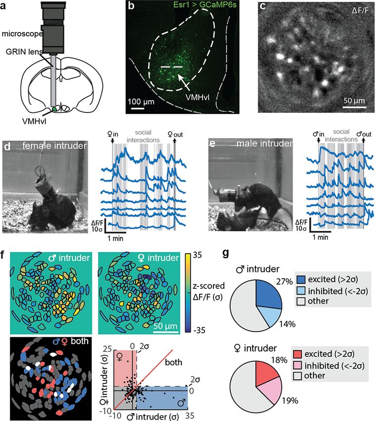
400                                                                                                          Microscopy, 2021, Vol. 70, No. 5

and single-cell level [4–6]. These optical tools have overcome prior       populations [22] during free behaviors, but these miniaturized two-
limitations with functional magnetic resonance imaging and multi-          photon systems are fundamentally limited to small fields of view
electrode array technologies, which exhibit poor spatial and/or tem-       and/or a low temporal resolution that preclude the study of neu-
poral resolution, require animals to be immobilized and are unable         ronal activity in hundreds to thousands of neurons under naturalistic
to target specific cell types. The advent of these new optical tools for   conditions.
in vivo calcium imaging has necessitated the development of novel              Fiber optic technology has also been adapted for one-photon
algorithms for translating the noisy fluorescent signals into mean-        imaging of neural dynamics. Flusberg et al. [1] developed a head-
ingful representations of neural activity. Several novel applications      mounted miniaturized microscope that enabled cellular-resolution
of matrix factorization combined with appropriate statistical model-       neural recordings as well as hemodynamic measurements in freely
ing now make it possible to automatically extract neuronal activity        behaving rodents. These fiber-based one-photon imaging technolo-
with increasing accuracy and minimal human intervention, thereby           gies were however limited by rigid and pixelated fibers, severely
enabling a scalable analysis of circuit activity at a single-neuron        constraining animal behaviors and resulting in low-resolution and
resolution from optical calcium activity signals [7–10].                   low-fidelity imaging signals. Fiber photometry is another technique
    In this review, we describe developments and advancements              that uses optical fibers to record changes in calcium activity in neural

                                                                                                                                                      Downloaded from https://academic.oup.com/jmicro/article/70/5/399/6324512 by guest on 26 October 2021
in optical tools for imaging neural activity in vivo. We focus on          circuits during free behaviors [23]. With fiber photometry, photons
miniaturized microscopy (miniscope) development for recording and          from the optical fibers are summed, resulting in a single bulk fluo-
manipulating neural circuit activity in freely behaving animals and        rescent photometry signal. Owing to the small spatial footprint of
review neuroscience research that has been enabled with these minis-       optical fiber ferrules, implanting multiple optical fibers into differ-
copes. Specifically, we will review the application of miniscope           ent brain regions is relatively straightforward and has been used
technology to the study of neural computations underlying stimulus         to collect simultaneous photometry signals from cortical and sub-
encoding, decision-making, and learning and memory to regulate a           cortical brain regions [24]. While fiber photometry-based imaging
diverse array of behavioral outputs, from naturalistic and instinc-        approaches are low cost and relatively easy to implement, a bulk
tive behaviors, such as food intake and sleep, to more complex             signal is inherently spatially and temporally lossy, precluding the
repertoires of behaviors, such as environmental exploration and            analysis of cellular-level neuronal dynamics and potentially resulting
reward-seeking. These powerful new tools have so far mostly been           in the misleading interpretation of brain circuit activity.
applied in mice, taking advantage of transgenic mouse lines to target
specific cell types and model human disease. Nevertheless, their use
                                                                           Development of a miniaturized microscope for
has increasingly extended to other less genetically tractable species
that serve as important animal models for basic and translational
                                                                           recording cellular-level neural circuit dynamics in
neuroscience research and offer unique species-specific advantages         freely behaving animals
including songbirds, rats, prairie voles and non-human primates            To overcome the limitations of the aforementioned optical tools
(NHPs).                                                                    and enable neuroscientists to record and monitor large-scale cal-
                                                                           cium dynamics at single-cell resolution in freely behaving rodents,
                                                                           the integrated miniaturized head-mounted microscope (miniscope)
Optical tools for imaging neural activity in vivo
                                                                           was invented and developed by a team at Stanford University [25].
Two-photon microscopy is an established method for functional              Miniscopes weigh ∼2 g and, when borne on the cranium of a freely
and morphological neural imaging in vivo. Two-photon microscopy            behaving rodent and used in conjunction with GCaMP, allow neuro-
is a laser-scanning technique that allows for optical sectioning to        scientists to visualize activity in hundreds of genetically defined neu-
exclude out-of-focus fluorescence, resulting in high-resolution three-     rons during natural behaviors. When paired with custom-designed
dimensional images of depths of up to 1 mm in brain tissue. The high-      optical GRIN lenses that are implanted in the brain, these minis-
resolution capabilities of two-photon imaging enable the investiga-        copes permit neuroscientists to target and image from deep brain
tion of subcellular dynamics such as calcium dynamics of dendritic         regions and from regions inaccessible to other large-scale recording
spines [11] and subcellular membrane potential [12]. Two-photon            technologies.
microscopy is also capable of larger-scale imaging and has been rou-           Miniscopes utilize a conventional epi-fluorescence microscope
tinely used to study cortical network activity [13,14] and has widely      design in which the objective serves to both deliver the excitation
been used to interrogate the cortical circuit underpinnings of sen-        light to the specimen and collect the fluorescence emitted by the
sory processing [15–17]. More recently, two-photon microscopy has          specimen to form an image of the brain region of interest (ROI).
also been combined with imaging through gradient refractive index          Excitation light and emission light are separated by a dichroic mir-
(GRIN) lenses to access deep brain regions such as the hypothalamus        ror assisted by additional excitation and emission bandpass filters
[18,19].                                                                   for further spectral cleanup (Fig. 1). Miniscopes predominantly use
    A major limitation of traditional two-photon imaging when              a GRIN optic as an objective as these offer large numerical apertures
studying neural circuit dynamics is the reliance on a head fixa-           in a small form factor, which is not readily obtained using discrete
tion preparation. While some naturalistic behaviors can be modeled         optical elements.
under head-fixed conditions, head fixation can introduce added con-            Building on the success of these early miniscopes, next-generation
founds such as stress [20]. Importantly, many naturalistic behaviors       miniscopes are now commercially available and include enhanced
such as social interaction and sleep behaviors are not suited for head     functionality. The nVistaTM system is a single-channel epifluores-
fixation. To overcome the limitation of traditional two-photon imag-       cence miniscope that has electronic focusing capabilities to enable
ing, efforts have been made to miniaturize two-photon systems by           simultaneous multiplane imaging, thus maximizing the number of
adapting fiber optic technology to enable two-photon imaging of            recorded cells. The nVokeTM system integrates simultaneous and
layer 2/3 dendritic calcium transients [21] and small-scale neuronal       sequential same field of view optogenetics, allowing researchers to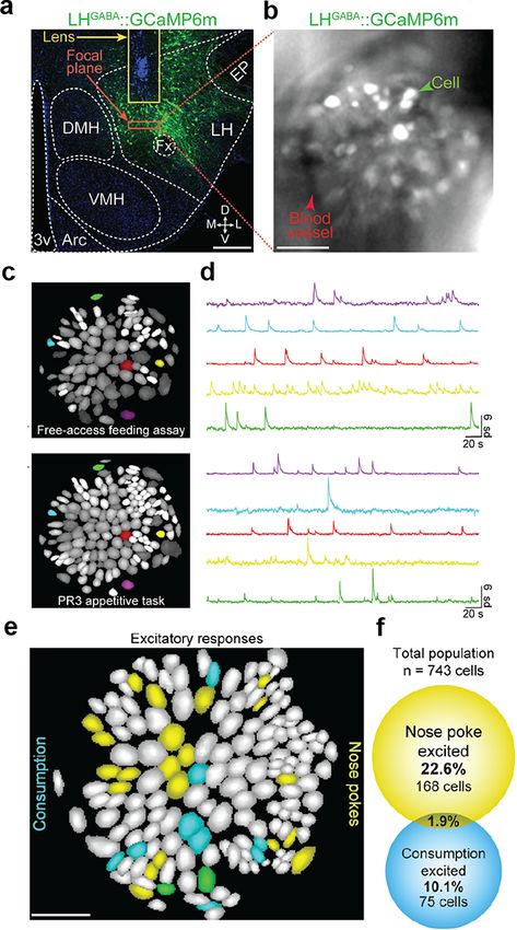
A.M. Stamatakis et al. Miniscopes for in vivo brain activity 401
Downloaded from https://academic.oup.com/jmicro/article/70/5/399/6324512 by guest on 26 October 2021
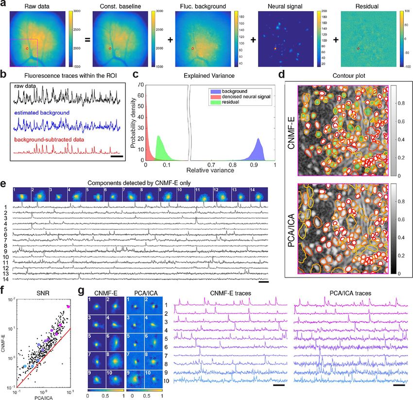
Fig. 1. Design and fabrication of an integrated fluorescence microscope. (a) Computer-assisted design of a first-generation epifluorescence miniscope. (b)
Assembled miniscope. Note the fine-threaded turret enabling manual focusing by the rotation of the image sensor mount. Figure reprinted from Ghosh et al.
[25].
casually link neural circuit activity with behaviors [26]. Finally,
the recently developed nVueTM system includes dual-color imaging
capabilities, enabling researchers to image from two brain signals
simultaneously.
Each of these miniscope systems has a nonintrusive data-
acquisition and control box that can be remotely controlled without
interfering with the animal’s behavior and/or the ongoing experi-
ment. The nVista, nVoke and nVue boxes are web-enabled enabling
direct data-streaming to the cloud (if allowed by the available net-
work capacity and bandwidth). They each comprise Graphics pro-
cessing unit (GPU)-based video processing, synchronization with
external data streams (such as behavioral video streams) and various
analog and digital input/output options (Fig. 2).
Extracting neural signals from noisy optical
calcium imaging data
Processing calcium activity movies into estimations of individual
neuron activity requires two main steps: (i) identifying the spatial
footprints of individual neurons and (ii) computing the time-varying
calcium activity signal for each neuron. The most straightforward
solution is to visually inspect the movie, manually draw ‘ROIs’ for
each neuron and then estimate each neuron’s calcium activity as the
mean ROI pixel intensity; however, this ‘manual ROI’ approach has
Fig. 2. Next-generation miniscope. Control and data-acquisition device for
several disadvantages. First, it is time-consuming as careful visual
the Inscopix nVista miniscope system allowing remote control via a web-
inspection and identification of ROIs can take several hours of expert browser-enabled GUI.
labor. Second, it does not account for out-of-focus fluorescent sig-
nals or leverage understanding of the underlying biosensor kinetics,
which can significantly degrade the accuracy of the extracted neural that while the discussion below focuses on algorithms for translating
signals. Finally, manual ROI selection is vulnerable to human bias pixel intensities to neural calcium dynamics, we should not forget the
and subjectivity that can both drift over time and vary across peo- importance of preprocessing data to correct for brain motion (which
ple, all of which can potentially confound the estimation of neural varies dramatically in magnitude depending on the brain region and
activity. surgical preparation), as well as deconvolution and event detection
To overcome the limitations of manual ROI selection, several [27].
automated algorithms have been developed over the past decade, The first majorly successful automated source extraction method
each improving the accuracy and speed of transforming calcium for calcium imaging was the development of ‘principal component
imaging movies into representations of neural circuit activity. Here, analysis–independent component analysis (PCA–ICA)’ by Mukamel
we briefly review and compare the two most common techniques et al. in 2009 [7]. The method comprises two main steps: first, a
currently used for processing miniscope calcium imaging data, as PCA to remove shot noise and reduce dimensionality, and second,
well as the software products available for their deployment. Note an (ICA) to identify the spatial footprints and signal dynamics of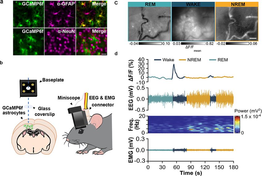
402                                                                                                           Microscopy, 2021, Vol. 70, No. 5

each neuron. PCA–ICA is computationally efficient and remarkably            interpreting meaningful neural circuit activity patterns. Dimension-
effective at cell detection without requiring any preconceptions of         ality reduction methods [30], generalized linear models [31] and
neurons’ spatial shape or temporal activity kinetics. There is, how-        recurring switching dynamical systems [32] are just a few examples
ever, an assumption that neurons’ spatial location and temporal             of computational techniques that are proving useful for uncovering
activity are sparse and statistically independent.                          latent patterns embedded in high-dimensional neural circuit activity
     PCA–ICA was originally developed for two-photon calcium                data. Analogous methods are being developed and used for quan-
imaging data, where a high z-resolution minimizes signal con-               tifying the equally rich behavior video data (for a nice summary of
tamination from out-of-focus neurons expressing fluorescent cal-            computational ethology methods, see ref. [33]). With miniscopes, it
cium indicators. Unfortunately, poorer z-resolution of one-photon           is now possible to combine these datasets and computational tech-
microscopy results in a much larger contribution of the fluores-            niques together toward a better understanding of how neural circuit
cent signal from neurons residing above and below the imaging               dynamics relate and give rise to freely moving behaviors [34].
plane, which acts to confound and obscure the activities of in-focus
neurons. To better account for this dynamic background signal in            Scientific applications in rodent models
one-photon data, constrained non-negative matrix factorization for

                                                                                                                                                       Downloaded from https://academic.oup.com/jmicro/article/70/5/399/6324512 by guest on 26 October 2021
microendoscopic imaging (CNMF-E) was proposed in 2018 [8]. In               Features of the miniscope described above have greatly expanded the
this approach, movies are factorized into four separate matrices: a         breadth of neuroscience research questions that can be asked. The
constant background, a fluctuating background, the neural signals           ability to monitor the activity of large populations of neurons during
and the residual (Fig. 3a and b). Factorization is achieved via opti-       naturalistic behaviors has been particularly advantageous as many
mization of several model constraints including the following: (i)          adaptive and translationally relevant behaviors are not compatible
neuron shapes are roughly spherical and of a certain diameter and           with conditions where movement is restricted. For example, animals
(ii) neural activity is well-described by an autoregressive model with      cannot sleep under head-fixed conditions, and social behaviors that
exponential decay constants matching our knowledge of biosensor             require dynamic back-and-forth interactions between individuals are
kinetics. Fluctuating background is computed for each neuron indi-          severely limited under restrained locomotion [35]. An important
vidually by considering a ring of pixels greater than the diameter          additional advantage of the miniscope technology is the ability to
of the neuron to approximate signal contributions from blurred              repeatedly track the same cells over time, which is critical to under-
neurons residing above or below the imaging plane. Compared to              stand how experience-induced plasticity can alter behaviors. This
PCA–ICA, the authors of CNMF-E showed evidence that it yields a             feature was first applied to studies of learning and memory [36] but
higher cell identification recall, with a higher signal-to-noise ratio of   has more recently been applied to studies of homeostatic drive, where
the extracted activity traces (Fig. 3d–f). However, these advantages        it is critical to track changes in neural activity alongside changes
come at a cost: CNMF-E tends to identify more false positives (lower        in internal states [37]. Finally, the combination of miniscopes with
precision) and is computationally slow and expensive compared to            tools to genetically and anatomically define cell types as well as
PCA–ICA.                                                                    GRIN lenses to bring the fluorescently encoded activity patterns of
     While PCA–ICA and CNMF-E both represent critical advances              cells above the surface of the brain is enabling neuroscientists to
in our ability to automatically detect neurons and extract calcium          decode the precise neural circuits that drive complex behavioral out-
activity from one-photon miniscope data, there remain limitations           puts [38]. Together, these features are unlocking experiments that
in efficiency and accuracy that will require continued innovation to        were not previously feasible to increase our understanding of neural
overcome. Recent attempts to improve background estimation and              circuit computations that regulate hunger, thirst, sleep, environment
dimensionality reduction for CNMF-E [28], as well as an entirely            exploration and social behaviors.
new method of matrix factorization based on leveraging robust esti-
mation theory and GPU implementation [9] show exciting promise              Hunger and thirst
in this regard.                                                             Hunger and thirst direct appetitive or consummatory actions toward
     In addition to method innovation, there is a need to standardize       maintaining body homeostasis at an optimal set point for normal
and package existing techniques into software tools and products for        functioning [39]. This set point is not static, as energy needs will
wide adoption by miniscope users. For those comfortable program-            change with survival security, internal energy state and desirability of
ming, the CaImAn computational toolbox offers several methods for           resources consumed [40]. However, how the brain receives and inte-
calcium imaging processing, including a Python version of CNMF-             grates signals to direct consumptive behaviors is not well understood.
E [10]. CNMF-E was recently implemented in C++ and can be                   The application of miniscope technology to this field has been advan-
used in the Inscopix Data Processing Software (IDPS) Graphical user         tageous as it has identified heterogeneous signals within deep brain
interface (GUI), Python application programming interface (API) or          regions that generate complex modulations of feeding and drinking
Matlab API. IDPS also offers PCA–ICA and several other prepro-              behaviors.
cessing tools (e.g. motion processing) in its GUI and APIs. CIAtah,             Cellular resolution imaging of genetically defined neuron popu-
written in Matlab, is another software package that allows users to         lations by miniscopes revealed previously unidentified heterogenous
access several different algorithms for one-photon calcium imaging          patterns of neuronal activity that regulate consumptive behaviors.
with a GUI [29]. As more neuroscientists adopt miniscope tech-              For example, when feeding was classified into constituent phases
nology for investigating the brain, it will be increasingly important       of food-seeking and consumption, miniscope experiments revealed
to develop easy-to-use software tools for processing, analyzing and         that subpopulations of γ-aminobutyric acid (GABA) neurons in
managing calcium data so that it can be interpreted without requiring       the lateral hypothalamus (LH) encode functionally discrete appet-
computer science expertise.                                                 itive and consummatory behaviors (Fig. 4) [41]. Miniscope imag-
     Of course, extracting neuronal signals from the noisy optical cal-     ing also revealed that bidirectional modulation within genetically
cium imaging movies is just the first step toward identifying and           defined cell types is responsible for the varied permutations of con-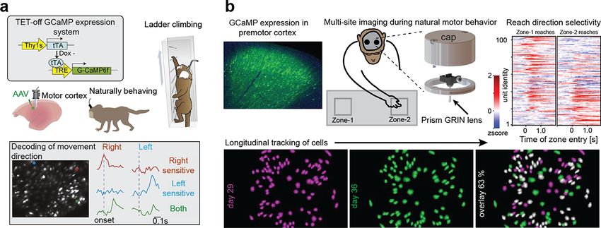
A.M. Stamatakis et al. Miniscopes for in vivo brain activity                                                                                                  403

                                                                                                                                                                      Downloaded from https://academic.oup.com/jmicro/article/70/5/399/6324512 by guest on 26 October 2021
Fig. 3. Comparison of PCA/ICA and CNMF-E for extracting neural signals from miniscope calcium movies. (a) An example frame of the raw data and its four
components decomposed by CNMF-E. (b) The mean fluorescence traces of the raw data, the estimated background activity and the background-subtracted data
within the segmented area (red box) in (a). (c) The distributions of the variance explained by different components over all pixels; note that estimated background
signals dominate the total variance of the signal. (d) The contour plot of all neurons detected by CNMF-E and PCA/ICA superimposed on the correlation image.
Green areas represent the components that are only detected by CNMF-E. The components are sorted in decreasing order based on their SNRs (from red to
yellow). (e) The spatial and temporal components of 14 example neurons that are only detected by CNMF-E (green areas in d). (f) The SNRs of all neurons
detected by both methods. Colors match the example traces shown in (g), which shows the spatial and temporal components of 10 example neurons detected
by both methods. Scale bar: 10 s. Figure reprinted from Zhou et al. [8] under the Creative Commons Attribution license. SNR: Signal-to-noise ratio.

sumptive decision-making, as glutamatergic neurons in the median                    For example, several studies observed heterogenous or transient
preoptic area (MPOA) bidirectionally respond to water intake [42]                   neuronal responses that were not observed in previous photometry
and clusters of glutamatergic neurons in the anterior peri-locus                    studies [45–48]. Together, these results have revealed complex pat-
coeruleus are activated or inhibited by food and water intake [43].                 terns of activity within genetically and anatomically defined circuits
Single-cell recordings of parabrachial neurons expressing transcrip-                that regulate feeding and drinking behaviors.
tion factor Satb2 have also shown that the same neurons can be                          It is critical that neural circuits that control food intake are
excited or inhibited by different taste stimuli [44]. Importantly,                  also capable of suppressing feeding behaviors when it is not imme-
some miniscope results have challenged earlier findings on hunger                   diately relevant for survival. Taking advantage of the miniscope’s
and thirst that originated from studies lacking single-cell resolution.             capability to repeatedly monitor the activity of neurons under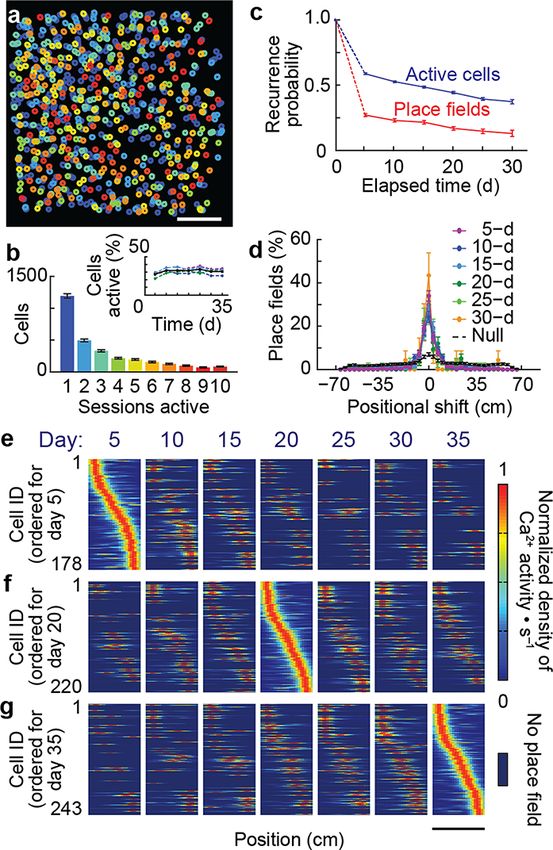
404                                                                                                              Microscopy, 2021, Vol. 70, No. 5

                                                                               of miniscope technology to complex experimental conditions where
                                                                               competing drive states can be assessed therefore has the ability to
                                                                               increase our understanding of how the brain integrates internal and
                                                                               external factors to generate behavioral responses that are adaptive
                                                                               under specific physiological states and environmental context.

                                                                               Sleep
                                                                               Sleep is a fundamental biological phenomenon widely observed in
                                                                               the animal kingdom. The regulation of sleep is primarily controlled
                                                                               by a network of nuclei located within the deep brain regions such as
                                                                               the hypothalamus, midbrain and the brainstem. Measuring the activ-
                                                                               ities of these sleep-regulating neurons has been technically challeng-
                                                                               ing due to a mix of different cell types activating at different brain

                                                                                                                                                          Downloaded from https://academic.oup.com/jmicro/article/70/5/399/6324512 by guest on 26 October 2021
                                                                               states, neurons located at deep brain regions and the requirement
                                                                               of nonrestrained recording for natural sleep–wake cycle. Moreover,
                                                                               traditional in vivo electrophysiology or photometry cannot clearly
                                                                               distinguish intermingled cell types, and two-photon imaging requires
                                                                               head restraint that is not conducive to sleep. Miniscope imaging
                                                                               overcomes these limitations and can be combined with electroen-
                                                                               cephalography (EEG) and Electromyography (EMG) to classify the
                                                                               three distinct phases of sleep—wakefulness, rapid eye movement
                                                                               (REM) sleep and non-REM (NREM) sleep [51]. Miniscope imaging
                                                                               has therefore become a powerful tool in studying sleep circuits.
                                                                                   The first application of using miniscope imaging to study sleep
                                                                               was published in 2015 and characterized the activity of glutamater-
                                                                               gic, GABAergic and cholinergic pontine neurons in sleep–wake reg-
                                                                               ulation [52]. Thereafter, scientists have applied miniscope imaging
                                                                               to identify the activity of different cell types across different sleep–
                                                                               wake nuclei, such as GABAergic neurons in the ventral tegmental
                                                                               area, neurotensinergic neurons in the midbrain [53,54] and many
                                                                               different inhibitory neurons controlling REM–NREM transitions
                                                                               [55–57]. Recently, it was also used to identify dynamic changes in
                                                                               astrocyte activity across brain states that was distinct from that of
                                                                               neurons (Fig. 5) [58]. This finding not only identifies a role for glial
                                                                               cells in sleep regulation but also highlights a direction of applying
                                                                               miniscope imaging to investigate the role of non-neural cell types
                                                                               in the regulation of behaviors. Neural mechanisms of sleep disorder
Fig. 4. Heterogeneous responses to consumptive behaviors. (a and b) Tar-       such as cataplexy were also studied by this approach, and abnor-
geting LH GABAergic neurons to study regulation of appetitive versus con-      mal activity of GABAergic neurons in the amygdala was found to
summatory behaviors. (c and d) Cell maps and corresponding example             be associated with emotion-induced cataplexy [59]. These findings
calcium traces showing that LH GABAergic neurons differently encode appet-     demonstrate the broad application of miniscope imaging in answer-
itive versus consummatory behaviors. (e and f) Miniscope imaging revealed
                                                                               ing the neural mechanisms of sleep–wake regulation and elucidating
that individual LH GABAergic neurons functionally encode either nose-poke
                                                                               the pathological mechanisms of sleep disorders.
responsive cells or lick-responsive cells, with a small (1.9%) overlap in LH
GABAergic neurons that encode both. Figure adapted from Jennings et al.            Miniscope technology can be used not only to study the sleep
[41].                                                                          circuit (how we sleep) but also to study the function of sleep (why
                                                                               we sleep). Most of the recent efforts have focused on REM sleep,
                                                                               given its significant role in learning, memory and neural plastic-
varying metabolic states and environmental conditions, Viskaitis               ity. Zhou et al. found the activity of hippocampal CA1 neurons
et al. demonstrated that steroidogenic factor 1 (SF1) neurons within           was greater in REM sleep than NREM sleep and wakefulness,
the ventral medial hypothalamus (VMH) differentially encode feed-              suggesting the activities of these memory-associated neurons are
ing versus threat and that the activity patterns of these neurons can          modulated by sleep states [60]. Adult-born neurons in the den-
exert opposing regulation over behavioral output. Specifically, the            tate gyrus (DG) are also important for learning and memory, and
feeding behavior was associated with low activity of these neurons,            miniscope imaging identified a role for these neurons in consol-
while a high activity was associated with threatening environments             idation of context–shock association during REM sleep [61,62].
as well as reduction in exploration and feeding [37]. While previous           Researchers not only examined known memory-associated neurons
studies had demonstrated a role for SF1-VMH neurons in both                    during sleep but also explored the role of sleep-regulatory neurons
feeding and defensive behaviors [49,50] it was unknown how SF1                 in memory formation. Izawa et al. found REM sleep–active melanin
neurons in the VMH differently encode rewarding versus threaten-               concentrating hormone–producing neurons in the LH are involved
ing stimuli to drive opposing behavioral responses. The application            in active forgetting, illustrating hypothalamic sleep regulators can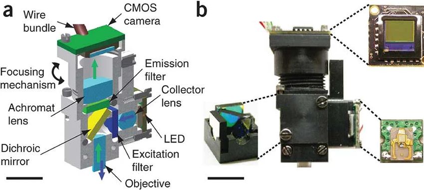
A.M. Stamatakis et al. Miniscopes for in vivo brain activity                                                                                               405

                                                                                                                                                                   Downloaded from https://academic.oup.com/jmicro/article/70/5/399/6324512 by guest on 26 October 2021
Fig. 5. Miniscope imaging to investigate the role of astrocytes in sleep regulation. (a) Expression of GCaMP6f within cortical astrocytes, scale bar, 20 mm. (b)
Schematic of miniscope imaging with EEG and EMG recordings. (c) Maximum projection dF/F images of calcium imaging in astrocytes during wake, NREM and
REM sleep. Scale bar, 100 mm. (d) An example of astrocyte calcium dynamics with corresponding EEG, EEG power spectrogram and color-coded brain states.
Figure adapted from Ingiosi et al. [58].

also be important for memory regulation [63]. Future studies uti-                     The hippocampus has been among the first and most studied
lizing the miniscope for different behavior tasks or combining with               brain areas related to learning and memory. The so-called place cells
optogenetic approaches may help further understand other unknown                  in dorsal CA1 (dCA1) have been indeed known for several decades
functions of sleep.                                                               [67], but the investigation of the neuronal activity was usually lim-
                                                                                  ited to few tens of cells per experimental subject, and therefore,
Learning and memory                                                               the data were too sparse to study them at the ensemble level. Ziv
Our everyday life involves multiple sensory experiences, includ-                  et al. [36] tracked thousands of neurons over weeks from different
ing exploring different places and interacting with animate and                   mice exploring familiar environments. They found that the dCA1
inanimate objects. Episodic memories reflect the ability to rec-                  coding had a day-to-day dynamism at the cellular level while pre-
ollect the temporal and spatial context of these experiences.                     serving spatial information in the ∼15–25% overlap between coding
Their formation requires rapid synaptic plasticity within the hip-                ensembles from any 2 days. In other hippocampal regions, such
pocampus and is gradually consolidated within the hippocampal–                    as the DG, the use of miniscope associated with machine learning
entorhinal/perirhinal cortex–neocortex for permanent storage [64].                demonstrated that, although each individual neuron carries only a
The consolidation units, named engrams, have been theorized for                   small amount of spatial information, the population activity pat-
over a century [64,65] as a functional entity enabling memory stor-               tern encodes orientation features such as position, speed and motion
age and retrieval through the dynamic interaction of a large number               direction (Fig. 6) [68]. Further analyses of similar experimental data
of neurons. While synaptic plasticity has been studied with elec-                 suggest that the day-scale ensemble dynamics might serve as times-
trophysiological techniques established in the 20th century [66],                 tamps for the formation of temporal information and to support the
the systematic investigation of large neuronal networks and their                 episodic memory [69,70].
modulation has been limited by the lack of supporting technologies                    Several recent studies [71,72] employed an activity-
until very recently. Notably, the visualization of calcium dynamics               dependent cell-labeling technology, the c-fos-tet-tag system [73], to
in freely moving animals with miniscopes has been largely used to                 tag neurons that are active during a specific memory formation task,
define the neural substrate of engrams, for their unique ability to               namely considered part of a given engram. The neural activity associ-
track the activity of large populations of neurons across time, while             ated with memory formation induces the early gene c-fos expression,
the experimental subjects are in a naturalistic environment.                      which in the c-fos-tTA transgenic mice induces activity-dependent
406                                                                                                                   Microscopy, 2021, Vol. 70, No. 5

                                                                                   fear conditioning (CFC) task to show that c-fos increased in PFC
                                                                                   neurons early on during the task, but the calcium activity in PFC in
                                                                                   response to the CFC shock was much higher 15 days after exposure
                                                                                   than 1–2 days after exposure. These data, combined with optoge-
                                                                                   netic experiments, suggest that the PFC engrams are generated early
                                                                                   on during CFC, but they become fully functional during the time.
                                                                                   These findings highlight the role of the PFC in the early stages of
                                                                                   memory formation and are in contrast with a previous model that
                                                                                   hypothesized that remote memory is stored in the cortex by a slow
                                                                                   transfer of hippocampal memory, rather than being primed at the
                                                                                   time of the memory formation. The ability to track the activity of
                                                                                   specific cell populations within complex circuits in freely moving
                                                                                   animals greatly enhanced the knowledge of learning and memory
                                                                                   mechanisms related to spatial orientation initially and of broader

                                                                                                                                                               Downloaded from https://academic.oup.com/jmicro/article/70/5/399/6324512 by guest on 26 October 2021
                                                                                   aspects of cognition such as emotions and social behaviors.

                                                                                   Social behavior
                                                                                   Social behavior is highly adaptive and critical for the survival of all
                                                                                   sexually reproducing species [74]. At its core, it can be described as
                                                                                   any form of communication or interaction with conspecifics; how-
                                                                                   ever, the appropriate display of social behaviors often requires the
                                                                                   dynamic processing of social cues as well as the subsequent output
                                                                                   of complex behavioral repertoires [75]. Moreover, social decisions,
                                                                                   such as the decision to cooperate or fight, are context- and stimulus-
                                                                                   specific, change across development as well as with experience, and
                                                                                   are dependent on internal states, further adding to the complexity
                                                                                   of social behaviors [76]. As a result, the neural mechanisms under-
                                                                                   lying the rapid integration of social cues with internal states to drive
                                                                                   complex social interactions are not well understood. The application
                                                                                   of miniscope technology to this field has been particularly advanta-
                                                                                   geous to elucidate the neural correlates of social behaviors because
                                                                                   its lightweight and flexible tether allows subjects to naturally inter-
Fig. 6. Miniscopes enable tracking large populations of neurons over weeks.        act with social conspecifics as well as for individual neurons to
(a) Map of calcium activity in mouse imaged for 45 days. Color code as in
                                                                                   be tracked under experimental conditions where changes in social
(b): of all the active cells in day 1, a fraction (represented by the histogram)
was active in the subsequent imaging sessions. Inset, notably, a constant
                                                                                   perception are hypothesized to occur.
fraction of all neurons detected over time was active each day. (c) Probability         An obvious difference in the display of social behaviors occurs
for a given neuron to be active (blue data) and to code for a place field (red     with the types of behaviors that are directed toward adult con-
data) in subsequent sessions declined with time. (d) The centroid shifts of        specifics of the opposite sex [77]. Specifically, upon encountering
identified cells were stable across days (color code days between sessions).       a conspecific, an individual must first encode the properties of the
(e–g) Example of place cells found on multiple sessions, ordered by place
                                                                                   stimulus (adult versus juvenile or male versus female) before selecting
fields’ centroid positions on day 5 (e), day 20 (f) or day 35 (g) (mean ± SEM,
                                                                                   the appropriate behavioral output (prosocial, defensive or non-
data from four mice). Figure adapted from Ziv et al. [36].
                                                                                   social/ignore) [78]. Although social stimulus perception is considered
                                                                                   a critical first step in the display of appropriate behavioral responses,
tTA expression under the control of the c-fos promoter. The system                 due to previous methodological limitations in the ability to repeat-
is inactive during the administration of doxycycline that can there-               edly track the activity patterns of neurons under different social
fore be used as a switch. In this way, it is possible to know what                 contexts, how the brain selectively encodes unique social stimulus
neurons participated in the memory formation and compare their                     properties is not well understood. To circumvent these limitations, Li
activity during the time with neighboring neurons that are not part                et al. used miniscope imaging to track the activity of neurons within
of the engram. Ghandour et al. [71] used this approach to moni-                    the medial amygdala (MeA), during naturalistic stimulus investiga-
tor the activity of hundreds of dCA1 neurons during a contextual                   tion [76]. Interestingly, their data revealed that in sexually naive
learning task, while selectively discriminating between engram (c-                 males and females, conspecific social cues were uniquely encoded
fos positive) and non-engram cells. By utilizing the selective tagging             within the MeA of both sexes, with the majority of neurons show-
approach combined with freely behaving calcium imaging, they were                  ing a selective increase in activity in response to the social stimulus
able to demonstrate that engram cells were more synchronized than                  and the remainder of neurons showing either decreases in activity
non-engram cells not only during the task but also during the post-                or non-selective responses to the social stimulus. Socially responsive
learning sleep and retrieval sessions, supporting the role of sleep in             neurons also displayed unique patterns of activity to specific cate-
memory consolidation.                                                              gories of social stimuli, but the activity pattern of individual neurons
    Using a similar approach, Kitamura et al. [72] labeled neurons                 was not consistent across interaction epochs. Instead, discrimination
in the prefrontal cortex (PFC) that were active during a contextual                of social stimuli was encoded at the population level, and the size of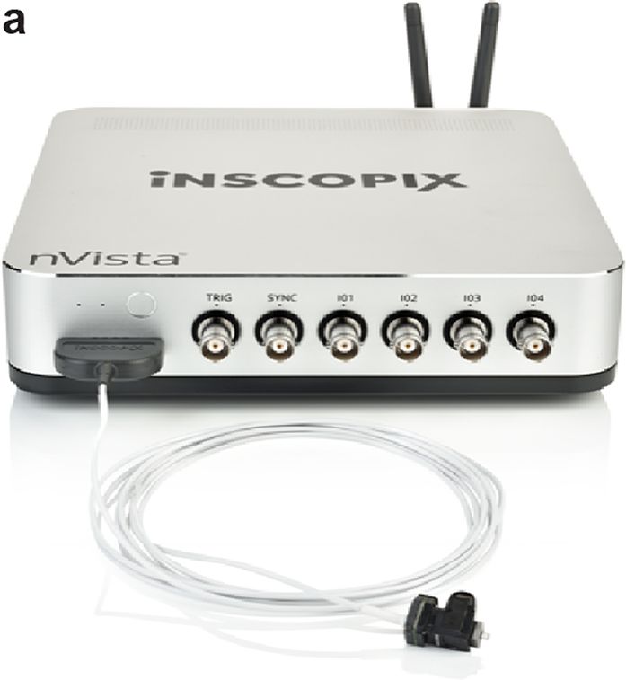
A.M. Stamatakis et al. Miniscopes for in vivo brain activity                                                                                                  407

the ensemble was inversely correlated with the latency to engage in                 the intruder. Moreover, cells activated by male and female con-
aggressive or reproductive behaviors [76]. These data indicate that                 specifics were largely distinct populations of neurons (Fig. 7f and
while encoding of social stimuli at the individual neuron level may                 g), and the specificity of neural stimulus encoding increased with
be noisy, encoding at the population level can accurately represent                 social experience [80]. Similar changes in population encoding of
social information to drive the appropriate behavioral response.                    social information following sexual experience have also been iden-
    The MeA projects to regions of the hypothalamus that have                       tified within non-hypothalamic nuclei such as the MeA [76] of mice
also been implicated in the regulation of aggression and reproduc-                  and the Nucleus Accumbens (NAc) [81] and are associated with
tive behaviors, such as the VMH and the MPOA [79], and studies                      changes in behavioral states toward conspecifics. In addition to sex-
utilizing miniscope imaging have begun to identify how neurons                      ual experience, repeated exposure to social stress can also impact
within these regions encode socially specific stimuli. Within the                   social stimulus encoding as chronic social defeat leads to increased
VMH of male mice, estrogen receptor-1 (Esr1+ ) expressing neu-                      activity of granule cells located within the ventral hippocampus as
rons showed increases in activity in response to both male and                      well as an increase in social avoidance behaviors [82]. Together,
female conspecifics (Fig. 7a–d) and that repeated exposure to male                  these observations enabled by miniscope imaging have revealed a
and female conspecifics activated similar neuronal ensembles across                 distributed network of brain regions where experience-dependent

                                                                                                                                                                      Downloaded from https://academic.oup.com/jmicro/article/70/5/399/6324512 by guest on 26 October 2021
trials, indicating that these neurons accurately encode the sex of                  plasticity underlies dynamic coding of social stimuli to drive adaptive

Fig. 7. Miniscope imaging during dynamic social interactions. (a) Schematic of miniscope imaging. (b) GCaMP6 expression within VMH Esr1+ cells and approx-
imate imaging plane. (c) Representative image from miniscope recording. (d and e) Representative frame from behavior video (left) and corresponding calcium
traces aligned to movie frame (right) from either a (d) reproductive encounter or (e) an aggressive encounter. (f) Cells showing significant changes in activity to
either a male or female intruder were largely distinct populations of neurons. (g) Male- and female-preferring cells responded with both increases and decreases
in neural activity. Figure adapted from Remedios et al. [80].
408 Microscopy, 2021, Vol. 70, No. 5
behavioral responses. The identification of plasticity within hypotha- Miniscope imaging is beginning to reveal novel mechanistic
lamic nuclei was particularly surprising because similar to other insights into debilitating neurological disorders, such as epilepsy,
instinctive behaviors such as feeding, innate social behaviors such in part through the ability to perform large-scale circuit dynamic
as mating and aggression were thought to be ‘hard-wired’ and fol- recordings under conditions that were previously inaccessible with
low a labeled line theory. Thus, the ability to repeatedly monitor the other methodologies. For example, although elevations in hip-
activity of cells has provided key insights into the understanding of pocampal calcium signaling have long been hypothesized to con-
how social information is represented within the brain. tribute to increased neuronal excitation and synchronization during
In addition to increasing our understanding of how social stim- seizure as well as seizure-induced brain damage [93], previous tech-
uli are encoded within the brain, the ability to repeatedly track cells nical limitations made it challenging to examine calcium dynamics in
over time with miniscope methods has been utilized to disentangle freely behaving animals undergoing seizure. By combining miniscope
neural correlates underlying social memory formation. In regard to imaging with an established model of epilepsy, kainic acid treatment
social memory, studies have focused on examining both how the [94] and assessments of seizure activity such as EEG and behavioral
memory of conspecifics are encoded in the brain and the location measures, Berdyyeva et al. were able to not only provide evidence
in space where social encounters have previously occurred. These supporting the hypothesis that calcium elevations are associated with
Downloaded from https://academic.oup.com/jmicro/article/70/5/399/6324512 by guest on 26 October 2021
studies have identified that social memories of conspecifics are stored neural and behavioral signatures of seizure but also identify novel
within the CA1 region of the ventral hippocampus [83], while pre- neural signatures that may better characterize the underlying dis-
limbic neurons that project to the NAc encode the location of spatial ease pathogenesis [89]. Specifically, they identified waves of calcium
encounters [84]. Within the PFC, neurons located in the prelimbic activity that occurred well before the onset of motor convulsions
region [84] and agranular insular cortex [85] are also activated dur- (average of 33 minutes prior) and had no consistent EEG pheno-
ing interactions with social stimuli, regardless of spatial location or type. Thus, it is possible that motor convulsions and EEG signatures
stimulus familiarity, suggesting that these neurons may also play typically used to study seizures may reflect the latent expression
a more general role in social processing, perhaps through interac- of central nervous system pathology and may result from seizure
tions with downstream regions associated with valence processing, propagation rather than initiation [89]. Moreover, when valproate,
motivation and social decision-making. a commonly prescribed seizure medication [95], was administered
A major goal within the social neuroscience field is to identify the prior to seizure induction, it was able to reduce behavioral symptoms
neural mechanisms that underlie the complexity of social behaviors of seizure (e.g. motor convulsions) but failed to alleviate aberrant cal-
that are not only critical for survival but also have large impacts on cium dynamics [89]. This latter finding is of particular importance,
human mental health. Taking advantage of the ability to study natu- given that current epilepsy treatments often fail and one possibility
ralistic behaviors in freely behaving animals, miniscope imaging has may be that therapeutics are developed to ameliorate the symp-
greatly increased our understanding of the neural correlates of social toms but fail to target the underlying pathology. Integrating calcium
behaviors along multiple steps of processing. Specifically, studies imaging with current assessments of seizures in animal models may
to date have identified experience-dependent computational patterns therefore improve the translatability of therapeutic compounds.
within the amygdala and hypothalamus that are associated with the The ability to repeatedly track the activity dynamics of individual
encoding of specific stimuli, regions of the hippocampus and cortex neurons is another advantage of miniscope imaging for translation-
that regulate social memory, and neuronal ensembles within the NAc ally relevant studies because it allows for pre- and post-disease states
that drive social motivation. Importantly, impairment in social func- to be compared, for changes in activity dynamics across the trans-
tioning is a comorbidity of many psychiatric diseases [86], but the gression of a disease states to be analyzed and for the ability of
specific circuit disruptions that lead to social behavior abnormalities therapeutic compounds to reverse aberrant circuit dynamics to be
are not well understood. Applying miniscope imaging to studies of assessed. These features may be especially important when trying
social functioning may thus have a translational impact, especially to understand the pathology underlying progressive neurodegener-
when combined with genetic models for disorders such as autism and ative disorders such as Parkinson’s disease where a gradual loss of
schizophrenia to identify pathophysiological mechanisms associated dopamine leads to motor deficits and the efficacy of current ther-
with aberrant social function [87,88]. apeutics to treat these motor deficits degrades over time [96]. A
hypothesized mechanism behind motor dysfunctions associated with
Parkinson’s disease is that they arise from an imbalance in the activ-
Translational applications ity of direct and indirect spiny projection neurons (SPNs) within the
In addition to the basic research applications described above, the striatum. More specifically, hypo- and hyperactivity of direct and
combination of miniscope imaging with animal models of disease indirect pathway SPNs is thought to contribute to movement inhi-
may prove to be a valuable tool for identifying novel biomarkers bition in the Parkinsonian state, while the inverse activity pattern
of disease and screening potential therapeutic compounds. Indeed, within SPNs is thought to underlie Levodopa (L-DOPA)-induced
recent studies using miniscopes to examine the pathogenesis under- dyskinesia [97]. However, direct evidence supporting this hypoth-
lying neurological disorders, such as epilepsy [89] and Parkinson’s esis had been lacking due to challenges in repeatedly monitoring the
disease [90], are providing novel mechanistic insights into circuit activity of direct and indirect SPNs across critical experimental time
abnormalities that lead to aberrant behavior phenotypes associated points.
with these conditions. Moreover, miniscope imaging can easily be By utilizing miniscopes to monitor the activity of direct and indi-
combined with pharmacological administration of therapeutic com- rect SPNs before and after DA depletion with 6-Hydroxydopamine
pounds [89–92] to test target engagement and perform predictive hydrobromide (6-OHDA), a classical model of Parkinson’s disease
efficacy assays, which together with an increased understanding of [96], Parker et al. were able to demonstrate that DA depletion
underlying disease pathology may help to improve the translatability indeed reduced the activity of direct pathway SPNs while increas-
of therapeutic compounds. ing the activity of indirect pathway SPNs. In addition, they alsoA.M. Stamatakis et al. Miniscopes for in vivo brain activity 409
demonstrated that the inverse of this activity pattern was true fol- particular model species for the brain functions and behaviors under
lowing L-DOPA-induced dyskinesia. Moreover, repeated miniscope study. Furthermore, understanding the similarities and differences
imaging also enabled them to identify more complex alterations in in circuit structure and function across a diverse range of species
striatal activity such as a reduction in activity coupling to motion is essential toward identifying common principles of brain function
onset and offset as well as a decrease in spatial activity cluster- that are more likely translatable to humans [104,105]. It is therefore
ing in indirect SPNs during the Parkisonian state. The opposite no surprise that significant effort has been made toward applying
impact on motor activity coupling and spatial activity clustering miniscopes beyond the mouse, and exciting progress has already
within indirect SPNs occurred following L-DOPA-induced dyski- been achieved in species ranging from songbirds to rats and prairie
nesia [90]. Together, these neural circuit signatures may act as voles [81,99,106–111].
biomarkers to facilitate the development of next-generation ther- Ultimately, to advance our understanding of higher-cognitive
apeutics that not only aim to restore activity balance within the function, complex behavior and mental health, a critical need exists
striatum but also to restore spatiotemporal patterns that are crit- to translate the latest miniscope-based technologies to research using
ical for normal motor function. These findings also suggest that NHPs. Historically, the NHP, rhesus macaque, has been the animal
therapeutics for chronic neurological conditions also need to target model of choice to study human-relevant brain functions due to well-
Downloaded from https://academic.oup.com/jmicro/article/70/5/399/6324512 by guest on 26 October 2021
treatment-induced neural adaptations in addition to the underlying documented similarities between macaques and humans in terms of
neural circuit abnormalities that lead to the onset of the disease. brain structures and complex behaviors [112,113]. More recently,
In addition to models described above, miniscope imaging has the common marmoset (Callithrix jacchus), which has long been rec-
also been used with animal models of chronic pain [29], addic- ognized as an important model for studying human-relevant social
tion [98,99], narcolepsy [100], anorexia [91], traumatic brain injury behaviors and vocal communications, has emerged as an attractive
[101] and genetic models of social deficits. The combination of alternative to the macaque, particularly due to their small size and
miniscope imaging with genetic models of psychiatric disorders is high reproductive efficiency, making them more amenable to genetic
particularly advantageous because it has the potential to connect manipulation [114–118]. Indeed, major efforts are now underway
gene–circuit interactions to aberrant behavioral patterns. For exam- toward studying marmoset brain structure and function and apply-
ple, by utilizing circuit-selective transgenic tools and a genetic mouse ing the latest gene editing technologies toward developing viable
model of maladaptive social behaviors, Kim et al. were able to genetic models of human brain disease [119,120].
demonstrate that disruption of the Arp2/3 complex, a cytoskeleton Significant progress has been made applying two-photon cal-
regulator of dendritic spines, specifically within PFC neurons that cium imaging in both marmosets and macaques, overcoming several
project to the basolateral amygdala alters neural encoding of social challenges associated with Adeno-associated viruses (AAV)-based
stimuli and leads to disruptions in social behaviors [87]. Thus, they expression of genetically encoded calcium indicators in the NHP and
identified how a genetic mutation within a specific component of imaging artifacts caused by movements of their larger volume brains
social circuitry was sufficient to disrupt the proper expression of [121–130]. These studies have in some cases achieved stable cellular-
adaptive social behaviors. Given that many psychiatric disorders resolution calcium imaging of large populations of neurons over
are highly heritable [102], understanding how genetic mutations several months of recordings, relying on transparent cranial win-
lead to specific circuit abnormalities may facilitate the discovery dows to enable imaging of the gyral cortex to depths of ∼500 µm.
of novel treatments with possible improved therapeutic efficacy. In Despite this important progress, several limitations of two-photon
addition, the continued development of genetic tools and ability to microscopy in NHPs remain including the following: (i) infection
combine these tools with miniscope technology for functional imag- risk of using cranial windows, which also have limited functional
ing will also be critical to decode the complex circuit interactions lifetimes and require daily maintenance, (ii) cumbersome alignment
that govern how an individual’s perception of the external world is between the microscope and the brain to maintain a consistent field
integrated with internal states to drive adaptive behaviors as well as of view and follow the same neurons across sessions, (iii) inability
how disruption of these circuits can lead to maladaptive behaviors to image the gyral cortex deeper than ∼500 µm or cortical and sub-
associated with central nervous system disorders. Finally, integra- cortical structures positioned deeper in the brain, and (iv) necessity
tion of miniscope technology with primate models, which may more for head restraint, which limits natural behaviors. Head-mounted,
closely resemble clinical features of human diseases, may also pro- one-photon miniscope calcium imaging is poised to overcome these
vide valuable insights into pathology underlying brain disorders [96] limitations and thereby significantly expand the brain regions and
and improve the successful translation of drugs into the clinic. behaviors that can be studied in NHPs.
The first successful demonstration of miniscope calcium imag-
ing in NHP was recently demonstrated in behaving marmosets (Fig.
Miniscope imaging in higher-order species 8) [131]. In this pioneering work, researchers developed and opti-
As reviewed above, enormous progress has already been made mized a viral strategy and surgical methods to express GCaMP and
applying miniscopes toward advancing our understanding of the chronically implant GRIN prism lenses into deep cortical layers of
neural circuit mechanisms underlying a vast array of brain func- the primary motor cortex. They relied on an AAV system that uti-
tions and behaviors. The most common animal model species used lizes tetracycline-controlled transcriptional activation to drive ade-
for this work so far has been the mouse, largely motivated by quate levels of GCaMP expression while providing a mechanism
the availability of transgenic lines (e.g. Cre recombinase lines) and (via doxycycline administration) to prevent overexpression [130].
other next-generation genetic and molecular technologies that have They were able to record from hundreds of neurons in deep lay-
enabled imaging and optogenetic manipulation of highly precise cell ers of the primary motor cortex while the marmosets engaged in
populations and circuits [103]. While the genetic tractability of a natural motor behaviors including a seated lever-pulling task and a
model species is an important consideration for the deployment of bidirectional arm-reaching task. As expected from previous electro-
these techniques, equally or more important is the relevance of a physiological studies in the primary motor cortex, neuronal calciumYou can also read