An overview of methods for production and detection of silver nanoparticles, with emphasis on their fate and toxicological effects on human, soil ...
←
→
Page content transcription
If your browser does not render page correctly, please read the page content below
Nanotechnology Reviews 2021; 10: 954–977
Review Article
Mohamed Mohamady Ghobashy*, Mohamed Abd Elkodous, Soha Hamdy Shabaka,
Sherif A. Younis, Dalal Mohamed Alshangiti, Mohamed Madani, Samera Ali Al-Gahtany,
Walid F. Elkhatib, Ayman M. Noreddin, Norhan Nady, and Gharieb S. El-Sayyad
An overview of methods for production and
detection of silver nanoparticles, with emphasis
on their fate and toxicological effects on human,
soil, and aquatic environment
https://doi.org/10.1515/ntrev-2021-0066
received July 13, 2021; accepted August 4, 2021
Abstract: Silver nanoparticles (AgNPs) have been exten-
sively used in various industries; however, this is accompa-
nied by several implications to humans and the environment.
This review focuses on different aspects of AgNPs including
the production and detection techniques, their fate, and
dynamics in response to different environmental factors. In
addition, this review illustrates the toxicity mechanism and
the interaction of AgNPs with different matrices, such as
* Corresponding author: Mohamed Mohamady Ghobashy,
Radiation Research of Polymer Chemistry Department, National
Center for Radiation Research and Technology (NCRRT), Atomic Graphical abstract: Practically, AgNPs are converted to the posi-
Energy Authority, P.O. Box 8029, Nasr City, Cairo, Egypt,
tively charged Ag+ ions by dissolution process and will react with
e-mail: Mohamed.ghobashy@eaea.org.eg,
the negatively charged oxygen and nitrogen atoms in the vital orga-
Mohamed_ghobashy@yahoo.com
Mohamed Abd Elkodous: Department of Electrical and Electronic nelles like DNA, mitochondrion, and the thiol group presented in
Information Engineering, Toyohashi University of Technology, protein structures and enzymes, which in terms interrupts the
Toyohashi, Aichi 441-8580, Japan; Center for Nanotechnology (CNT), normal cell reproduction, and finally, the death of cell will occur
School of Engineering and Applied Sciences, Nile University, according to the toxicity limit of (Ag+) silver ions level of each organ.
Sheikh Zayed, Giza 16453, Egypt
Soha Hamdy Shabaka: Marine Environment Department,
National Institute of Oceanography and Fisheres, NIOF, Egypt
Sherif A. Younis: Analysis and Evaluation Department, Egyptian
Petroleum Research Institute, Nasr City, Cairo 11727, Egypt; Liquid
Chromatography Unit, Central Laboratories, Egyptian Petroleum Ayman M. Noreddin: Faculty of Pharmacy, Department of Pharmacy
Research Institute, Nasr City, Cairo 11727, Egypt Practice and Clinical Pharmacy, Galala University, New Galala City,
Dalal Mohamed Alshangiti, Mohamed Madani: Department of Suez, Egypt; Department of Internal Medicine, School of Medicine,
Physics, Collge of Science and Humanities-Jubail, Imam University of California, Irvine, CA 92697, United States of America
Abdulrahman Bin Faisal University, Jubail, Saudi Arabia Norhan Nady: Polymeric Materials Research Department, City of
Samera Ali Al-Gahtany: Faculty of Science, Department of Physics, Scientific Research and Technological Applications (SRTA-city),
University of Jeddah, Jeddah 23218, Saudi Arabia Alexandria 21934, Egypt
Walid F. Elkhatib: Microbiology and Immunology Department, Gharieb S. El-Sayyad: Drug Radiation Research Department,
Faculty of Pharmacy, Ain Shams University, African Union National Center for Radiation Research and Technology (NCRRT),
Organization St., Abbassia, Cairo, Egypt; Department of Egyptian Atomic Energy Authority (EAEA), P.O. Box 8029, Nasr City,
Microbiology and Immunology, Faculty of Pharmacy, Cairo, Egypt; Chemical Engineering Department, Military Technical
Galala University, New Galala City, Suez, Egypt College (MTC), Egyptian Armed Forces, Cairo, Egypt
Open Access. © 2021 Mohamed Mohamady Ghobashy et al., published by De Gruyter. This work is licensed under the Creative Commons
Attribution 4.0 International License.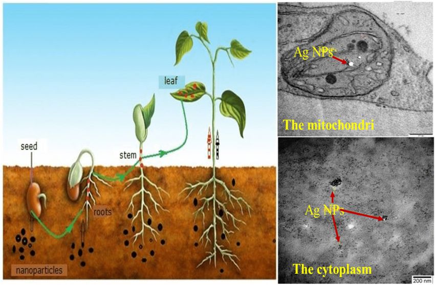
Overview of methods for production and detection of AgNPs 955
aquatic environment, soil, crops, and humans. Reduction some reviews dealt with the toxicity of AgNPs, there was a
measures and future research are discussed. lack of information about the mechanism of the ecotoxi-
cology in addition to the lack of reduction measures. This
Keywords: silver ions, toxicity mechanism, AgNPs, human
review aims to depict how the manufacturing techniques
health, aquatic environment, soil, crops
of AgNPs and receiving environment affect the fate and
impact of AgNPs on the aquatic environment and humans;
in addition, reduction measures are suggested.
1 Introduction
Silver nanoparticles (AgNPs) are one of the fastest-growing
products in the nanotechnology industry, due to their 2 Overview of the different
distinctive physicochemical properties and antimicrobial
activity [1–4]. Accordingly, the use of AgNPs has become
applications used for the
extensive with an estimated global production of approxi- synthesis of AgNPs
mately 500 tons per year [5]. AgNPs have been widely used
in medical applications, including wound dressings, con- AgNPs have been used in various applications since
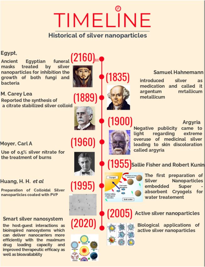
traceptive devices, surgical instruments, and bone pros- ancient times, for instance, they were used for the con-
theses [6–8], in addition to water purifications and indoor servation of mummies in Ancient Egypt [28] (Figure 1).
air quality management [9,10]. Moreover, AgNPs are used Nowadays, silver is used in smart nano-systems by devel-
in the aquaculture industry for rapid disease detection, oping AgNPs for various functions, e.g., imaging contrast,
vaccines, hormones, nutrients, and nanosensors [11,12]. drug delivery, cell targeting, etc. [29]. By searching the
Eventually, AgNPs enter the soil and aquatic environment Scopus for the words “AgNPs synthesis,” one can find
mainly through wastewater effluents, accidental spillages, 42,100 publications covering the period between 2160
industrial runoffs, and agricultural drainage water, where BC and 2020. Varied preparation methodologies of AgNPs
they exhibited substantial toxic effects on different organ- have been reported, including, chemical, physical, bio-
isms and humans. synthesis, and photochemical methods. Forty two per-
However, to date, there have not been conclusive cent of the published researches used chemical methods,
statements about their toxicity due to the lack of studies while physical, biological, and photochemical syntheses
on the fate of AgNPs under laboratory conditions [13]. represented 33, 18, and 7%, respectively, of the scholarly
Three main mechanisms that explain AgNPs toxicity have output (Figure 2).
been suggested: (1) AgNPs can directly damage cell mem-
branes due to the nano-size (physical impact), (2) AgNPs
and silver ions generate reactive oxygen species (ROS), and
(3) AgNPs can release Ag ions. The latter mechanism was 2.1 Chemical synthesis
suggested by many studies [14–17]. Some researchers sug-
gest that AgNPs could serve as a “Trojan horse,” avoiding Chemical synthesis is mainly used for the preparation of
common barriers, releasing Ag+ ions, and causing damage AgNPs since it allows for the preparation of stabilizing
to the cells [18,19]. AgNPs are converted from Ago form to monodisperse AgNPs with various nanostructure shapes.
dissolution or ionization (Ag+) form [20]. Some researches Usually, the AgNPs were synthesized chemically based
indicate that cysteine is vital to remove Ag+ ion toxicity on the following three major compounds: (i) silver nitrate
which is a free chelating agent for (Ag+) ions [21]. While (AgNO3) as silver precursors, (ii) an appropriate reducing
other results are somewhat conflicting, all evidence sug- agent, and (iii) a capping agent [30,31]. The production
gests that both silver ions and AgNPs cause toxicity in of colloidal silver solutions from silver ions reduction
human cells [22,23]. Several research papers evaluated involves two steps: nucleation and grain growth. It has
the antimicrobial and antifungal activity of AgNPs [24,25] been shown that the shape and size of the synthesized
which was attributed to the release of Ag+ to the medium AgNPs depend largely on these stages. The nucleation
[26,27]. Several studies used AgNPs for many applications, and grain growth of the first nuclei can be regulated by
but they did not study their side effect on the environment. changing the parameters of the reaction, like ion precur-
There remain wide information gaps concerning the poten- sors, temperature, pH, and types of both stabilizing and
tial risks of exposure to AgNPs, considering the increas- reducing agents. In several studies, stabilizing and redu-
ingly rising proportion of AgNPs in our societies. Although cing agents are the same. For example, AgNPs can be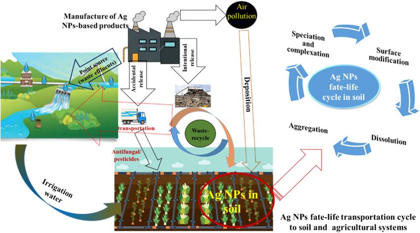
956 Mohamed Mohamady Ghobashy et al. Figure 1: Historical timeline of AgNPs. prepared by thermally reducing silver ions in the exis- triangular silver nanoplates were synthesized in situ by tence of polymer. This process is called in situ AgNPs heating a mixture of PVP and AgNO3 dissolved in N-methyl- formation [32]. Spherical AgNPs were synthesized in situ pyrrolidone (NMP) at a temperature of 100°C [34]. Another with controllable size and high monodispersity by heat- nanostructure form of silver nanocubes was produced in the ing a mixture of polyvinylpyrrolidone and silver nitrate existence of PVP as a stabilizer and ethylene glycol (EG) as a [PVP]/[AgNO3] with weight ratios of 5:1, 10:1, and 1:20 reducing agent of AgNO3 [35]. This process is known as the at a temperature of 70°C [33]. In this case, PVP severed “Polyol” methodology; in that case, EG acted as both a both as a stabilizing and reducing agent. It has been reducer and solvent, while PVP served as a stabilizer. [35] shown that the shape and size of the obtained AgNPs are showed that PVP and its molar ratio, compared to AgNO3, strongly affected by the PVP weight fraction in the mixture played important roles in determining the amount, size, and of [PVP]/[AgNO3]. Significant amounts of monodisperse geometric form of the product. According to [36], the
Overview of methods for production and detection of AgNPs 957
Figure 2: The published research articles on the topic of synthesis of AgNPs. The databases were collected from “Scopus” using the keyword
“AgNPs,” up to 30 May, 2021.
experimental conditions, such as different temperatures 2.2 Physical synthesis
ranges of the mixture (PEG, PVP, and AgNO3), affected
the size and shape of the obtained AgNPs, where at heating In brief, the physical synthesis method of AgNPs typically
rates of 1 and 7.5°C min−1, the mean sizes of AgNPs were 42 uses physical energies such as arc discharge and electric
and 18 nm, respectively. They were inclined to be more and thermal powers to generate narrow-sized AgNPs in
mono-dispersive, which means that rapid nucleation with powder form [49]. Generally, AgNPs may be synthesized
temperature occurred in a short time. The in situ colloidal via evaporation/condensation through a tube furnace at
AgNPs formation from the reduction of their salts depends ambient conditions. Lee and Kang formed silver nano-
on the nucleation and grain growth. It was also revealed crystallites 9.5 nm by the thermal decomposition of a
that the shape and the size of synthesized AgNPs are mainly complex of Ag-oleate at a temperature of 290°C [50].
dependent on experimental parameters like reactants ratio
[37], the temperature of reaction [38], and pH [39].
A microwave irradiation method is another fast method,
known also as one-pot, which is used to synthesize colloidal 2.3 Biological and green synthesis
AgNPs from the reduction of AgNO3 solution at a tempera-
ture within 80–128°C [40–42]. The reducing agent in the The biosynthesis of AgNPs has been explored by utilizing
microwave irradiation method may be natural such as various biological agents; bacteria have received great
cuminum cyminum [43] and glucose [44]. Cai et al. [45] interest through both the extracellular and intracellular
used the microwave irradiation method to synthesize a uni- synthesis pathways due to the ease of bacterial evolution,
form size (20 nm) of AgNPs coated by polyacrylic acid. The short generation period, and light culture procedures
source of the silver ions was AgNO3 and monoethanolamine [51]. Fast biosynthesis of AgNPs can occur with a non-
(MEA) was used as a reducing reagent. Liu et al. demon- pathogenic bacterium, Thiosphaera pantotropha, seeded
strated that the microwave irradiation approach assisted the with a solution of 2 mM AgNO3 [52]. This bacterium has
synthesis of a uniform structure of colloidal silver nano-rods an unusual ability to use nitrogen oxide or nitrate as an
in the absence of a polymer or surfactant [46]. In contrast, electron acceptor and was able to perform heterotrophic
the synthesis of the AgNPs using the one-pot method was nitrification exhibiting the activity of both nitrate-reduc-
characterized by different levels of aggregation and a variety tase enzymes (NaR) and nitrite-reductase enzymes (NiR).
of particle shapes by adjusting the time of microwave irra- In another study, the use of Penicillium oxalicum fungal
diation [47]. In conjunction, AgNPs can be synthesized metabolites for the extracellular biosynthesis of AgNPs
using the emulsions method, where the reactants of the from AgNO3 solution was reported [53]. Morphology of
metal precursor and reduction agents were put in two the obtained AgNPs yielded an irregular spherical shape
immiscible phases [6,48]. with high variability in a particle diameter ranging from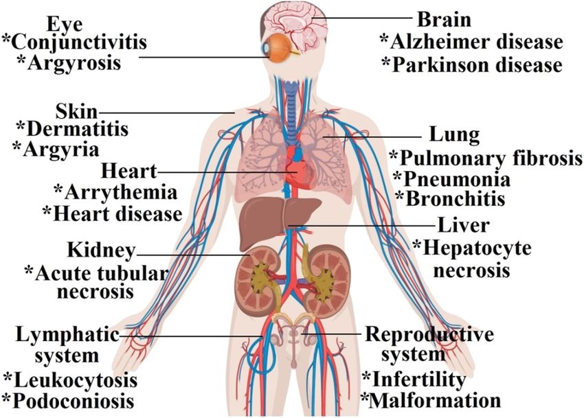
958 Mohamed Mohamady Ghobashy et al.
60 to 80 nm. Green synthesis is a new alternative AgNPs reaction time. Almost all silver particles have a small size,
synthesis eco-friendly approach. This eco-friendly tech- high surface area, and, therefore, great toxic potential [58].
nique used biological agents, plants agents, or microbial Localized surface Plasmon resonance (SPR) of AgNPs
agents which can act as capping and reducing agents at depends on the shape, size, and mutual interactions
the same time. AgNPs synthesized by green chemistry between particles of silver in close proximity [59]. The
offers a novel and potential alternative to chemically shape and size of AgNPs can tune the AgNPs Plasmon
synthesized nanoparticles. In the green process of AgNPs peak in the range of 393–738 nm [60].
synthesis, the biological agents not only reduce the silver
salts, but also can form a protected layer on the surface
of AgNPs; beside, they can act as reducing agents. This
protected layer has several advantages such as it (i) pre- 4 Toxicity and fate of AgNPs in
vents the agglomeration of the nanoparticles, (ii) reduces
AgNPs dissolution and their toxicity, and (iii) improves
aquatic systems
their antimicrobial property [54].
For Drinking Water Quality (DWQ), the World Health
Organization (WHO) noted that there are insufficient
data to deduce a health benefit for silver in drinking
water. These guidelines state that “amounts of up to
2.4 Photochemical synthesis
0.1 mg L−1 of silver can be accepted without health hazard.”
Silver usage is governed by the National Secondary Drinking
Another one-step in situ AgNPs formation is carried out
Water Regulations of the US Environmental Protection
by the irradiation technique. The γ-irradiation method at
Agency (EPA). In sources discussing drinking water, the
a dose of 30 kGy provided a convenient and uniform
permissible silver contaminant level is 0.1 mg L−1, a non-
reduction process in the existence of PVP as a stabilizer
enforceable standard due to potential health effects, such
[55]. The effect of γ-irradiation stems from the water radi-
as skin discoloration. In aquatic environments, the stability,
olysis that releases six species (˙OH, ˙H, H2O2, ehy, H2, and
and therefore toxicity, of AgNPs is inseparable from the
O2) where three of them are powerful reducing agents
chemistry of water, including parameters like dissolved
such as ˙H, ehy, and H2. Furthermore, UV-initiated photo-
organic matter (DOM), pH, ionic strength, and composition
reduction has been reported by Huang and Yang for the
[61]. High ionic strength encourages the agglomeration of
synthesis of AgNPs via AgNO3 photoreduction in the exis-
nanoparticles by screening double-layer electrostatic repul-
tence of collagen, PVP, citrate, and polyacrylic acid,
sion among similar particles, thereby reducing dissolution,
which served as stabilizing agents [56].
toxicity, and ROS production of (Ag+) ions [62].
The effect of inorganic aquatic chemistry on the sta-
bility of AgNPs (precipitation, dissolution, and aggrega-
tion) and their bacterial viability was elucidated by [63].
3 Physicochemical properties of Little is known about both the mobility and ultimate fate
AgNPs of AgNPs in freshwater ecosystems. Jin and coauthors
prepared various mixtures of anions and cations dis-
AgNPs can be obtained in different sizes, shapes, and solved in water at a fixed ionic strength. The results indi-
surface charges (positive, neutral, and negative) depending cated that the AgNPs seemed to be in a highly dispersed
on the method of synthesis. The stabilizers and capping form in the ions with the absence of Mg2+ and Ca2+. With
agents are usually used to modify the AgNPs’ surface Mg2+ and Ca2+ ions’ presence, AgNPs’ aggregation was
charges, beside influencing their physicochemical proper- enhanced regardless of the other ions’ presence due to
ties [57]. The shape of AgNPs can be of isotropic structure the divalent ions such as Mg2+ and Ca2+ reducing the
(0D) or anisotropic structure, such as 1D, 2D, and 3D. The electrostatic repulsion among AgNPs that have negative
morphologies of anisotropic AgNPs exhibit new physico- zeta potentials in aqueous media. The negative charge of
chemical properties due to the high surface area when com- AgNPs is due to the adsorption of anions on the silver
pared to isotropic AgNPs of inorganic NPs. Additionally, surface, like sulfate hydroxide and chloride. Usually,
several significant parameters control the anisotropic mor- AgNPs in water systems are not only found in metallic
phology of AgNPs during seed processes such as the con- form, but also in a salt form such as silver sulfide (Ag2S)
centration of precursors, reaction temperature, pH, and and silver chloride (AgCl) (Figure 3) [64].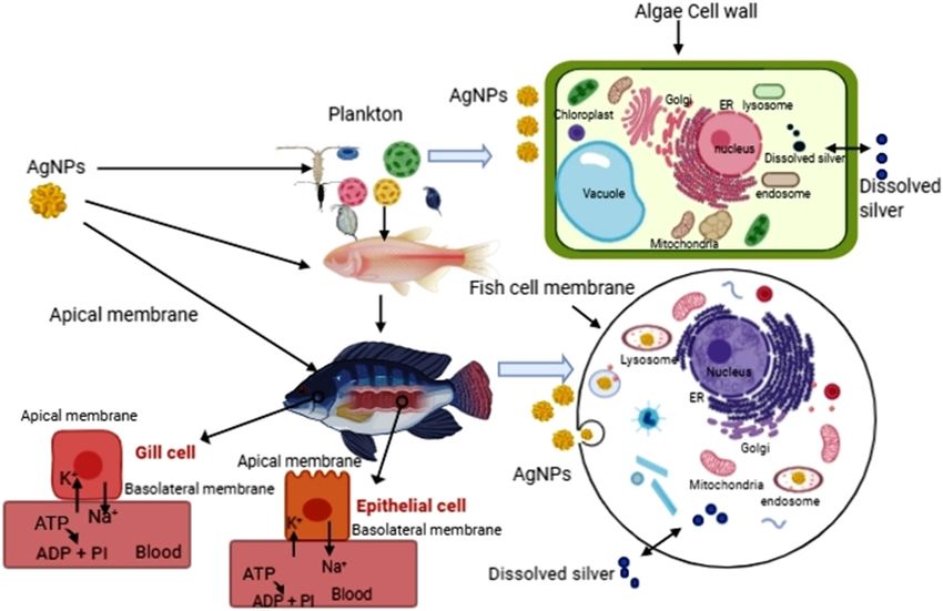
Overview of methods for production and detection of AgNPs 959
Figure 3: Behavior of AgNPs in freshwater ecosystem.
The dissolution of AgNPs by the oxidation process Li et al. [69] evaluated the stability of AgNPs citrate
has been reported in several studies. Kittler et al. [65] coated in natural freshwater from six separate sites. The
prepared AgNPs stabilized with poly(vinylpyrrolidone) results showed that citrate-coated AgNPs remained stable
(PVP) and citrate to gain the silver particles’ different in low-salinity waters due to the impacts of DOM which
surface fictionalization and studied the dissolution of promoted the stability of NPs (Figure 4). Free ions con-
coated silver particles in water at three temperatures of centrations of sulfide S22 −, chlorine Cl−, and sulfate SO4−
5, 25, and 37°C for several days. The results obtained in waters of high salinity cause rapid dissolution and
suggested that depending on their surface fictionaliza- sedimentation of citrate-coated AgNPs. Also, the results
tion (if citrate or PVP) and reaction temperature, the revealed that AgNPs remain stable for a long period
degree of silver dissolution was higher for nanoparticles in waters of low salinity. AgNPs can cause serious impli-
of PVP-stabilized silver than nanoparticles of citrate-sta- cations on the environment and organisms in freshwater
bilized silver. This could be due to the citrate layer which ecosystems than in estuarine or seawater systems. Zou
serves as a chemical shield to reduce the outgoing silver et al. [70] investigated the effect of natural organic matter
ions. Liu and Hurt [66] measured the rate of silver ions’ (NOM) and DO in natural and synthetic freshwaters on
dissolution from citrate-stabilized AgNPs in different tem- the stability and dissolution of AgNPs for seven days; aggre-
peratures and pH values. The results indicated that the gations of AgNPs in synthetic freshwater were observed,
rate of silver ions’ dissolution increased with temperature where they resulted from the contraction of the electric
and decreased with the reduction of pH or with the addi-
tion of fulvic acid or humic acid. The results confirmed
that organic compounds such as fulvic or humic acids
in natural waters do not dissolve silver. Gao et al. also
observed lower toxicity of AgNPs in water samples as
the concentration of DOM increased [67,68]. The effects
of different environmental parameters, such as the sali-
nity dissolved oxygen (DO), temperature, and pH, on the
dissolution rate of citrate-stabilized AgNPs have been
studied. High salinity promotes silver particle aggrega-
tion and hinders silver dissolution [66]. In addition, the
increase of DO and temperature and lower pH values
enhanced the rate of silver dissolution. Figure 4: The effect of DOM on AgNPs’ dissolutions.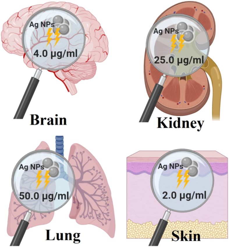
960 Mohamed Mohamady Ghobashy et al.
double layers, followed by the dissolution of silver NPs. pathways by interacting with protein molecules, entering
Nevertheless, the maximum concentration of dissolved silver the cell directly through endocytosis or diffusion to damage
(Agdis) significantly decreased from 356.5 to 272.1 mg L−1 the mitochondria and generate ROS that damages DNA and
under anoxic conditions. The addition of NOM mitigated causes necrosis and apoptosis [78,79]. Many studies have
the aggregation, prevented the oxidative dissolution effect, measured silver ions toxicity in freshwater fish [80,81]. LC10
and enhanced the AgNPs’ transformation into Ag2S due to was reported at concentrations of 0.8 μg L−1 for several fish
the adsorbed layers’ formation of NOM. Similarly, the inhibi- species [82]. However, physiological changes like blood
tion of oxidative dissolution occurred in oxygen-deficient acidosis causing circulatory collapse and death were
natural freshwaters compared with oxygenated freshwaters, recorded in fathead minnows and trout at higher concen-
resulting in a decrease in the concentration of Agdis from trations [83,84]. The effects and fate of AgNPs in rainbow
137.6 and 57.0 mg L−1 to 83.3 and 42.4 mg L−1, respectively, trout juveniles (Oncorhynchus mykiss) were investigated
in two natural freshwater locations. This indicates that (Figure 5), where they were exposed to 50 μg L−1 AgNPs
AgNPs pose more serious environmental risks in fresh- (20 nm) and dissolved 1 μg L−1 of silver ions [85]. The
waters. As silver ions are found to be one of the main results showed that water with a high organic carbon level
causes of the toxicity of AgNPs, the toxicity of Ag ions is (7 mg L−1) encouraged the production of bioaccumulated
strongly linked to the viability of nanoparticles [71]. The AgNPs in the livers and gills of fish. AgNPs of 10–80 nm
exposure of the freshwater alga Microcystis aeruginosa to influence the development of early life stage negative
AgNPs led to toxic repercussions and reduction of α chlor- effects that involve deformities of the spinal cord, cardiac
ophyll and membrane damages [72]. The toxicity was due arrhythmia, and zebrafish death [86,87]. AgNPs often
to the dissolved Ag ions, which emerged from the inter- accumulate in the tissues of the liver and gills, which
nalized AgNPs that directly targeted the photosynthetic impairs fish’s ability to cope with low oxygen value and
system of the alga. AgNPs can readily transform once in causes oxidative stress [88]. The exposure of the African
water depending on many environmental factors, which catfish, Clarias gariepinus, to AgNPs’ (40 nm) quantity of
included complexation with organic and inorganic spe- 10 and 100 μg L−1 caused serious hepatotoxic effects after
cies, agglomeration, and oxidizing changes which all con- being exposed for 15 days [89]. Rajkumar et al. [90]
tribute to the fate of AgNPs in water bodies. exposed rohu (Labeo rohita) to 5–100 mg kg−1 of AgNPs
for seven days. The results showed a substantial reduction
in hematological parameters compared to control samples.
Filter feeders tend to accumulate AgNPs with other
food. A toxicity study of AgNPs on embryonic develop-
5 Toxicity of AgNPs in aquatic ment of the oyster, Crassostrea virginica, revealed adverse
organisms impacts on embryonic development [91]. Multiple cel-
lular mechanisms linked to silver ions and AgNPs were
Silver ions (Ag+) have high reactivity with anionic and observed at concentrations of 100 mg L−1 in juvenile sea
sweater species as well as with negative ligands found urchin, with series of cellular responses like spherocyte
either on DOM or living cell surfaces [73]. Organic matter and amebocyte cell coagulation, oxidative stress, and
(OM) and sulfide may dominate silver speciation in fresh- expression of 70 kDa chaperone [92].
water systems and could reduce its bioavailability [74]. Phytoplankton is the primary producer in the food
Neutral chemical complexes like AgCl (aq.) and AgHS chain of aquatic ecosystems. Accordingly, studying the
(aq.) are formed at lower salt concentrations, and AgNPs effect of AgNPs on their vitality and dynamics is a priority,
dissolution occurs slowly [73]. With the increase of sali- where they are the first target for most pollutants in sea-
nity in oceans, the supply of silver ions changes while water. The toxic effect of AgNPs on marine phytoplankton
AgCl becomes dominant [73]. has been extensively reported [93]. Interestingly, the expo-
Aquatic organisms exposed to AgNPs causing cyto- sure of phytoplankton to AgNPs not only caused toxic
toxic and genotoxic impacts can reach humans via the effects, but also showed taxa-specific effects, where the
food chain [75]. The general mechanism of animal toxi- composition of the community changed in response: nega-
city relies on its phase of transformation in environ- tively diminishing cyanobacterial functions at concentra-
mental and biological products. Silver ions release the tions of Ag ions ≥200 ng L−1 and altering the domains
oxidative force of the surface and they interact with bio- of dinoflagellate and their composition concentration of
molecules like lipids, proteins, nucleic acids, etc. [76,77]. Ag ions at a 2,000 ng L−1; either decrease or increase of
Silver NPs can produce toxins by triggering signaling diatom (Climacosphenia and Nitzschia, Navicula) and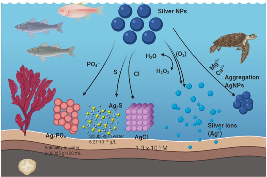
Overview of methods for production and detection of AgNPs 961
Figure 5: The effect of AgNPs’ dissolutions on aquatic organisms.
Dinoflagellate (Prorocentrumand Gyrodinium and Gymno- of sewage sludge is utilized in agricultural soil as fertilizers
dinium) [94]. Other research showed that the particle size in various countries, e.g., UK, USA, and Egypt. In some
of AgNPs was important in determining the hazardous other European countries, these sewages are incinerated.
impact of AgNPs in the gills and intestines of adult zebra- Naturally formed AgNPs have also been documented in
fish [95]. Zebrafish have a similar genetic composition to soil and aquatic environments through the reduction of geo-
human cells, and their larvae and embryos are transparent genic (Ag+) traces in the existence of (S2−) ions, as a reducing
which allows for easier observations. A prominent Ag+ agent, under some atmospheric conditions, e.g., dark/light,
deposition in the basolateral membranes for 20 nm Ag temperature, and anoxic/aerobic conditions [98].
silver particles disrupted the Na+/K+ ion channel, as Ag+ When AgNPs reach natural soil systems, they are
can compete for Na+ and disrupt the function and integrity exposed to many transformation mechanisms like disso-
of the channel; this was confirmed by a reduction in ATPase lution, oxidative stress, aggregation, agglomeration, desta-
activity, the 20 nm particles caused significantly higher bilization, chlorination, and sulfidation reactions. The soil
inhibition and disruption than the larger size particles. environmental conditions (soil texture, moisture, ionic
strength, pH, inorganic/organic matter, microbial diver-
sity, etc.) can significantly affect the surface properties
and fate-life cycle of AgNPs, in terms of their surface
6 The fate-life cycle of AgNPs charge, size, shape, agglomeration, uptake, migration,
in soil and dissolution processes [99]. For example, the sulfur-
rich soil contaminated with AgNPs can initiate a sulfida-
It is known that AgNPs can reach soil from the discharge tion reaction to form Ag°/Ag2S core–shell particles with low
of waste liquid effluents or sludges during their synthesis biological toxicity relative to AgNPs [100,101]. This is because
or industrial production (either intentionally or uninten- the solubility of Ag2S particles is lower than the AgNPs them-
tionally) and/or the disposal and recycling of goods con- selves; hence, the dissolution process and release of toxic
taining AgNPs (Figure 6) [96,97]. Industrially, once AgNPs silver ions into the environment are limited.
are discharged in waste streams, they accumulate in sewage It was also demonstrated that sedimentary humic
sludge in advanced waste treatment plants. A large portion acids (SHAs, especially aliphatic-based SHAs) accelerated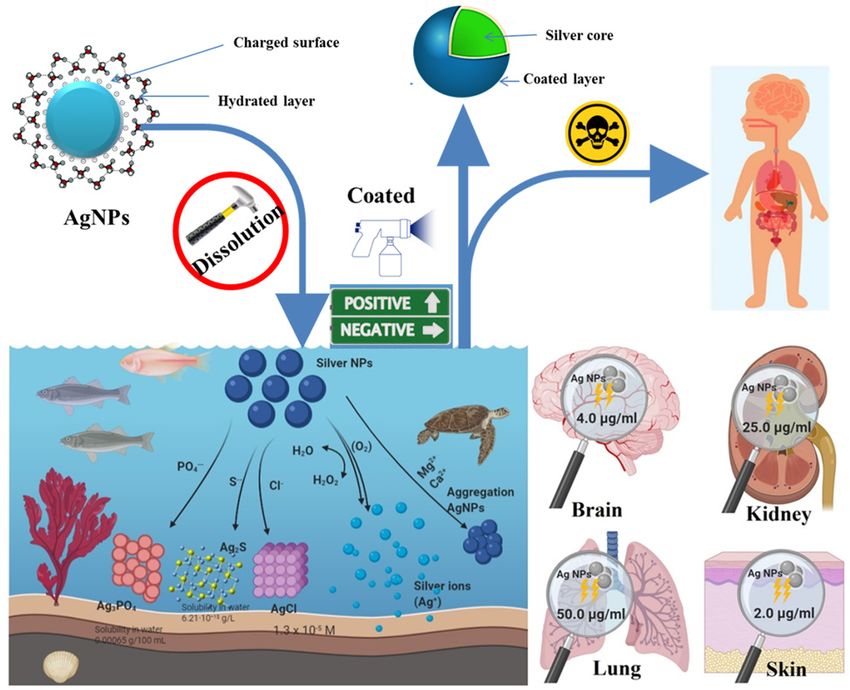
962 Mohamed Mohamady Ghobashy et al. Figure 6: The fate-life cycle of nanoparticles (e.g., silver nanoparticles; AgNPs) from industrialization to soil and agriculture systems. the formation of colloidally stabilized AgNPs by the reduc- species like metallic AgCl and AgNPs emerged after two tion of silver ions in soil when allowed to react for 25 days days of incubation from the initial aging of silver to soil at 22°C or 3 h at 90°C [102]. It was noted that an increase of [108]. These results indicate that the soil nature and com- ionic strength leads to an increase in the aggregation of position significantly affected the fate of AgNPs and their AgNPs, i.e., hydrodynamic radius, and their dissolution associated risks to environments. However, it is note- process to Ag+ ions with a high toxicity effect (alleviate worthy that the related toxicity of these transformed spe- oxidative stress) on Escherichia coli (E. coli) [103]. High cies in the in vivo cells is still not clear. chloride contents can also mediate significant changes in From the above findings, it can be concluded that the the toxicity and morphology of AgNPs by forming AgCl0(s) fate-life cycles (transformation, migration, dissolution, bridging and species of negatively charged Ag Clx(x−1) with aggregations, and toxicity) of AgNPs and their intrinsic less toxicity on E. coli. Similarly, an increase of monova- characteristics and toxicity in the soil are governed by lent (K+ or Na+) cations could alter the behavior of AgNPs, many parameters, such as the disposal routes (liquid or e.g., morphological transformation, size, dissolution, and sewage solids) and the surrounding environmental con- aggregation, and their related toxicity to Caenorhabditis ditions. These parameters should be accurately consid- elegans (C. elegans) [104]. In particular, the higher concen- ered while assessing AgNPs’ toxicity on the environment tration of K+ or Na+ cations (from 1–10 mM) significantly and human health. decreased the size of AgNPs (
Overview of methods for production and detection of AgNPs 963
Figure 7: Migration paths of nanomaterials (e.g., silver nanoparticles; AgNPs) from soil to the plant organs and the corresponding TEM
images of AgNPs in the mitochondria (leaf cells) and the cytoplasm (stem cell) of wormwood (Artemisia absinthium) [113].
to plant organs, (2) absorption/uptake by the plant roots, (50–2,000 mg kg–1) for 98 days due to the cell damage
then migration to other organs, and (3) the direct migra- and accumulation of AgNPs in shoots and edible por-
tion/movement to plant organs and their localization in tions, confirming the unsafety of crops [117]. In another
the epidermal or xylem cells [113,114]. These distribution/ work, the chemical transformation (homeostasis) of com-
migration pathways from soil/water to plant cells are mercial nanoparticles of Ago and Ag2S into Ag+ ions
known to be size-, concentration-, and AgNPs’ physico- inside the roots/epidermis of wheat (Triticum aestivum)
chemical features-dependent, along with the nature of was also reported as a result of the gene defense function
crops and the soil structure (composition and thickness). of plants [118].
Generally, the major findings in this area suggested These observations demonstrated the complexity of
that the toxicity effects of AgNPs in agriculture are mainly assessing AgNPs’ toxicity in crops due to their transfor-
dose-dependent [98]. Also, AgNPs in irrigated water can mation mechanisms, which depend on AgNPs’ shape and
move and penetrate soil layers up to 5 cm thickness, then concentrations and the nature of plant cells. Hence, in
migrate to seed tissues within the first 24 h of irrigation ecotoxicological investigations, it is important to define
[113]. Also, AgNPs can quickly diffuse and translocate another monitoring system for the toxicity effect of AgNPs
into plant cells during germination, as confirmed by (or other nanomaterials) based on different biological
TEM analysis of leaf and stem cells of wormwood grown biomarkers (enzymes, fatty acids, lipids, nutrients, etc.)
in soil contaminated by AgNPs (Figure 7). Such observa- to assess the protection of edible plants for the sake of
tion indicates that the crops with fibrous roots are more human health.
susceptible to uptake toxic nanoparticles from contami- Soils are complex matrices containing wide biodiver-
nated soil [113]. sity of microbial communities and organic/inorganic con-
The seedling growth in grass shoots and roots of tents, which control the fate-life cycle and toxicity of
Lolium multiflorum can be inhibited when exposed to AgNPs in soil [119]. In this regard, it was demonstrated
a high concentration of small-sized gum arabic (GA)- that the negative impacts of AgNPs on soil microbial com-
coated AgNPs (6 nm) due to the cell damage resulting munities are dependent on concentration, size, agglom-
from the toxicity effect of Ag+ ions released from the eration, dissolution, the transformation of AgNPs, and
AgNPs’ dissolution [115]. However, such toxicity effect their residence time in soil, as well as the chemical nature
can be decreased by sunlight irradiation that induces and texture properties of the soil itself [120,121].
irreversible aggregation of AgNPs [116]. For example, the toxicity of AgNPs significantly decreased
Abnormalities in the levels of antioxidant enzymes in the microbial community in the soil in the case of (i) a
and fatty acids in peanut plants were also observed sulfur-rich soil, i.e., transformation to sulfurized Ag2S
upon exposure to citrate-caped AgNPs in sandy soil from low-solubility [122] and (ii) the presence of fulvic964 Mohamed Mohamady Ghobashy et al. acid as a reducing agent, i.e., dissolution decreased [123,124]. in relation to the generation of ROS in A549 cells. Ag NPs Further, it was reported that the low concentrations of caused ROS formation in the cells, a reduction in their AgNPs (
Table 1: The summary of the recent reports of the cytotoxicity of silver NPs against different cell lines

Preparation route           Particle     Shape               Concentration         Coating materials       Tested        Cytotoxicity assay Recorded effect   Duration of      References
                            size (nm)                        (µg mL−1)                                     cell line                                         incubation (h)

Green synthesis using      24–54         Spherical           63.257                Eucalyptus              MCF-7         MTT assay         50% inhibition of 48               [141]
Eucalyptus tereticornis                                                            tereticornis leaf                                       total cell number
leaf extract                                                                       extract
Biological synthesis       17–50         Spherical           48                    Chitosan                HepG2         – MTT assay       50% inhibition of 24               [142]
using chitosan as                                                                                                        – Trypan blue     total cell number
capping and reducing                                                                                                     exclusion assay
agent
Green synthesis using      72.77         Nonuniform          – 14.96 for           Delonix regia leaf      – A459        MTT assay         50% inhibition of 48               [143]
Delonix regia leaf extract               (anisotropic)       A549 and              extract                                                 total cell number
                                                             – 15.96 for SiHa                              – SiHa
Biological synthesis
966 Mohamed Mohamady Ghobashy et al.
Apparently, after entering the cell, AgNPs usually pro-
duce ROS [150]. After increasing ROS levels, GSH level
dramatically decreases, and LDH increases in the medium,
which eventually leads to apoptosis [151]. Moreover, redox
homeostasis can be affected by ROS generation at the
intracellular level. Consequently, protein carbonylation
and lipid peroxidation take place. Simultaneously, anti-
oxidant enzyme activity and the glutathione level are
decreased. Thus, protein-bound sulfhydryl group deple-
tion, antioxidant enzyme activity, and glutathione level
promote apoptosis [152]. Therefore, apoptosis-mediated
cell death is the main cytotoxic impact of silver NPs
[153]. Apoptotic pathways like AKT, p53, and MAPK which
activate cell death are also reported [154].
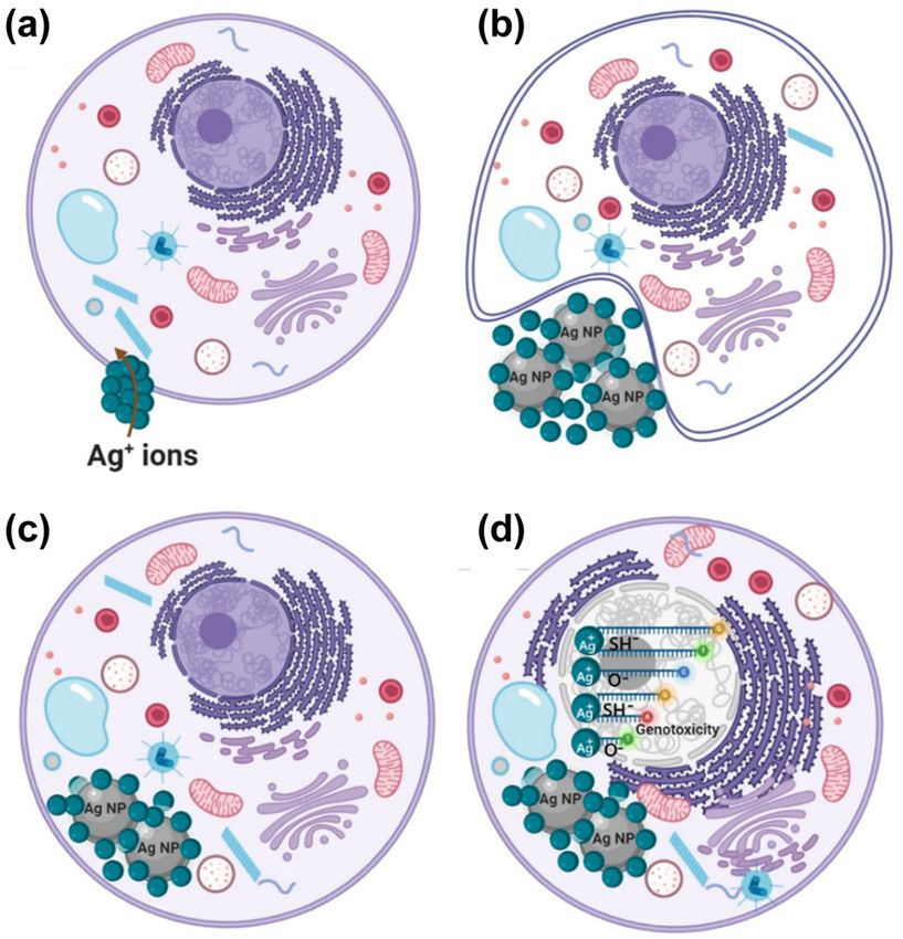
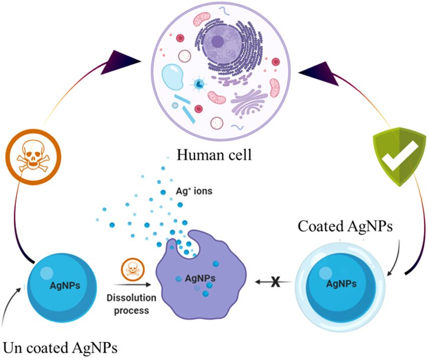
8.1 Trojan horse effect
The Trojan horse effect was suggested as the toxicity
mechanism of AgNPs [155]. Some metal oxide nanoparti-
Figure 8: Toxicity of AgNPs and silver ions on the normal cells and
cles affect lung epithelial cells by this effect [156].
Trojan horse effect. (a) cell defense response to Ag+ (b) engulf AgNPs
According to the Trojan horse theory, if silver NPs with and Ag+ ions (c) excite of Ag ions inside cell and (d) death cell occur.
very small size (≤40 nm) cross through the cell mem-
brane, they may continuously release (Ag+) silver ions
once inside [157]. Inside the cell, the silver ions (Ag+) utilized by some crops or different living animals; the pre-
can form ROS and cause lipid peroxidation [158]. Ag+ sent AgNPs can arrive at the food series [166]. Through the
ions attach to the host cell, consumed by the cells before past ten years, it was believed that AgNPs and silver ions
reaching the vital organelles inside the normal cells [159] are nonlethal to animal and human cells, but severe
as shown in Figure 8a. In a defense response to the Ag+ argyria and blue skin coloration were observed after con-
ions, the normal cells secrete the reductase enzyme to tact with nano silver-based materials [138].
reduce the dangerous effect of Ag+ ions and finally engulf The principal AgNPs’ uptake probabilities inside the
silver NPs, which also carries the liberated Ag+ ions out- human body are by the first-line defense: skin by direct
side their surface [160] as displayed in Figure 8b. AgNPs contact, or the respiratory region through inhalation,
with the formed Ag+ ions inside the normal cells are con- or finally through the gastrointestinal tract by AgNPs-
sidered to be the beginning point of the toxic behavior contaminated foods [167]. Thus, AgNPs’ quantity, config-
and the hazardous effect starting inside the normal cell uration, and exterior adjustment perform an essential
[15] as exhibited in Figure 8c. Figure 8b and c displayed function in human organs [168]. AgNPs present in the
the Trojan horse effect [161]. respiratory region can transfer to the lymph stream,
Finally, the positively charged Ag+ ions will react with then the blood circulation [168]. Recent studies showed
the negatively charged oxygen and nitrogen atoms in the that AgNPs can move into the blood-brain barrier [169]
vital organelles like DNA, mitochondrion, and the thiol and penetrate cell membranes [129 thus accumulating in
group presented in protein structures and enzymes, which vital organs and biological systems [129]. Because AgNPs
in turn interrupts the normal cell reproduction, and finally, were the subject of various earlier investigations, the
the death of cell will occur [162] as presented in Figure 8d. potentially dangerous impacts of AgNPs were revealed.
The evaluation of AgNPs toxicity is essentially affected It is significant to examine the lethal effect more strongly.
by their movement inside the human body [163]. Addition- Accordingly, Figure 9 shows the toxic impact of AgNPs
ally, because of the high surface area of AgNPs, pollutants and Ag ions on human organs and their related diseases.
may be adsorbed on the surface of AgNPs [164]. In the The different diseases in various human organs were dis-
synthesis of nanomaterials-based compounds, a growing played in Figure 9, after the exposure to the toxic levels
aggregation of AgNPs in the water and/or the atmosphere of AgNPs in drinking water, determined to be above
may happen [165]. Also, AgNPs can be absorbed and 5.0 µg kg−1 body weight/day (according to the EPA) [170].Overview of methods for production and detection of AgNPs 967
Figure 9: The related diseases after exposure to the toxic level of silver NPs.
Despite a notable uptake within the normal cells, catalase, and glutathione peroxidase activity. On the
AgNPs possessed an entirely irrelevant hazardous effect contrary, above 4.0 µg mL−1, AgNPs revealed a notable
on kidney cells at concentrations more than 25.0 µg mL−1 decrease in the levels of all the brain enzymes, reduced
[171] as displayed in Figure 10. Brain cells subjected to glutathione, and total glutathione [172] as shown in
2.0 µg mL−1 of AgNPs did not reveal any important differ- Figure 10. In severe oral and dermal toxicity experiments,
ence in the levels of total glutathione, reduced gluta- none of the tested models displayed any unusual symp-
thione, glutathione reductase, superoxide dismutase (SOD), toms or death at a dosage level less than 2.0 µg mL−1 [173]
as exhibited in Figure 10. After examining the cytotoxicity
of AgNPs (50 μg mL−1) on the lung cells, the results indi-
cate that there was no difference in the level of surfactant
protein-B in the bronchoalveolar lavage. After seven days
of the introduction of AgNPs (at concentration more than
50 µg mL−1), an increase in bronchoalveolar lavage cell
numbers and a reduction in lung function were recog-
nized [174].
Scientific information on the potentially harmful
effects of AgNPs on human health severely lags behind
their exponentially growing applications in consumer
products [175]. In assessing the toxic risk of AgNP usage,
the liver, as a detoxifying organ, is particularly impor-
tant. Different studies [176,177] were aimed to explore
the toxicity mechanisms of nano and ionic forms of silver
on human cells. Their results showed that silver ions
and AgNPs reduced cell viability in a dose-dependent
manner. The IC50 values of silver ions and AgNPs were
0.5 and 50 mg L−1, respectively. AgNPs affected the trans-
form toxic metabolites (TTM); the LDH leakage and inhi-
bition of albumin synthesis, along with decreased alanine
transaminase (ALT) activity, indicated that treatment with
Figure 10: The toxicity levels of AgNPs in different organs. either AgNP or Ag ions resulted in membrane damage and968 Mohamed Mohamady Ghobashy et al.
reduced the cell function of human liver cells. Evaluation Although an increasing number of analytical techniques
of oxidative stress markers demonstrating depletion of glu- and methods are becoming available for the detection, char-
tathione (GSH), increased ROS production, and increased acterization, and quantification of AgNPs, their application
SOD activity indicated that oxidative stress might contribute to complex samples is still very limited and far from being
to the toxicity effects of nano and ionic forms of silver. The incorporated into routine analysis. AgNPs can be trans-
observed toxic effect of AgNPs on human hepatic cells was formed into different four complex environmental matrices
substantially weaker than that caused by ionic silver, while during anaerobic treatment of wastewater and post-proces-
the uptake of nano and ionic forms of silver by HepG2 cells sing of sewage sludge such as Ag carbonate (Ag2CO3), Ag
was nearly the same [176]. oxide (Ag2O), Ag sulfide (Ag2S), and “bulk” AgCl [182]. To
rid silver ions complexion, additional development of stan-
dard ICP methods was needed to get information about
inorganic nanoparticles, the use of ICP-MS in combination
with field-flow fractionation (FFF) separations or in single-
9 Different methods for silver ions particle detection mode are finding their way in the most
detection recent analytical approaches, because of the supplementary
information that can provide [183]. In addition, there have
Different detection methods for Ag+ ions detection were been advances in determining the speciation of Ag and the
developed for the nanomolar (nM) level. There are several transformation products of AgNPs using X-ray absorption
strategies for silver (Ag+) ions detection that rely on the spectroscopy (XAS) and X-ray absorption near edge struc-
combination of metal ion analysis with enzymatic or oxi- ture (XANES) techniques: [184] used microalga Coccomyxa
dative amplifying strategies. It was recognized and devel- actinabiotis to take up and cope with Ag+ that was detected
oped as a powerful tool for improving the sensitivity of using XAS; X-ray diffraction over the concentration range of
metal ion detection. The biosensors approach gained 10–7 to 10–2 M. Lombi et al. [182] used XANES spectroscopy
attention due to their high sensitivity, less time consump- to investigate the behavior and transformation of AgNPs.
tion, and operational convenience [178]. These methods XANES data were collected at the Materials Research Colla-
are based on nucleic acid interaction with metal ions in borative Access Team (MRCAT) beamline 10-ID, Sector 10
very low concentrations which can be detected. located at the Advanced Photon Source (APS), Argonne
For instance, in the past few years, significant advances National Laboratory (ANL), Argonne, IL [185].
have been made in the analysis of nanoparticles using
single-particle ICP-MS methods, and methods have also
been developed for analysis in tissues. These methods
were developed due to the increased production of nano- 9.1 Biosensors
particles which leads to increased volume of these sub-
stances in the environment, in particular in sewage and Ono et al. [186] stated that silver ions (Ag+) could selec-
sewage sludge, and also in water, sediments, and soils. tively connect via coordinating bonds with cytosine (C)
The methods must be widely available, uncomplicated, molecules to form a strong C–Ag+–C framework and
and inexpensive, as well as accurate and reliable to become transform single-stranded DNA into the double-helix struc-
commonly used by both research and control laboratories. ture. The C–Ag+–C interaction is highly selective because
Hence, it is necessary to develop some new methodological the C–C mismatching interaction with silver ions is stronger
solutions and subsequent applications for analytical prac- than other metal ions. Xie et al. [187] used an electroche-
tice. It would allow researchers to obtain repeatable and mical-based biosensors method to design a fluorescent
reliable results which could provide the basis for adequate FAM-labeled DNA for silver ions detection. The interaction
analytical interpretation [179]. Mitrano et al. [180] tracked of cytosine and silver ions (C–Ag+–C) in the existence of
the AgNPs’ dissolution at environmentally relevant concen- graphene oxide results in the mismatch of C–C that leads
trations in the laboratory, natural, and processed waters to increasing the intensity of FAM fluorescence and a red
using single-particle ICP-MS. The track changes in particle shift of the emission wavelength (λem) of FAM fluorescence.
diameter over time for 60 and 100 nm Ag NPs coated by Li et al. [188] prepared a sensitive silver ions detec-
citrate, tannic acid, and PVP were quantitatively demon- tion method based on a fluorescence biosensor using
strated using ICP-MS by direct measurement of Ag+ (aq.). cytosine (C). Since oligo-1 implies C–C mismatches in
Montano et al. [181] used ICP-MS for fast detection time cytosine (C) molecules, the existence of Ag+ ions can be
((∼500 μs)) of Ag+ (aq.) in very low concentration (ng L−1). collected to form pairs of C–Ag+–C that result in a bluntOverview of methods for production and detection of AgNPs 969
terminus with a structure of double helix. The obtained (AuNPs) with furfuryl alcohol (Fu-AuNPs). The silver ion
double-helix shape can be destroyed by exonuclease III (Ag+) detection occurred within the limit of 12 nM.
to release silver ions and trigger DNA. The released silver Selva Sharma et al. [194] developed a colorimetric
ions bind with (oligo-1) and (oligo-2) that may be pro- sensor based on Ascorbic Acid (AA) and AuNPs for silver
duced in the digestion cycles and promote the plentiful ions’ detection at concentrations of 2–28 μM in aqueous
G-quadruplex DNA generation. Hybridization N-methyl- solutions. The mechanism of sensing depends on the Ag+
mesoporphyrin IX (NMM) fluorochrome with the G-quad- reduction to Ago on the surface of AuNPs/AA. The reduc-
ruplex DNA allows Ag+ to be detected in the concentration tion of silver ions resulted in the SPR blue shift from
from 5 up to 1,500 pM L−1, with a limit of detection at AuNPs at 512 nm and it is accompanied by a new peak
2 pM L−1. observed at around 385 nm.
Xu et al. [189] developed a colorimetric approach Ghobashy and Mohamed [195] prepared a nanocom-
for silver ions (Ag+) detection based on the interaction posite membrane of Cu–(PAAc/PVA) by gamma radiation
among Methylene Blue (MB) and C-rich Single-Stranded for Rapid Colorimetric Detection (RCD) of silver Ag+ and
DNA (ssDNA). When the MB was absorbed on the S-SDNA mercury Hg2+ ions associated with significant changes of
surface, the MB color changed from blue to purple. How- color of the Cu–(PAAc/PVA) membrane from yellow to
ever, in the existence of Ag+ ions, the specific C–Ag+–C dark green and pale gray color, respectively. A detection
pair is formed and removed the interaction among S- limit of Hg2+ and Ag+ as low as 10−5 and 10−6 M, respec-
SDNA and MB, returning to the blue color. For DNA tively, occurred.
duplexes, pairs of cytosine–cytosine (C–C) can catch Wang et al. [196] describe a simple colorimetric sensor
Ag+ exclusively to form base pairs C–Ag+–C. Based on employing cationic polymer single-stranded AuNPs and
this feature, Li et al. [190] developed a method for silver ligand of DNA (ssDNA) to detect silver ions (Ag+). The
ions’ detection by adding cysteine that removed base cationic polymer is combined with ssDNA leading to AuNP
pairs C–Ag+–C because it binds to Ag+ instead of cytosine. aggregation, thus changing color. When the silver ions are
The amount of free cysteine was critical for colorimetric present, they bind to cytosine (C) that prevents polymers’
detection using ABTS–H2O2 (ABTS = 2, 2′-azinobis-(3-ethyl- interaction with ssDNA. The AuNPs strategy used as a
benzthiazoline-6-sulfonate)). Li et al. [191] developed a bio- colorimetric sensor for silver ions is in several studies
sensor based on electro-chem-iluminescence (ECL) of Ru [197–200]. Another nonmetal ion used as a colorimetric
(bpy)2(mcbpy-O-Su-ester)(PF6)2 for a highly sensitive and sensor of silver ions is carbon dot. Carbon dots (CDs)
selective Ag+ detection. This process, based on deoxyribo- have fluorescence emission properties that quench when
nucleic acid (DNA tetrahedron TS primer (STTS)), consists its surface is attached to metal ions such as silver ions
of three hybridized oligonucleotides forming three dual- [201–203]. Murugesan et al. [204] synthesized CDs with
stranded DNAs, close to a Y-shaped DNA structure. The strong fluorescent emission at 479 nm when excited over
formation of DNA-TS makes signal intensity change of Ru 370 nm. This fluorescent emission was quenched in the
(bpy)2(mcbpy-O-Su-ester)(PF6)2 at different concentrations existence of silver ion (Ag+) in an aqueous solution.
of Ag+ ions.
9.2 Chemical sensors
10 Recommendations for future
research
The highly sensitive chemical colorimetric sensor approach
for silver ions’ detection in the Picomolar (pM) range was The toxicity of AgNPs is caused by the release of (Ag+)
established by Gao et al. [192]. This approach uses Pt nano- silver ions. Silver widely disperses throughout the body
cubes coated by PVP as artificial peroxidases. The peroxi- and in laboratory animals regardless of form or exposure
dase substrates generate a colored signal that diminishes and can cross both the placental and blood-brain bar-
the existence of Ag+ ions. This colorimetric approach will riers. Silver, in exposed humans, was found abundantly
achieve an 80 pM as an ultralow detection limit and a across the body. To limit the toxicity of (Ag+) silver ions
10–2–104 nM as a wide dynamic range. released from AgNPs, it is recommended to coat AgNPs to
Alizadeh et al. [193] described a colorimetric approach prevent their dissolution (Figure 11).
for silver ions’ (Ag+) detection that comprises measur- AgNPs coating is an excellent technique to improve
ing the changes in SPR of modified gold nanoparticles their advantages and limit their toxicity. Of course, the970 Mohamed Mohamady Ghobashy et al.
stable than the other studied type (with unspecified
coating) in both water and simulated biological fluids.
The release of Ag ions into interstitial or lysosomal fluids
appears to be negligible as determined by ICP analysis in
these simulated fluids. Samberg et al. (2010) identified
the toxicity of AgNPs in vitro and in vivo human epidermal
keratinocytes [209]. The cells were subjected to different
concentrations of coated and uncoated silver carbon,
individually. The cells subjected to uncoated silver showed
decreased viability. On the contrary, there was no toxicity
observed in the cells treated with coated silver carbon [210].
11 Conclusion
Figure 11: A scheme explaining how to limit the toxicity of silver ions. Although AgNPs have been extensively used in several
applications including water treatment, health, and
coating technique can enhance the AgNPs’ stability via industry, silver ions immobilized by AgNPs are harmful
the electrostatic stabilization between silver particles and to humans and the environment. Various preparation
can reduce their particles’ agglomeration. One of the methodologies and the effect of environmental factors
main functions of coating is to avoid the cytotoxic impact largely affect the fate of AgNPs (precipitation, dissolu-
of AgNPs on living cells [205]. Most of the coating sub- tion, aggregation). These observations demonstrated the
stances have proved successful in stabilizing AgNPs, pre- complexity of assessing AgNPs’ toxicity in crops due to
serving their distinctive structure, and reducing silver their transformation mechanisms, which are dependent
ions dissolution which are major contributing factors of on the AgNPs’ shape and concentration and the nature of
AgNPs toxicity. The role of the coating depends heavily plant cells. The previous research study deals with the
on the characteristics of the coating substances, including toxicity of AgNPs and related diseases in the human body
organic coating materials such as synthetic polymer and confirmed that the (Ag+) ions can induce serious effects
natural polymer and inorganic coating materials such as on biological macromolecules like DNA and mitochon-
carbon dot, TiO2, graphene, etc. Zook et al. investigated dria through some actions such as induced cytotoxicity,
the dissolution of AgNPs in cell culture medium (DMEM) genotoxicity, immunological responses, and eventually
and observed a fractional dissolution of various polymer- cell death. Some researchers suggested that AgNPs could
coated AgNPs. The dissolution was increased in the biolo- serve as a “Trojan horse,” avoiding common barriers and
gical medium in comparison to inorganic salt solutions releasing Ag+ ions, and causing damage to the cells. In a
which is presumably owing to the complexation of the defense response to the Ag+ ions, the normal cells had
liberated silver ions [206]. been secreted by the reductase enzyme to reduce the
The behavior of polymer-coated AgNPs in two simu- dangerous effect of Ag+ ions, and finally engulfs AgNPs
lated biological fluids representative of the fluids present which also carries outside their surface the liberated Ag+
in lungs, as inhalation is regarded as an important uptake ions. The toxicity mechanism of AgNPs depends on the
route in humans [207], was studied by Stebounova et al. degree of silver ions’ dissolution from their nanoparticles
[208]. The results showed that the initial concentration of form; the AgNPs’ coating alerts their cytotoxicity. The
nanoparticles has a significant impact on their stability coated AgNPs stabilized with PVP and other organic com-
and sedimentation. The authors use Dejaguin–Landau– pounds could decrease the dissolution of coated silver
Verwey–Overbeek (DLVO) theory as a basis for theoretical particles in water for several days than AgNPs uncoated.
calculations explaining nanoparticles’ behavior in solu-
tion, and their experimental results concur with the theory Acknowledgments: All the figures presented in this review
used. According to DLVO theory, the stability of particles article were created with BioRender.com. The authors
is determined by the net electrostatic surface interactions appreciate the support of BioRender.com software.
of the particles and their Van der Waals forces. Polymer-
coated AgNPs (with higher surface charge) were more Funding information: The authors state no funding involved.You can also read