Hedgehog: functions and mechanisms
←
→
Page content transcription
If your browser does not render page correctly, please read the page content below

Downloaded from genesdev.cshlp.org on September 22, 2015 - Published by Cold Spring Harbor Laboratory Press

REVIEW

Hedgehog: functions and mechanisms
Markku Varjosalo and Jussi Taipale1
Department of Molecular Medicine, National Public Health Institute (KTL), and Genome-Scale Biology Program,
Biomedicum Helsinki, Institute of Biomedicine and High Throughput Center, Faculty of Medicine, University of Helsinki,
Helsinki FI-00014, Finland

The Hedgehog (Hh) family of proteins control cell                           (chicken)—were cloned in 1993 (Echelard et al. 1993;
growth, survival, and fate, and pattern almost every as-                    Krauss et al. 1993; Riddle et al. 1993; Chang et al. 1994).
pect of the vertebrate body plan. The use of a single mor-                  Cloning of the first Rattus rattus (rat) and human Hh
phogen for such a wide variety of functions is possible                     genes were reported shortly thereafter, in 1994 and 1995,
because cellular responses to Hh depend on the type of                      respectively (Roelink et al. 1994; Marigo et al. 1995). The
responding cell, the dose of Hh received, and the time                      vertebrate genome duplication (Wada and Makabe 2006)
cells are exposed to Hh. The Hh gradient is shaped by                       has resulted in expansion of the Hh genes, which can be
several proteins that are specifically required for Hh pro-                 categorized into three subgroups: the Desert Hedgehog
cessing, secretion, and transport through tissues. The                      (Dhh), Indian Hedgehog (Ihh), and Sonic Hedgehog (Shh)
mechanism of cellular response, in turn, incorporates                       groups (Echelard et al. 1993). The Shh and Ihh subgroups
multiple feedback loops that fine-tune the level of signal                  are more closely related to each other than to the Dhh
sensed by the responding cells. Germline mutations that                     subgroup, which in turn is closest to Drosophila Hh.
subtly affect Hh pathway activity are associated with                       Avians and mammals have one Hh gene in each of the
developmental disorders, whereas somatic mutations ac-                      three subgroups, but due to another whole-genome du-
tivating the pathway have been linked to multiple forms                     plication (Jaillon et al. 2004) and further rearrangements,
of human cancer. This review focuses broadly on our                         zebrafish has three extra Hh homologs, one in the Shh
current understanding of Hh signaling, from mecha-                          subgroup: tiggywinkle hedgehog (Twhh) (Ekker et al.
nisms of action to cellular and developmental functions.                    1995), and two others in the Ihh group; echidna hedgehog
In addition, we review the role of Hh in the pathogenesis                   (Ehh) (Currie and Ingham 1996); and qiqihar hedgehog
of human disease and the possibilities for therapeutic                      (Qhh) (Fig. 1A; Ingham and McMahon 2001).
intervention.                                                                  Components of the Hh signal transduction pathway
                                                                            have been identified primarily using Drosophila genetics
The origin of the name Hedgehog derives from the short                      (for example, see Lee et al. 1992; Alcedo et al. 1996; van
and “spiked” phenotype of the cuticle of the Hh mutant                      den Heuvel and Ingham 1996; Burke et al. 1999; Cham-
Drosophila larvae. Mutations in the Hh gene were iden-                      oun et al. 2001; Jacob and Lum 2007b). Mechanisms by
tified by Nusslein-Volhard and Wieschaus (1980) in their                    which the Hh signal is transduced has been further char-
large-scale screen for mutations that impair or change                      acterized using Drosophila and mouse cell culture mod-
the development of the fruit fly larval body plan. Dro-                     els (Fig. 1B,C; e.g., see Kinto et al. 1997; C.H. Chen et al.
sophila Hh DNA was cloned in the early 1990s (Lee et al.                    1999; Chuang and McMahon 1999; Taipale et al. 2000;
1992; Mohler and Vani 1992; Tabata et al. 1992; Tashiro                     Lum et al. 2003a; Nybakken et al. 2005; Varjosalo et al.
et al. 1993). In addition to Drosophila, Hh genes have                      2006). In both vertebrates and invertebrates, binding of
also been found in a range of other invertebrates includ-                   Hh to its receptor Patched (Ptc) activates a signaling cas-
ing Hirudo medicinalis (leech) and Diadema antillarum                       cade that ultimately drives the activation of a zinc-finger
(sea urchin) (Chang et al. 1994; Shimeld 1999; Inoue et al.                 transcription factor (Ci in Drosophila, GLI1–3 in mam-
2002). It is important to note that the model organism                      mals), leading to the expression of specific target genes
Caenorhabditis elegans (roundworm) has no Hh ortho-                         (Huangfu and Anderson 2006; Jacob and Lum 2007a; Var-
log, even though it has several proteins homologous to                      josalo and Taipale 2007).
the Hh receptor Ptc (Kuwabara et al. 2000).                                    Although many of the key components are conserved
   Hh orthologs from vertebrates—including Mus mus-                         in vertebrates, the mammalian Hh signaling pathway is
culus (mouse), Danio rerio (zebrafish), and Gallus gallus                   incompletely understood and harbors some differences
                                                                            and additional pathway components (see below). It was
[Keywords: hedgehog; signal transduction; cancer; development; tran-
scription]
                                                                            long thought that the main difference between Dro-
1
 Corresponding author.                                                      sophila and mammalian Hh signaling was that mam-
E-MAIL jussi.taipale@helsinki.fi; FAX 358-9-1912-5554.                      mals had multiple orthologs of many pathway compo-
Article is online at http://www.genesdev.org/cgi/doi/10.1101/gad.1693608.
Freely available online through the Genes & Development Open Ac-            nents, including Hh, Ptc, and Ci. However, the roles of
cess option.                                                                mammalian orthologs of two critical components of the

2454       GENES & DEVELOPMENT 22:2454–2472 © 2008 by Cold Spring Harbor Laboratory Press ISSN 0890-9369/08; www.genesdev.org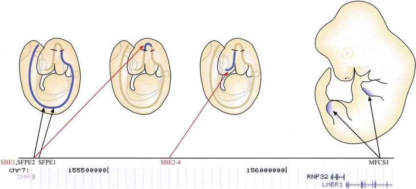
Downloaded from genesdev.cshlp.org on September 22, 2015 - Published by Cold Spring Harbor Laboratory Press
Hedgehog
al. 2006; Varjosalo et al. 2006). This suggests that the
mechanisms of Hh signal transduction from the receptor
to the Ci/GLI transcription factors have evolved differ-
entially after separation of the vertebrate and inverte-
brate lineages approximately 1 billion years ago (Hedges
2002; Varjosalo and Taipale 2007).
Developmental functions and expression
of mammalian Hh proteins
The Hh proteins act as morphogens controlling multiple
different developmental processes (Fig. 2). All mamma-
lian Hh proteins are thought to have similar physiologi-
cal effects—the differences in their roles in development
result from diverse pattern of expression (McMahon et
al. 2003; Sagai et al. 2005).
Dhh expression is largely restricted to gonads, includ-
ing sertoli cells of testis and granulosa cells of ovaries
(Bitgood et al. 1996; Yao et al. 2002; Wijgerde et al. 2005).
Consistent with its expression in a very narrow tissue
range, Dhh-deficient mice do not show notable pheno-
types is most tissues and are viable. However, males are
infertile due to complete absence of mature sperm (Bit-
good et al. 1996).
Ihh is specifically expressed in a limited number of
tissues, including primitive endoderm (Dyer et al. 2001),
gut (van den Brink 2007), and prehypertrophic chondro-
cytes in the growth plates of bones (Vortkamp et al.
1996; St-Jacques et al. 1999). Approximately 50% of
Ihh−/− embryos die during early embryogenesis due to
poor development of yolk-sac vasculature. Surviving em-
bryos display cortical bone defects as well as aberrant
chondrocyte development in the long bones (St-Jacques
et al. 1999; Colnot et al. 2005). Homozygous hypomor-
phic mutations of IHH in humans cause acrocapitofemo-
ral dysplasia, a congenital condition characterized by
bone defects and short stature (Hellemans et al. 2003).
Shh is the most broadly expressed mammalian Hh sig-
naling molecule. During early vertebrate embryogenesis,
Figure 1. (A) Phylogram illustrating the evolution of the Hh
Shh expressed in midline tissues such as the node, no-
proteins. The different Hh proteins were aligned using Prank-
tochord, and floor plate controls patterning of the left–
ster (Loytynoja and Goldman 2005). Hh subgroups are indicated
by a color code, as follows: Dhh (blue), Shh (green), and Ihh (red). right and dorso-ventral axes of the embryo (Sampath et
(B) The central conserved components of the Hh signaling path- al. 1997; Pagan-Westphal and Tabin 1998; Schilling et al.
way and their role in forward signaling. Positively and nega- 1999; Watanabe and Nakamura 2000; Meyer and Roelink
tively acting pathway components are in green and red, respec- 2003). Shh expressed in the zone of polarizing activity
tively. Note that most interactions between components are (ZPA) of the limb bud is also critically involved in pat-
inhibitory. The conserved kinases involved in regulation of Ci/ terning of the distal elements of the limbs (Riddle et al.
GLI processing from activator forms (Ci/GLI-A) to repressor 1993; Chang et al. 1994; Johnson et al. 1994; Marti et al.
forms (Ci/GLI-R) are casein kinases (CKs) 1␣ and 1, glycogen 1995). Later in development, during organogenesis, Shh
synthase kinase-3 (GSK3), and protein kinase A (PKA). (C)
is expressed in and affects development of most epithe-
The four negative (red) and two positive (green) transcriptional
lial tissues (Fig. 2).
feedback loops of the Hh pathway. Ci/GLI-positive feedback to
itself is mediated by GLI1 in mammals. HIP and FoxA2 are only Deletion of Shh leads to cyclopia, and defects in ven-
found in vertebrates, and Engrailed (En) has been characterized tral neural tube, somite, and foregut patterning. Later
as a regulator of Hh only in Drosophila. Both Drosophila and defects include, but are not limited to, severe distal limb
mammalian names of the components are given separated by a malformation, absence of vertebrae and most of the ribs,
slash. and failure of lung branching (Chiang et al. 1996; Liting-
tung et al. 1998; Pepicelli et al. 1998).
Drosophila pathway, the protein kinase Fused (Fu) and The different Hh ligands often act in the same tissues
the atypical kinesin Costal2 (Cos2), appear not to be con- during development, and can function partially redun-
served (Chen et al. 2005; Merchant et al. 2005; Svard et dantly (Fig. 2). For example, Shh and Ihh act together in
GENES & DEVELOPMENT 2455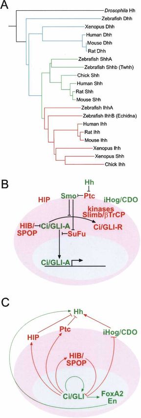
Downloaded from genesdev.cshlp.org on September 22, 2015 - Published by Cold Spring Harbor Laboratory Press

Varjosalo and Taipale

Figure 2. Shh controls mouse development from an embryo to an adult. (Top) The embryo cartoons show aspects of expression of the
Hh target gene patched (blue) during mouse embryonic development. (Bottom) Bars show approximate embryonic stages when Shh,
Ihh, and/or Dhh (color code in bottom left) control developmental processes in the indicated tissues or cell types. The approximate
embryonic stage is indicated by dpc and Theiler stage (TS) (Theiler 1989). References: the role of Hh in early embryogenesis prior to
TS 15 (Chiang et al. 1996; Zhang et al. 2001; Astorga and Carlsson 2007); limb development (Ahn and Joyner 2004); cranial neural crest
(Jeong et al. 2004); cardiac septation (Goddeeris et al. 2008); gastrointestinal system (Madison et al. 2005); bladder (Haraguchi et al.
2007); lung (White et al. 2007); prostate (Berman et al. 2004); pancreas (Hebrok et al. 2000); testis development (Yao et al. 2002); retina
(Sigulinsky et al. 2008); kidney (Hu et al. 2006); hair (St-Jacques et al. 1998; Jeong et al. 2004); taste buds (Miura et al. 2001); ear
(Riccomagno et al. 2002); ovary (Wijgerde et al. 2005; Pangas 2007); tooth (Cobourne et al. 2001, 2004); bone growth (St-Jacques et al.
1999); cerebellum growth (Hatton et al. 2006; Sillitoe and Joyner 2007).

early embryonic development, and their combined loss                       Two independent enhancers—Shh floor plate en-
phenocopies the loss of the Hh receptor component                       hancer 1 (SFPE1) and SFPE2, located at −8 kb and in
Smoothened (Smo), leading to early embryonic lethality                  intron 2, respectively—act to direct reporter expression
due to defects in heart morphogenesis and extraembry-                   exclusively to the floor plate of the hindbrain and spinal
onic vasculogenesis (Zhang et al. 2001; Astorga and                     cord (Epstein et al. 1999). A third element in intron 2,
Carlsson 2007).                                                         Shh brain enhancer 1 (SBE1), directs reporter expression
                                                                        to the ventral midbrain and caudal diencephalon. The
                                                                        more distal elements SBE2, SBE3, and SBE4, which are
Regulatory elements affecting mammalian Hh
                                                                        located >400 kb upstream of the Shh transcription start
expression
                                                                        site (TSS) drive reporter expression in the ventral fore-
Of the mammalian Hh genes, only the mechanisms con-                     brain. The combined activity of these enhancers appears
trolling Shh expression have been studied in detail. The                to cover all regions of Shh transcription along the ante-
expression pattern of Shh is the result of the combined                 rior-posterior axis of the mouse neural tube (Jeong et al.
action of multiple enhancer-elements, which act inde-                   2006).
pendently to control Shh transcription in different tis-                   The enhancer controlling Shh expression in the ZPA
sues and expression domains. Both local-acting and very                 of limb buds, mammals–fish conserved sequence 1
distal elements have been identified (Fig. 3).                          (MFCS1), is located even further upstream of the start

2456      GENES & DEVELOPMENT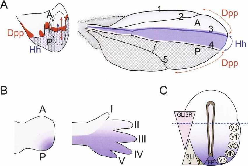
Downloaded from genesdev.cshlp.org on September 22, 2015 - Published by Cold Spring Harbor Laboratory Press

                                                                                                                         Hedgehog

Figure 3. Regulation of mammalian Shh gene expression. (Top) Enhancer-elements driving expression of the mouse Shh gene in
different neural domains (left) and in posterior margin of the embryonic limb buds (right). Approximate expression domains of the
elements are indicated by blue color. Black lines perpendicular to the neural tube indicate zona limitans intrathalamica (ZLI) and
midbrain–hindbrain junction. (Bottom) Known genes in the ∼1 Mb genomic region upstream of the human Shh gene (University of
California at Santa Cruz genome browser, assembly 36). Note that only one transcriptional start site of another gene appears to be
between the most distal conserved Shh enhancer (MFCS1) and the Shh gene itself.

site, at −1 Mb in intron 5 of the Lmbr1 gene (Sharpe et al.         activity is controlled by conserved transcriptional regu-
1999; Lettice et al. 2003; Sagai et al. 2004). This element         lators whose DNA-binding specificity is currently not
is the only enhancer in Shh that has been analyzed also             known.
by loss-of-function studies (Sagai et al. 2005), which con-
clusively demonstrate that MFCS1 is necessary for Shh
expression in mouse ZPA. Consistently in humans,
                                                                    Hh processing and secretion
germline mutations within the conserved MFCS1 ele-
ment cause congenital limb malformations character-                 After translation, Hh undergoes multiple processing
ized by preaxial polydactyly (Lettice et al. 2003). Inter-          steps that are required for generation and release of the
estingly, the MFCS1 sequence is not conserved in limb-              active ligand from the producing cell. The mechanisms
less vertebrates such as snake, limbless lizard, and newt           involved in Hh processing and secretion are evolution-
(Sagai et al. 2005). Although the SBE2–4 and MFCS1 el-              arily conserved (see Burke et al. 1999; Amanai and Jiang
ements are physically far from Shh, the TSS of the region           2001; Chamoun et al. 2001; Ingham and McMahon 2001;
upstream of Shh contains very few genes, and only one               Caspary et al. 2002; Dai et al. 2002; Ma et al. 2002).
well-described TSS exists between the MFCS1 and the                    After the signal sequence is removed, the Hh molecule
TSS of Shh (Fig. 3). Given the diverse expression pattern           undergoes a cleavage catalyzed by its own C-terminal
of Shh, it is likely that a number of other enhancer-ele-           domain that occurs between conserved glycine and cys-
ments remain to be identified in this “gene-poor” region.           teine residues (Fig. 4; Lee et al. 1994; Porter et al. 1996).
   Although many enhancers that drive Shh expression                First, the peptide bond between these residues is rear-
have been identified, very little is known about the spe-           ranged to form a thioester. Subsequently, a hydroxyl-
cific transcription factors that control their activity. The        oxygen of cholesterol attacks the carbonyl of the thioes-
temporal and spatial expression pattern of FoxA2 sug-               ter, displacing the sulfur and cleaving the Hh protein
gests that it could induce Shh expression (Chang et al.             into two parts, a C-terminal processing domain with no
1997; Epstein et al. 1999) in the midline. Consistently,            known signaling activity and an N-terminal Hh signal-
conserved binding sites for FoxA2 and Nkx6 are required             ing domain (HhN) of ∼19 kDa that contains an ester-
for SFPE2 activity (Jeong and Epstein 2003). The Nkx2.1             linked cholesterol at its C terminus (Porter et al. 1996).
homeodomain protein has also been suggested as a likely             The cholesterol modification results in the association
candidate regulating Shh expression in ventral forebrain            of HhN with the plasma membrane. Subsequently, a pal-
(Jeong et al. 2006).                                                mitic acid moiety (Pepinsky et al. 1998) that is required
   No known consensus binding sites for transcription               for HhN activity is added to N terminus of Hh by the
factors are affected by the mutations in the MFCS1 limb             acyltransferase Skinny hedgehog (Ski, HHAT in humans)
enhancer, and the mutations are not clustered close to-             (Chamoun et al. 2001; Lee et al. 2001; Buglino and Resh
gether. However, the severity of the polydactyly pheno-             2008). The resulting fully active HhN signaling molecule
type correlates negatively with the conservation of                 is thus modified by cholesterol at its C terminus and
nucleotide at the mutation sites, suggesting that MFCS1             palmitate at its N terminus (Chamoun et al. 2001; Lee

                                                                                                GENES & DEVELOPMENT          2457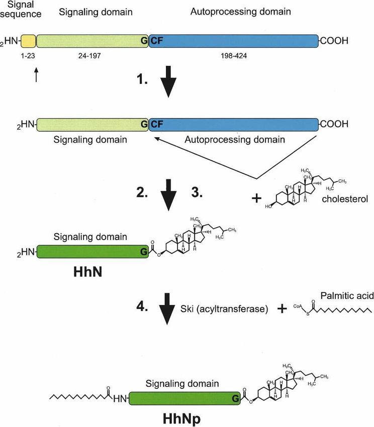
Downloaded from genesdev.cshlp.org on September 22, 2015 - Published by Cold Spring Harbor Laboratory Press

Varjosalo and Taipale

                                                                    transported across a field of cells lacking the heparan
                                                                    sulfate synthesizing enzymes of the EXT/tout velu (ttv)/
                                                                    brother of tout velu (botv)/sister of tout velu (sotv) fam-
                                                                    ily (Bellaiche et al. 1998; Lin et al. 2000; Bornemann et
                                                                    al. 2004; Han et al. 2004a; Koziel et al. 2004). The sub-
                                                                    strates of ttv involved in this process appear to be the
                                                                    glypicans (glycosylphosphatidylinositol-linked HSPGs)
                                                                    Dally and Dally-like (Han et al. 2004b). Dally and Dally-
                                                                    like also affect Hh signaling by facilitating binding of Hh
                                                                    to cell surfaces (Nakato et al. 1995; Lum et al. 2003a;
                                                                    Han et al. 2004b).
                                                                       Whether Hh is transported as individual molecules or
                                                                    assembled into larger particles prior to transport is not
                                                                    clear. Several lines of evidence support the role of large
                                                                    lipid/protein particles in long-range Hh transport. First,
                                                                    Hh staining of receiving cells displays a punctate pattern
                                                                    (Panakova et al. 2005). In addition, soluble Shh multim-
                                                                    ers that contain lipids and that have strong signaling
                                                                    potency have been described in mammalian cells (Zeng
                                                                    et al. 2001), and it has been reported that Drosophila
                                                                    Hh is transported in lipoprotein particles (Panakova
                                                                    et al. 2005; Callejo et al. 2006). Recent genetic evidence
                                                                    also suggests that Hh may be secreted in two different
                                                                    forms, the first of which diffuses poorly and acts at a
                                                                    short range. The second form is “packaged” for long-
Figure 4. (A) Hedgehog protein maturation. Hh protein under-        range transport, and its formation requires the cytoplas-
goes multiple processing steps: (1) the signal sequence is          mic membrane-scaffolding protein Reggie-1/flotillin-2
cleaved; (2) the C-terminal domain of the Hh polypeptide cata-      (Katanaev et al. 2008).
lyzes an intramolecular cholesteroyl transfer reaction, resulting      Multiple studies have analyzed the role of cholesterol
in (3) the formation of a C-terminally cholesterol-modified N-      modification in Hh transport in vivo, with conflicting
terminal Hh signaling domain (HhN). This causes association of
                                                                    results suggesting that cholesterol either aids or hinders
HhN with membranes, which facilitates the final modification
step 4, the addition of a palmitic acid moiety to the N terminus
                                                                    Hh transport (for example, see Lewis et al. 2001; Dawber
by the acyltransferase Skinny hedgehog, resulting in the forma-     et al. 2005; Gallet et al. 2006; Li et al. 2006). These stud-
tion of dually modified Hh signaling domain (HhNp).                 ies are complicated because the protein expression levels
                                                                    of the different mutant forms of Hh need to be constant
                                                                    in order to rule out dose effects. In addition, interpreta-
and Treisman 2001). For clarity, we refer to this protein           tion of the results is made even more difficult by the fact
as Hh hereafter.                                                    that Hh protein lacking cholesterol modification is
                                                                    soluble, and thus its secretion does not require Dis-
                                                                    patched and it can escape the producing cell without
                                                                    being palmitoylated (Mann and Beachy 2004) and could
Formation of the Hh gradient
                                                                    even become palmitoylated later during transport or at
Although Hh is tightly associated with the plasma mem-              the receiving cell. Thus, genetic experiments alone can-
brane, it is able to act directly over a long range (Roelink        not conclusively determine the role of cholesterol modi-
et al. 1995; Briscoe et al. 2001; Wijgerde et al. 2002). In         fication in Hh activity and transport. In contrast, analy-
both Drosophila and vertebrates, the secretion of Hh                sis of the role of the palmitate modification in Hh trans-
from the producing cell requires the activity of the 12-            port is more straightforward, as palmitoylation can be
span transmembrane protein, Dispatched (Disp). Disp,                selectively prevented either by mutation of Ski, or mu-
like Ptc, belongs to the bacterial RND (Resistance-Nodu-            tation of the palmitoylated N-terminal cysteine of the
lation-Division) family of transport proteins. Loss of              Hh proteins. Such experiments indicate that palmitoyla-
Disp leads to accumulation of Hh in the producing cells             tion is required for Hh activity in Drosophila (Burke et
and failure of long-range signaling (Burke et al. 1999; Ma          al. 1999), and for generation of soluble multimeric Hh
et al. 2002).                                                       protein complexes and long-range signaling in verte-
   Distances over which Hh has been shown to act are                brates (Chen et al. 2004).
∼50 µm in Drosophila wing imaginal disc and ∼300 µm                    Several mechanisms are used to control the shape and
in vertebrate limb bud (Zhu and Scott 2004). How Hh                 size of the Hh gradient (for review, see Teleman et al.
moves over a such a long distance is still not clear, and           2001). Very high levels of Hh can induce Hh expression
could involve passive diffusion, active transport, and/or           in responding cells both in Drosophila and in mammals
transcytosis. Genetic evidence points to a role of heparan          (Tabata et al. 1992; Roelink et al. 1995; Methot and
sulfate proteoglycans in this process, as Hh cannot be              Basler 1999). This increases the local concentration of

2458      GENES & DEVELOPMENT
Downloaded from genesdev.cshlp.org on September 22, 2015 - Published by Cold Spring Harbor Laboratory Press

                                                                                                                        Hedgehog

Hh near the original source. Hh also induces the expres-           results in complete activation of the Hh pathway (Good-
sion of its receptor Ptc, which internalizes Hh and tar-           rich et al. 1997), suggesting that Ptc is the functional
gets it to the lysosomes for degradation (Chen and Struhl          ortholog of Drosophila Ptc. Ptc has been proposed to
1996; Incardona et al. 2000; Gallet and Therond 2005).             function as a permease to affect the transmembrane
This negative feedback loop restricts the spreading of the         movement and/or concentration of small molecules that
Hh signal through tissues. Vertebrates also have an ad-            then either agonize or antagonize Smo (Taipale et al.
ditional transmembrane protein, Hedgehog-interacting               2002). Supporting this hypothesis, Smo activity can be
protein (HIP), which is also induced by Hh signaling and           modulated by many synthetic small molecules (Chen et
binds to and further reduces the range of movement of              al. 2002b; Frank-Kamenetsky et al. 2002) and natural
Hh (Chuang and McMahon 1999; Jeong and McMahon                     products, including the steroidal alkaloids cyclopamine
2005).                                                             and jervine (Chen et al. 2002a). These compounds were
                                                                   identified by Keeler and Binns (1966) as active ingredi-
                                                                   ents in Veratrum californicum, a plant whose ingestion
Hh signal transduction                                             by sheep led to an outbreak of cyclopia in US midwest in
                                                                   the 1950s. The clue that these compounds antagonize
Hh receptor
                                                                   Shh signaling came from the observation that the still-
In addition to the glypical dally-like, which acts both in         born lambs have a phenotype that is strikingly similar to
Hh transport and as an accessory receptor, the binding of          that of Shh mutant mouse embryos (Chiang et al. 1996).
Hh to responding cells is facilitated by the transmem-                The structural similarity between cyclopamine and
brane proteins Cdo and Boc (iHog and boi in Drosophila)            sterols (Cooper et al. 1998) suggests that endogenous ste-
(Lum et al. 2003a; Tenzen et al. 2006; Yao et al. 2006).           rols might regulate Smo activity. This hypothesis is also
These proteins act positively in the pathway, binding to           supported by genetic evidence, as disruption of embry-
Hh via conserved fibronectin repeats (Yao et al. 2006)             onic cholesterol synthesis leads to developmental mal-
and increasing Hh association with its signaling receptor          formations that strikingly mimic Hh mutants (Kelley et
Ptc (Tenzen et al. 2006; Yao et al. 2006). The expression          al. 1996; Cooper et al. 1998). Oxysterols (Corcoran and
levels of Cdo and Boc are down-regulated in response to            Scott 2006) and vitamin D3 derivatives (Bijlsma et al.
Hh signaling, resulting in yet another negative feedback           2006) have been suggested to be the endogenous metabo-
that limits pathway activity (Fig. 1C).                            lites that modulate Smo activity. Of these, vitamin D3
   In the absence of Hh ligand, Ptc catalytically inhibits         appears to bind to Smo (Bijlsma et al. 2006) based on its
the activity of the seven-transmembrane-span receptor-             ability to compete against binding of labeled cyclopa-
like protein Smo (Taipale et al. 2002). Binding of Hh to           mine (Chen et al. 2002a).
Ptc results in loss of Ptc activity, and consequent acti-             Based on the fact that increased activity of oncogeni-
vation of Smo. Smo then transduces the Hh signal to the            cally activated Smo proteins correlates with their in-
cytoplasm (Stone et al. 1996; Taipale et al. 2002). This           creased resistance to cyclopamine, it was suggested that
general model is based on the genetic observations that            Smo exists in active and inactive conformational states
loss of Hh or Smo cause similar phenotypes, and that Ptc           (Taipale et al. 2000). Similarly, experiments in Dro-
loss results in a phenotype that is similar to strong over-        sophila suggest that dSmo can exist in two conforma-
expression of Hh. Epistasis analyses in turn indicate that         tional states (Zhao et al. 2007). However, the activity of
Ptc acts downstream from Hh and upstream of or parallel            all small molecules found to activate or inhibit Smo ap-
to Smo (Ingham et al. 1991; Alcedo et al. 1996; van den            pear to be specific for vertebrate Smo proteins, suggest-
Heuvel and Ingham 1996). Binding of Hh to Ptc, in turn,            ing that mechanisms of action of Drosophila and mam-
was determined using purified Shh and cultured cells               malian Smo may be different. Stronger evidence for this
overexpressing Ptc (Stone et al. 1996; Fuse et al. 1999).          comes from both structural and functional analyses,
   By inferring the protein levels of ligand-bound and un-         which indicate that Smo C-terminal domain has evolved
bound Ptc from gene expression, Casali and Struhl (2004)           differentially in vertebrates and invertebrates.
suggested that the activity of the pathway depends on                 Several lines of evidence suggest that the cytoplasmic
the ratio between these two forms. However, the fact               components and the mechanism of Hh signal transduc-
that increasing the level of Ptc protein decreases cellular        tion have diverged between Drosophila and mammals.
responsiveness to Hh (see Bailey et al. 2002; Taipale et al.       In the following section, we will first discuss the mecha-
2002) indicates that it is the absolute amount of unli-            nism of intracellular Hh signal transduction in Dro-
ganded Ptc in a cell that controls pathway activity. This          sophila, which is fairly well understood. We will then
mechanism, together with the induction of Ptc by Hh                discuss the evidence suggesting that Drosophila and
results in gradual desensitization of cells to Hh and al-          mammals appear to use different components and
lows cells to accurately interpret the wide range of Hh            mechanisms in transducing the Hh signal between Smo
concentrations present in morphogenetic gradients.                 and the Ci/GLI transcription factors.
   In vertebrates, Ptc exists as two isoforms, Ptc and Ptc2.
Mice deficient in Ptc2 are viable, but develop alopecia
                                                                   Intracellular Hh signaling in Drosophila
and epidermal hypoplasia and have increased tumor in-
cidence in the presence of one mutant allele of Ptc (Lee           In the absence of Hh, Ptc keeps Drosophila Smo in an
et al. 2006; Nieuwenhuis et al. 2006). Loss of Ptc, in turn,       unphosphorylated state. Unphosphorylated Smo is

                                                                                               GENES & DEVELOPMENT          2459
Downloaded from genesdev.cshlp.org on September 22, 2015 - Published by Cold Spring Harbor Laboratory Press
Varjosalo and Taipale
cleared from the cell surface via endocytosis and is de- spond to Hh because Ci levels are post-transcriptionally
graded in lysosomes (Jia et al. 2004; Zhang et al. 2004). down-regulated due to the expression of hib (Hh-induced
After Hh stimulation, Smo is hyperphosphorylated and MATH and BTB protein; SPOP in vertebrates), a protein
its endocytosis and degradation are blocked. Phosphoryla- that acts as a substrate recognition subunit for the Cul3
tion can be mimicked by mutation of the phosphoryla- E3 ubiquitin ligase. In contrast to Slimb-mediated ubiq-
tion sites to negatively charged residues or by mutating uitinylation, which leads to partial Ci degradation, the
adjacent positively charged arginine clusters to alanine. hib/Cul3-mediated ubiquitinylation causes complete
Based on these observations, Zhao et al. (2007) suggested degradation of Ci (L. Zhang et al. 2006). Expression of hib
that phosphorylation neutralizes the positive charge of increases in response to Hh, providing another negative
the dSmo C terminus and induces a conformational feedback mechanism to this pathway (Fig. 1C; Kent et al.
switch in the C-terminal cytoplasmic tail and conse- 2006; Q. Zhang et al. 2006).
quent dimerization or multimerization of dSmo. How
these events lead to activation of downstream signaling
Divergence of pathway components and mechanisms
pathway components is not understood (Zhao et al. 2007).
dSmo C terminus binds directly to the kinesin-like Despite the conservation of the Hh signaling pathway
protein Cos2, which acts as a scaffolding protein, bring- and many of its roles in development between inverte-
ing together multiple cytoplasmic components of the brate and vertebrate species (Ingham and McMahon
pathway (Jia et al. 2003; Lum et al. 2003b; Ogden et al. 2001; Taipale and Beachy 2001), the mechanisms by
2003; Ruel et al. 2003). These include the full-length which Smo regulates the Ci/GLI transcription factors ap-
transcriptional activator form of Ci, CiA (155 kDa) (Rob- pears to be distinct between Drosophila and mammals
bins et al. 1997), and multiple serine–threonine kinases, (Huangfu and Anderson 2006; Varjosalo and Taipale
including a kinase that specifically acts on the Hh patha- 2007).
way, Fused (Fu) (Therond et al. 1996) and the multifunc- The relatively rapid evolution of some components of
tion kinases PKA, GSK3, CKI␣, and CKI (for review, the Hh pathway, including Smo, Cos2, and Fu, is appar-
see Aikin et al. 2008). ent at sequence level. The C-terminal domains of verte-
In the absence of Hh, CiA is hyperphosphorylated by brate Smo proteins are significantly shorter than those of
the combined action of PKA, which acts as a priming invertebrates and lack the main phosphorylation regions
kinase, and GSK3 and the casein kinases, which further described below. In addition, the two mammalian or-
phosphorylate the primed substrate (Fig. 1B). The hyper- thologs of Cos2, Kif27, and Kif7 have none of the unique
phosphorylation promotes recognition of CiA by the sequence characteristics of Cos2 that differentiate Cos2
ubiquitin E3 ligase Slimb (-TrCP in vertebrates) (Jiang from the kinesin family of motor proteins. Based on se-
and Struhl 1998), leading to the generation of a truncated quence, Kif7 and Kif27 appear to be functional molecular
transcriptional repressor form of Ci, CiR (75 kDa) (Y. motors, whereas Cos2 has apparently lost its ability to
Chen et al. 1999; Price and Kalderon 1999, 2002; Wang et bind ATP and function as a motor protein. The closest
al. 1999; Jia et al. 2002, 2005). In addition to promoting mammalian homolog of Drosophila Fu is also highly di-
CiR formation, Cos2 regulates Ci by tethering it to the verged, and significant homology between these proteins
cytoplasm and preventing its nuclear translocation (C.H. can be seen only in the protein kinase domain itself
Chen et al. 1999; G. Wang et al. 2000). (Murone et al. 2000).
In the presence of Hh, Sno accumulates and the bind- Drosophila Smo activation is coupled to the hyper-
ing of Cos2 to Smo prevents conversion of CiA to CiR phosphorylation of 26 serine/threonine residues located
(Hooper 2003; Jia et al. 2003). However, this mechanism within the C-terminal cytoplasmic tail by PKA and CKI
alone is not sufficient to fully activate the pathway, as (Jia et al. 2004; Zhang et al. 2004; Apionishev et al. 2005).
some CiA is still retained in the cytoplasm by another None of these PKA or CKI phosphorylation sites are con-
protein, Supressor of Fused [Su(Fu)] (Pham et al. 1995; served in vertebrate Smo. The vertebrate Smo C termini
Methot and Basler 2000). Genetic evidence from Dro- lacks one of the two known Cos2-binding domains (Jia et
sophila indicates that full activation of the pathway in al. 2003), and the region homologous to the other domain
response to Hh requires the Fu protein kinase, which (Lum et al. 2003b) is dispensable for mouse Smo (mSmo)
blocks the negative influence of Su(Fu) on Ci (Ohlmeyer function (Varjosalo et al. 2006). Drosophila Cos2, or
and Kalderon 1998; Lefers et al. 2001; Lum et al. 2003b). mouse Kif7 or Kif27 do not appear to bind to mSmo or
Upon entering the nucleus, CiA binds to specific se- GLI proteins or affect Shh signaling when overexpressed
quences (Kinzler and Vogelstein 1990; Hallikas et al. in NIH-3T3 cells (Varjosalo et al. 2006). Furthermore,
2006) in promoter and enhancer regions and controls the loss of the Fu protein kinase—which forms a tight com-
transcription of the Hh target gene(s). plex with Cos2 and is required for Hh signaling in Dro-
In Drosophila, cellular responsiveness to Hh is con- sophila—appears not to impair Hh signaling in mice
trolled by modulating the expression of Ci. In the poste- (Chen et al. 2005; Merchant et al. 2005). Taken together,
rior compartment of the wing disc, Hh and its receptor this evidence suggests that the Cos2–Fu complex, which
components are expressed, but target genes are not acti- is centrally important in Drosophila, plays little or no
vated, as Ci mRNA expression is repressed by Engrailed role in mammalian Hh signaling. Instead, it appears that
(Eaton and Kornberg 1990). Cells posterior to the mor- mammalian Hh signaling critically depends on Su(Fu)
phogenetic furrow of Drosophila eye, in turn, fail to re- (Svard et al. 2006)—which has a minor role in Drosophila
2460 GENES & DEVELOPMENTDownloaded from genesdev.cshlp.org on September 22, 2015 - Published by Cold Spring Harbor Laboratory Press
Hedgehog
(Ohlmeyer and Kalderon 1998)—and on several compo- tracellular signal-regulated kinase-1 (MEK-1), Akt, and
nents involved in formation of the primary cilia, which DYRK1 (Mao et al. 2002; Riobo et al. 2006a,b). From our
either do not have Drosophila orthologs or whose or- studies and previous analyses of the Hh pathway, it ap-
thologs appear not to function on the Drosophila Hh pears that Hh does not regulate the activity of any
pathway (Nybakken et al. 2005). known kinase toward a generic substrate. Thus, the
Primary cilium is an organelle that protrudes from the mechanism by which Hh signal is transduced appears
surface of most vertebrate cells. Genetic evidence sug- not to depend on activation of pathway-specific kinases,
gesting a role for primary cilium in mammalian Hh sig- but on regulation of access of substrates to relatively
naling comes from studies that found that mutations of generic multifunctional kinases.
several proteins required for its formation, including In conclusion, the mechanisms of mammalian Hh sig-
Kif3a, Ift88, and Ift172, result in embryonic phenotypes naling have clearly diverged from those of Drosophila.
characteristic of the loss of Shh signaling (Huangfu et al. This suggests that even signal-transduction mechanisms
2003; Park et al. 2006; Caspary et al. 2007; Vierkotten et of conserved signaling pathways have not been “locked”
al. 2007). Subsequent studies have linked these proteins early in evolution, and that they can be subject to evo-
to the processing of the GLI transcription factors (May et lutionary change. The divergence of the Hh pathway—
al. 2005; Caspary et al. 2007). Some experiments suggest arguably the last major signaling pathway to evolve—is
that primary cilium would act as a “signaling center” also relevant to the evolution of multicomponent signal-
where the biochemical events of signal transduction ing pathways. Some pathways, such as the Notch path-
take place. It has been reported that activated mamma- way, where the same protein (Notch) functions as a re-
lian Smo accumulates to primary cilia in response to Shh ceptor and a transcriptional coactivator are relatively
treatment (Corbit et al. 2005); in the absence of Shh, this simple and consist of a small number of pathway-spe-
accumulation is prevented by Ptc (Rohatgi et al. 2007). cific components (Artavanis-Tsakonas et al. 1999; Pires-
Other components involved in Hh signaling, including daSilva and Sommer 2003). Other pathways, such as the
Su(Fu) and unprocessed GLI proteins, have also been lo- Hh signaling pathway in Drosophila are more complex.
calized to the primary cilium (Haycraft et al. 2005). In addition to many multifunctional proteins, the Hh
Drosophila lacking centrioles, and all microtubule- pathway consists of 11 known specific components: Hh,
based structures derived from them, including centro- Skinny hedgehog (Ski), Dispatched, iHog/boi, Ptc, Smo,
somes, cilia, and flagella develop almost normally, indi- Cos2, Fu, Su(Fu), and Ci (Burke et al. 1999; Chamoun et
cating that cilia are not required for Drosophila Hh sig- al. 2001; Lum and Beachy 2004). The emergence of the
naling (Basto et al. 2006). In contrast, the genetic studies Cos2–Fu system in invertebrates suggests that such mul-
described above have clearly established that mamma- ticomponent pathways may evolve by insertion of novel
lian Hh signaling depends on a process that requires proteins between existing pathway components.
components involved in formation of primary cilia.
However, this evidence is also consistent with a model
Regulation of GLI activity
where some other microtubule-linked process that is
critical for Hh signaling is disrupted by loss of these pro- In contrast to the differences in signaling between Smo
teins. In addition, the fraction of cellular Hh pathway and GLI, the activities of the GLI proteins themselves
components found in the primary cilium at any given are regulated similarly to Ci—with the added complexity
time appears small. Thus, it remains to be established that the activator and repressor functions of Ci are di-
what role cilia play in mammalian Hh signaling and vided in mammals to three GLI proteins, GLI1–3 (Jacob
whether localization of the pathway components to cilia and Briscoe 2003; Ruiz i Altaba et al. 2007). GLI1 and
is required for signaling. GLI2 are responsible for most activator functions and
The lack of effect of the closest mammalian homolog have similar activities at protein level (Bai and Joyner
of Drosophila Fused on Hh signaling suggests that 2001). Whereas loss of GLI2 is embryonic lethal (Mo et
other—mammalian-specific—kinases act on this path- al. 1997; Ding et al. 1998; Matise et al. 1998), GLI1 is
way. We recently identified two such kinases, DYRK2 dispensable for normal development (Park et al. 2000).
and MAP3K10, which are required for Shh signaling in GLI1 expression is induced by Hh ligands, and its func-
NIH-3T3 cells (Varjosalo et al. 2008). Of these, DYRK2 tion appears to be primarily to provide positive feedback
directly phosphorylates GLI2 and GLI3 and induces their and to prolong cellular responses to Hh. GLI3, in turn,
degradation. MAP3K10, in turn, appears to act in a more functions primarily as a repressor (B. Wang et al. 2000;
indirect fashion, binding to and phosphorylating mul- Litingtung et al. 2002), and its loss or mutation leads to
tiple other proteins that regulate the Hh pathway, in- limb malformations in mice and humans (Vortkamp et
cluding GSK3, DYRK2, and Kif3a (Nagata et al. 1998; al. 1991; Schimmang et al. 1992).
Varjosalo et al. 2008). Because of the many connections GLI activity appears to be regulated by Hh in a way
of MAP3K10 to different pathway components, its that is very similar to that observed in Drosophila. In the
mechanism of action is likely to be complex, and re- absence of Hh, GLI3 is phosphorylated, recognized by
quires further study. In addition to DYRK2 and -TrCP, and proteolytically processed to a truncated re-
MAP3K10, it has been reported that other vertebrate- pressor form (B. Wang et al. 2000; Pan et al. 2006).
specific kinases regulate Shh signaling. These include Whether similar processing of GLI2 results in complete
protein kinase C-␦ (PKC␦), mitogen-activated protein/ex- degradation or generation of a truncated repressor form is
GENES & DEVELOPMENT 2461Downloaded from genesdev.cshlp.org on September 22, 2015 - Published by Cold Spring Harbor Laboratory Press
Varjosalo and Taipale
unclear (Pan et al. 2006; Wang and Li 2006). Addition of
Shh leads to inhibition of processing and accumulation
of full-length forms of both GLI2 and GLI3.
Dose-, time-, and context-dependent responses to Hh
The developmental processes that the Drosophila and
vertebrate Hh signaling pathways regulate appear re-
markably conserved (Ingham and McMahon 2001). At
the cellular level, the effects of Hh range from growth
and self-renewal to cell survival (Liu et al. 1998; Rowitch
et al. 1999), differentiation, and/or migration. During
embryogenesis, the Hh cascade is used repeatedly and in
different tissues to induce a large number of develop-
mental processes. The ability of a single morphogen to
affect almost every part of the vertebrate body plan is Figure 5. (A) Hh acts both directly and indirectly to pattern the
made possible by the fact that cellular responses to Hh Drosophila wing imaginal disc. (Left) Hh activates decapen-
depend on the type of responding cell, the dose of Hh taplegic (dpp; red) at the anterior side of the A–P boundary of the
received, and the time the cell is exposed to Hh (see imaginal disc, which diffuses into and patterns both A and P
compartments (red arrow). Hh (blue) also acts directly to pattern
below). At the molecular level, the diverse cellular re-
the anterior compartment close to the A–P boundary. (Right)
sponses are effected by induction of different sets of tar- Adult wing showing the regions derived from the anterior (A,
get genes. Among the genes regulated tissue specifically top) and posterior compartment (P, bottom, shaded), and the
by Hh signaling are those encoding other secreted signal- regions patterned by Dpp (red arrows) and Hh (blue color, be-
ing proteins, including bone morphogenetic protein 4 tween wing veins 3 and 4). (B) Shh has a similar role in anterior-
(BMP4) (Astorga and Carlsson 2007), fibroblast growth posterior patterning of the distal elements of vertebrate limbs
factor 4 (FGF4) (Laufer et al. 1994), and vascular endo- and in specifying digit identity (roman numerals). (C) Time and
thelial growth factor (VEGF)-A (Pola et al. 2001), genes dose-dependent action of Shh. The gradient of Shh (blue color)
involved in cell growth and division (e.g., N-Myc) (Oliver emanating from the notochord (not shown) and floor plate (FP)
et al. 2003), and many transcription factors that are es- progressively defines five different neuronal subtypes in the
ventral neural tube. The Shh protein gradient is converted to
sential for animal development, including members of
gradient of GLI activities shown on the left. GLI1 and GLI2
the Myod/Myf, Pax, Nkx, Dbx, and Irx families (Pierani (bottom) act as transcriptional activators, whereas GLI3 func-
et al. 1999; Gustafsson et al. 2002; Jacob and Briscoe tions as a repressor (GLI3R, top). (MN) Motoneuron; (V0–V3)
2003; Vokes et al. 2007). The total number of genes that interneurons. Dotted line indicates the dorsal limit of the do-
Hh regulates is only beginning to be discovered: A num- main patterned by the Shh gradient. Data adapted from Fuccillo
ber of expression profiling studies have identified several et al. (2006).
novel target genes (for example, see Xu et al. 2006; Vokes
et al. 2007), and our genome-wide in silico analyses iden-
tified 42 conserved enhancer modules with two or more ments and controls the growth and patterning of the en-
GLI sites in the human genome (Hallikas et al. 2006). tire wing. Dpp expression is normally repressed by CiR,
The genes that are induced by Hh in many tissues, in and its activation only requires that this repression is
turn, are generally involved in positive and negative lifted. Therefore, very low levels of Hh suffice to induce
feedback to the pathway itself and include Hib, GLI1, dpp expression (Methot and Basler 1999). The short and
Ptc, and HIP (Fig. 1C). As Ci and the GLI proteins act as intermediate range effects of Hh require induction of tar-
repressors in the absence of Hh and activators in its pres- get genes such as collier (col) and engrailed (en), whose
ence, many of the target genes also behave similarly, expression require CiA function and higher levels of Hh
being repressed in the absence of Hh and induced in its (Methot and Basler 1999; Hooper 2003).
presence. Shh has an analogous role in controlling vertebrate
limb patterning. Shh expressed by the ZPA located at the
Hh acts both directly and indirectly to pattern tissues posterior margin of developing limb buds diffuses to ad-
jacent tissues, forming a temporal and spatial gradient
During the development of the Drosophila wing imagi- that specifies the anterior–posterior pattern of the limbs
nal disc, posterior (P) compartment cells express and se- (Fig. 5B).
crete the Hh protein (Fig. 5A). The secreted Hh then
induces the expression of target genes in cells located in
the anterior (A) compartment. Hh acts both directly at
Time and dose dependency of the Hh response
intermediate range to pattern the anterior wing tissues
close to the A–P boundary and indirectly over long range The effect of Hh dose on target tissue responses is best
by inducing the BMP family morphogen decapentaplegic characterized in the specification of cell identities in the
(dpp) (Basler and Struhl 1994; Tabata and Kornberg 1994). ventral neural tube (Jessell 2000; Patten and Placzek
Dpp diffuses bidirectionally into both A and P compart- 2000; Marti and Bovolenta 2002). During neural tube de-
2462 GENES & DEVELOPMENTDownloaded from genesdev.cshlp.org on September 22, 2015 - Published by Cold Spring Harbor Laboratory Press
Hedgehog
velopment, Shh protein diffuses from the notochord and not interfere with the possible role of Hh as a stem cell
floor plate, creating a concentration gradient across the factor (Berman et al. 2002; Kimura et al. 2008).
ventral neural tube (Fig. 5C). Different doses of Shh The best-characterized role for Hh signaling in adults
within this gradient specify five neuronal subtypes at is in the reproductive system, and Hh proteins are ex-
precise positions along the floor plate–roof plate axis. pressed and required for maturation of the germ cells in
Initially, Shh induces Class II homeodomain (e.g., multiple species. In Drosophila ovary, Hh acts as a so-
Nkx2.2, Nkx6.1) (Pierani et al. 1999; Jacob and Briscoe matic stem cell factor, directly controlling the prolifera-
2003) and represses Class I homeodomain (Pax6, Pax7, tion and maintenance of ovarian somatic stem cells
Irx3, and Dbx1/2) transcription factors. Cross-repressive (Zhang and Kalderon 2001). In mammals, Ihh and Dhh
interactions between these factors then act to sharpen produced by granulosa cells act as paracrine factors to
the expression boundaries and to subsequently direct induce target gene expression in the developing theca
cells to differentiate into specific lineages (Briscoe and cell compartment. This suggests that hedgehog signaling
Ericson 2001). regulates the theca cell development in growing follicles
The activity of Shh as a morphogen was initially (Wijgerde et al. 2005). Dhh also has a role in the regulat-
thought to be due to concentration-dependent responses, ing the development and function of the somatic cells of
but the duration of Shh signal seems also to critically the testis (Bitgood et al. 1996; Yao et al. 2002).
affect cellular responses. Both during neural tube and
limb development, the pattern of cellular differentiation
Aberrant Hh signaling in disease
is controlled not only by the amount but also by the time
of Shh exposure (Briscoe and Ericson 2001; Ahn and Joy- Loss of Hh signaling activity during vertebrate embryo-
ner 2004; Harfe et al. 2004). The changing of the concen- genesis causes severe developmental disorders including
tration or duration of Shh seem to have an equivalent holoprosencephaly, polydactyly, craniofacial defects,
effect on intracellular signaling. and skeletal malformations (Muenke and Beachy 2000;
Chick neural cells convert different concentrations of Hill et al. 2003; McMahon et al. 2003; L. Zhang et al.
Shh into time-limited periods of signal transduction, 2006). It is now also becoming evident that components
such that signal duration is proportional to Shh concen- required for the function of primary cilia are required in
tration (Dessaud et al. 2007). This depends on the gradual mammalian Shh signaling (Huangfu et al. 2003). It is
desensitization of cells to Shh caused by up-regulation of therefore possible that Hh signaling may also be altered
patched (Ptc) (Taipale et al. 2002). Thus, in addition to its in human syndromes caused by defects in cilia, includ-
role in shaping the Shh gradient (Chen and Struhl 1996; ing Meckel, Bardet-Biedl and Kartagener syndromes,
Briscoe et al. 2001; Jeong and McMahon 2005), Ptc par- polycystic kidney disease, and retinal degeneration (Pan
ticipates cell-autonomously in gradient sensing. This et al. 2005; Kyttala et al. 2006).
mechanism integrates Shh signal strength over time, al- On the other hand, aberrant activation of Hh signaling
lowing cells to more accurately determine their distance can cause basal cell carcinoma (BCC, the most common
from the Hh source—resulting in more robust patterning type of skin cancer) (Hahn et al. 1996; Johnson et al.
of the nervous system. 1996), medulloblastoma (a childhood cancer with an in-
variably poor prognosis) (Goodrich et al. 1997; Berman et
al. 2002), and rhabdomyosarcoma (Table 1; Kappler et al.
Role of Hh signaling in young and adult mammals
2004). These tumor types occur at an increased rate in
The multiple roles of Hh signaling in embryonic pattern- patients or mice with germline mutations in Ptc, and
ing are discussed above and reviewed in more detail in sporadic cases are often associated with mutations in the
McMahon et al. (2003). Much less is known about the Hh pathway components Ptc, Smo, or Su(Fu), or more
roles played by Hh in pupal development and in main- rarely, the amplification of GLI1.
taining homeostasis of tissues during adult life. Aberrantly activated Shh signaling has also been sug-
During maturation of mouse pups, Ihh signaling is im- gested to play a role in other cancers, such as glioma,
portant for bone growth. Permanent deletion of Ihh in breast, esophageal, gastric, pancreatic, prostate, and
chondrocytes of mice carrying conditional and inducible small-cell lung carcinoma (see Table 1 for references).
null alleles of Ihh results in permanent defects in bone With the exception of rare GLI1 amplifications found in
growth, inhibiting proliferation and promoting differen- gliomas (Kinzler et al. 1987), the mutational basis of Hh
tiation of chondrocytes, leading to dramatic expansion of pathway activation in these types of cancer has not been
the hypertrophic zone (Razzaque et al. 2005; Maeda et al. ascertained.
2007) and truncation of long bones. Interestingly, similar Multiple lines of evidence suggest that Hh acts to pro-
phenotype was observed with treatment of young mice mote cancer by directly regulating cellular growth and/
with Smo antagonist for just 48 h (Kimura et al. 2008). In or survival. Loss of one ptc allele causes larger body size
adults, Hh pathway controls bone homeostasis; activa- in mice (Goodrich et al. 1997) and in humans (Gorlin
tion of the Hh pathway in osteoblasts leads to bone re- 1987). Several common human single nucleotide poly-
sorption, and conversely, Hh inhibition protects aging morphisms affecting body height map close to Hh path-
mice against bone loss (Mak et al. 2008; Ohba et al. way components, including Ihh, Ptc, and Hip (Lettre et
2008). Adult mice seem to tolerate Hh antagonists well, al. 2008; Weedon et al. 2008), suggesting that individual
suggesting that short-term Hh pathway inhibition might variation in height is determined in part by the strength
GENES & DEVELOPMENT 2463Downloaded from genesdev.cshlp.org on September 22, 2015 - Published by Cold Spring Harbor Laboratory Press
Varjosalo and Taipale
Table 1. Cancers linked to aberrant Shh signaling CGNPs and in medulloblastoma cells, which are
Cancer type References
thought to be derived from CGNPs (Hatton et al. 2006).
Basal cell carcinoma (BCC) (Hahn et al. 1996;
Johnson et al. 1996) Potential for therapeutic intervention
Medulloblastoma (Goodrich et al. 1997;
Berman et al. 2002) As the Hh pathway in BCC and medulloblastoma is of-
Rhabdomyosarcoma (Hahn et al. 1996; ten affected at the level of Ptc or Smo, small molecule
Kappler et al. 2004) antagonists should act at/or downstream from these
Glioma (Kinzler et al. 1987) components (Taipale et al. 2000). Furthermore, several
studies have shown that Smo can be targeted by small
Breast cancer (Kubo et al. 2004) molecule drugs, and that antagonizing Smo could pro-
Esophageal cancer (Berman et al. 2003;
vide a way to interfere with tumorigenesis and tumor
Watkins and Peacock 2004)
Gastric cancer (Berman et al. 2003)
progression. The most commonly used antagonist of the
Pancreatic cancer (Thayer et al. 2003) Hh pathway is the plant alkaloid cyclopamine (Taipale
Prostate cancer (Karhadkar et al. 2004; et al. 2000). Cell-based high-throughput screening has
Sanchez et al. 2004) revealed several distinct classes of antagonists, which,
Small-cell lung cancer (Watkins et al. 2003) like cyclopamine, bind to Smo. These include SANTs
Biliary tract cancer (Berman et al. 2003) 1–4 (Chen et al. 2002b); KAAD-cyclopamine (Taipale et
Bladder cancer (Hamed et al. 2004) al. 2000), compound-5 and compound-Z (Borzillo and
Oral cancer (Nishimaki et al. 2004) Lippa 2005), and Cur-61414 (Frank-Kamenetsky et al.
Mutations in Hh pathway components have been identified in 2002). Although one phase I clinical trial has already
BCC, medulloblastoma, rhabdomyosarcoma, and glioma (top). reported promising results of Hh pathway antagonist in
advanced BCC (Garber 2008), further clinical studies are
needed to establish which of these antagonists are suit-
of negative feedback loops that fine-tune Ihh signaling
able for therapeutic use. As it has been proposed that
during bone growth. Hh pathway controls growth also
autocrine Shh expression is required for growth of some
during embryonic development—transgenic mice that
cancers (Dahmane et al. 1997; Karhadkar et al. 2004), and
overexpress ptc are consistently smaller than control
stromal cell-derived Shh can also activate the Hh path-
mice, but remarkably well proportioned, illustrating that
way in tumors (Becher et al. 2008), it might also be pos-
Hh signaling controls growth in many tissues. However,
sible to treat tumors with Shh-specific monoclonal an-
whether this growth effect is direct or indirectly caused
tibodies. In fact, one such antibody, 5E1, has been shown
by altered placental or vascular development is unclear.
to block the growth of some tumors, including small-cell
In development of midbrain and forebrain, Shh is cru-
lung carcinoma (Watkins et al. 2003). In addition to tar-
cial in driving the rapid, extensive expansion of the early
geting tumors that themselves have hyperactive Hh
brain vesicles. The action of Shh is mediated through
pathways, antagonists of the Hh pathway could also af-
coordination of cell proliferation and survival (Britto et
fect growth of tumors that use Hh ligands to induce an-
al. 2002). In addition, Shh has been implicated in regu-
giogenesis (Pola et al. 2001; Nagase et al. 2008) or recruit
lating cell proliferation and survival in a number of other
other types of stromal cells supporting tumor growth.
cell types, including retinal precursor cells (Jensen and
Further studies are needed to characterize the role that
Wallace 1997), myoblasts (Duprez et al. 1998), optic
Shh plays in such tumor–host interactions.
nerve astrocytes (Wallace and Raff 1999), cerebellar gran-
Because adults can tolerate inhibition of the Hh path-
ule cells (Dahmane and Ruiz i Altaba 1999), and neural
way (Berman et al. 2002; Kimura et al. 2008), specifically
crest cells (Ahlgren and Bronner-Fraser 1999).
blocking Hh signaling offers an effective treatment for
The molecular mechanisms by which Shh controls
the various cancers originating from aberrant Hh path-
growth are beginning to be unraveled. In vitro studies
way activation. However, systemic treatment of pediat-
have shown that the Shh protein up-regulates N-myc
ric tumors such as medulloblastoma may not be feasible
expression in cerebellar granule neuron progenitor
due to the severe effects that transient inhibition of the
(CGNP) cultures and that N-myc overexpression pro-
Hh pathway has on bone growth (Kimura et al. 2008).
motes CGNP proliferation even in the absence of Shh
(Kenney et al. 2003). N-myc is thought to promote pro-
liferation of CGNPs synergistically with cyclins D and E
Perspective
(Knoepfler et al. 2002), whose expression is also regu-
lated by Shh (Duman-Scheel et al. 2002). The Hh signaling pathway was first identified in Dro-
Direct evidence for the role of N-myc in pathway-as- sophila 16 yr ago. Subsequently, it has taken its rightful
sociated cancer comes from a study of Shh pathway-in- place among the major signaling pathways controlling
duced medulloblastoma formation in mice, where it was animal development, being found to regulate the mor-
shown that the disruption of N-myc, but not c-myc, in- phogenesis of a variety of tissues and organs during the
hibits cellular proliferative responses to Shh (Hatton et development of organisms ranging from Drosophila to
al. 2006). This provides in vivo evidence that N-myc human (McMahon et al. 2003). In addition, the Hh path-
plays a central role in Shh-mediated proliferation in way has been linked to multiple forms of human cancer,
2464 GENES & DEVELOPMENTYou can also read