Molecular Mechanism of Immunoglobulin Gene Conversion In Chicken DT40 Cells
←
→
Page content transcription
If your browser does not render page correctly, please read the page content below

Technische Universität München
               GSF - Forschungszentrum für Umwelt und Gesundheit
                        Institut für Molekulare Strahlenbiologie
                    Direktor: apl. Prof. Dr. Jean-Marie Buerstedde
                                          Neuherberg

    Molecular Mechanism of Immunoglobulin Gene Conversion
                              In Chicken DT40 Cells

                                  Huseyin Saribasak

Vollständiger Abdruck der von der Fakultät Wissenschaftszentrum Weihenstephan für
Ernährung, Landnutzung und Umwelt der Technische Universität München zur Erlangung
des akademischen Grades eines

                     Doktors der Naturwissenschaften (Dr. rer. nat.)

genehmigte Dissertation.

              Vorsitzender:                   Univ.- Prof. Dr. Siegfried Scherer
               Prüfer der Dissertation:
                                           1. Univ.- Prof. Dr. Wolfgang Wurst
                                           2. apl. Prof. Dr. Jean-Marie Buerstedde

Die Dissertation wurde am 15.05.2006 bei der Technischen Universität München
eingereicht und durch die Fakultät Wissenschaftszentrum Weihenstephan für Ernährung,
Landnutzung und Umwelt am 04.09.2006 angenommen.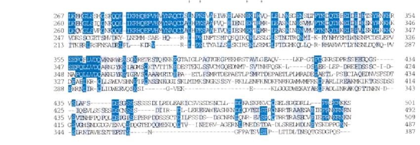
Summary

    Immunoglobulin (Ig) gene conversion is one of three B cell specific processes which
create the repertoire of antigen receptors in B cells. The process involves the unidirectional
transfer of sequences from pseudogene V segments into the rearranged V gene. There are
general and lymphoid-specific trans-acting factors as well as cis-acting elements involved
in this process.
    Gene conversion is most likely initiated by AID mediated cytosine deamination. If the
resulting uracils need to be further processed by uracil glycosylase (UNG), UNG
inactivation should block gene conversion and induce transition mutations. We report in
this thesis that is indeed the phenotype in the B cell line DT40. Ig gene conversion is
almost completely extinguished in the UNG-/- mutant and large numbers of transition
mutations at C/G bases accumulate within the rearranged Ig light chain gene. The mutation
rate of UNG-/- cells is about seven times higher than of pseudo V gene deleted (ψV-) cells
in which mutations arise presumably after uracil excision. In addition, UNG-/- cells show
relatively more mutations upstream and downstream of the VJ segment. This suggests that
hypermutating B cells process AID-induced uracils with approximately one seventh of
uracils giving rise to mutations depending on their position.
    Besides gene conversion DT40 is also a good tool to analyze somatic hypermutation.
Whereas in human and mice all of the nucleotides are target for mutations, in DT40 C/G
bases are the main targets. Proliferating cell nuclear antigen (PCNA) coordinates DNA
synthesis during replication. Mono-ubiquination of PCNA by RAD18 is induced by DNA
damage and leads to the engagement of translesion DNA polymerases in S. cerevisiae. In
this thesis we showed that RAD18 is sensitive to DNA damaging agents and somatic
mutations are reduced in the absence of RAD18. The results demonstrate that PCNA
ubiquitination by RAD18 as well as by some other factors in vertebrate cells are needed,
most likely by recruitment of error-prone translesion polymerases.

                                             ii
Zusammenfassung

   Immunglobulin (Ig) Genkonversion ist einer der drei B-Zellspezifischen Prozesse,
welche für die große Variabilität der Antigenrezeptoren in den B-Zellen verantwortlich
sind. Maßgebend für den Prozess ist der unidirektionale Transfer von Sequenzen eines
strangaufwärts gelegenen V-Segment-Pseudogens in das exprimierte, umgebaute V-
Region-Gen. Der Prozess läuft unter der Beteiligung allgemeiner und lymphoid-
spezifischer trans-wirkender Faktoren, sowie cis-wirkender Elemente ab.
   Genkonversion wird wahrscheinlich durch die AID-Vermittelte Desaminierung von
Cytosin eingeleitet. Wird das resultierende Uracil im weiteren Verlauf durch Uracil
Glykosylase (UNG) modifiziert, sollte die Inaktivierung von UNG die Genkonversion
stoppen und Transition-Mutationen einführen. In dieser Doktorarbeit konnte nachgewiesen
werden, dass in einer UNG-/- Mutante der B-Zelllinie DT40 die Genkonversion fast
komplett zum Erliegen kommt und sich Transitionen an C/G Basen im umgebauten Ig Gen
der leichten Kette häufen. Die Mutationsrate von UNG-/- Zellen ist ungefähr siebenmal
höher als in Zellen mit einer Deletion der V-Pseudogene (ψV-), bei welchen Mutationen
vermutlich nach dem Ausschneiden des Uracils auftreten. Darüber hinaus zeigen UNG-/-
Zellen relativ mehr Mutationen stromaufwärts und stromabwärts des VJ-Segments. Das
weist darauf hin, dass in Hypermutierenden B-Zellen etwa ein Siebtel des durch AID
eingeführten Uracils in Abhängigkeit von seiner Position zu Mutationen führt.
   Neben der Genkonversion lässt sich in DT40 auch die somatische Hypermutation
untersuchen. Während beim Menschen und in der Maus alle Nukleotide Ziel für
Mutationen sind, werden in DT40 hauptsächlich C/G Basen mutiert. „Proliferating cell
nuclear antigen“ (PCNA) koordiniert die DNA-Synthese während der Replikation. Mono-
Ubiquitinierung von PCNA durch RAD18 wird durch Schädigung der DNA eingeleitet und
führt zur Rekrutierung der Translesion DNA Polymerasen in S. cerevisiae. In der
vorliegenden Arbeit haben wir gezeigt, dass RAD18 sensitiv für DNA schädigende
Agenzien ist und somatische Mutationen in Abwesenheit von RAD18 abnehmen. Die
Ergebnisse veranschaulichen, dass die Ubiquitinierung von PCNA durch RAD18 und
einige andere Faktoren in Zellen von Wirbeltieren für die somatische Hypermutation
notwendig ist, wahrscheinlich weil es Error-Prone Translesion Polymerasen rekrutiert.

                                           iii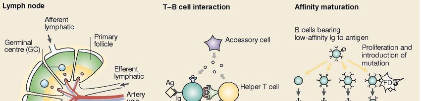
Abbreviations:

AID              Activation Induced Cytidine Deaminase
AP               Apurinic/ apyrimidinic
APOBEC-1         Apolipoprotein B mRNA Editing Catalytic Polypeptide 1
BDT              Big Dye Terminator
BSR              Blasticidine S Resitance gene
CIP              Calf Intestine Phosphatase
CSR              Class Switch Recombination
C region         Immunoglobulin Constant region
dNTP             Deoxynucleotide Triple Phosphate
DMSO             Dimethyl sulfoxide
D region         Immunoglobulin Diversity region
DSB              Double Strand Break
EDTA             Ethylene di-Amine Tetra Acetic Acid
EF               Elongation Factor
EST              Expressed Sequence Tag
EtBr             Ethidium Bromide
FBS              Fetal Bovine Serum
GC               Gene Conversion
GPT              Guanine Phosphoribosyl Transferase
HR               Homologues Recombination
4-HT             4 Hydroxy Tamoxifen
Ig               Immunoglubulin
IRES             Internal Ribosome Entry Site
J region         Immunoglobulin Joining region
LB               Luria Broth
NHEJ             Non Homologues End Joining

                                          iv
PBS        Phosphate Buffer Saline
pKS (+)    pBluescript
PCR        Polymerase Chain Reaction
Pol        Polimerase
PRR        Post-Replication Repair
SDS        Sodium Dodecyl Sulphate
sIgM       Surface Immunoglobulin M
SHM        Somatic Hypermutation
S region   Switch region
SOB        Hanahan's Broth
TAE        Tris Acetic Acid ETDA
TE         Tris EDTA
TLS        Trans Lesion Synthesis
UNG        Urasil DNA Glycosylase
V region   Immunoglobulin Variable region
2YT        2 x Yeast Extract Tryptone

                             v
Table of Contents

Summary                                                                   ii

Zuzammenfassung                                                           iii

Abbreviations                                                             iv

INTRODUCTION:

I.    Immunity and Generation of Antibody Diversity                        1
      A. V(D)J Recombination                                               1
      B. Somatic Hypermutation                                             2
      C. Class Switch Recombination                                        4
II.   Chicken B-Cell Repertoire Development and Bursal B-Cell Line DT40    5
      A. Immunoglobulin Gene Conversion                                    5
      B. Chicken B Cell Development                                        7
      C. DT40                                                              7
III. Molecular Mechanism of Gene Conversion                                9
      A. Factors involved in initiation                                    9
         1. AID                                                            9
         2. UNG                                                           11
      B. The effect of homologous recombination (HR)                      12
      C. Somatic Hypermutation Besides Gene Conversion in DT40            13
           * Pseudogene (ψV) Knockout                                     13
      D. Translesion Synthesis (TLS)                                      16
IV. Objectives:                                                           18

MATERIALS

I.    Instruments                                                         19
II.  Experimental Kits                                                    19
III. Oligonucleotides                                                     19

                                             vi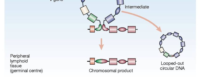
IV. Enzymes                                           21
V.    Immuno-staining antibodies & anti-antibodies    22
VI. DNA Size Marker for electrophoresis               22
VII. Bacterial Strain                                 22
VIII. Mammalian Cell Line                             22
IX. Plasmids                                          22

METHODS

I.    Molecular Biology                               23
      A. Culture of E.coli                            23
      B. E.coli DH5α competent cell preparation       23
      C. Polymerase Chain Reaction (PCR)              25
      D. Analysis of DNA by electrophoresis           25
      E. Purification & Gel Purification of DNA       25
      F. DNA Ligation                                 26
      G. Transformation                               26
      H. Colony PCR                                   26
      I.   Plasmid preparation                        27
      J. Restriction Enzyme Digestion                 28
      K. First strand cDNA synthesis                  28
      L. Determination of DNA and RNA concentration   28
      M. Genomic DNA Isolation                        29
      N. Total RNA Isolation                          29
      O. Cell Extract Preparation                     30
II.   Cell Culture                                    31
      A. Basic Cell Culture Conditions                31
      B. Thawing of the cells                         31
      C. Freeze down of cells                         31
      D. Transfection                                 31
      E. Identifying Targeted Events By PCR           32

                                              vii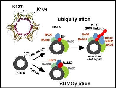
F. Subcloning of DT40 cells                    32
      G. Analysis of IgM expression by FACS          33
      H. Drug Resistance Marker Recycling            33
      I.   Sorting:                                  33
      J. Colony Survial Assay                        34

RESULTS

Section I. UNG                                       35

I.    Genomic Identification of UNG Locus            35
II.   Vector Construction                            35
      A. Knockout Vectors                            35
      B. Complementation Vector                      38
III. Knockout and Complementation of UNG             38
IV. UNG Activity Test                                39
V.    Quantification of Surface IgM Levels           41
      A. Ig Reversion Assay                          41
      B. Ig Mutation Assay                           42
VI. Ig Light Chain Sequencing                        45

Section II. RAD18                                    51

I.    Genomic locus of RAD18                         51
II.   Knockout of RAD18                              52
III. Mutagen Sensitivity Test                        53
IV. Quantification of Surface IgM Levels             54
V.    Ig Light Chain Sequencing                      55

DISCUSSION                                           56

REFERENCES                                           62

Acknowledgements                                     74
Publication List                                     75

                                              viii
INTRODUCTION:
I. Immunity and Generation of Antibody Diversity:
The function of the immune system is to protect vertebrates from foreign substances.
Two types of responses have evolved to accomplish this task: Innate and adaptive
(acquired) immunity. Innate immunity involves hereditary components which provide an
immediate "first-line" of defense to frequently encountered pathogens whereas acquired
immunity involves particular type of cells and proteins which provide a protection to
particular type of agents (Tortora et al., 1995).
A component of the blood, white blood cells are produced in the bone marrow and help
to defend the body against infectious disease and foreign materials as part of the immune
system. Lymphocytes are a type of white blood cells and they are the primary effectors in
the adaptive immune system. There are two broad categories of lymphocytes, namely T
cells and B cells. T cells are chiefly responsible for cell-mediated immunity whereas B cells
are primarily responsible for humoral immunity (antibody related). Antibodies
(immunoglobulins-Ig) are proteins synthesized and secreted by B cells during immune
response within processes like phagocytosis or inflammation. Each antibody recognizes a
specific antigen unique to its target (Janeaway et al., 2001).
To fight against such a variety of invaders, the immune system needs to generate vast
numbers of receptors. To do this, immune system has evolved different strategies like
V(D)J recombination, somatic hypermutation (SHM) and class switch recombination
(CSR).
A. V(D)J Recombination:
As a primary immune repertoire, V(D)J recombination (Fig. 1) involves combinatorial
gene rearrangement of functional immunoglobulin genes (Tonegawa et al, 1983). It is
antigen independent and occurs in the bone marrows. Initiated by lymphoid and sequence
specific recombination activating genes (RAG1 and RAG2) (Schatz et al., 1988; Schatz et
al., 1989), it recruits Non Homologous End Joining (NHEJ) mechanism for resolution
(Chaudhuri et al., 2004). B cells then migrate to secondary lymphoid organs like spleen and
lymph node, where they can undergo further antigen driven Ig gene diversification through
Somatic Hypermutation and Class Switch Recombination.
1B. Somatic Hypermutation (SHM):
Immunoglobulin molecules from the primary immune repertoire generally bind
antigens with only modest affinity and specificity; so a fine-tuning of the antibody response
is necessary for complete recognition and destruction of the antigen (Papavasiliou et al.,
2002). This is provided by another lymphocyte specific process that is known as Somatic
Hypermutation (Fig. 1) (Lederberg, 1959). After Brenner and Milstein proposed a model in
which nucleotide point mutations arise as a consequence of DNA synthesis errors that are
generated during repair of localized breaks (Brenner et al., 1966), Weigert and his
colleagues, were the first to observe hypermutations in a study using mouse λ light chains
at the protein level (Weigert et al., 1970).
Figure 1. Immunoglobulin heavy chain gene locus and generation of antibody
diversity. Solid boxed represent coding regions, and ovals switch regions. In bone
marrow mainly V(D)J recombination occurs as a primary immune repertoire. Further
diversification is introduced in peripheral lymphoid tissues by mecahisms of Somatic
Hypermutation and Class-Switch Recombination (Modified from Kinoshita et al., 2001).
2Somatic Hypermutation is triggered when the surface Ig of B cell encounters with an
antigen. Following this, point mutations are introduced at a high rate specifically into
immunoglobulin V genes of activated B cells (Kinoshita et al., 2001). In the germinal
centers, successive cycle of these mutations and selection lead B cells produce antibodies
with higher affinity which is called as affinity maturation (Fig. 2) (Siskind et al., 1969).
Owing to the strong effects of selection, the true rates of SHM in vivo are unclear. However
it is thought generally that SHM occurs at an extraordinarily high frequency (10-3 bases per
generation). This frequency is almost one million times higher than the level of mutation in
housekeeping genes (Martin et al., 2002). Transition mutations occur more frequently than
transversions (Golding et al., 1987) and using a huge collection of mutation data it is found
out that there is a hotspot motif for SHM (Rogozin et al., 1992). That is, mutations of G-C
base pairs that are in RGYW (R=A or G, Y=C or T, W=A or T) and the complementary
WRCY motifs occur at a high frequency. These mutational properties are probably the
result of the biochemical specificities of the mutator enzymes (Martin et al., 2002).
Figure 2. Immune response in a lymph node. When B cells in the primary follicle are
activated by invading antigens and helper T cells , they begin to proliferate rapidly to form a
germinal center. In germinal centers, T-B cell interaction mainly occurs through T cell
receptor (TCR), processed antigen on major histocompatibillty complex (MHC) on B cells,
and with ligand and receptor of CD40. This interaction activates T cells and accessory cells
to secrete several cytokines to modulate B-cell response. These signals induce the
expression of AID and thereafter SHM and CSR starts. B cells expressing Ig with a high
affinity to given antigens are selected by follicular dendritic cells (FDC), which hold non
processed antigens on the surface. Low affinity immunoglobulins die by apoptosis
(Modified from Kinoshita et al., 2001).
3One of the questions for SHM is how the target genes are selected for mutations. In
several studies, it has been shown that transcription of the target locus is essential for
somatic hypermutation (Peters et al., 1996; Fukita et al., 1998), and that the frequency of
mutation directly correlated to the rate of transcription (Goyenechea et al., 1997; Bachl et
al., 2001). Mutations start downstream of the promoter and lie around 1.5kb to 2 kb along
Ig locus (Lebecque et al., 1990). In addition, Ig enhancer as well as transcription regulatory
elements were also shown to be important for SHM (Betz et al., 1994; Klix et al., 1998;
Michael et al., 2003).
It could well be that SHM start with a single strand (Brenner et al., 1966) or a double
strand DNA breaks (Bross et al., 2000; Papavasiliou et al., 2000) or both (Sale et al., 1998),
although the association of double strand breaks are controversial (Bross et al., 2002; Chua
et al., 2002).
Mutations can arise actively from the error-prone processing of DNA breaks or
passively from the absence of normal repair machineries (Papavasiliou et al., 2002). It has
been documented in many articles that part of the Base Excision Repair and Mismatch
Repair System is involved in SHM (reviewed in Martin et al., 2002; Neuberger et al.,
2005). The proteins shown to be important for SHM up to now are including a lymphoid
specific factor Activation Induced Cytidine Deamniase (AID) (Muramatsu et al., 2000),
Uracil DNA Glycosylase (UNG) (Di Noia et al., 2002; Rada et al., 2002), MSH2 (Rada et
al., 1998; Rada et al., 2004) as well as error-prone polymerases like pol η (Zeng et al.,
2001) and REV1 (Simpson et al. 2003; Jansen et al., 2006).
C. Class Switch Recombination:
Class Switch Recombination (Fig. 1) allows the expression of antibodies that have the
same antigen specificity but are of a secondary isotype like (IgG, IgA, or IgE). The
immunoglobulin locus contains CH genes, which encode proteins that are capable of
different effector functions (Chaudhuri et al., 2004). CSR involves recombination between
switch regions, which are highly repetitive GC-rich sequences of 1-10 kb in length that lie
upstream of Ig C-region genes (Martin et al., 2002). Signal transduction through surface
IgM, CD40 and cytokine receptors of a B cell induce the reaction of CSR and the repair
step is mediated by non-homologues end-joining repair system (Kinoshita et al., 2001).
4Mechanistically, CSR is a deletional recombination event like V(D)J recombination,
however because neither consensus nor homologues sequence is generally found around
junctions, it is different from it. Despite being distinct processes targeted to distinct Ig
regions, CSR and SHM are similar in that they both occur in antigen-simulated B cells,
require transcription as well as AID for initiation (Chaudhuri et al., 2004). The comparison
between SHM and CSR is summarized in Table 1.
Table 1. Comparison between SHM and CSR
SHM CSR
Target V region and its flanks S region and its flanks
Transcription Required Required
DNA Cleavage Required Required
Ig Enhancer Required Dispensable
Repair Phase Translesion Synthesis NHEJ
II. Chicken B-Cell Repertoire Formation and Bursal B-Cell Line DT40:
A. Immunoglobulin Gene Conversion:
Immunoglobulin gene diversification and the ontogeny of B lymphocytes differ species
to species (Reynaud et al., 1994). Although humans and mice develop B lymphocytes in the
bone marrow by V(D)J recombination, in most of the species like chicken, cow, and rabbit
since there is only one single functional V and J segments it is only to ensure the assembly
and expression of a single functional gene (Arakawa et al., 2004). Therefore, the main
diversity as a primary immune repertoire is introduced into the rearranged V(D)J segments
by the mechanism so called gene conversion (GC) in the organ bursa of Fabricious in
chicken (Fig. 3) (Reynaud et al., 1987). It is a kind of homologues recombination event and
it uses pseudo V genes (pseudogene) upstream of the functional V gene. Chicken light
chain includes 25 pseudogenes whereas heavy chain includes 80 pseudogenes. All
pseudogenes lack promoter, leader exon or V(D)J recombination signal sequences. Only a
few of the pseudogenes contain stop codons or frameshift mutations, but quite a few are
5truncated in their 5’ or 3’ ends (Reynaud et al., 1989). Unlike V(D)J recombination or
CSR, after gene conversion pseudogenes do not disappear that is, it is not a cut & paste
mechanism but a copy & paste mechanism (Reynaud et al., 1987; Carlson et al., 1990).
Only the pseudogenes on the same chromosome are used as donors (Carlson et al., 1990)
and pseudogenes that are either more homologous, closer or in the opposite orientation to
the rearranged V segment are preferred (McCormack et al., 1990; Sayegh et al., 1999).
Conversion tracts range from 8 bp to around 200 bp (McCormack et al., 1990). The 5' ends
of the gene conversion tracts always begin in regions of homology between the pseudogene
donor and recipient V segment, whereas the 3' ends can occur in regions of non-homology
and often encompass nucleotide insertions or deletions (McCormack et al., 1990).
Figure 3. Chicken Ig gene diversification. Antibody diversity in chicken mainly
involves gene conversion and somatic hypermutation. Chicken has only a single V and J
segment in the light chain locus which can be functionally rearranged by V(D)J
recombination. The rearranged VJ segment is diversified by segmental gene conversion
using pseudo V genes as donors. Rearranged VJ segments also undergoes somatic
hypermutation in the bursa and spleen.
6B. Chicken B Cell Development:
The bursa of Fabricius plays a central role for chicken B cell development which can be
classified into pre-bursal, bursal and post-bursal stages (Arakawa et al., 2004). In the pre-
bursal stage the first DH-JH joints in the heavy chain locus can be detected in the yolk sac
on embryonic day 5 or 6 and V(D)J recombination at the locus is virtually complete by
embryonic day 15 (Reynaud et al., 1992). Cells which have already completed V(D)J
recombination at the heavy and light chain loci, can be detected in the blood, spleen,
thymus and even in non-lymphoid organs. During the bursal stage, the mature bursa of
Fabricius is composed of approximately 10 000 lymphoid follicles and presents the major
site for B cell proliferation and repertoire formation. Between embryonic days 10 and 15
each follicle is seeded by oligoclonal B stem cells (Pink et al., 1985). Gene conversion in
bursal B cells is initiated around embryonic day 15 even in the absence of environmental
antigen (Mansikka et al., 1990) and most likely it continues until the bursa involuted 4-6
months after hatching. In addition, SHM at low frequency also contributes to bursal Ig gene
diversification (Arakawa et al., 2002a). In the post busal stage adult chicken generate
germinal centers in the spleen in response to antigen. Germinal center formation is clearly
detectable by day 7 after antigen stimulation and begins to wane 14 days after
immunization. The Ig genes of antigen-activated B cells are diversified both by gene
conversion and somatic hypermutation in the very early phase of the germinal center
reaction (Arakawa et al., 1996). In the later stage, gene conversion is down-regulated, and
most modifications are somatic hypermutations. That means antibody maturation in
chicken involves first GC and then subsequently SHM (Arakawa et al., 1998).
C. DT40:
Chicken immunoglobulin gene conversion is difficult to study using primary bursal B
cells, as these cells survive only a short time during in vitro culture (Bezzubova et al.,
1994). It was, however found out that, the ALV induced lymphoma line DT40 (Baba et al.,
1985) seems to be arrested at the stage of bursal B cells and continues Ig light chain gene
conversion during in-vitro cell culture (Buerstedde et al., 1990; Kim et al., 1990).
Although wild-type DT40 cells are dominantly surface (s) IgM(+), spontaneously
arising sIg(-) subclones can be isolated which contain frameshifts in the rearranged light
7chain V segment. Repair of these frameshifts by pseudogene templated gene conversion
events lead to re-expression of sIg (Buerstedde et al., 1990). This reversion from sIg(-)
status to sIg(+) status can be used to quantify gene conversion efficiency by Ig reversion
assay (Arakawa et al., 2004a).
In a remarkable study done by Buerstedde and Takeda in 1991 it was realized that
transfected gene constructs can be integrated into their endogenous loci at higher ratios
(Buersttede et al., 1991). This was surprising, since transfection of mammalian or murine
cell lines leads to integration of the gene constructs pedominantly at random chromosamal
positions (Smithies et al., 1985; Thomas et al., 1986). This efficient gene targeting appears
to reflect an intrinsic character of chicken B cells, which is shared by most analyzed
chicken B cell lines and not by any non-B cell lines (Yamazoe et al., 2004). It is still not
understood whether the increased ratio of gene targeting in chicken B cell lines is related to
gene conversion activity of bursal B cells. It would be highly attractive to find out a factor
which enhances the integration of the recombining substrates (Bezzubova et al., 1994).
Besides efficient targeting, DT40 cells possess a number features that make it elegant.
First, DT40 cells exhibit relatively a stable character in both karyotype and phenotype even
during longer periods of cell culture (Sale et al., 2004). This stable character is a great
advantage over murine ES cells. Thus the analysis of genetic networks on a cellular level
by targeting of multiple genes is possible. Second, since DT40 has a rapid growth rate
(doubling time ~10 h) cell culture assays like colony formation is greatly facilitated. Third,
since the cloning efficiency of wild type cells is nearly 100%, isolation of stably transfected
cells as well as subcloning of cells is easily done (Yamazoe et al., 2004). And lastly, not
only for analysis of Ig gene conversion but many researchers in the fields of B cell antigen
receptor signaling, cell cycle regulation, apoptosis, histon acetylation, homologues
recombination as well as DNA repair nowadays use DT40 efficiently (Winding et al.,
2001).
8III. Molecular Mechanism of Gene Conversion:
The studies published in recent years have shed light on the mechanism of Ig gene
conversion greatly. We can categorize the mechanism into several stages for simplicity:
A. Factors involved in initiation:
1. AID:
Immunoglobulin gene diversification mechanisms SHM, CSR and GC were always
thought to be different until one AID comes out. In 1999, Honjo and his colleagues have
identified a novel protein, Activation Induced Cytidine Deaminase (AID) that is
specifically expressed in CSR active cells (Muramatsu et al., 1999). In one later study, the
same group has shown that not only CSR requires AID but also SHM depends on it in
mouse (Muramatsu et al., 2000). In the same year another study showed that mutations in
AID cause an immunological disorder hyper IgM syndrome due to lack of CSR and SHM
(Revy et al., 2000). These findings have aroused big excitement between the scientists.
Because this was the first evidence that two distinct processes were under the control of one
single protein. Later our group has shown that GC requires AID too (Arakawa et al.,
2002b). That means one AID was enough to unite and rule them all (Fugmann et al., 2002;
Nussenzweig et al., 2004).
AID is a rather small protein (~200 aa) that makes cytidine (C) to uracil (U) change
either on DNA or RNA. At first, it was suggested that AID is a RNA-editing enzyme since
there is a homology between AID and APOBEC-1 (Muramatsu et al., 2000). However,
although still indirect, most experimental evidence support a role of AID in the deamination
of single-stranded DNA in the immunoglobulin V and switch regions but not double-
stranded DNA (Bransteitter et al., 2003; Chaudhuri et al., 2003; Dickerson et al., 2003;
Ramiro et al., 2003; Pham et al., 2003; Peterson-Mahrt et al., 2002). It is under intense
research to identify the target and action of AID. The two models of AID was described in
Fig. 4 for gene conversion.
AID has the ability to mutate all the genes and it must therefore be regulated to avoid
widespread genomic damage. The controls that target AID specifically to Ig genes include
tissue and stage-specific expression, restricted nuclear residence, interactions with specific
factors (Schotz et al., submitted) and accessibility of the target sequence (Barreto et al.,
2005). Therefore AID must be tightly controlled particularly in the nucleus. Several studies
9showed that AID localizes predominantly in the cytoplasm (Rada et al., 2002b; Ito et al.,
2004) and AID has a functional C-terminal nuclear export signal (NES) that actively
exports AID from nucleus (McBride et al., 2004; Ito et al., 2004; Brar et al, 2004) and it has
an nuclear import signal (NLS) in the N-terminus (Ito et al., 2004). Reynaud and colleagues
have reported that some chaperones keep AID in the cytoplasm until B receptor stimulation
actively translocates AID to the nucleus (Reynaud et al. 2003).
In order to identify the minimal requirements of AID function and to understand the
hypermutation activity in non-lymphoid cells, AID was ectopically expressed in fibroblasts
(Yoshikawa et al., 2002) and hybridomas (Martin et al., 2002b). The results showed that
AID is the only B-cell specific factor required to generate hypermutations.
Although there are many factors involved in SHM, CSR and GC identified, only
Replication Protein A (RPA) has shown to be physically interacting with AID (Chaudhuri
et al., 2004). To be able characterize partners of AID, a series of mutant analysis have been
done. These studies showed that C terminal of AID is essential for CSR but not for GC and
SHM (Ta et al., 2003; Barreto et al., 2003). Vice versa N-terminal has been shown to be
important for SHM (Shinkura et al., 2004).
Figure 4. DNA editing and RNA editing models of AID action. In the DNA editing
model, AID changes C to U by DNA deamination. A single strand break is then generated
after uracil base excision by uracil DNA glycosylase (UNG) and APE endonuclease or
through a mismatch repair reaction. In the RNA editing model, AID edits a codon in a
mRNA which encodes a DNA modifying enzyme. This change leads to the translation of
an active protein which then introduces a DNA alteration in the rearranged V segment.
102. UNG:
Uracil may arise as an aberrant base in DNA either through misincorporation of dUMP
opposite adenine during DNA replication (Brynolf et al., 1978; Tye et al., 1978) or by
spontaneous deamination of cytosine residues in DNA generating a U:G mispair (Lindahl,
1993; Nilsen et al., 2000). If left not repaired, these uracils may give rise to transition
mutations C to T or G to A, in a subsequent replication (Duncan et al., 1982; Impellizzeri et
al., 1991) or may result in mutant proteins after transcriptional bypass by RNA polymerase
(Viswanathan et al., 1999).
Uracil residues in DNA are substrates for base excision repair (Lindahl, 2000). In this
repair, uracil DNA glycosylase (UNG - in all organisms) as well as SMUG1, TDG and
MBD4 (in human and mice) are the responsible enzymes to remove uracils from DNA
(Lindahl, 1974; Pearl, 2000). Mammalian UNG gene encodes two isoforms, nuclear
(UNG1) and mitochondrial (UNG2), which are generated via transcription from different
promoters and alternative RNA splicing (Nilsen et al., 1997).
Although UNG has been identified more than 30 years since now, a link between UNG
and Ig gene diversification has never been thought. It was just 4 years ago Neuberger and
his colleagues have published three papers and discovered uracils as the main intermediates
of antibody diversity (Petersen-Mahrt et al., 2002; DiNoia et al., 2002; Rada et al., 2002).
Later, it was also showed in the human patients that are deficient for UNG that CSR is
decreased and SHM is perturbed (Imai et al., 2003). However, direct evidence for the
presence of uracils in DNA that were produced by AID, came just recently in the cells that
are induced to express AID (Martomo et al., 2005).
The DNA deamination model of antibody diversification predicts that recruitment of
AID to the Ig locus causes localized deamination of dC → dU; repair of this uracil in DNA
then leads to SHM, GC or CSR depending on the pathway used for resolving the dU lesion,
as well as on the precise location of the lesion (either the IgV domain or in the vicinity of
the switch region) (Neuberger et al., 2003). In the case for gene conversion (Fig. 4), these
uracils are removed by UNG, and the abasic sites are repaired in a recombination mediated
manner and pseudogenes as donors.
11B. The effect of homologous recombination (HR):
Studies in yeast S. cerevisiae have shown that gene conversion and targeted integration
depends on the RAD52 pathway and these genes mediate the repair of double-strand break
(DSB) repair by homologous recombination (Haber et al., 1999). RAD52 epistasis group
encode proteins which function either in the recognition of the double-strand break
(RAD50, MRE11 and XRS2) or in the promotion of homology search and strand invasion
(RAD51, RAD52, RAD54, RAD55, RAD57) (Symington, 2002).
The mechanism of chicken Ig gene conversion and HR might be related, because both
processes occur at high levels in DT40 and other chicken B-cell lines (Buerstedde et al.,
1990). Analysis of HR in vertebrates was initiated by the discovery of mammalian
orthologs of the yeast recombination gene RAD51 (Shinohara et al., 1993). Homologues of
the RAD51, RAD52 and RAD54 genes were first cloned from chicken bursal cells by
reverse PCR using degenerate primers derived for conservative sequence motifs of the
yeast proteins. Gene disruptions in DT40 revealed that RAD51 deficient cells do not
survive, most likely because they cannot repair replication induced DSBs (Sonoda et al.,
1998). In contrast to the severe recombination and repair defect of the S. cereviseae RAD52
mutants, the DT40 RAD52 deficient cells have only modestly reduced targeted integration
frequencies and are not hyper-sensitive to DNA damage (Yamaguchi-Iwai et al., 1998).
However, RAD54 deficient DT40 cells are highly X-ray sensitive compared to wild type
cells, have 100 fold decreased targeted integration ratios and also show reduced Ig light
chain gene conversion activity (Bezzubova et al., 1997). The disruption of the Nbs1 gene
which is the counterpart of the yeast XRS2 gene produces a phenotype very similar to the
RAD54 phenotype in DT40 (Tauchi et al., 2002). Vertebrates have five RAD51 paralogues
(RAD51B, RAD51C, RAD51D, XRCC2 and XRCC3) and DT40 mutants of each of these
genes have reduced targeted integration frequencies and DSB repair deficiencies (Takata et
al., 2001). Interestingly, some of the functions of the XRCC3 may be compensated by
RAD52, because inactivation of XRCC3 in cells lacking RAD52 results in chromosomal
instability and cell death (Fujimori et al., 2001).
The studies of the RAD52 pathway in DT40 prove that DSB repair is well conserved
during eukaryotic evolution and that targeted integration in vertebrate cells is a side effect
of DSB repair. The high ratios of targeted integration in DT40 most likely reflects the up-
12regulation of the RAD52 pathway in bursal B cells compared to other chicken and
mammalian cells. This increased general homologous recombination activation is required
for efficient Ig gene conversion, but it cannot explain how gene conversion events are
specifically initiated in the Ig loci.
C. Somatic Hypermutation Besides Gene Conversion in DT40:
In wild type DT40 cells besides gene conversion there seems to occur also single point
mutations close to GC tracts. These non templated mutations were first suggested as they
may arise in consequence of errors in recombination (Buersttedde et al., 1991; Reynaud et
al., 1987). However, they were also suggested in several studies that these are not related
with gene conversions (Sale, 2004; Carlson et al., 1990). This led the idea that SHM and
GC might be mechanistically related since both of the processes occur mainly in
immunoglobulin V gene (Reynaud et al., 1987; Maizels, 1995; Weill et al., 1996). Direct
evidence for the relationship between SHM and GC came with the inactivation of the
RAD51 paralogues in DT40 cells. Normally, these proteins involved in HR but knockout
study revealed a reduction in frequency of the GC events along with increase in the
frequency of SHM (Sale et al., 2001). This finding showed exactly that both GC and SHM
can occur in the same cell line and that the initiating events of GC and SHM are related but
they differ in the manner by which most likely U:G mispairs are resolved.
• ψV) Knockout:
Pseudogene (ψ
In order to get further insight into how SHM and GC is being regulated and to get
intermediates of gene conversion reaction, we deleted ψV genes in DT40 (Arakawa et al.,
2004b). The results demonstrated that the deletion of the nearby pseudogene donors
abolishes Ig gene conversion in DT40 and activates a mutation activity which closely
resembles Ig hypermutation (SHM). The features shared between the new activity and
SHM include 1) AID dependence, 2) a predominance of single nucleotide substitutions, 3)
distribution of the mutations within the 5' transcribed region, 4) a preference for hotspots
and 5) Ig gene specificity. The only difference with regard to Ig hypermutation in vivo is
the relative lack of mutations in A/T bases and a predominance of transversion mutations in
the ψV knock-out clones. However, this difference is also seen in
13Figure 5. Ig light chain sequence analysis of the ψV knock-out clones Mutation
profiles of the AIDRψV- and AIDRψVpartial clones. All nucleotide substitutions identified
in different sequences in the region from the leader sequence to the J-C intron are
mapped onto the rearranged light chain sequence present in the AIDR precursor clone.
Mutations of the AIDRψV- and the AIDRψVpartial clones are shown above and below the
reference sequence, respectively. Deletions, insertions and gene conversion events are
also indicated. Hotspot motifs (RGYW and its complement WRCY) are highlighted by
bold letters.
14hypermutating EBV transformed B cell lines (Bachl et al., 1996; Faili et al., 2002) and
DT40 mutants of RAD51-paralogues (Sale et al., 2001) indicating that part of the Ig
hypermutator activity is missing in transformed B cell lines.
The induction of Ig hypermutation by the blockage of Ig gene conversions supports a
simple model explaining how hypermutation and recombination is initiated and regulated
(Fig. 6). At the top of the events is a modification of the rearranged V(D)J segment which
is either directly or indirectly induced by AID. The default processing of this lesion in the
absence of nearby donors or in the absence of high homologous recombination activity
leads to Ig hypermutation in form of a single nucleotide substitution (Fig. 6, right side).
However, if donor sequences are available, processing of the AID induced lesion can be
divided into a stage before strand exchange, when a shift to Ig hypermutation is still
possible and a stage after strand exchange when the commitment toward Ig gene
conversion has been made (Fig. 6, left side). Whereas completion of the first stage requires
the participation of the RAD51 paralogues, the second stage involves other recombination
factors like the RAD54 protein.
Figure 6. A model explaining the regulation of Ig gene conversion and Ig
hypermutation. AID is tightly regulating both GC and SHM tightly in DT40 cells.
15D. Translesion Synthesis (TLS):
DNA damage that stalls replication poses a major threat to genomic stability and
cellular viability. Several strategies of post-replication repair (PRR) have evolved to allow
a cell to tolerate or repair such damage, thereby allowing replication to be completed
(Simpson et al., 2003). These strategies can be broadly divided into two groups: translesion
DNA synthesis and homologues recombination. We tried to explain HR in the previous
section. In this section we will focus on the relationship between TLS and antibody
diversity.
Studies from yeast have shown that TLS as well as error free damage avoidance are
controlled by a complex system known as the RAD6 pathway (Ulrich, 2004). RAD6 itself
encodes a ubiquitin conjugating enzyme (E2) (Jentsch et al., 1987) that is recruited to DNA
by the ubiquitin ligase (E3) RAD18 (Bailly et al., 1997). Ubiquitination by RAD6-RAD18
stimulates PCNA switch from replicative to TLS (Jonsson et al., 1997; Hoege et al., 2002).
PCNA forms a homo-trimeric sliding clamp around the DNA of the replication fork
controlling the access and processivity of replicative polymerases (Fig. 7) (Stelter et al.,
2003). Whereas lysine 164 of yeast PCNA can be modified either by mono- or poly-
ubiquitin or by SUMO, only mono-ubiquitination of PCNA was observed following MMS
treatment or UV irradiation of a human cell line (Hoege et al., 2002; Kannouche et al.,
2004; Watanabe et al., 2004). Mono-ubiquitination of human PCNA requires the human
RAD18 homologue and increases the affinity of PCNA for the translesion DNA
polymerase, Polη (Kannouche et al., 2004; Watanabe et al., 2004) and REV1 (Garg et al.,
2005).
A link between SHM and error-prone repair has been first suggested by Brenner and
Milstein (Brenner at al., 1966). However a possible answer for the question how error
prone repair could induce SHM came just recently with the idea of DNA deamination
model of AID. As we have discussed earlier in this model AID converts C to U within Ig V
gene. The resulting uracils are then recognized either by UNG or mismatch repair system
leading to mutations at G/C and A/T bases, respectively (Di Noia et al., 2002; Rada et al.,
2004). During and after the recognition process or uracils, it seems TLS is somehow
engaged to the system. Several studies about low fidelity polymerases and SHM, done in
recent years showed remarkable results. Polη deficient human and murine B cells (Zeng et
16al., 2001; Delbos et al., 2005; Martomo et al., 2005) and a REV1 disrupted mouse and
DT40 mutant showed an altered spectrum and a decreased frequency of Ig mutations
(Jansen et al., 2006; Simpson et al., 2003). To study TLS in DT40, AIDR ψV- cells are the
best candidates, because these cells diversify their Ig genes mainly by somatic
hypermutation (Arakawa et al., 2004b).
Figure 7. Model for ubiquitination and SUMO modification of PCNA. Two sites for
modifications are amino acids at K127 and K164. DNA-damage induces mono-
ubiquitination of PCNA at K164 which is catalyzed by RAD6 and RAD18. In the
absence of DNA damaging agents PCNA is modified by SUMO in S phase. (Modified
from Hoege et al., 2002)
17IV. Objectives:
Many studies suggest that hypermutation and switch recombination is initiated by AID-
induced cytosine deamination in the rearranged V(D)J segments, and the resulting uracils
are removed by UNG (Muramatsu et al., 2000; Di Noia et al., 2002; Imai et al., 2003). It is
an interesting question whether gene conversion, in analogy to hypermutation and switch
recombination, requires the processing of uracils by UNG. It was reported that expression
of an Ugi transgene in DT40 reduced gene conversion to 30% of the wild-type level (Di
Noia et al., 2004), suggesting that uracil glycosylase activity enhances gene conversion.
However this phenotype remains difficult to interpret, because contrary to expectation no
evidence for an increased mutation rate was found and it was unclear how efficiently and
specifically Ugi transgene expression inhibited UNG activity. Therefore, our first objective
includes disruption of the UNG gene in DT40 and analysis of the phenotype with regard to
gene conversion.
PCNA mono-ubiquitination by RAD18 has been shown to be important for DNA
damage tolerance in yeast (Ulrich et al., 2004). Although it has been reported RAD18 is
dispensable for hypermutation in DT40 (Simpson et al., 2005), we wondered this also holds
true in a background which mainly diversifies V region by hypermutations (Arakawa et al.,
2004b). Thus our second objective includes knockout of RAD18 in DT40 (AIDR ψV-) cells.
As an experimental overview, the targeting constructs were prepared, transfections were
done and phenotypes were analyzed by either Ig reversion and or Ig mutation assay as well
as by sequencing of the immunoglobulin V genes.
18MATERIALS :
I. Instruments:
• PCR machines: GeneAmp PCR System 9700 [Applied Biosystems, CA; USA]
• Centrifuges: Heraeus [Kendro Lab. Products, Osterode, GERMANY]
• Electrophoresis chambers: Horizontal & Vertical [Bio-Rad Lab., CA; USA]
• Gel Visualization: Gel Doc 2000TM [Bio-Rad Lab., California; USA]
• Spectrophotometer: BioPhotometer [Eppendorf GmbH, Hamburg; GERMANY]
• Incubators: Heraeus [Kendro Lab. Products, Osterode; GERMANY]
• Culture Shaker: Innova 4430 [New Brunswick Scientific, Nürtingen; GERMANY]
• Electroporator: Gene Pulse XcellTM [Bio-Rad Lab., CA; USA]
• Vacuum Pump: [Vacuubrand GmbH, Wertheim; GERMANY]
• Vacuum Manifold: [Millipore GmbH, Schwalbach; GERMANY]
• Analytical FACS: BDTM LSRII Flow Cytometer [Becton Dickinson, CA; USA]
• Quantitative FACS: MoFloTM Cell Sorter [Dako Colorado Inc., Colorado; USA]
• Light Microscope: Axiovert 25 [Zeiss, Göttingen; GERMANY]
• Cell Viability Analyzer: Vi-CellTM [Beckman Coulter GmbH, Krefeld; GERMANY]
• Phosphoimager: Fuji, FLA-3000 [Fuji Photo Film Corp., Tokyo; JAPAN]
• DNA Sequencer: ABI 3730 DNA Analyzer [Applied Biosystems & Hitachi; Hitachi
High Tech. Corp., Tokyo; JAPAN.
II. Experimental Kits:
• PCR Purification: [Qiagen GmbH, Hilden; GERMANY]
• Gel Extraction: [Qiagen GmbH, Hilden; GERMANY]
• DNA Ligation: Version 2.1 [Takara Bio Inc., Shiga; JAPAN]
• Plasmid Isolation (Mini): [Qiagen GmbH, Hilden; GERMANY]
• Plasmid Isolation (Maxi): [Qiagen GmbH, Hilden; GERMANY]
• First strand cDNA Synthesis: Super Script IIITM [Invirogen GmbH, Karlsruhe;
GERMAY]
III. Oligonucleotides:
Oligomers (5’ to 3’) used throughout the study were summarized in Table 2.
[Invitrogene GmbH, Karlsruhe; GERMANY].
19Table 2. The primers used in the study.
1. Cloning:
GGG CTC GAG GCT ATG ATC GGG CAG AAG ACG CTG CAT TGC
UR15 XhoI site
UNG
5’ target arm
GGG GGA TCC ACC TCT TAG TTC CTG CGC ATC CGC TCC AGC
UR16 BamHI site
GGG GGA TCC GTC TTC ACT TGG ACA CAG ATG TGC GAC ATC
UR17 BamHI site
UNG
3’ target arm
GGG TCT AGA AAC CGT CTG CAG GAC GTG GTG GCG TTT CCT
UR18 XbaI site
GGG GCT AGC GCC ACC ATG ATC GGG CAG AAG ACG CTG CAT T
UR21 NheI site
UNG
cDNA
GGG AGA TCT GGG CTG TTT GCA CCC ATC AGG CCC CAG AGC
UR22 BglII site
GGG AAG CTT TGG GAA ATA CTG GTG ATA GGT GGA T
VL51 HindIII site
Ig Gene
GGG TCT AGA CCT CTC AGC TTT TTC AGC AGA ATA ACC TCC
VL50 XbaI site
TCC GCC AAA GCT TGG GCT CCT CTC CTC CTG
VL31 HindIII site
Ig Gene
cDNA GGG TCT AGA GTC AGC ACT AGT TCA GTG TCG TGT TT
VL36
XbaI site
EF6 GGG AAG CTT CGG AAG AAA GAA GCT AAA GAC CAT C
HindIII site
EF1α
EF7 GGG ACT AGT AGA AGA GCG TGC TCA CGG GTC TGC C
SpeI site
202. Target Screening:
UR1 CCA GTC CCG CCG CCG CTG CCT TCC CTC CGG
UR7 GAT GTC GCA CAT CTG TGT CCA AGT GAA GAC
UNG
UR23 AGC GTT CCT CCG GGC TTC GGG GAG AGC TGG
UR26 ACA CCG TGT ACC TCT TCC TCT CCT CGG CCA
18RA2 CAG TGA ATG AGG GGA AAG CCT GGC GGA AAG
RAD18 18RA3 AGC AGC AGT TCT CCA GAG GCT GGT AGC AAA
18RA5 TGA AAC CAT GAC CAT TTT GGT TGT GGG TGC
BS1 CGA TTG AAG AAC TCA TTC CAC TCA AAT ATA CCC
Marker Genes
(Bsr & Gpt)
GP1 TGT TGA TAT CCC GCA AGA TAC CTG GAT TGA
3. Sequencing:
VL56 GAC TGG AGG ATC TTA GAG GTC TTT TCC AAC
Ig Gene
VL154 GTG CGT GCG GGG CCG TCA CTG ATT GCC GTT
Universal UC1 AGC GGA TAA CAA TTT CAC ACA GGA
Primers
UC2 CGC CAG GGT TTT CCC AGT CAC GAC
4. UNG activity detection oligomer:
VL207 [FITC] – TCA CCT GCT CCG GGG GTG GCA GUT ACG GCT GGA AGT
TAC TAT TAT GGC TG
IV. Enzymes:
• DNA Polymerases:
Expand Long System [Roche Diagnostics GmbH, Mannheim; GERMANY]
Pfu Ultra Hotstart [Stratagene, California; USA]
• Restriction Enzymes: [New England Biolabs GmbH, Frankfurt; GERMANY]
• Proteinase K: [Qiagen, Hilden; GERMANY]
21• Calf Intestinal Phosphatase: [New England Biolabs GmbH, Frankfurt;
GERMANY]
• RNaseA: [Qiagen, Hilden; GERMANY]
V. Immuno-staining antibodies & anti-antibodies:
In order to quantify the surface IgM on B cells, Mouse Anti Chicken IgM (Clone M-
1) and Goat Anti Mouse IgG (H+L)-RPE Human Adsorbed were used. [Southern
Biotech. Ass. Inc., Biozol, Eching, Germany]
VI. DNA Size Marker:
A DNA marker which was manually prepared from φX174 DNA-HaeIII digest and
λ DNA-HindIII digest was used throughout the study [New England Biolabs GmbH,
Frankfurt; GERMANY]. It was prepared as follows:
λ DNA (500 ng/µl) 400 µl
φX174 DNA (1000 ng/µl) 100 µl
6X Loading buffer 334 µl
MiliQ water 1166 µl
The mixture was incubated for 15 min at 70oC without mixing and then it was taken
into ice for 5 min.
VII. Bacterial Strain:
During cloning of the constructs E. coli-DH5α cells were used as competent cells.
VIII. Mammalian Cell Line:
A chicken B-cell line DT40 was used to transfect constructs.
IX. Plasmids:
UNG: In order to construct vectors for knockout and complementation for UNG study,
pBluescript II KS (+) phagemid vector [Stratagene, California; USA] was used. In this
vector blue/white selection is possible and it has ampicillin resitance gene (ampR).
RAD18: RAD18 targeting vectors were requested from group of Prof. Dr. Shunichi
Takeda, Kyoto University, Japan (Yamashita et al., 2002).
22METHODS :
I. Molecular Biology:
A. Culture of E.coli:
Luria Broth (LB)* agar plates and 2YT Broth* medium were used for culture of E.coli.
The plates were kept at 4°C for about 1 month. For longer storages glycerol stocks were
prepared in a following way: (2 volumes (v.) of cells + 1 v. of 50% glycerol). Stocks
were frozen at -80°C deep freezer.
2YT Broth: The mixture was autoclaved and 1ml ampicillin (250mg/ml) was added to
the mixture before use.
Bacto tryptone 16 g
Yeast extract 10 g
NaCl 5g
10N NaOH 250 µl
Distilled water 1000 ml
LB Agar Plates: The mixture was autoclaved. Before pouring onto petri plates, 1 ml
ampicillin (250mg/ml) and 1 ml 4% X-gal was added to the mixture.
LB Broth Base 20 g
TM
Bacto Agar 15 g
Distilled water 1000 ml
B. E.coli DH5α competent cell preparation:
1) A streak of bacterial strain DH5α was made on a LB agar plate without antibiotics for
single colony isolation. The plate was incubated over night at 37oC.
2) A single colony was picked up and was cultured in 5 ml L broth or SOB broth*
medium at 37oC for over night.
3) 1 ml of SOB broth was taken and the background was measured at OD600.
4) 2.5 ml of overnight culture was transferred into 500 ml of SOB broth which had been
kept at 25oC and bacteria were cultured at 25oC.
5) OD600 was measured every 30 min or every 1 h.
6) When the OD600 was 0.4, the bacterial culture was cooled down immediately by ice
and was kept on ice for 10 min.
7) The culture was transferred into the large centrifuge tubes and was centrifuged at
3000 rpm at 4oC for 10 min.
238) Supernatant was discarded and the pellet was resuspended into 330 ml of ice-cold
FTB*. Vortex should not be used during this step.
9) The culture was kept on ice for 10 min, and was centrifuged at 3000 rpm at 4oC for
10 min.
10) Supernatant was discarded and the pellet was resuspended into 50 ml of ice-cold
FTB. Vortex should not be used. 3.5 ml of DMSO was added, mixed gently.
11) 400 µl of aliquots was dispensed into 1.5 ml tubes. Tubes were then frozen
immediately by liquid nitrogen and the competent cells were kept at -80oC.
12) The efficiency of competent cells was tested by using 1 ng of Bluescript plasmid for
transformation to one competent cell tube (400 µl). After heat shock, 600 ml of L broth
was added to the tube. Before spreading, 10 µl (1/100) and 100 µl (1/10) of bacterial
liquid was taken onto agar plates. The left (89/100) was centrifuged for 1 min at 13000
rpm. Then, the bacteria were spreaded by a loop.
13) The number of colonies was counted. The titer of competent cells was calculated
(Titer of competent cells is the number of colonies which would be produced by
transfection of 1 µg plasmid).
* SOB Broth: MgCl2 and MgSO4 were added after autoclave of mixture.
Bacto tryptone 20 g
Yeast extract 5g
NaCl 0.585 g
KCl 0.l86 g
Distilled water 1000 ml
MgCl2 Final 10 mM
MgSO4 Final 10 mM
* FTB: The pH was adjusted to 6.7 after KCl and MnCl2.4H2O treatments respectively.
0,5 M PIPES 20 ml
CaCl2.2H2O 2.2 g
Distilled Water 1000 ml
KCl 18.64 g
MnCl2.4H2O 10,88 g
24C. Polymerase Chain Reaction (PCR):
For the amplification of genomic DNA, hot start was preferred in order to prevent
unspecific bands. For this reason, upper and lower mixes were prepared separately and
upper mix was added onto lower mix at 80oC after pre-incubation on the machine. In
the case for high fidelity polymerases, since pfu Ultra was inhibited by a taq-directed
antibody no upper and lower mixes were prepared.
Lower Mix: Upper Mix:
DNA 1 – 100 ng 10x Expand buffer 0.5 µl
dNTP mix 0.2 mM Expand System 0.5 U
Primer forward 5 pmol 10X Crezol Red 1 µl
Primer reverse 5 pmol MiliQ water rest
10x Expand buffer 0.5 µl
MiliQ water rest
Total 5 µl Total 5 µl
Amplifications were done by using Long-range PCR protocol as follows:
Pre-denaturation 93oC, 2 min
Denaturation 93oC, 10 sec
Annealing
Extension
65oC, 30 sec
o
68 C, 5 min*
} 35 cycles
* 20 sec. increases after each cycle
Final extension 68oC, 7 min
D. Analysis of DNA by electrophoresis:
Up to the purpose, 0.8% to 2% agarose gel which contains 0.1 µg/ml EtBr was used to
visualize DNA by Gel Doc 2000TM [Bio-Rad Lab., California; USA]. DNA samples
were run for 35 min at 120 V. For gel purification, they were kept around 1 h in
electrophoresis. In the case for UNG activity test 15% TBE-urea polyacrylamide gel
was used and the DNA samples were run at 200 V for 40 min.
E. Purification & Gel Purification of DNA:
After PCR or restriction enzyme digestion, DNA was purified by Qiagen PCR
Purification kit [Qiagen GmbH, Hilden; GERMANY Qiagen] according to
manufacturer’s instructions.
25F. DNA Ligation:
Ligation was done by using a kit [Takara Bio Inc., Shiga; JAPAN] according to
manufacturer. In principle, around 1 volume of vector (100ng/µl), 9 volume of insert
(100ng/µl) and 10 volume of kit were used.
G. Transformation:
A heat shock protocol was preferred during transformation of plasmids into bacterial
cells:
1) The plasmid (>1ng) was added into 50-400 µl of competent cells, depending on the
efficiency of the cells.
2) The tube was incubated on ice for 20 (-30) min and then was heat shocked at 42oC
for 45 sec on a heat block, and was incubated again on ice for 2 min.
3) 2YT Broth was added onto tube until the mixture reaches 1 ml and the tube was
incubated at 37oC for at least 10 min.
4) After mixing, 100 µl (1/10) was taken from bacterial liquid and was put onto half of
LB agar plate. The rest (9 /10) was centrifuged at 13.000 rpm for 1 min, the supernatant
was discarded and the rest (~50 µl) was put onto another half of the plate. The cells
were then spreaded by using L-loop.
7) The plates were incubated overnight at 37oC.
H. Colony PCR:
The ligation of the DNA was first checked by colony PCR. Single bacterial colonies
were taken into U-Bottom 96 well plates which contains 20 µl 2YT Broth. From this, 1
µl was used as a DNA template for PCR.
Mix: PCR Programme:
DNA 1 µl Pre-denaturation 94oC, 1 min
10X Expand buffer 1 µl
Denaturation 94oC, 20 sec
dNTP mix 0.2 mM
Annealing 55oC, 1 min
Primer forward 5 pmol
Primer reverse 5 pmol Extension 72oC, 2 min
Expand System 0.5 U Final extension 72oC, 6 min
10X Crezol red 1 µl
MiliQ water rest *Amplifications were done 30 cycles.
Total 10 µl
26I. Plasmid preparation:
During construction of the vectors for small number of samples, plasmid extraction was
done by using a kit according to manufacturer’s protocol [Qiagen GmbH, Hilden;
GERMANY]. After constructs were confirmed by enzyme digest, a maxi-prep was done
[Qiagen GmbH, Hilden; GERMANY]. For higher number of samples or during cloning
of Ig gene PCR products for sequencing, large-scale miniprep preparation was used by
using deep-well blocks and plates as follows:
1) The bacterial colonies were picked up into a 96 well U-Bottom plate each well
containing 70µl of 2YT Broth. 25µl of each individual sample were transferred into two
96 well blocks containing 1.3 ml 2YT Broth.
2) The blocks were incubated overnight at 37 oC at 220 rpm.
3) The next day, the blocks were centrifuged at 3300 rpm for 10 min.
4) Supernatant was discarded by tapping and 80 µl of Solution-I* was added into each
well. The pellet was resuspended, vortexed and incubated for 1 min.
5) 80 µl of Solution-II* was added and vortexed immediately. The samples were
incubated for an additional 5 min at room temperature.
6) 80 µl Solution-III* was added and vortexed.
7) After this, the deep well block was centrifuged at 3.300 rpm for 5 min. (During this
time Millipore FB plate was placed in the bottom, and NA lysate clearing plate was put
on top of the manifold.
8) 200 µl of supernatants were transferred onto the clearing plate.
9) The vacuum was adjusted to not exceed 8 inches Hg in order to ensure uniform
filtration. The vacuum was applied for 5 min until the lysate is drawn.
10) The FB plate was taken from inside the manifold and placed on top of the empty
manifold. A plastic tray was placed in the bottom of the manifold for waste.
11) 150 µl of Binding Solution* was added to wells and mixed immediately by
pipetting. Vacuum was applied at full strength for 2 min.
12) 200µl of 80% ethanol was added to each well. The vacuum was applied at full
strength for 2 min. The waste accumulated in the plastic tray at the bottom of the
manifold was discarded and the FB plate was centrifuged at 1.000 rpm for 30sec. (This
step was repeated two times)
13) The FB plate was put on top of a new 96-well microtiter plate and 100µl of 1x TE
Buffer was added to each well. The plate was incubated for 5 min and centrifuged at
3500 rpm for 5 min. The plasmid was kept at -20OC.
27Solution-I* Solution-II*
50X TE 1 ml 10 N NaOH 10 ml
Distilled Water 49 ml Distilled Water 500 ml
RNaseA (100mg/ml) 30 µl 20 % SDS 2,5 µl
Solution-III* Binding Solution*
Glacial asetik asit 70 ml Potassium Iodide 100 g
Distilled Water 70 ml Distilled Water 70 ml
5 M Potassium Acetate 360 ml
J. Restriction Enzyme Digestion:
Before ligation: To prepare vector and insert fragments, DNA samples were incubated
at 37oC for at least 3 h. After ligation: DNA samples were incubated for 1 h to check
cloning. In general 1-5 U of enzyme was enough to digest 1µg of DNA in 1 h.
Mix, prior to ligation: Mix, for test digestion:
DNA 3-5 µg DNA 0.5 µg
10X Buffer 4 µl 10X Buffer 2 µl
100X BSA 0.4 µl 100X BSA 0.2 µl
Enzyme 1 (and 2)* 2 (-4) µl Enzyme 1 (and 2)* 0.5 (-1) µl
MiliQ water rest MiliQ water rest
Total 40 µl Total 20 µl
K. First strand cDNA synthesis:
Super Script III (Invitrogen) was used to synthesize first strand cDNA according to
manufacturer’s intructions.
L. Determination of DNA and RNA concentration:
The purity and the concentration of the DNA were checked by a spectrophotometer
BioPhotometer [Eppendorf GmbH, Hamburg; GERMANY]. Generally, the intensity of
absorbance at the wavelength 260 nm was used for calculating concentration and the
ratio of 260/280 nm was used to estimate the purity. For pure DNA samples this value
should be between 1.8, 2.0.
28You can also read


