High-fat diet-activated fatty acid oxidation mediates intestinal stemness and tumorigenicity
←
→
Page content transcription
If your browser does not render page correctly, please read the page content below
Article

High-fat diet-activated fatty acid oxidation mediates
intestinal stemness and tumorigenicity
Graphical abstract                                             Authors
                                                               Miyeko D. Mana, Amanda M. Hussey,
                                                               Constantine N. Tzouanas, ...,
                                                               Maria M. Mihaylova, Alex K. Shalek,
                                                               Ömer H. Yilmaz

                                                               Correspondence
                                                               miyeko.mana@asu.edu (M.D.M.),
                                                               ohyilmaz@mit.edu (Ö.H.Y.)

                                                               In brief
                                                               Mana et al. demonstrate that a high-fat
                                                               diet enhances intestinal stemness and
                                                               tumorigenicity through a PPAR-FAO
                                                               program. The PPAR family members
                                                               d and a redundantly activate a robust FAO
                                                               program in stem cells where loss or
                                                               inhibition of CPT1a (the mitochondrial
                                                               long-chain FAO rate-controlling step)
                                                               dampens these HFD effects.

Highlights
d   HFD augments intestinal stemness through PPARd and
    PPARa

d   A PPAR-FAO program enhances stemness and
    tumorigenicity in a HFD

d   Loss or inhibition of Cpt1a-mediated FAO blunts the HFD-
    enhancing effects in ISCs

d   Early intestinal tumors arising from HFD ISCs are highly
    sensitive to FAO inhibition

           Mana et al., 2021, Cell Reports 35, 109212
           June 8, 2021 ª 2021 The Authors.
           https://doi.org/10.1016/j.celrep.2021.109212                                       ll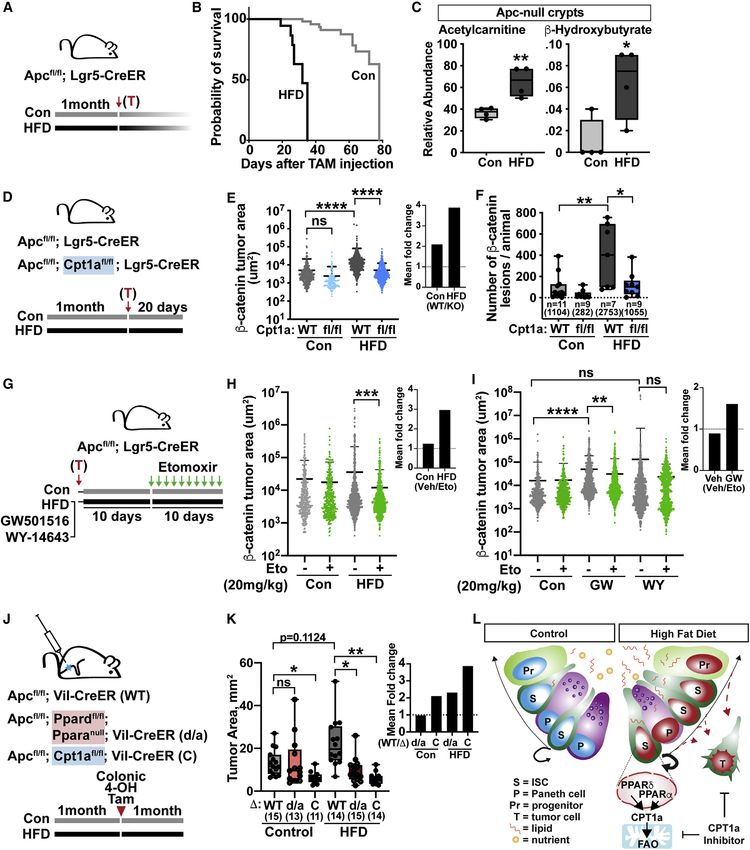
ll
                                                                                                                       OPEN ACCESS

Article
High-fat diet-activated fatty acid oxidation
mediates intestinal stemness and tumorigenicity
Miyeko D. Mana,1,2,* Amanda M. Hussey,1 Constantine N. Tzouanas,3,4,5,6 Shinya Imada,1 Yesenia Barrera Millan,2
Dorukhan Bahceci,1,14 Dominic R. Saiz,2 Anna T. Webb,1,15 Caroline A. Lewis,7 Peter Carmeliet,8,9,10
Maria M. Mihaylova,11,12 Alex K. Shalek,3,4,5,6 and Ömer H. Yilmaz1,4,13,16,*
1Department of Biology, The David H. Koch Institute for Integrative Cancer Research at MIT, Massachusetts Institute of Technology,

Cambridge, MA 02139, USA
2School of Life Sciences, Arizona State University, Tempe, AZ 85287, USA
3Ragon Institute of MGH, MIT, and Harvard, Cambridge, MA 02139, USA
4Broad Institute of MIT and Harvard, Cambridge, MA 02142, USA
5Institute for Medical Engineering & Science, Department of Chemistry, and Koch Institute for Integrative Cancer Research, Massachusetts

Institute of Technology, Cambridge MA 02139, USA
6Program in Health Sciences & Technology, Harvard Medical School, Boston, MA 02115, USA
7Whitehead Institute for Biomedical Research, Cambridge, MA 02142, USA
8Laboratory of Angiogenesis and Vascular Metabolism, Center for Cancer Biology, VIB, and Department of Oncology, Leuven Cancer

Institute, KU Leuven, Leuven 3000, Belgium
9State Key Laboratory of Ophthalmology, Zhongshan Ophthalmic Center, Sun Yat-Sen University, Guangzhou 510060, Guangdong, P.R.

China
10Department of Biomedicine, Aarhus University, Aarhus 8000, Denmark
11Department of Biological Chemistry and Pharmacology, Ohio State University, Columbus, OH 43210, USA
12The Ohio State University Comprehensive Cancer Center, Columbus, OH 43210, USA
13Department of Pathology, Massachusetts General Hospital and Harvard Medical School, Boston, MA 02114, USA
14Present address: Department of Pathology, University of California, San Francisco, San Francisco, CA 94143, USA
15Present address: Department of Cell and Molecular Biology, Karolinska Institutet, Stockholm, Sweden
16Lead contact

*Correspondence: miyeko.mana@asu.edu (M.D.M.), ohyilmaz@mit.edu (Ö.H.Y.)
 https://doi.org/10.1016/j.celrep.2021.109212

SUMMARY

Obesity is an established risk factor for cancer in many tissues. In the mammalian intestine, a pro-obesity
high-fat diet (HFD) promotes regeneration and tumorigenesis by enhancing intestinal stem cell (ISC)
numbers, proliferation, and function. Although PPAR (peroxisome proliferator-activated receptor) nuclear re-
ceptor activity has been proposed to facilitate these effects, their exact role is unclear. Here we find that, in
loss-of-function in vivo models, PPARa and PPARd contribute to the HFD response in ISCs. Mechanistically,
both PPARs do so by robustly inducing a downstream fatty acid oxidation (FAO) metabolic program. Phar-
macologic and genetic disruption of CPT1A (the rate-controlling enzyme of mitochondrial FAO) blunts the
HFD phenotype in ISCs. Furthermore, inhibition of CPT1A dampens the pro-tumorigenic consequences of
a HFD on early tumor incidence and progression. These findings demonstrate that inhibition of a HFD-acti-
vated FAO program creates a therapeutic opportunity to counter the effects of a HFD on ISCs and intestinal
tumorigenesis.

INTRODUCTION                                                           factor that directly affects intestinal regeneration and tumori-
                                                                       genesis. For example, we and others have shown that constit-
Obesity increases the incidence and mortality of many types of         uents of a Western diet (a diet rich in fat and sugar), such as
cancer, including those of the intestine (Calle and Kaaks, 2004;       excess fatty acids (Beyaz et al., 2016; Fu et al., 2019; Park
Lauby-Secretan et al., 2016). Many factors contribute to how           et al., 2016), cholesterol (Wang et al., 2016), high-fructose
obesity elevates colon cancer risk, including changes in sys-          corn syrup (Goncalves et al., 2019), as well as low vitamin D
temic growth factors (Andres et al., 2015; Calle and Kaaks,            (Peregrina et al., 2015), contribute to intestinal tumor formation
2004; Mah et al., 2014), visceral adipose tissue (Huffman              through distinct mechanisms. These different dietary interven-
et al., 2013), the microbiome (Arkan, 2017; Schulz et al.,             tions mimic aspects of obesogenic diets in humans and in-
2014), bile acids (Fu et al., 2019), inflammation (Grivennikov         crease tumorigenesis. Understanding precisely how each one
et al., 2012; Poullis et al., 2004), and dietary constituents (Beyaz   does so will provide novel insights into how early cancers
et al., 2016; Wang et al., 2018; Yang et al., 2005). Diet in partic-   form or whether metabolic programs imposed by such diets
ular stands out as an important environmental and lifestyle            can be exploited therapeutically.

                                                                        Cell Reports 35, 109212, June 8, 2021 ª 2021 The Authors. 1
                     This is an open access article under the CC BY-NC-ND license (http://creativecommons.org/licenses/by-nc-nd/4.0/).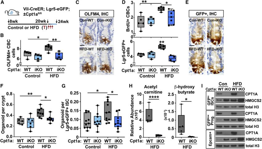
ll
      OPEN ACCESS                                                                                                                    Article

   The intestinal epithelial lining is at the frontline of digestion and   cholesterol trafficking, and enteroendocrine cell function (Daoudi
absorption and is maintained by actively cycling Lgr5+ intestinal          et al., 2011; Oliver et al., 2001; Poirier et al., 2001). In particular,
stem cells (ISCs) that are located at the base of intestinal crypts        CPT1A-mediated FAO is a common metabolic process strongly
(Barker et al., 2007) and respond to dietary cues (Alonso and Yil-         activated by the PPAR family members a and d (Mascaró et al.,
maz, 2018). These Lgr5+ ISCs reside in a supportive microenvi-             1998). We observed previously that HFD-derived or GW
ronment, or niche, comprised of small-intestine Paneth cells               agonist-treated ISCs and progenitors significantly upregulate
(Rodrı́guez-Colman et al., 2017; Sato et al., 2011), colonic               genes involved in FAO (Beyaz et al., 2016); however, the functional
deep secretory cells (Sasaki et al., 2016), stromal immune cells           role of FAO in mediating aspects of the HFD or PPAR response in
(Biton et al., 2018; Lindemans et al., 2015), and mesenchymal              ISCs and tumors that arise from them is unclear. Here we investi-
cell types (Degirmenci et al., 2018; McCarthy et al., 2020;                gate the necessity of Ppard and Ppara and their FAO metabolic
Shoshkes-Carmel et al., 2018) that provide instructive signals             program as drivers of ISC adaptation to a HFD and whether
to stem cells. Lgr5+ ISCs can respond directly or indirectly to            HFD-imposed FAO is a therapeutic vulnerability for the genesis
diet through their niche. For example, low-calorie diets activate          and progression of early intestinal adenomas.
Lgr5+ ISCs indirectly via the Paneth cell niche (Igarashi and Guar-
ente, 2016; Yilmaz et al., 2012) and directly by engaging a fatty          RESULTS
acid oxidation (FAO) program in Lgr5+ ISCs (Mihaylova et al.,
2018). Ketone bodies, which are terminally oxidized products               PPARd and PPARa contribute redundantly to the effects
of FAO, mediate some of the direct effects of diet on ISCs by              of a HFD in ISCs
modulating Notch signaling (Cheng et al., 2019). Thus, diverse             To determine the in vivo role of Ppard in driving ISC adaptation in
strategies exist that enable ISCs to coordinate tissue adaptation          response to HFD feeding (the macronutrient content is listed in
with dietary cues.                                                         Figure S1A), we generated a Ppardfl/fl; Vil-CreER strain to selec-
   We and others recently proposed that a pro-obesity high-fat             tively delete Ppard in a tamoxifen-inducible manner in the intes-
diet (HFD) increases intestinal tumorigenesis by inducing many             tinal epithelium. At 6–7 weeks of age, Ppardfl/fl; Vil-CreER
direct changes in Lgr5+ ISCs, which are the cells of origin for a          (Ppard-iKO) or Ppardfl/fl (wild type [WT]) were treated with tamox-
majority of early dysplasias in the intestine (Barker et al., 2009;        ifen, and then cohorts were placed on a control diet or HFD (Fig-
Beyaz et al., 2016; Mah et al., 2014). These HFD-mediated alter-           ures 1A and S1A). After 24–28 weeks, small intestinal crypts from
ations in Lgr5+ ISCs include expansion of their numbers per                control-fed Ppard-WT and -iKO mice were equally clonogenic in
crypt, higher proliferation, and enhanced regenerative capacity            an organoid assay for stemness. However, although HFD crypts
(Beyaz et al., 2016), and this expansion in the pool of proliferative      were more clonogenic than controls, loss of Ppard led to a
ISCs might contribute to how a HFD elevates cancer risk. We                modest decrease (Figure 1B), indicating that PPARd contributes
also demonstrated mechanistically that ISCs from HFD-fed                   partially to HFD-stimulated crypt clonogenicity (Figures 1B and
mice and organoids exposed exogenously to dietary lipids                   1C; Beyaz et al., 2016). Separately, we used an alternate deletion
engage a peroxisome proliferator-activated receptor (PPAR)                 strategy where Ppard was ablated after the establishment of
transcriptional program and that pharmacologic activation of               HFD-induced obesity (Figures S1B and S1C). Loss of Ppard in
PPARd with a high-affinity agonist, GW501516 (GW), was suffi-              this model had no effect on crypt clonogenicity (Figures S1B
cient to recapitulate many aspects of the HFD ISC phenotype,               and S1C). In contrast to the mild effects of PPARd on HFD ISC
including augmentation of their tumorigenic potential, thereby             activity in organoid assays, long-term Ppard loss prevented
linking lipid sensing with stemness and tumorigenicity.                    expansion and proliferation of OLFM4+ and Lgr5-EGFP+ ISCs
   PPARs include a family of lipid-activated nuclear receptors (a,         in the HFD state and partially restored Paneth cell numbers (Fig-
d, and g) that form obligate heterodimers with the retinoid X re-          ures S1D–S1G). Given that some features of the HFD state, like
ceptor (RXR) (Barak et al., 2002; Krey et al., 1993) and, upon             ISC numbers, but not elevated function, are counteracted by
ligand stimulation, which includes dietary fats or their deriva-           Ppard loss, these data raise the possibility that functional redun-
tives, drive PPAR-specific transcriptional programs (Barish                dancy among PPAR family members in the HFD state may
et al., 2006; Ordentlich et al., 2001). Although our initial study         compensate for loss of Ppard.
(Beyaz et al., 2016) implicated PPARd signaling in the HFD intes-             To explore this possibility in more detail, we assessed how
tinal phenotype, the in vivo necessity of this program remains             expression of Ppara and Pparg and their target genes changed
unsettled. Furthermore, GW has been reported to be a highly se-            with loss of Ppard in control and HFD intestines (Figure 1D). By
lective agonist for PPARd, but it is plausible that some of its ef-        qRT-PCR and RNAscope in situ hybridization (ISH), deficiency of
fects occur through activation of other PPAR family members,               Ppard led to altered expression of Ppara and Pparg transcripts
requiring in vivo loss-of-function experiments to clarify the role         and PPAR targets such as Pdk4, Cpt1a, Mgll, Fabp1, and Hmgcs2
of PPARs in the HFD intestine. Finally, PPAR family members                in HFD crypts compared with WT controls (Figures 1D, S1H, and
have overlapping downstream targets (Forman et al., 1997)                  S1I). We generated Ppardfl/fl; Vil-CreER; Lgr5-eGFP mice, a model
and, because of functional redundancy of PPARs, it may be                  that disrupts Ppard in a tamoxifen-inducible and intestine-specific
possible to identify and target common downstream effectors                manner and allows flow cytometry isolation of Lgr5-EGFPhi ISCs
that become active in the HFD state for therapeutic purposes.              or Lgr5-EGFPlow progenitors, to further probe how PPAR targets
   Intestinal adaptation to a HFD may involve multiple downstream          change in sorted control and HFD ISCs or progenitors at the pro-
roles of PPAR targets. PPARd, for instance, plays critical roles in        tein level after Ppard loss. Ppard loss did not affect the protein
intestinal physiology through regulation of lipid absorption,              abundance of CPT1A and HMGCS2 in Lgr5-EGFP+ ISCs and

2 Cell Reports 35, 109212, June 8, 2021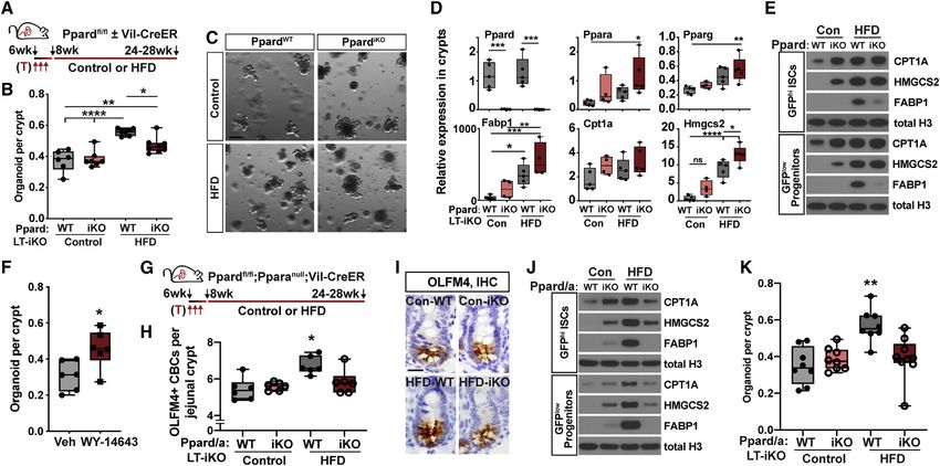
ll
Article                                                                                                                                          OPEN ACCESS

Figure 1. PPARd and PPARa function redundantly to enhance stem cell activity in a HFD
(A) Schematic of intestinal Ppard conditional deletion with Villin-CreERT2 (Ppard-iKO), including timeline of tamoxifen (T) injections for long-term deletion, diet,
and tissue collection.
(B and C) Quantification of organoids per crypt from WT (Ppardfl/fl) or Ppard-iKO mice in (A) (B), accompanied by representative bright-field images (C). LT-iKO
indicates deletion of intestinal Ppard prior to establishment on the diet. Scale bar, 200 mm.
(D) qRT-PCR of transcripts amplified from crypts collected from WT (Ppardfl/fl) or Ppard-iKO mice on chow or HFD. Transcripts were normalized to b-actin.
(E) Immunoblot of flow-sorted ISCs (Lgr5-EGFPhi) and progenitors (Lgr5-EGFPlow) from WT and Ppard-iKO (Ppardfl/fl; Villin-CreERT2; Lgr5-eGFP-IRES-CreERT2)
mice on a control diet or HFD. Villin-CreERT2; Lgr5-eGFP-IRES-CreERT2 animals were used as WT controls.
(F) Quantification of organoids per crypt from mice injected daily for 30 days with the PPARa agonist WY-14643 (4 mg/kg) or vehicle.
(G) Schematic of the intestinal Ppard conditional allele with Villin-CreERT2 crossed to Ppara-null (Ppard/a-iKO), including timeline of T injections, diet, and tissue
collection. Villin-CreERT2 transgenic mice were used as WT controls.
(H) Quantification of OLFM4+ cells per jejunal crypt. Each data point represents quantification of 20+ crypts from one animal (n = 6).
(I) Representative images of OLFM4+ immunohistochemistry (IHC) from (H). Scale bar, 20 mm.
(J) Immunoblot blot of flow-sorted ISCs (Lgr5-EGFPhi) and progenitors (Lgr5-EGFPlow) from WT and Ppard/a-iKO mice on a control diet or HFD. Ppard/a-iKO mice
carried Lgr5-eGFP-IRES-CreERT2 for isolation of ISC and progenitor populations. Villin-CreERT2; Lgr5-eGFP-IRES-CreERT2 animals were used as WT controls.
(K) Quantification of organoids per crypt from mice in (G).
To compare differences in diet, control WT mice were fed chow in (B)–(D) and a purified control diet in (E) and (H)–(K). In (B), (D), (F), and (K), each data point
represents the average of 2–3 technical replicates of one animal. *p < 0.05, **p < 0.01, ***p < 0.005, ****p < 0.0001, one-way ANOVA.

progenitors (Figure 1E), consistent with the notion that other                       mice with a PPARa inhibitor, GW6471 (Figure S1M; Xu et al.,
PPARs may be involved in the HFD ISC response. However,                              2002). Notably, treatment with the PPARa inhibitor reduced the
FABP1 mRNA and protein expression differed with Ppard loss                           clonogenicity of HFD Ppard-iKO crypts to control levels and
on the HFD (Figures 1D and 1E); FABP1 protein levels, but not                        had no effect in the control or HFD WT groups (Figure S1M), indi-
mRNA levels, respond to the HFD in a PPARd-dependent manner,                         cating that PPARa and PPARd are required for elevated HFD
illustrating that mRNA levels need not dictate protein expression                    crypt function in the organoid assay. To further characterize
and that FABP1 likely undergoes post-transcriptional regulation                      the in vivo necessity of Ppara, we used a previously generated
to account for these differences.                                                    whole-body null mouse model (Lee et al., 1995) that was fed a
    Next we turned our attention to Ppara, which, like Ppard, plays                  control diet or a HFD. Ppara loss by itself mitigated the HFD-
a critical role in regulating FAO metabolism (Evans et al., 2004).                   enhancing effects on ISC numbers and crypt clonogenicity
In vivo activation of PPARa for 30 days with an agonist, WY-                         (Figures S1N and S1P) but had no effect on their proliferation
14643 (Kliewer et al., 1994), not only increased expression of                       (Figure S1O). These findings indicate that PPARd and PPARa
PPAR targets but also enhanced OLFM4+ and Lgr5-EGFP+ ISC                             have distinctive and redundant roles in supporting intestinal pro-
numbers and crypt organoid clonogenicity, demonstrating that                         genitor cell adaptation to a HFD.
activated PPARa and PPARd have similar effects on stem cell                             To further examine the effects of Ppard and Ppara loss in vivo,
activity (Figures 1F and S1J–S1L; Beyaz et al., 2016). To test                       we generated compound mice with inducible deletion of intestinal
whether Ppara compensated for Ppard loss in HFD organoids,                           Ppard on a Ppara whole-body null background (Lee et al., 1995),
we treated control and HFD crypts from WT and Ppard-iKO                              Ppardfl/fl; Pparanull; Vil-CreER (Ppard/a-iKO). We then treated WT

                                                                                                                    Cell Reports 35, 109212, June 8, 2021 3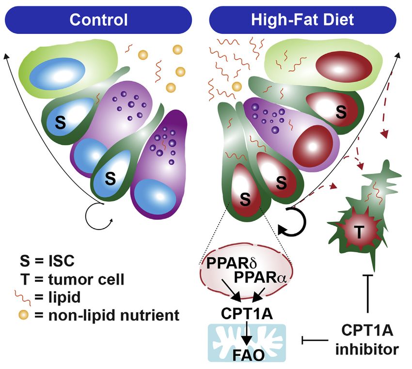
ll
OPEN ACCESS Article
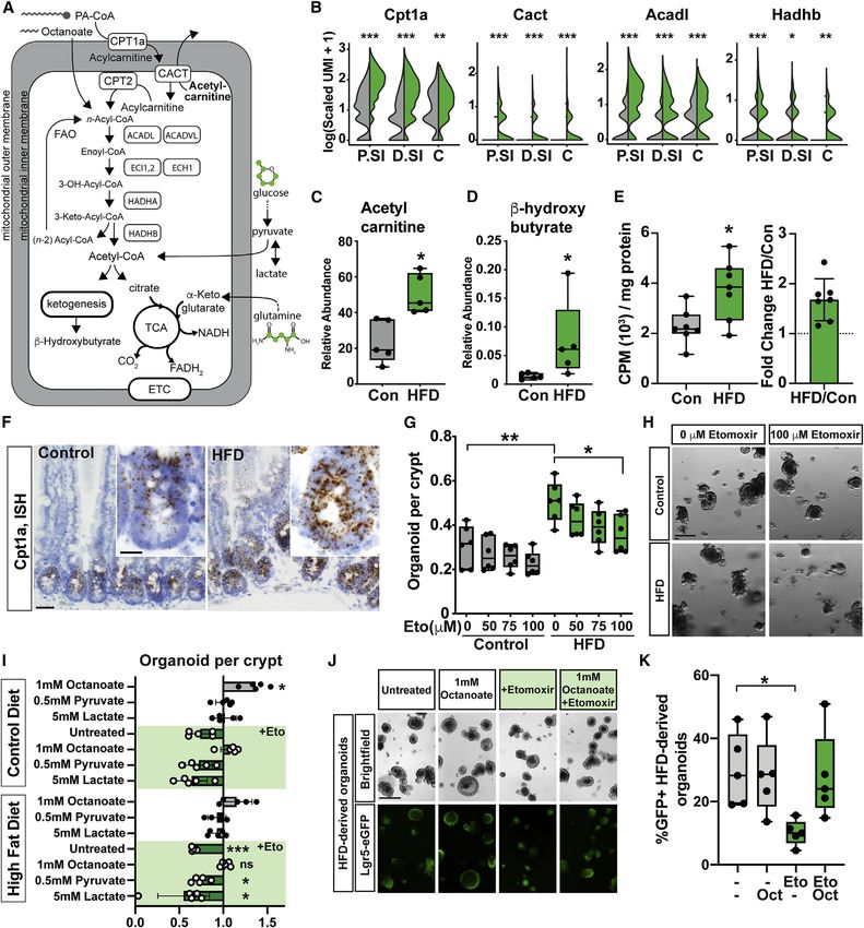
Figure 2. HFD fuels FAO in ISCs
(A) Schematic of mitochondrial FAO involving key processes, proteins, metabolites, and fuel inputs.
(B) Log(UMIs per 10K + 1) expression of genes integral to the FAO in ISCs from control-fed (gray, n = 2) or HFD-fed mice (green, n = 3) (P.SI, proximal small
intestine; D.SI, distal small intestine; C, colon). Benjamini-Hochberg-corrected p values: *p < 102, **p < 105, ***p < 1010.
(C and D) Relative abundance of the FAO-mediated metabolites acetylcarnitine (C) and BOHB (D) isolated from freshly harvested crypts and measured by liquid
chromatography-mass spectrometry (LC-MS).
(E) FAO activity (counts per minute [cpm]) of metabolized 3H-palmitic acid normalized to total protein in crypts from control and HFD-fed mice, accompanied by
the fold change calculated as the ratio of HFD cpm per control cpm.
(F) Single-molecule RNA in situ hybridization (ISH) of Cpt1a in WT tissue from control and HFD-fed mice. Scale bars, 50 mm and 20 mm (inset).
(G and H) Quantification of organoids per crypt from control or HFD-fed mice grown in culture (ADMEM medium) with increasing concentrations of the irreversible
CPT1A inhibitor etomoxir (Eto) (G), accompanied by representative images (H). Scale bar, 200 mm.
(legend continued on next page)
4 Cell Reports 35, 109212, June 8, 2021ll
Article OPEN ACCESS
and compound mutant (Ppard/a-iKO) mice with tamoxifen and es- and b-hydroxybutyrate (BOHB), known products of FAO, were
tablished cohorts on a control diet or HFD for 24 weeks prior to more abundant in HFD crypts (Figures 2C and 2D). We then
analysis (Figures 1G and S1A). As observed with the Ppara mutant exposed HFD and control crypts to tritium-labeled palmitate to
and inhibitor studies, in vivo loss of Ppara and Ppard was neces- measure production of tritiated H2O, which serves as a proxy for
sary to prevent increased crypt clonogenicity (Figure 1K) in acute FAO rates in cells (Manning et al., 1990; Nieman et al., 2011),
and chronic loss of intestinal Ppard in the Ppara-null background. and observed that HFD crypts were nearly 2-fold more capable
In addition, loss of both PPARs blocked expansion of OLFM4+ of FAO than controls (Figure 2E). Given that HFD primes for FAO
ISCs in vivo (Figures 1H and 1I) as well as induction of the PPAR metabolism, we wanted to find out how the cellular capacity of
program in Lgr5-EGFPhi ISCs and GFPlow progenitors, as seen in the crypt to use other fuel sources is affected using isotope-
immunoblots or transcriptional analysis (Figures 1J, S1T, and labeled substrates for glucose and glutamine. We incubated con-
S1U). ISC proliferation and Paneth cell numbers were also main- trol and HFD crypts with U13C-glucose or U13C-glutamine and
tained at control WT levels (Figures S1R and S1S). Utilizing then measured formation of labeled downstream metabolites.
Ppardfl/fl; Pparanull; Lgr5-CreER; Rosa-LSL-LacZfl/fl engineered Interestingly, with U13C-glucose, we observed that HFD crypt cells
mice to further test the in vivo role of Ppars on ISCs, we found incorporated fewer carbons into lactate and citrate than controls,
that loss of Ppard and Ppara reduced lineage output specifically supporting the notion that glucose metabolism is diminished in
in the HFD state (Figures S1V and S1W). Last, compound Ppard HFD ISCs and progenitors (Figures S2E–S2I). However, neither
and Ppara loss prevented HFD crypt survival and regeneration af- diet showed a preference for U13C-glutamine as an alternate fuel
ter a lethal dose of radiation (Figures S1X and S1Y). Our data illus- source (Figures S2J–S2L). These data demonstrate that HFD
trate that PPARd and PPARa overlappingly contribute to the HFD crypts increase FAO and concomitantly decrease glucose
response in intestinal stem and progenitor cells. oxidation.
In our prior study (Beyaz et al., 2016), we had functionally vali- Given that CPT1A, the rate-controlling enzyme in FAO, is highly
dated that the PPARd agonist GW was sufficient to emulate expressed in HFD ISCs by gene expression (Figure 2B), mRNA
many of the effects of the HFD on ISCs. However, given that ISH (Figure 2F), and protein immunoblots (Figures 1E and 1J),
PPARd and PPARa play redundant roles in the HFD ISC we wanted to find out whether FAO inhibition would attenuate
response, some effects of GW may act through stimulation of the HFD-induced increase in organoid formation. We incubated
PPARa. To address this scenario, we treated WT, Ppard-iKO, crypts from control and HFD-fed mice in organoid assays with
Ppara-null, and Ppard/a-iKO organoids with GW for 8 days and increasing concentrations of etomoxir, an irreversible inhibitor
performed RNA sequencing to probe the specificity of GW in of CPT1A, to ascertain the role of FAO as an effector of the HFD
engaging a PPAR or FAO transcriptional program (Beyaz et al., response (Figures 2H and 2I). Although etomoxir had no effect
2021). Although GW treatment in WT and Ppara-null mice (geno- on the ability of control crypts to form organoids, treatment with
types with intact PPARd) led to strong gene set enrichment for etomoxir dampened HFD crypt clonogenicity in a dose-depen-
signatures related to PPAR signaling pathways and lipid/fatty dent manner (Figures 2G and 2H). Similarly, PPARd (GW) or
acid metabolism, Ppard deficiency reduced, but did not entirely PPARa (WY-14643, WY) agonist treatment in the organoid assay
eliminate, these signatures as double Ppard/a-deficiency did, boosted FAO rates and sensitivity to etomoxir-mediated FAO in-
nor did Ppard deficiency block the increase in PPAR transcrip- hibition (Figures S2M and S2N), highlighting the functional impor-
tional targets (Figures S1Z–S1BB). These findings indicate that tance of FAO as a downstream target of the HFD-PPAR axis.
at least some of GW’s activity involves PPARa. Addition of octanoate, an 8-carbon fatty acid that bypasses
CPT1A-mediated entry into mitochondria, counteracted the
FAO drives the ISC HFD and PPAR response inhibitory effects of etomoxir on HFD crypt clonogenicity and
Upon ligand stimulation, PPARd and PPARa induce a robust tran- Lgr5-eGFP+ organoids (Figures 2I–2K). Although exogenous oc-
scriptional program that drives fatty acid uptake and breakdown tanoate had no additional effect on HFD crypt clonogenicity, likely
through FAO (Finck et al., 2002; Kersten et al., 1999; Wang et al., because these crypts are already in a state of elevated FAO, stim-
2003). To understand the extent to which HFD activates a FAO ulation of control crypt organoid formation with exogenous octa-
transcriptional program (Figure 2A), we performed single-cell noate was observed (Figures 2I–2K and S2O–S2Q). In contrast,
RNA sequencing on control and HFD crypts extracted from three lactate or pyruvate, glycolytic metabolites that feed into the TCA
regions of the intestine: the proximal and distal small intestine and cycle to undergo oxidation, fail to alleviate the effects of FAO inhi-
the colon (Figures S2A–S2C). We confirmed that PPAR target and bition on HFD crypt clonogenicity (Figure 2I).
FAO genes are highly enriched throughout the intestine in these
HFD ISC populations compared with controls (Figures 2B and CPT1A-mediated FAO promotes HFD ISC stemness
S2D). In addition, through liquid chromatography-mass spectrom- Next we sought to investigate the in vivo necessity of FAO meta-
etry (LC-MS), we found that metabolites such as acetylcarnitine bolism in control and HFD ISCs using tamoxifen-inducible and
(I) Quantification of organoids per crypt from control or HFD-fed mice, grown in RPMI 1640 basal medium with or without Eto (37.5 mM) and supplemented with
octanoate (1 mM), sodium pyruvate (0.5 mM), or sodium-L-lactate (5 mM).
(J) Representative images of HFD-derived Lgr5-EGFP+ organoids in (I). Scale bar, 200 mm.
(K) Quantification of HFD-derived Lgr5-eGFP+ organoids grown in the presence of octanoate (1 mM) or Eto (37.5 mM).
In (B)–(I), mice were fed a purified control diet or HFD. In (E), (G), and (I), each data point represents the average of 3+ technical replicates from one animal. *p <
0.05, **p < 0.01, ***p < 0.005; ns, not significant; one-way ANOVA.
Cell Reports 35, 109212, June 8, 2021 5ll
OPEN ACCESS Article
Figure 3. CPT1A-mediated FAO promotes HFD ISC stemness
(A) Schematic of intestinal Cpt1a conditional deletion with Villin-CreERT2; Lgr5-eGFP-IRES-CreERT2 (Cpt1a-iKO), including timeline of T injections, diet, and
tissue collection. Villin-CreERT2; Lgr5-eGFP-IRES-CreERT2 animals were used as controls.
(B and C) Quantification of OLFM4+ cells per jejunal crypt (B) with representative images (C). Scale bar, 20 mm.
(D and E) Quantification of BrdU+ and EGFP+ CBC cells per jejunal crypt (D) with representative images (E). Scale bar, 20 mm.
(F and G) Quantification of organoid per crypt (F) or Lgr5-EGFPhi ISC (G) from control or HFD-fed mice carrying WT or deleted copies of Cpt1a (iKO).
(H) Relative abundance of the FAO-mediated metabolites acetylcarnitine and BOHB isolated from freshly harvested crypts of HFD-fed mice and measured by LC-
MS.
(I) Immunoblot blot of flow-sorted ISCs (Lgr5-EGFPhi), progenitors (Lgr5-EGFPlow), and epithelial crypt (EpCAM+) cells from WT and Cpt1a-iKO mice on a control
diet or HFD.
(B)–(E) were reprinted from Beyaz et al. (2021). In (A)–(I), mice were fed a purified control diet or HFD. In (F) and (G), each data point represents the average of 3+
technical replicates from one animal. *p < 0.05, **p < 0.01, ***p < 0.005, ****p < 0.0001, one-way ANOVA.
intestine-specific Cpt1afl/fl; Vil-CreER; Lgr5-eGFP mice (Fig- (6–7 months) Cpt1a loss had mild effects on intestinal differenti-
ure 3A). Consistent with our prior study (Mihaylova et al., ation, as assessed by quantification of Lysozyme+ Paneth cell
2018), acute deletion of Cpt1a for 4 weeks in control intestines numbers, ChromograninA+ (CgA+) neuroendocrine cell, or Alcian
had no effect on OLFM4+ or Lgr5-EGFP+ ISC numbers (Beyaz blue+ goblet cell numbers (Figures S3B–S3D and S3H–S3J).
et al., 2021; Figures 3B–3E) or on stem and progenitor cell prolif- Although a HFD decreases Paneth cell numbers, as we noted
eration, as assessed with a 4-h pulse of bromodeoxyuridine previously (Beyaz et al., 2016), it does so in a CPT1A-dependent
(BrdU+) (Figures 3D and S3A). However, Cpt1a deletion in HFD manner because long-term Cpt1a loss rescues this decline (Fig-
intestines restored ISC numbers (Figures 3B and 3D) and prolif- ure S3H). Finally, Cpt1a loss prevented expansion and prolifera-
eration to control levels (Figures 3D and S3A) and blocked the or- tion of stem/progenitor cells in the HFD crypt (Figures S3A and
ganoid-enhancing effects of a HFD on crypts (Figure 3F) and S3G).
sorted Lgr5-EGFPhi ISCs (Figure 3G). Using a Lgr5-CreER; As expected, loss of Cpt1a in HFD crypts led to many meta-
Rosa-LSL-LacZfl/fl lineage tracer, loss of Cpt1a reduced b-galac- bolic changes compared with controls. FAO from surplus free
tosidase (b-gal) lineage tracing from control and HFD stem cells fatty acids (FFAs) provided by the HFD, for example, generates
but with a greater reduction in HFD crypts (Figures S1V and acetyl-coenzyme A (CoA), which can feed into ketogenesis and
S1W). In addition, long-term Cpt1a loss (Figures S3E and S3F) the TCA cycle (Figure 2A). Loss of Cpt1a led to decreased FAO
prior to initiation of a HFD also blunted the regenerative effects in HFD crypts (Figure S3Q) and resulted in significant reductions
of a HFD on crypts in the organoid assay (Figure S3K) and in levels of FAO-associated metabolites, such as acetylcarnitine
following injury from ionizing radiation (Figures S3L and S3M), and BOHB (Figures 3H and S3N). Moreover, the abundance of
indicating that HFD-induced FAO in ISCs and progenitors pro- the TCA intermediate citrate was lower in HFD Cpt1a-iKO crypts
motes recovery after damage. Acute (1 month) and long-term compared with controls, presumably due to loss of FAO-derived
6 Cell Reports 35, 109212, June 8, 2021ll
Article OPEN ACCESS
Figure 4. HFD-initiated tumors are metabolically vulnerable to FAO disruption
(A) Schematic of the Apc tumor model with Lgr5-eGFP-IRES-CreERT2 driving Apcfl/fl excision specifically in Lgr5-CreERT2 stem cells upon T induction (25 mg/
kg) 1 month after establishment on a control diet or HFD.
(B) Kaplan-Meier survival curve of the mice in (A).
(C) Relative abundance of the FAO-mediated metabolites acetylcarnitine and BOHB isolated from freshly harvested crypts of Apcfl/fl; Villin-CreERT2 mice and
measured by LC-MS.
(D) Schematic of Apcfl/fl; Lgr5-eGFP-IRES-CreERT2 tumor initiation model with or without the Cpt1afl/fl allele. Mice were put on a control diet or HFD for 1 month
prior to a single T injection (25 mg/kg), followed by 20 days before tissue collection.
(legend continued on next page)
Cell Reports 35, 109212, June 8, 2021 7ll
OPEN ACCESS Article
acetyl-CoA (Figure S3P), whereas pyruvate levels increased 4 days after Apc loss in Apcfl/fl; Vil-CreER mice (Figures S4B
concomitantly, representing an adaptation to decreased flux and S4C) and as predicted from the expression of Cpt1a and
through the TCA cycle (Figure S3O). Notably, although loss of Hmgcs2, have higher levels of FAO-associated metabolites like
Cpt1a dramatically diminished crypt BOHB levels, it also led to acetylcarnitine and BOHB (Figure 4C). Third, loss of Cpt1a in
a compensatory boost in HMGCS2 protein, independent of mice on a HFD significantly reduced the number and size of
diet, in sorted EpCAM+ intestinal epithelial cells, ISCs, and pro- spontaneous Apc-null adenomas formed in a loss-of-heterozy-
genitors (Figure 3I). A possible explanation for this observation gosity model compared with Cpt1a-WT control counterparts
is that FAO disruption through Cpt1a deletion reduces acetyl- (Figures S4K and S4L). Fourth, genetic disruption of Cpt1a in
CoA feeding into ketogenesis, which accounts for the diminish- Cpt1afl/fl; Apcfl/fl; Lgr5-eGFP-IRES-CreER mice, where a single
ment of BOHB, whereas FFAs continue to stimulate sensors tamoxifen dose concomitantly deletes Cpt1a and Apc in Lgr5+
such as PPARd and PPARa to transcribe genes involved in ISCs, dramatically dampened the numbers and area of b-cate-
FAO and lipid metabolism (Figures S3R and S3S), including nin-positive tumors 20 days after tamoxifen administration in
HMGCS2 (a known PPAR target) (Figure 3I). This upregulation mice on an HFD for 7 weeks, whereas there was no effect on
of HMGCS2, after FAO disruption, permits crypt BOHB levels control diet tumors (Figures 4D–4F and S4D). Furthermore,
to remain stable on a control diet and to maintain at least basal HFD tumors were enriched for greater numbers of Cpt1a-
levels in HFD crypts (Figure S3N). This finding is consistent competent escapers than what was observed in the controls,
with our recent studies implicating BOHB as a key metabolite highlighting an important role of FAO in HFD tumor maintenance
that regulates intestinal stemness and dietary responses (Cheng (Figures S4E and S4F). Last, to address how Cpt1a dampens
et al., 2019; Gebert et al., 2020). Overall, these findings support HFD-mediated tumorigenesis, we administered three doses of
the notion that a shift toward FAO in a HFD accounts for many of tamoxifen over 8 days to Apc-iKO mice to maximize Cpt1a dele-
the metabolic and functional changes that occur in ISCs. tion. Acute Cpt1a loss reduced HFD-stimulated proliferation, as
measured by Ki67, which also corresponded to decreased DNA
Intestinal tumors that arise on a HFD are sensitive to damage and apoptosis, as assessed by gH2AX and C-Casp3
FAO inhibition immunofluorescence, respectively (Figures S4G–S4J). Although
A pro-obesity HFD induces many changes in intestinal stem and these data highlight that a HFD boosts adenomatous prolifera-
progenitor cells, such as expanding their numbers, proliferation, tion in a CPT1A-dependent manner, it is unclear whether the
and function by engaging a robust PPAR-FAO program. It is decrease in DNA damage or apoptosis with Cpt1a loss in an
possible, for example, that tumors arising from ISCs in a HFD HFD stems indirectly from reduced proliferation or tumor size
state retain a similar sensitivity to FAO inhibition as their non-tu- or directly from Cpt1a loss itself.
mor counterparts, raising the question of whether a HFD creates Although co-deletion of Cpt1a and Apc in Lgr5+ HFD ISCs
a therapeutic opportunity to exploit FAO dependencies in these reduced tumor initiation and burden when FAO was disrupted
tumors. To assess this possibility, we utilized conditional Apcfl/fl; genetically at the beginning of tumor formation, we turned our
Lgr5-eGFP-IRES-CreER (Apc-iKO) mice where tamoxifen focus to investigating whether FAO inhibition in established ade-
administration leads to loss of both copies of Apc in Lgr5+ nomas would also have growth-retarding effects in a HFD. To test
ISCs and rapid tumor formation, permitting study of early intes- this scenario, we induced Apc loss in Apc-iKO mice, maintained
tinal tumor formation in response to diet (Figure 4A; Barker et al., cohorts on a control diet or HFD for 20 days, and then mice
2009; Beyaz et al., 2016; DeClercq et al., 2015). were injected with daily doses of etomoxir starting on day 11 (Fig-
We placed Apc-iKO mice on a control or HFD for 1 month, and ure 4G). Analogous experiments were also performed with PPARd
then induced Apc excision with a single tamoxifen injection. As and PPARa agonists (GW and WY, respectively) to understand
expected, HFD feeding dramatically accelerated mortality in the role of PPARs in this process (Figure 4G). While HFD and
this model compared with the control condition (Figure 4B; PPARa and PPARd agonist treatments boosted tumor numbers
Beyaz et al., 2016; Park et al., 2016). Many lines of evidence indi- (Figure S4N), etomoxir had no effect on adenoma multiplicity
cate that HFD-stimulated FAO mediates this accelerated tumor- because tumors were initiated prior to etomoxir treatment. Eto-
igenesis. First, Apc-null tumors and Lgr5-EGFP+ tumor cells moxir, however, did blunt the growth-enhancing effects of HFD
from HFD mice demonstrate heightened expression of Cpt1a and GW, but not WY, on adenoma size (Figures 4H, 4I, S4M,
and Hmgcs2 by ISH and immunoblots relative to controls (Fig- and S4N), indicating that the HFD and these agonists mediate
ures S4A and S4B). Second, HFD tumors, as determined some of their tumorigenic effects via FAO.
(E and F) Quantification of the area (E) and number (F) of b-catenin+ adenomas in the proximal half of the intestine.
(G–I) Schematic of Apcfl/fl; Lgr5-eGFP-IRES-CreERT2 tumor progression model (G). Mice receive a single T injection (25 mg/kg) to induce adenoma formation.
The following day, cohorts are established on a control diet or HFD (H) or treated with daily injections of vehicle, GW501516 (GW; 4 mg/kg), or WY-14643 (WY;
4 mg/kg) (I). The diet regimen or agonist administration was extended for 20 days. On days 11–20 before tissue collection, mice received daily injections of the
CPT1A inhibitor Eto (20 mg/kg). Shown is quantification of the b-catenin+ tumor area in mice under HFD conditions (H) and GW or WY administration (I) according
to the regimens in (G). The inset plots represent the mean area fold change between non-Eto and +Eto recipients of a given treatment.
(J) Schematic of endoscopy-guided colonic injections with 4-OH Tamoxifen (Tam) 1 month after start of the control diet or HFD.
(K) Quantification of tumor area 1 month after injection. The mean fold change of WT versus mutant is depicted in the right graph inset.
(L) Model of ISC adaptation to a HFD: PPARd and PPARa instruct a transcriptional program in response to a HFD that promotes CPT1A-mediated FAO. In contrast
to a control diet, genetic loss or inhibition of Cpt1a on a HFD disrupts tumor initiation and progression.
Control mice were fed chow in (B) and (I) and a purified control diet in (C), (E) ,(F), (H), and (K). *p < 0.05, **p < 0.01, ***p < 0.005, ****p < 0.0001, one-way ANOVA.
8 Cell Reports 35, 109212, June 8, 2021ll
Article OPEN ACCESS
To test the effect of decreased FAO in HFD-induced ade- 2017; Schell et al., 2017). Subsequently, Bensard et al. (2020)
nomas in the colon (Figure 2B), we generated Apcfl/fl; Cpt1afl/fl; have shown that loss of Mpc1 leads to a pro-tumorigenic pheno-
Vil-CreER and, similarly, Apcfl/fl; Ppardfl/fl; Pparanull; Vil-CreER type (Bensard et al., 2020). A shared feature of Mpc1 loss and
mice and injected 4-OH tamoxifen using an endoscopy-guided HFD-activated PPAR signaling is that both interventions restrict
approach to induce adenoma formation specifically in the distal the participation of pyruvate in the TCA cycle and compensate
colon (Figures 4J and 4K). Ppard; Ppara loss in colonic ade- by boosting FAO. With this metabolic shift, Mpc1 loss, like a
nomas significantly diminished tumor size in a HFD-dependent HFD, enhances the contribution of fatty-acid-derived acetyl-
manner, analogous to their roles in actuating the HFD response CoA to the TCA cycle in ex vivo cultures (Schell et al., 2017).
in intestinal stem and progenitor cells (Figure 4K). Cpt1a loss We have shown here and previously (Mihaylova et al., 2018)
decreased tumor size irrespective of diet, but there was a that FAO inhibition blunts the ISC-enhancing effects of a HFD
much larger reduction on the HFD, consistent with the notion and PPAR agonists, but deciphering how much of the ISC and
that HFD tumors are more sensitive to Cpt1a inhibition (Fig- pro-tumorigenic phenotype associated with Mpc1 deletion oc-
ure 4K). Furthermore, Cpt1a-null tumor cells, which were curs though FAO versus other sequelae of altered carbohydrate
strongly selected against (Figure S4O), were less proliferative metabolism will be an important future endeavor.
than their Cpt1a competent counterparts (Figure S4P). These An intriguing similarity of fasting (Mihaylova et al., 2018) and a
data demonstrate that a HFD drives intestinal tumorigenesis in HFD is that both interventions augment ISC function through
part by shifting tumor cell metabolism toward FAO metabolism engagement of a PPAR-FAO program. However, fasting and
and creates a therapeutic opportunity for tumor prevention or HFD are believed to have very different effects on health and
treatment by targeting the FAO pathway in the HFD state. cancer incidence. Although both diets strongly induce FAO for
utilization of circulating FFAs as an energy source, the extent
DISCUSSION of fatty acid saturation of the mitochondrial FAO machinery is
quite different between these two diets, which may ultimately
Here we propose that a HFD enhances intestinal stemness and affect tumor formation. In a HFD, ISCs are exposed to surplus
tumorigenicity by engaging a PPAR-FAO program. Although amounts of FFAs, whereas in fasting, ISCs need to scavenge en-
we have suggested previously that PPARd is the main PPAR ergy from a limited quantity of FFAs to satisfy their energy needs.
family member that underlies the HFD ISC response, we now In a HFD, excessive amounts of FFAs likely overwhelm the mito-
modify this view because PPARa also contributes to ISC regula- chondria and lead to generation of reactive lipids and reactive
tion in a pro-obesity high-lipid environment. Compensation of oxygen species that have a cumulative deleterious effect over
PPARd loss by PPARa is compatible with an earlier study that time, which does not occur with fasting, where FFAs are much
proposed that PPARd inhibits PPARa activity, underscoring the less abundant.
complexity of PPAR biology in physiological states such as A remaining question is which factor(s) downstream of FAO
HFD-induced obesity (Shi et al., 2002). Because it can be thera- contribute to the ISC-enhancing effects of FAO. We recently
peutically challenging to inhibit this redundancy in HFD ISCs, discovered that the ketone body BOHB plays a part in regu-
both of these PPARs converge on and elicit a robust FAO lating ISC homeostasis and their dietary responses. BOHB is
program where pharmacologic or genetic disruption of CPT1A- a terminally oxidized lipid byproduct of FAO that has energetic
mediated FAO blunts the HFD-enhancing effects on ISCs or tu- and signaling properties (Newman and Verdin, 2017; Puchal-
mors that arise from them (Figure 4L). Disruption of this PPAR- ska and Crawford, 2017) and further serves as a class 1 his-
FAO axis has minimal effects on baseline intestinal homeostasis tone deacetylase inhibitor to reinforce the NOTCH program
(Figures 1 and S1), in contrast to other regulators of lipid meta- in ISCs (Cheng et al., 2019). BOHB levels increase dramatically
bolism, such as PRDM16 and HNF4a/g (Chen et al., 2020; Stine in ISCs from ketotic states induced by HFDs (Figure 2D), fast-
et al., 2019). One possible explanation for this discrepancy is that ing (Mihaylova et al., 2018), and ketogenic diets (KDs) (Cheng
PPARs, by sensing lipid availability, permit ISCs to adapt to et al., 2019), and all of these diets are associated with
diverse dietary states, especially ketotic states induced by an improved ISC function. Future studies will need to delineate
HFD, fasting, and ketogenic diets. PPARs, in the setting of a the precise roles of mitochondrial activity and ketone bodies,
HFD, augment FAO in ISCs as a response to surfeit dietary lipids, such as the energetic and NOTCH signaling properties of
but their genetic loss does not compromise basal FAO or ISC BOHB, in accounting for the similarities and differences be-
function (Figures 1J and 1K). Different from PPARs, PRDM16 tween HFD, fasting, and KD regimens on intestinal stemness
and HNF4a/g play an obligatory role in regulating basal FAO and tumorigenicity.
because their loss significantly hampers intestinal stemness in From a therapeutic perspective, the precise role of Ppard in
a FAO-dependent fashion (Chen et al., 2020; Stine et al., 2019). intestinal tumorigenesis has been a matter of debate, in part
Another important implication of our work is in regard to the because of the commonly used agonist GW and different ge-
role of FAO in stem cell biology and intestinal tumorigenesis netic models utilized to model Ppard loss, the latter of which
(Shapira and Christofk, 2020). A PPARd-FAO axis is necessary has been reviewed previously (Beyaz and Yilmaz, 2016; Wag-
for maintaining the HSC pool (Ito et al., 2012), and FAO governs ner and Wagner, 2020). For example, GW treatment acceler-
neural progenitor quiescence (Knobloch et al., 2017). It has also ates intestinal tumorigenesis in an Apcmin model (Gupta
been proposed that limiting mitochondrial pyruvate metabolism et al., 2004) and promotes metastasis in orthotopic colon can-
by deletion of mitochondrial pyruvate carrier 1 (Mpc1) in the in- cer cell line models (Wang et al., 2019). Based on our initial
testine and hair follicles promotes stemness (Flores et al., work showing that GW recapitulates the HFD ISC response
Cell Reports 35, 109212, June 8, 2021 9ll
OPEN ACCESS Article
(Beyaz et al., 2016), we suggested that PPARd signaling B RNA-seq data processing and differential expression
coupled a HFD with enhanced intestinal stemness. However, analysis
as we find here, GW also has PPARa-stimulating properties B scRNA-seq Seq-Well library preparation
(Figures S1Z–S1BB), raising the possibility that at least B Seq-Well data alignment, filtering, and analysis
13 13
some of its effects on ISCs and early tumors are through B Metabolomics, U C-glucose and U C-glutamine
PPARa. A previous study demonstrated that PPARa in loss- tracing, and LC/MS methods
of-function and agonist treatment experiments enhances B Fatty acid oxidation assay
and attenuates DSS/chemically-induced AOM intestinal B Irradiation experiments
tumorigenesis, respectively, which is different from what we B Orthotopic injection into the colon
observe in the HFD Apc loss-of-function model (Luo et al., d QUANTIFICATION AND STATISTICAL ANALYSIS
2019). One possibility is that DSS/AOM promotes tumorigen-
esis by not only transforming intestinal epithelial cells but also SUPPLEMENTAL INFORMATION
by inciting inflammation and that PPARa has a protective role
in inflammation-driven tumorigenesis, a process relevant to in- Supplemental information can be found online at https://doi.org/10.1016/j.
flammatory bowel disease. Future lines of investigation will celrep.2021.109212.
need to decipher how PPARs mediate intestinal tumorigenesis
in inflammation and obesity and in tumors with complex ge- ACKNOWLEDGMENTS
netics beyond that achieved with DSS/AOM or Apc loss.
Importantly, induction of FAO is a shared downstream and We thank the Whitehead Institute Metabolite Profiling Core Facility, the White-
targetable feature of the HFD-induced PPARd/a program that head Institute Flow Cytometry Core, and the Swanson Biotechnology Center
at the Koch Institute, which encompasses the Flow Cytometry, Histology, and
can be exploited to ameliorate the stemness and cancer conse-
Genomics & Bioinformatics Core facilities (NCI P30-CA14051). We thank Charlie
quences of this obesity model. Wang et al. (2019) found that a Whittaker for analysis and helpful discussions regarding RNA sequencing data.
HFD-activated Nanog program facilitates colon cancer metas- We thank the Department of Comparative Medicine for mouse husbandry
tasis in a PPARd-dependent manner (Wang et al., 2019). It is support. We thank Sven Holder and members of the Hope Babette Tang
possible, for example, that FAO actuates some of the down- (1983) Histology Facility for substantial histology support. We thank Heaji Shin
stream effects of Nanog and offers a therapeutic metabolic and Chia-Wei Cheng for comments on the manuscript, Kerry Kelley for labora-
tory management, and Liz Galoyan for administrative assistance. M.D.M.
target for colon cancer progression and metastasis in this dietary
received support from the American Cancer Society (PF-16-202-01-NEC), an
model. Additionally, epithelial FAO may have non-cell-autono-
MIT/Ludwig Center for Molecular Oncology postdoctoral fellowship in metas-
mous effects that influence the tumor microenvironment in a tasis/cancer research, and an MIT/Koch Institute quinquennial cancer research
HFD state, leading to disease progression (Ringel et al., 2020). fellowship and receives current funding from NIH 1K22CA241083-01. C.N.T. is
Finally, as interest in exploiting diet-induced metabolic vulnera- supported by a Fannie and John Hertz Foundation fellowship and a National Sci-
bilities grows and as our understanding of pathways that govern ence Foundation graduate research fellowship (1122374). P.C. is supported by
these approaches expands (Kanarek et al., 2020; Lien and Van- grants from Methusalem funding (Flemish government), the Fund for Scientific
Research-Flanders (FWO-Vlaanderen), and the European Research Council
der Heiden, 2019; Tajan and Vousden, 2020), exploration of
(ERC advanced research grant EU- ERC743074). M.M.M. is supported by
whether FAO inhibition can be leveraged therapeutically in NIH R00 AG054760 and the American Federation of Aging Research (AFAR).
HFD or PPAR agonist-stimulated tumors will be compelling to A.K.S. is supported by the Pew-Stewart Scholars Program for Cancer
pursue. Research. Ö.H.Y. is supported by R01CA211184, R01CA034992, and
U54CA224068; a Pew-Stewart Trust scholar award; the Kathy and Curt Marble
cancer research award; a Koch Institute-Dana-Farber/Harvard Cancer Center
STAR+METHODS Bridge Project grant; and AFAR. C.N.T., A.K.S., and Ö.H.Y. receive support
from the MIT Stem Cell Initiative through Fondation MIT.
Detailed methods are provided in the online version of this paper
and include the following:
AUTHOR CONTRIBUTIONS
d KEY RESOURCES TABLE
M.D.M. and A.M.H. designed, conducted, and interpreted the experiments
d RESOURCE AVAILABILITY with support from Ö.H.Y. C.N.T. and A.K.S. conducted and analyzed
B Lead contact scRNA-seq. S.I., D.B., Y.B.M., D.R.S., and A.T.W. provided experimental sup-
B Materials availability port. C.A.L. analyzed and interpreted the LC-MS data. P.C. and M.M.M. pro-
B Data and code availability vided mouse strains. M.M.M. contributed to interpretation of the Ppar experi-
d EXPERIMENTAL MODEL AND SUBJECT DETAILS ments. M.D.M., A.M.H., and Ö.H.Y. wrote the manuscript, and all authors
edited the manuscript.
d METHOD DETAILS
B Crypt isolation and culture
B qRT-PCR and in situ hybridization DECLARATION OF INTERESTS
B Immunoblotting
The authors declare no competing interests.
B Immunohistochemistry (IHC) and immunofluorescence
(IF)
Received: June 28, 2020
B Isolation of ISCs and flow cytometry Revised: March 1, 2021
B Loss of heterozygosity Accepted: May 12, 2021
B Lineage tracing analysis Published: June 8, 2021
10 Cell Reports 35, 109212, June 8, 2021ll
Article OPEN ACCESS
REFERENCES Chen, L., Vasoya, R.P., Toke, N.H., Parthasarathy, A., Luo, S., Chiles, E.,
Flores, J., Gao, N., Bonder, E.M., Su, X., and Verzi, M.P. (2020). HNF4 Regu-
Aicher, T.P., Carroll, S., Raddi, G., Gierahn, T., Wadsworth, M.H., Hughes, lates Fatty Acid Oxidation and Is Required for Renewal of Intestinal Stem Cells
T.K., Love, C., and Shalek, A.K. (2019). Seq-Well: A Sample-Efficient, Portable in Mice. Gastroenterology 158, 985–999.e9.
Picowell Platform for Massively Parallel Single-Cell RNA Sequencing. Methods Cheng, C.-W., Biton, M., Haber, A.L., Gunduz, N., Eng, G., Gaynor, L.T., Tripa-
Mol. Biol. 1979, 111–132. thi, S., Calibasi-Kocal, G., Rickelt, S., Butty, V.L., et al. (2019). Ketone Body
Alonso, S., and Yilmaz, Ö.H. (2018). Nutritional Regulation of Intestinal Stem Signaling Mediates Intestinal Stem Cell Homeostasis and Adaptation to Diet.
Cells. Annu. Rev. Nutr. 38, 273–301. Cell 178, 1115–1131.e15.
Anders, S., and Huber, W. (2010). Differential expression analysis for sequence Colnot, S., Niwa-Kawakita, M., Hamard, G., Godard, C., Le Plenier, S., Hou-
count data. Genome Biol. 11, R106. bron, C., Romagnolo, B., Berrebi, D., Giovannini, M., and Perret, C. (2004).
Andres, S.F., Santoro, M.A., Mah, A.T., Keku, J.A., Bortvedt, A.E., Blue, R.E., Colorectal cancers in a new mouse model of familial adenomatous polyposis:
and Lund, P.K. (2015). Deletion of intestinal epithelial insulin receptor attenu- influence of genetic and environmental modifiers. Lab. Invest. 84, 1619–1630.
ates high-fat diet-induced elevations in cholesterol and stem, enteroendo- Daoudi, M., Hennuyer, N., Borland, M.G., Touche, V., Duhem, C., Gross, B.,
crine, and Paneth cell mRNAs. Am. J. Physiol. Gastrointest. Liver Physiol. Caiazzo, R., Kerr-Conte, J., Pattou, F., Peters, J.M., et al. (2011). PPARb/d acti-
308, G100–G111. vation induces enteroendocrine L cell GLP-1 production. Gastroenterology
Arkan, M.C. (2017). The intricate connection between diet, microbiota, and 140, 1564–1574.
cancer: A jigsaw puzzle. Semin. Immunol. 32, 35–42. DeClercq, V., McMurray, D.N., and Chapkin, R.S. (2015). Obesity promotes
Barak, Y., Liao, D., He, W., Ong, E.S., Nelson, M.C., Olefsky, J.M., Boland, R., colonic stem cell expansion during cancer initiation. Cancer Lett. 369,
and Evans, R.M. (2002). Effects of peroxisome proliferator-activated receptor 336–343.
delta on placentation, adiposity, and colorectal cancer. Proc. Natl. Acad. Sci. Degirmenci, B., Valenta, T., Dimitrieva, S., Hausmann, G., and Basler, K.
USA 99, 303–308. (2018). GLI1-expressing mesenchymal cells form the essential Wnt-secreting
Barish, G.D., Narkar, V.A., and Evans, R.M. (2006). PPAR delta: a dagger in the niche for colon stem cells. Nature 558, 449–453.
heart of the metabolic syndrome. J. Clin. Invest. 116, 590–597.
Dobin, A., Davis, C.A., Schlesinger, F., Drenkow, J., Zaleski, C., Jha, S., Batut,
Barker, N., and Clevers, H. (2010). Lineage tracing in the intestinal epithelium. P., Chaisson, M., and Gingeras, T.R. (2013). STAR: ultrafast universal RNA-seq
Curr. Protoc. Stem Cell Biol Chapter 5, Unit 5A.4. aligner. Bioinformatics 29, 15–21.
Barker, N., van Es, J.H., Kuipers, J., Kujala, P., van den Born, M., Cozijnsen, el Marjou, F., Janssen, K.-P., Chang, B.H.-J., Li, M., Hindie, V., Chan, L., Lou-
M., Haegebarth, A., Korving, J., Begthel, H., Peters, P.J., and Clevers, H. vard, D., Chambon, P., Metzger, D., and Robine, S. (2004). Tissue-specific and
(2007). Identification of stem cells in small intestine and colon by marker inducible Cre-mediated recombination in the gut epithelium. Genesis 39,
gene Lgr5. Nature 449, 1003–1007. 186–193.
Barker, N., Ridgway, R.A., van Es, J.H., van de Wetering, M., Begthel, H., van Evans, R.M., Barish, G.D., and Wang, Y.-X. (2004). PPARs and the complex
den Born, M., Danenberg, E., Clarke, A.R., Sansom, O.J., and Clevers, H. journey to obesity. Nat. Med. 10, 355–361.
(2009). Crypt stem cells as the cells-of-origin of intestinal cancer. Nature
457, 608–611. Finck, B.N., Lehman, J.J., Leone, T.C., Welch, M.J., Bennett, M.J., Kovacs, A.,
Han, X., Gross, R.W., Kozak, R., Lopaschuk, G.D., and Kelly, D.P. (2002). The
Bensard, C.L., Wisidagama, D.R., Olson, K.A., Berg, J.A., Krah, N.M., Schell,
cardiac phenotype induced by PPARalpha overexpression mimics that
J.C., Nowinski, S.M., Fogarty, S., Bott, A.J., Wei, P., et al. (2020). Regulation of
caused by diabetes mellitus. J. Clin. Invest. 109, 121–130.
Tumor Initiation by the Mitochondrial Pyruvate Carrier. Cell Metab. 31, 284–
300.e7. Flores, A., Schell, J., Krall, A.S., Jelinek, D., Miranda, M., Grigorian, M., Braas,
D., White, A.C., Zhou, J.L., Graham, N.A., et al. (2017). Lactate dehydrogenase
Beyaz, S., and Yilmaz, Ö.H. (2016). Molecular Pathways: Dietary Regulation of
activity drives hair follicle stem cell activation. Nat. Cell Biol. 19, 1017–1026.
Stemness and Tumor Initiation by the PPAR-d Pathway. Clin. Cancer Res. 22,
5636–5641. Forman, B.M., Chen, J., and Evans, R.M. (1997). Hypolipidemic drugs, polyun-
saturated fatty acids, and eicosanoids are ligands for peroxisome proliferator-
Beyaz, S., Mana, M.D., Roper, J., Kedrin, D., Saadatpour, A., Hong, S.-J., Ba-
activated receptors alpha and delta. Proc. Natl. Acad. Sci. USA 94, 4312–
uer-Rowe, K.E., Xifaras, M.E., Akkad, A., Arias, E., et al. (2016). High-fat diet
4317.
enhances stemness and tumorigenicity of intestinal progenitors. Nature 531,
53–58. Fu, T., Coulter, S., Yoshihara, E., Oh, T.G., Fang, S., Cayabyab, F., Zhu, Q.,
Beyaz, S., Mana, M.D., and Yilmaz, Ö.H. (2021). High-fat diet activates a Zhang, T., Leblanc, M., Liu, S., et al. (2019). FXR Regulates Intestinal Cancer
PPAR-d program to enhance intestinal stem cell function. Cell Stem Cell 28, Stem Cell Proliferation. Cell 176, 1098–1112.e18.
598–599. Gebert, N., Cheng, C.-W., Kirkpatrick, J.M., Di Fraia, D., Yun, J., Schädel, P.,
Birsoy, K., Wang, T., Chen, W.W., Freinkman, E., Abu-Remaileh, M., and Sa- Pace, S., Garside, G.B., Werz, O., Rudolph, K.L., et al. (2020). Region-Specific
batini, D.M. (2015). An Essential Role of the Mitochondrial Electron Transport Proteome Changes of the Intestinal Epithelium during Aging and Dietary Re-
Chain in Cell Proliferation Is to Enable Aspartate Synthesis. Cell 162, 540–551. striction. Cell Rep. 31, 107565.
Biton, M., Haber, A.L., Rogel, N., Burgin, G., Beyaz, S., Schnell, A., Ashenberg, Gierahn, T.M., Wadsworth, M.H., 2nd, Hughes, T.K., Bryson, B.D., Butler, A.,
O., Su, C.-W., Smillie, C., Shekhar, K., et al. (2018). T Helper Cell Cytokines Satija, R., Fortune, S., Love, J.C., and Shalek, A.K. (2017). Seq-Well: portable,
Modulate Intestinal Stem Cell Renewal and Differentiation. Cell 175, 1307– low-cost RNA sequencing of single cells at high throughput. Nat. Methods 14,
1320.e22. 395–398.
Buescher, J.M., Antoniewicz, M.R., Boros, L.G., Burgess, S.C., Brunengraber, Goncalves, M.D., Lu, C., Tutnauer, J., Hartman, T.E., Hwang, S.-K., Murphy,
H., Clish, C.B., DeBerardinis, R.J., Feron, O., Frezza, C., Ghesquiere, B., et al. C.J., Pauli, C., Morris, R., Taylor, S., Bosch, K., et al. (2019). High-fructose
(2015). A roadmap for interpreting (13)C metabolite labeling patterns from corn syrup enhances intestinal tumor growth in mice. Science 363, 1345–1349.
cells. Curr. Opin. Biotechnol. 34, 189–201. Grivennikov, S.I., Wang, K., Mucida, D., Stewart, C.A., Schnabl, B., Jauch, D.,
Calle, E.E., and Kaaks, R. (2004). Overweight, obesity and cancer: epidemio- Taniguchi, K., Yu, G.-Y., Osterreicher, C.H., Hung, K.E., et al. (2012). Ade-
logical evidence and proposed mechanisms. Nat. Rev. Cancer 4, 579–591. noma-linked barrier defects and microbial products drive IL-23/IL-17-medi-
Chen, Y., Lun, A.T.L., and Smyth, G.K. (2016). From reads to genes to path- ated tumour growth. Nature 491, 254–258.
ways: differential expression analysis of RNA-Seq experiments using Rsu- Gupta, R.A., Wang, D., Katkuri, S., Wang, H., Dey, S.K., and DuBois, R.N.
bread and the edgeR quasi-likelihood pipeline. F1000Res. 5, 1438. (2004). Activation of nuclear hormone receptor peroxisome proliferator-
Cell Reports 35, 109212, June 8, 2021 11You can also read