Practical Management of Iron Overload Disorder (IOD) in Black Rhinoceros (BR; Diceros bicornis) - MDPI
←
→
Page content transcription
If your browser does not render page correctly, please read the page content below
animals
Review
Practical Management of Iron Overload Disorder
(IOD) in Black Rhinoceros (BR; Diceros bicornis)
Kathleen E. Sullivan, Natalie D. Mylniczenko , Steven E. Nelson Jr. , Brandy Coffin and
Shana R. Lavin *
 Disney’s Animal Kingdom® , Animals, Science and Environment, Bay Lake, FL 32830, USA;
 Kathleen.E.Sullivan@Disney.com (K.E.S.); Natalie.Mylniczenko@Disney.com (N.D.M.);
 Steven.E.Nelson@Disney.com (S.E.N.J.); Brandy.Coffin@Disney.com (B.C.)
 * Correspondence: Shana.Lavin@Disney.com; Tel.: +1-407-938-1572

 Received: 29 September 2020; Accepted: 26 October 2020; Published: 29 October 2020

 Simple Summary: Black rhinoceros under human care are predisposed to Iron Overload Disorder
 that is unlike the hereditary condition seen in humans. We aim to address the black rhino caretaker
 community at multiple perspectives (keeper, curator, veterinarian, nutritionist, veterinary technician,
 and researcher) to describe approaches to Iron Overload Disorder in black rhinos and share learnings.
 This report includes sections on (1) background on how iron functions in comparative species and
 how Iron Overload Disorder appears to work in black rhinos, (2) practical recommendations for
 known diagnostics, (3) a brief review of current investigations on inflammatory and other potential
 biomarkers, (4) nutrition knowledge and advice as prevention, and (5) an overview of treatment
 options including information on chelation and details on performing large volume voluntary
 phlebotomy. The aim is to use evidence to support the successful management of this disorder to
 ensure optimal animal health, welfare, and longevity for a sustainable black rhinoceros population.

 Abstract: Critically endangered black rhinoceros (BR) under human care are predisposed to
 non-hemochromatosis Iron Overload Disorder (IOD). Over the last 30 years, BR have been documented
 with diseases that have either been induced by or exacerbated by IOD, prompting significant efforts
 to investigate and address this disorder. IOD is a multi-factorial chronic disease process requiring
 an evidence-based and integrative long-term approach. While research continues to elucidate the
 complexities of iron absorption, metabolism, and dysregulation in this species, preventive treatments
 are recommended and explained herein. The aim of this report is to highlight the accumulated evidence
 in nutrition, clinical medicine, and behavioral husbandry supporting the successful management
 of this disorder to ensure optimal animal health, welfare, and longevity for a sustainable black
 rhinoceros population.

 Keywords: chelation; ferritin; hemochromatosis; hemosiderosis; oxidative stress; phlebotomy;
 transferrin saturation

Table of Contents
1.    Introduction

      1.1.     Why and how do we know BR have problems accumulating iron?
      1.2.     Why does iron overload matter to wellness?
      1.3.     How does IOD work in BR?
      1.4.     What health complications connected to iron are of concern?
2.    Evidence-Based Veterinary Practice: Technical and Clinical Aspects

Animals 2020, 10, 1991; doi:10.3390/ani10111991                                www.mdpi.com/journal/animals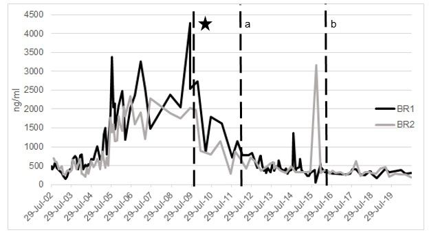
Animals 2020, 10, 1991                                                                                2 of 28

      2.1.     Clinical Signs
      2.2.     Diagnostic Testing

               2.2.1.    Serum iron
               2.2.2.    Total iron binding capacity (TIBC) and transferrin saturation
               2.2.3.    Ferritin
      2.3.     Perspectives on Integrative Monitoring
      2.4.     Inflammatory Biomarker Connections to IOD
      2.5.     Recommendations for Diagnosis
3.    Evidence-Based Nutrition Practices

      3.1.     Could we make the diet more like the wild?
      3.2.     Has diet ever changed the impact of IOD?
      3.3.     What are the best practices for feeding black rhinos?
4.    Treatment and Prevention

      4.1.     Hematologic Sampling Recommendations
      4.2.     Therapeutic Large Volume Phlebotomy
      4.3.     Techniques for VTLVP
      4.4.     Pharmacologic Chelation Therapy
      4.5.     History of Synthetic Chelation for BR
5.    Animal Husbandry and Operant Conditioning Practices
6.    Conclusions

1. Introduction
      Black rhinoceroses (BR; Diceros bicornis) under human care are predisposed to non-hemochromatosis
Iron Overload Disorder (IOD; see Section 1.3 How does IOD work in BR?) with laboratory and
histopathologic evidence of cellular injury, necrosis, and clinical signs similar to human iron overload
disorders [1,2]. BR are native to eastern and central Africa and are Critically Endangered [3]. Poaching has
reduced the wild population by >90% since 1970, and ~240 individuals are managed under human care
with ~87 individuals in North America [4]. Over the last 30 years, BR have been documented with diseases
that have either been induced by or exacerbated by IOD, prompting significant efforts in diagnostic,
treatment, and prevention strategies [2,5,6].
      Iron overload is an abnormal and chronic imbalance of iron metabolism with iron accumulation
occurring over the course of years, saturating iron transport proteins and leading to organ damage and
failure (reviewed in [7]). BR can live many years with IOD and typically do not show overt signs of
illness until late in disease progression resulting in a shortened life span and reduced fecundity in this
endangered species [8]. A lack of acute clinical symptoms in this species is not an honest marker of
animal health nor iron balance.
      IOD is a multi-factorial disease process requiring an evidence-based and integrative approach
for successful prevention and treatment. The aim of this report is to highlight accumulated evidence
supporting the successful management of this disorder to ensure optimal animal health, welfare,
and longevity. Specifically, as representatives of the Center for the Study of Iron in Rhinos (CSI-R),
the authors will provide specific practical recommendations to treat this disorder in BR. Strategies and
recommendations build on the collective expertise of colleagues and empirical data from experiences
in BR nutrition, clinical veterinary medicine, husbandry, and operant conditioning. This report also
integrates evidence from human medicine (Figure 1), as iron overload is a common clinical problem [9],
and the management of the disorder has been studied extensively; thus, we use these learnings to
supplement our strategies in BR under human care.
authors will provide specific practical recommendations to treat this disorder in BR. Strategies and
recommendations build on the collective expertise of colleagues and empirical data from experiences
in BR nutrition, clinical veterinary medicine, husbandry, and operant conditioning. This report also
integrates evidence from human medicine (Figure 1), as iron overload is a common clinical problem
[9], and
Animals the10,management
2020, 1991 of the disorder has been studied extensively; thus, we use these learnings
3 of 28
to supplement our strategies in BR under human care.
Figure 1.
Figure 1. Regulation
Regulation of iron homeostasis in humans (adapted and simplified from from Knutson
Knutson and and
Wessling-Resnick 2003)
Wessling-Resnick 2003) [10].
[10]. Dietary iron is absorbed in the duodenum, part of the small intestine.
After regulated
After regulated passage
passage into
into the
the body,
body, iron
iron isis transported
transported primarily
primarily on on transferrin
transferrin (TF)
(TF) to
to the
the bone
bone
marrowfor
marrow forredred blood
blood cell production.
cell production. As redAs bloodredcells
blood cells are
are broken downbroken down bymacrophages
by phagocytic phagocytic
macrophages
where iron is where
containediron in
is contained in ferritin
ferritin (black (black
circles), ironcircles), iron is recycled
is primarily primarilytorecycled
the boneto the bone
marrow.
Excess
marrow. iron is stored
Excess iron isinstored
the liver, mainly
in the liver, within
mainly ferritin protein,protein,
within ferritin until needed. Hepcidin,
until needed. the iron
Hepcidin, the
regulating peptide
iron regulating hormone
peptide produced
hormone by the
produced byliver, blocks
the liver, entryentry
blocks of iron
of from the small
iron from intestine
the small and
intestine
release of iron
and release from
of iron macrophages
from macrophages by by
signaling
signaling the
theinternalization
internalizationofoftransport
transportprotein
protein ferroportin.
Once
Once iron has entered the body, there is no route of excretion except forms of bloodloss.
iron has entered the body, there is no route of excretion except forms of blood loss.
1.1. Why and How Do We Know BR Have Problems Accumulating Iron?
BR under human care are inevitably housed differently than their wild counterparts, which impacts
homeostasis (i.e., physiological regulation and stability) through diet, environment, and management
of social and behavioral interactions. The generally accepted etiology of IOD in BR is that the natural
environment and diet of this browser species cannot be replicated. Additionally, there are possible
genetic and physiological factors. Under managed care, BR consume more iron than would be available
in the wild, and we cannot duplicate the complexity of African browse species, including chemical
properties such as iron-binding capacity [11]. Furthermore, a predisposition to iron overload in BR
may be due to genetic mutations affecting iron regulation and red blood cell (RBC) fragility [1,2,12–16].
Based on consistently elevated iron biomarkers and virtually every necropsy in BR under managed
care in the past 60 years indicating moderate to massive iron deposition in multiple organs, BR are
susceptible to excessive accumulation of iron [8]. Indeed, the amount of time under managed care
correlated with iron accumulation in BR [1,14–17].
1.2. Why Does Iron Overload Matter to Wellness?
Iron plays an important role in free radical biology and pathology, which is a key factor in tissue
damage in many pathological conditions [18]. Free iron is a catalyst for the formation of reactive oxygen
species (ROS) such as peroxide and superoxide, causing inflammation and disease [19]. Excess iron
increases the likelihood of infection and may reactivate dormant blood and tissue microbes that also
cause inflammation [20]. Several factors affect iron signaling, including body iron load, circulating vs.
stored iron concentrations, pathological conditions, and dietary habits [21]. Predicting outcomes and
timelines is challenging, as the progression of IOD depends not only on the input of iron, but also onAnimals 2020, 10, 1991 4 of 28
individual animal history, including pregnancy, parasite load, and confounding oxidative stressors,
including those associated with metabolic syndrome.
1.3. How Does IOD Work in BR?
IOD does not manifest in a similar manner as hereditary hemochromatosis (HH) does in
humans [22], and the terms IOD and hemochromatosis in BR often are used interchangeably, which is
inaccurate. Instead of hemochromatosis, IOD in BR appears comparable to a compilation of multiple
other forms of iron overload in humans (e.g., transfusion or thalassemia linked iron overload or
RBC disorders) [23,24], via intoxication of iron into the body. The excessive iron, dietary in origin in
BR, loads in tissues. Iron is primarily loaded in the BR spleen, liver, bone marrow, small intestine,
and lung tissues [1,2]. Iron in excess can result in damage to these tissues, inflammation, and immune
responses [8]. While the high iron levels from diets under human care (see Section 3. Evidence-Based
Nutrition Practices) is a known contributor to loading, any potential malfunction(s) of known hormonal
and genetic regulators of iron overload are unclear in BR [1,12,14].
On the other hand, HH is a human disease, not dietary in origin. Instead, HH and related variants
are the result of particular mutations of genes causing defects in the iron control system known as the
hepcidin/ferroportin iron regulatory axis [24]. HH shows iron deposition in primarily liver like BR,
but also heavily in pancreas and heart tissues [10]. IOD in BR also appears to differ from HH as the
iron-regulating hormone hepcidin shows a degree of functionality in BR as evidenced by histological
evaluation of where iron is loaded in the liver (i.e., macrophages instead of hepatocyte cells) [1,23].
In HH, macrophages do not load iron due to hepcidin dysfunction; hepcidin dynamics in BR have not
been evaluated yet due to assay challenges (see Section 2.3. Perspectives on Integrative Monitoring).
Many factors, however, can influence hepcidin regulation of iron loading in vivo, including RBC
turnover rates, kidney function (erythropoietin), and dietary iron bioavailability [23].
When necropsies are conducted in BR, hemosiderin deposits are evident in tissues (secondary iron
overload), especially the liver (defined as multi-organ hemosiderosis); [2,25]. Hemosiderin is created in
iron overload when ferritin, the iron storage protein, is damaged in a cell and abnormally metabolized
for deposition [26–28]. Hemosiderin is also formed through complex physiological processes involving
heme breakdown and iron storage [26]. Ferritin is shaped like a tiny (nano) cage which captures
and holds iron (typically ~2000 atoms) [27,28]. When it takes on too much iron (>4000 atoms) when
there is excess, the cage can expand and be altered to both become stuck in place (insoluble) and
expose iron to create damaging reactions (reactive oxidant chemistry) [27,28]. An epidemiological
investigation of BR iron status is required to paint a comprehensive picture of disorder machinations
and progression. Such an investigation is difficult and technically not feasible in this species without
correlations of antemortem (before death) liver iron concentrations and serum iron measures with
post-mortem findings.
1.4. What Health Complications Connected to Iron Are of Concern?
Regardless of husbandry, history, pedigree, and/or seemingly normal complete blood count and
blood chemistry profile (which does not include an iron panel), an animal with too much iron is prone
to oxidative stress, inflammation, tumor formation, and infection (reviewed in [19,29,30]). The presence
of hemosiderin is abnormal and is a contributing factor in the occurrence of many diseases across taxa.
Deposits of hemosiderin in tissue are damaging to the function of that organ, and elevated iron leads
to a myriad of health problems, including bacterial gum disease [31–33], insulin resistance [34,35],
neurologic disease (reviewed in [36,37]), Salmonella infection [38], Toxoplasma gondii infection [39],
inflammatory bowel disease [40], and ulcerative skin disease [41]. Furthermore, excess iron and
hemosiderosis have been implicated in increased susceptibility to septicemia in a variety of taxa [42,43].
Multiple studies of necropsies and serum iron, transferrin saturation, and ferritin in BR (defined
in Section 2. Diagnostic Testing) corroborate significant multi-organ iron deposits (reviewed in [2])
contributing to high morbidity and associated mortality in this species [2,6,15,44]. Further study isAnimals 2020, 10, 1991 5 of 28
warranted to investigate the connections between the presence of high levels of circulating iron via
IOD and BR disease and pathologies with known iron connections.
2. Evidence-Based Veterinary Practice: Technical and Clinical Aspects
2.1. Clinical Signs
Signs of iron overload are generic and can include lethargy, decreased appetite, reproductive
depression, and behavioral changes. Animals are prone to comorbidities (simultaneous diseases),
and IOD can be undetected in some animals until post-mortem examination or hidden because of
secondary disease issues. Additionally, some believe that iron overload is not a significant threat
as it does not cause overt or seemingly acute disease, as hemochromatosis does with obvious acute
dysfunctions, such as hepatopathy, defined as an abnormal or diseased state of the liver. Iron overload
is much more subtle at the onset and is more pervasive as overload progresses.
2.2. Diagnostic Testing
There is no single definitive test for iron overload in rhinoceros. Instead, there is a combination
of serum biochemical markers that are shown to be appropriate tools, which give us information
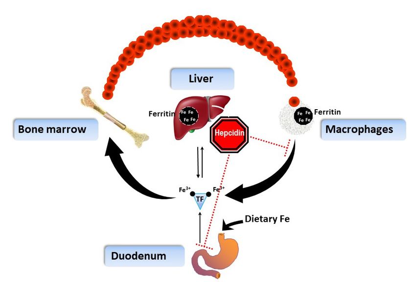
about individual animals and their general iron status [45] (Figure 2). Necropsy results have helped
validate the use of these tests (reviewed in [2,8]). This strategy holds true in other species as well,
including humans, where the details of iron metabolism and disease are well-studied [18]. It is
important to have these applicable and useful measurements for a meaningful clinical assessment,
and thus documentation of iron loading and general inflammatory state. The following parameters,
in combination and with clinical context, allow us to evaluate an animal over time and observe trends
in iron saturation status:
1. Serum iron (the amount of unbound iron in the blood)
2. Total iron-binding capacity (TIBC; indirect transferrin level)
3. Transferrin saturation (the percentage of TIBC occupied by iron) [46]
4. Ferritin (iron carrier protein; used with the exclusion of other diseases that may elevate ferritin)
Animals 2020, 10, x 6 of 29
Black rhino iron balance annual assessment
• Diet
-Test pellets, hay for iron concentrations; minimize iron
-Maximize browse, fiber sources
Transferrin Saturation (TSAT)
-Limit high vitamin C foods
• Body condition
-Body Condition Score > 3/5: consider diet reduction, increase activity
> 60%
Hemoglobin: > 8 g/dL; hematocrit: > 32%
< 60%
Voluntary phlebotomy training
Rule out alternative explanation (e.g., infectious disease)
Serum ferritin
Wild BR = 290.54 ± 247.4 ng/ml
< 750 ug/L > 750 ug/L or trending upward
Voluntary phlebotomy training Induction phase phlebotomy: 1-2 L / week
Retest 3-6 months Maintenance phase phlebotomy: 1 L as needed to maintain TSAT < 60%
Retest 3-6 months
Figure
Figure 2. Decision
2. Decision Tree
Tree forfor approachingiron
approaching iron overload
overload disorder
disorder(IOD) assessment
(IOD) assessmentin Black rhinoceros
in Black rhinoceros
(BR) under human care. Serum ferritin in wild BR are from Miller et al. 2016 [47].
(BR) under human care. Serum ferritin in wild BR are from Miller et al. 2016 [47].
2.2.1.2.2.1.
Serum Serum Iron—Review
Iron—Review andComparative
and Comparative Evaluation
Evaluation
IronIron
is anisimportant
an important
metalmetal required
required by thebybody,
the body, but
but it is it is regulated
highly highly regulated and actively
and actively sequestered
sequestered by cells to limit oxidative stress [48]. There is no process in animals to excrete iron;
by cells to limit oxidative stress [48]. There is no process in animals to excrete iron; therefore,
therefore, excesses remain and must be stored or contained (Figure 1) [49]. Higher than normal serum
iron with low total iron-binding capacity (e.g., 100% saturation) indicates more iron is circulating in
the body than can be contained.
Iron levels are abnormal when they reach or surpass the upper limit of the reference range in
any group of animals. Serum iron values in wild BR (which we consider ‘normal’) were found to
show a wide range of potential normality, 225.6 ± 106.7 µg/dL (n = 194) [47]. Serum iron values in BRAnimals 2020, 10, 1991 6 of 28
excesses remain and must be stored or contained (Figure 1) [49]. Higher than normal serum iron with
low total iron-binding capacity (e.g., 100% saturation) indicates more iron is circulating in the body
than can be contained.
Iron levels are abnormal when they reach or surpass the upper limit of the reference range in
any group of animals. Serum iron values in wild BR (which we consider ‘normal’) were found to
show a wide range of potential normality, 225.6 ± 106.7 µg/dL (n = 194) [47]. Serum iron values in BR
under human care have a reported reference range of 204 ± 54.1 µg/dL (n = 374) [50], and from Kansas
State University Diagnostic Lab, a reference range of 84–341 µg/dL has been reported (Kansas State
Veterinary Diagnostic Laboratory; Manhattan, KS, USA). Alone, iron cannot be used to determine
overload, but levels over the normal range should concern the clinician and prompt investigation into
these other parameters. Elevated levels have been associated with overload as well as chronic liver
disease [46].
Elevated iron suggests opportunities for chronic oxidative stress to organ systems. In excess,
unbound iron is toxic and produces hydroxyl radicals and can damage cellular proteins, lipids,
and nucleic acids directly [48,51], which can cause cellular, and thus, tissue damage. Iron also plays an
important catalytic role in free radical pathology and oxidative damage that is observed in almost all
major iron loaded and non-iron loaded diseases such as cardiovascular, neurodegenerative, hepatic,
and renal diseases, as well as in cancer and aging [52]. With chronicity in humans, iron deposition
(hemosiderosis or secondary iron overload) will occur in the heart, brain, kidney, liver, joints, skin,
and endocrine system [49,53–59]. This chronic iron deposition in 60 BR necropsied was in the liver,
spleen, bone marrow, and lungs, with some found in the small and large intestine, lymph nodes,
and endocrine glands [2]. Lung involvement is unique to BR [1,37,58,60]. Hemosiderosis causes
disease in several other species, but notably primates (e.g., callitrichids [61], lemurs [60]), birds [62],
bats [63], and dolphins [64,65]) and is associated secondarily with other infectious and inflammatory
diseases [66,67].
2.2.2. Total Iron-binding Capacity (TIBC) and Transferrin Saturation—Review and Comparative
Species Evaluation
Transferrin is the main protein that binds iron for transport and can be measured directly through
immunochemical assays or indirectly through lower-cost colorimetric assays and molar calculation of
TIBC; usually, the latter is used (TIBC) and reflects the amount of transferrin available to bind iron in
circulation [68]. Under inflammatory conditions, this value decreases [69]. The UIBC (unsaturated
iron-binding capacity) may also be determined as part of TIBC assessment methods. Recent work noted
challenges with a methodology of assaying TIBC in BR involving pH shifts; however, different methods
may have more success [70,71]. While there are a wide variety of methods possible for TIBC or
transferrin, assessments validate the equivalence of either approach [68,72,73]. Alone, TIBC is not
meaningful until compared to serum iron, where the ratio known as transferrin saturation or TSAT
(100× serum iron/TIBC) corresponds to a saturation level that is a much better indicator of overload in
the individual.
Typically, normal serum TSAT is 20–50% in mammals [42,43,46,74]. Johnson et al. [65] suggested
that >60% is a level of concern for overload in dolphins, while in humans, anything >45% is a
trigger [21,75]. In iron overload, TSAT is usually elevated >50% before serum ferritin consistently
increases [76]. However, elevated levels of transferrin saturation are a clear indication of iron
dysregulation and increased load. TSAT is a more sensitive indicator of initial iron loading than ferritin
and given as a first-line test in humans but never evaluated in isolation [71]. It is also recommended
to take blood samples before morning feeding, so serum iron is not impacted by iron diet intake,
which can impact the TSAT [71].Animals 2020, 10, 1991 7 of 28
2.2.3. Ferritin
Iron is vital for life, and as mentioned above, it is critical to sequester iron when in excess;
cellular ferritin performs that function of detoxification [77]. In fact, ferritin is solely an iron storage
protein [49]. If iron were not present or available, ferritin levels could not be elevated, especially as there
is a distinctly characterized genetic signaling pathway for regulating its production in each cell [78,79].
Hyperferritinemia in humans is defined by serum values >400 µg/L in males and >300 µg/L in females,
but without clinical context, the value alone is difficult to interpret [80]. In BR, wild animal values
are 290.5 ± 247.4 ng/mL [47]; therefore, consistently increasing or elevated values (e.g., >750 ng/mL;
Figure 2) suggest an abnormal state. Ferritin itself is also a well-established marker of inflammation,
and its dynamic nature shifts with non-iron storage-related diseases, sometimes resulting in episodic
rather than chronic elevation [49]. However, given that there are no other diseases species-wide other
than iron storage accounting for hyperferritinemia to the degree observed, it is very reasonable to
weigh heavily on the chronic contribution of iron to elevated levels of ferritin as part of a holistic
approach to IOD. Additionally, the continued upward rise of ferritin over time or a sustained elevated
level is not likely with a fluctuating inflammatory condition such as a wound or infection. There is a
limit on the production of ferritin (called apoferritin before iron is held within its nanocage structure),
as a body can only produce so much of the protein at a time, so extremely high levels in circulation
would indicate either a human error in measurement or the leakage of ferritin from iron-damaged
cells [27]. This leakage is important as it further releases iron in an unbound form causing greater,
continual damage [77]. Necropsies in BR show cellular injury and necrosis with evidence of released
intracellular ferritin [2], which accounts for high serum ferritin concentrations, greatly exceeding
apoferritin rates of synthesis [2,23]. While serum ferritin is not a perfect representation of iron stores,
and hematologists in human iron studies have long worked to characterize iron carrier turnover and
dynamics, ferritin and its measurement is still inextricably tied to further understanding iron overload.
The most practical and reliable ferritin test in BR is a commercially available equine assay
(developed by [81]; available at the Kansas State Veterinary Diagnostic Laboratory (KSVDL). It has been
shown to correlate well with BR ferritin in multiple studies [81,82]. Recent research has verified that
although ferritin can vary across species, BR ferritin was found to be 91.4% identical to horse ferritin
on a protein basis [23,83]. Based on the very high degree of homology between BR and horse L-ferritin
genetically, it is likely that the polyclonal antibodies raised against horse L-ferritin will cross-react with
BR ferritin, and the KSVDL assay is appropriate, and the data are valuable. The reference values the
laboratory reports, however, are not ‘normal’, but rather represent all reported values from submissions
in animals under human care, which are biased heavily with iron-overloaded animals.
2.3. Perspectives on Integrative Monitoring
The recent notion that ferritin should be a stand-alone diagnostic and that its value in monitoring
IOD should be dismissed remains problematic [70,84,85]. In fact, in veterinary medicine, few diseases
utilize a single serum biochemical marker for the diagnosis, let alone progression, of a disease state
(e.g., renal disease). Changes in elevated ferritin (all cases >500 ng/mL) were considered not predictive
of disease relative to onset of clinical signs in IOD cases; in those cases highlighted, multiple potential
factors were involved (disease state, diet, environment, and unknowns) that understandably showed a
variable pattern of ferritin across time [84]. Regardless, all elevated ferritin levels still indicated a state of
iron overload, even if fluctuating but without the context of other holistic information, including TSAT
or the ever-challenging antemortem liver biopsy. Herein, we have sufficient evidence-based information
to support the continued use of ferritin, as outlined, as a part of a practical diagnostic and for monitoring
of IOD in BR. It is important to characterize ferritin dynamics across time in the context of other assays.
In humans, there is considerable informed knowledge on iron metabolism and regulation,
especially as more signaling molecules like hepcidin were discovered [86]. However, the overall
basic monitoring strategy has not changed. A number of groups have attempted to measure BR
hepcidin directly with no published success [87,88], and as noted previously, iron deposition in tissuesAnimals 2020, 10, 1991 8 of 28
demonstrates hepcidin has some degree of functionality in BR [1,23]. While the search remains for more
informative diagnostics, including hyaluronic acid, micro RNA, microbial gut communities, and labile
plasma iron measurement [89,90], none has shown clear promise as a new IOD marker beyond those
that continue to be the benchmark currently across species. Therefore, continued monitoring of
known direct iron-related panel parameters, including ferritin and transferrin saturation, is sensible.
These tests are relatively inexpensive, and longitudinal information on individuals will demonstrate
and elucidate patterns of loading.
Several other diagnostic tools have been used in the evaluation of iron disease or patient status
affected by iron disease. Biopsies for histology and tissue iron concentration have been used in many
species but require invasive and technically-challenging procedures to obtain samples antemortem.
In the case of liver iron concentration, a larger piece of tissue is required than feasible to collect from
BR unless post-mortem. Liver iron concentration has also fallen out of favor in human medicine due to
the risk of complications [91].
2.4. Inflammatory Biomarker Connections to IOD
Oxidative damage and inflammation, which is inevitable with elevated iron, has had the attention
of many rhinoceros researchers [70,82,92]. While these markers also do not indicate IOD, they can
imply the negative effects of IOD and may connect with disease monitoring. Biomarkers that show
some statistical significance over ‘normal’ include serum amyloid A (SAA), tumor necrosis factor
α (TNFα), glucose to insulin ratios [82], superoxide dismutase, glutathione, and reactive oxygen
metabolites in serum measured via electrophoresis (specifically α2 macroglobulin) [70,93]. Additional
specific testable biomarkers include C-reactive protein (CRP), ceruloplasmin, and haptoglobin [94,95],
but ceruloplasmin has not been formally tested, and haptoglobin has shown no obvious benefit
in BR [81]. In assessing four animals across eight years at Disney’s Animal Kingdom® (n = 91
samples/animal), correlations were poor between ferritin or transferrin saturation with ceruloplasmin
and haptoglobin (0.000002 < R < 0.26), despite animals with well-documented IOD [45]. Some of these
biomarkers may have utility in holistic patient assessment and may help provide a ‘bigger picture’ of
total patient health but are not specific or sensitive enough to provide a diagnosis of IOD versus another
inflammatory disease. While trend evaluation may be useful, relative values are not understood at
this time. Additionally, these measurements do not allow immediate clinically useful information,
and many may not be commercially available at this point. Another avenue of investigation based
on some success in human medicine would be the impact of α-lipoic acid in protecting BR from the
induced oxidative damage of iron overload [96,97]. While these assays are not the current solution
to understanding IOD in BR, future research may circle back to how they are connected to iron
accumulation, potentially as diagnostic technologies evolve.
2.5. Recommendations for Diagnosis
In summary, iron overload can be assumed with elevated serum ferritin levels when corroborated
by elevated serum iron and transferrin saturation, especially when confirmed through long-term
monitoring. As a general reference, average transferrin saturation and serum ferritin concentrations
in free-ranging BR are 34% and 180 ng/mL, respectively [42]. Black rhino in a free-ranging situation
tested with the same assay used by KSVDL in the US were also found to have serum iron ranging from
225.6 ± 106.7 µg/dL and ferritin values ranging 290.5 ± 247.4 ng/mL (n = 194) [47]. Further medical
evaluation and testing and diet evaluation are recommended if transferrin saturation is elevated to
>60% and serum ferritin concentrations are >750 ng/mL consistently, or if these values are trending
upward (Figure 2).
3. Evidence-Based Nutrition Practices
Nutrition is an integral component of preventing IOD; thus, the assessment of dietary iron
concentration and other nutritional factors is critical for iron balance in BR. BR likely evolved with aAnimals 2020, 10, 1991 9 of 28 low-iron diet due to low iron concentrations (300 ppm) as well as a poor representation of the nutrient and plant defense chemical compositions (e.g., polyphenols and alkaloids) found in wild browse [100]. The source of high iron concentrations in diets is pelleted feed, as dietary constituents and soil contamination can produce feeds high in iron, even when formulated to be low in iron (e.g., 4000 ng/mL and transferrin saturation at 100%) [45]. Within a month post-diet change, serum ferritin decreased in all four animals, regardless of concomitant phlebotomy [50]. The other two younger animals showed reduced IOD biomarkers. Currently, both animals show no evidence of IOD compared to age-matched conspecifics under a more traditional diet. These results highlight the significant impact that diet modification can have on iron loading in the BR (Figures 3 and 4).
including two rhinos that were markedly iron-overloaded (ferritin >4000 ng/mL and transferrin
saturation at 100%) [45]. Within a month post-diet change, serum ferritin decreased in all four
saturation at 100%) [45]. Within a month post-diet change, serum ferritin decreased in all four
animals, regardless of concomitant phlebotomy [50]. The other two younger animals showed reduced
animals, regardless of concomitant phlebotomy [50]. The other two younger animals showed reduced
IOD biomarkers. Currently, both animals show no evidence of IOD compared to age-matched
IOD biomarkers. Currently, both animals show no evidence of IOD compared to age-matched
conspecifics under a more traditional diet. These results highlight the significant impact that diet
conspecifics
Animals 2020, 10,under
1991 a more traditional diet. These results highlight the significant impact that diet
10 of 28
modification can have on iron loading in the BR (Figures 3 and 4).
modification can have on iron loading in the BR (Figures 3 and 4).
Figure 3. Transferrin Saturation (%) across time of two male black rhinoceros (BR1, BR2) at Disney’s
Figure 3. Transferrin
Figure 3. Transferrin Saturation (%)
®. * Saturation (%) across
across time
time of
of two
two male
male black
black rhinoceros
rhinoceros (BR1,
(BR1, BR2)
BR2) at
at Disney’s
Disney’s
Animal
Animal Kingdom
Kingdom ® . * Major
Major diet
diet change
change occurred
occurred on
on 11
11 October
October 2009
2009 for
for both
both animals.
animals. a
a Phlebotomy
Phlebotomy
Animal Kingdom . * Major dietb change occurred on 11 October 2009 for both animals. Phlebotomy
® a
began onBR1—March
began BR1—March 4,2012; 2012;b Phlebotomybeganbegan onBR2—20
BR2—20 May2016.
2016.
began on
on BR1—March 4, 4, 2012; b Phlebotomy
Phlebotomy began on on BR2—20 May
May 2016.
Figure4.4. Serum
Figure Serum ferritin
ferritin (ng/mL)
(ng/mL) across
across time
time of
of two
two male
male black
black rhinoceros
rhinoceros (BR1,
(BR1, BR2)
BR2)at
atDisney’s
Disney’s
Figure 4.
Animal Serum ®
Kingdom ferritin
. * (ng/mL)
Major diet across
change time of
occurred two
on 11male black
October rhinoceros
2009 for both (BR1,
animals. a Phlebotomy
BR2) at Disney’s
Animal Kingdom®. * Major diet change occurred on 11 October 2009 for both animals. a Phlebotomy
Animal
began onKingdom ®. * Major
BR1—4 March 2012; b Phlebotomy
diet change occurred
beganonon11BR2—20
OctoberMay
20092016.
for both animals. a Phlebotomy
began on BR1—4 March 2012; b Phlebotomy began on BR2—20 May 2016.
began on BR1—4 March 2012; b Phlebotomy began on BR2—20 May 2016.
3.3. What Are the Best Practices for Feeding Black Rhinos?
3.3. What Are the Best Practices for Feeding Black Rhinos?
3.3. What Are the Best Practices
An appropriate diet for forBRFeeding Black Rhinos?
maximizes animal welfare, lifespan, and reproductive output.
An appropriate
Recommendations diet
for diet
feeding for BR maximizes animal welfare, lifespan, and reproductive output.
An appropriate for must consider notanimal
BR maximizes only iron but also
welfare, the many
lifespan, and interconnected
reproductivenutrients
output.
Recommendations
and metabolic concernsfor feeding must consider not only iron but also the many interconnected
Recommendations for which
feeding may exacerbate
must consideriron-related
not only issues,
iron butsuchalso
as obesity.
the many Based on empirical
interconnected
nutrients the
evidence, andfollowing
metabolic concerns which may exacerbate iron-related issues, such as obesity. Based
nutrients and metabolicspecific
concernsfeeding
whichguidelines for BR are
may exacerbate recommended
iron-related issues,[50]:
such as obesity. Based
on empirical evidence, the following specific feeding guidelines for BR are recommended [50]:
•on empirical evidence,
Iron should the following
be limited. specific feeding
Iron concentration in theguidelines for BR are recommended
diet is recommended [50]:mg/kg
to not exceed 300
dry matter (DM) or about 6 g of iron per day for a 1300 kg BR fed 1.5% BW in DM [5]. Based on
the availability of feed items such as low-iron pelleted feed and browse (Table 1), a dietary iron
concentration less than 300 mg/kg (dry matter basis) is a practical recommendation for BR [5,108].
Consider testing exhibit soil and vegetation to ensure they are not significant sources of iron in the
diet of BRs [5,105].Animals 2020, 10, 1991 11 of 28
• Monitor individual body weight and body condition. Feed 1–3% of body weight (BW) on an as
fed basis, 1–2% on a dry matter basis. Maintaining appropriate body weight is critical as iron
imbalance and obesity are presumed to be related; iron balance also is implicated with metabolic
syndrome and associated negative health impacts [82]; however, the exact mechanism is not
yet clear. Individual assessment of BR for bodyweight regularly across time (ideally weekly)
and tracking with diet consumption is recommended [50,100]. Body condition scoring systems
(e.g., a 1–5 scoring system based on ~seven body areas) are subjective, with varied recommendations
on what is considered ideal depending on housing conditions along with medical and physiological
considerations. Typically, a score of 3–4 out of a 5-point scale is considered ideal under human
care. [109,110]. To optimize health, adjust diets as needed to ensure animals are within a target
body weight range set for the individual animal.
• Feed at least twice daily. It is recommended to feed pellets in two feedings each day with forage
to ensure maximum absorption of macronutrients [108]. Iron is not the only nutrient to consider
in feeding complex diets to rhinos under human care. A single feeding would not be ideal
for multiple reasons, including digestive efficiency, microbial community maintenance, satiety,
and natural foraging behaviors.
• Feed appropriate pelleted feeds. The pellets milled for zoo animals vary widely in nutrient
composition, and not all available pellets are appropriate for browsing species. Pellet formulations
for BR are recommended to be high in fiber and low in starch and soluble sugars (Neutral Detergent
Fiber (NDF) = 40–60%) [5]. Starch and sugar must be limited as these items can be associated with
severe dental plaque, have metabolic impacts, and contribute to obesity [50,111,112]. A maximum
of one-third of total calories is recommended to come from a pelleted concentrate. This limit avoids
high pellet inclusion rates, which could be negative for dental health, body weight, and proper
digestive health due to lack of long particle fibers [99].
• Alfalfa hay should be limited. This recommendation is due to its high protein, calcium, and iron,
which can also create diarrhea and colic [5,50,108]. Conversely, low-quality hay (straw, wet/moldy,
low nutrient content) is also not recommended due to the risk of intestinal impaction and/or colic.
The iron in alfalfa is also held in a potentially highly bioavailable form (plant ferritin) [50].
• Maximize browse and provide access to hay. Preferably high quality roughage, ideally grass
hay — not legume-based, as well as clean water and salt ad libitum [108]. Browse options may
vary based on season and region, with options to freeze or ensile [110,113]. As browse best
approximates the natural physical form of BR diets; it has the potential for iron-binding [114].
• Total dietary vitamin E concentrations should be 150–200 IU/kg diet. Extra supplementation may be
necessary in addition to vitamin E in pelleted feed dependent on serum evaluation [17]. Vitamin E
is a critical antioxidant that protects against ROS created by and including iron [50,115,116]. As BR
lack some natural antioxidant production, ensuring dietary alpha-tocopherol (vitamin E) serves as a
necessary preventive [50,115,116].
• Phosphorus levels in the serum should be monitored and supplemented where appropriate.
BR have a predisposition to deficiency and continued concern for hemolytic issues; additionally,
there is a link between phosphorus and iron metabolism [8,117]. Supplementation of monosodium
phosphate and/or wheat bran in addition to phosphorus provided in a pelleted diet is recommended
based on serum assessment. Naturally low phosphorus carriage in BR RBC (2–5% other
mammals) [116] is thought to be connected to RBC fragility and potentially elevated RBC
turnover [8,14,23]. In support of supplementation, higher levels of dietary phosphorus have been
documented to combat anemic hemolytic crises in this species [8,23,117].
• The calcium to phosphorus (Ca:P) ratio of the diet should be 2:1 (no less than 1:1). A well-formulated
pellet will provide appropriate calcium and phosphorus to meet the nutrient requirements of BR.
An appropriate ratio eliminates the need for calcium supplementation, which can be contaminated
with iron [102]. Grass hay typically is 1:1 and alfalfa 3:1, the latter of which can lead to
hypercalcemia and hypophosphatemia. The amount of phosphorus added as a supplementAnimals 2020, 10, 1991 12 of 28
should not unbalance the Ca:P ratio in the diet, in the amounts recommended based on body
weight. The diet is balanced primarily with the pelleted portion, which is the majority of the
dry matter of the diet and typically has the optimal 2:1 ratio (Table 1). Inverted serum Ca:P
ratios are incredibly rare in rhinos; instead, hypercalcemia cases are far more common. As black
rhinos physiologically appear to have an increased need for phosphorus, which is utilized for
RBC turnover, they appear able to maintain serum Ca:P ratios of 2:1 despite a potential intake
between 1:1 to 2:1.
• Avoid non-specific mineral supplements and mineral salt blocks. Plain salt blocks have minimal
to no iron content and are appropriate [50].
• Limit high vitamin C diet items (e.g., citrus fruits). Also, avoid feeding these foods at the same
time as pellets due to increased iron availability in the presence of acidic foods such as vitamin C
(Table 1) [118].
• Training and enrichment diet items should be low in sugar, starch, and iron. Target less than
10% of the total diet comprising of training and enrichment foods. Take into consideration
high-sugar, high-starch, and high-iron items (such as molasses-based foods), which often are
included in balanced diets for BR (Table 1) [50].
Table 1. Moisture (%), dry matter (DM;%), iron (ppm DM), and vitamin C (mg/100 g as fed; AF),
calcium (%DM), and phosphorus (%DM) values in example diet items commonly used for black
rhinoceros. Feed composition can be quite variable depending on harvest location, manufacturer,
and season. Nutrients included in the table are not sufficient to balance an animal’s diet or to evaluate
inclusion as training or enrichment food items; thus, it is recommended to consult a nutritionist.
For example, although bananas are lower in iron and vitamin C than cauliflower, bananas are high in
starch (~23% DM) and thus should be limited in favor of metabolic health. Another cautionary example
is appreciating the high moisture in produce such as leafy greens, which dilutes nutrient concentrations
in the AF product. Thus, produce items generally are not used to balance nutrient concentrations such
as calcium or phosphorus.
Dry
Moisture Iron Vitamin C Calcium Phosphorus
Matter
mg/100 g
Food Item % % ppm (DM) * (%DM) (%DM)
(AF) **
Pelleted feed examples:
Mazuri ADF 25
10.5 89.5 652 nd 1.51 0.95
Herbivore diet
Mazuri ADF 16
8.2 91.8 490 nd 1.16 0.84
Herbivore diet
Mazuri Browser
10.4 89.6 222 nd 1.22 0.68
Rhino Cube 5Z1P
Produce examples:
Cucumber (raw,
97.6 2.4 86 3.2 0.89 1.35
whole)
Carrot (raw, whole) 88.9 11.1 35 5.9 0.31 0.25
Celery (raw, whole) 95.1 4.9 29 3.1 0.98 0.51
Sweet potato (raw,
79.8 20.2 22 2.4 0.36 0.29
whole)
Apple (raw, whole) 88.0 12.0 11 4.6 0.05 0.08Animals 2020, 10, 1991 13 of 28
Table 1. Cont.
Dry
Moisture Iron Vitamin C Calcium Phosphorus
Matter
mg/100 g
Food Item % % ppm (DM) * (%DM) (%DM)
(AF) **
Produce with higher vitamin C or iron level examples:
Green Leaf lettuce
94.9 5.1 278 9.2 0.74 0.66
(fresh, raw)
Spinach (fresh, raw) 91.4 8.6 264 28.1 1.02 0.73
Romaine (fresh,
95.6 4.4 152 11.5 0.73 0.67
raw)
Green beans (fresh,
92.2 7.8 101 12.2 0.55 0.53
raw)
Cantaloupe melon
93.1 6.9 74 36.7 0.11 0.17
(fresh, whole)
Cauliflower (raw,
94.0 6.0 60 48.2 0.55 0.75
whole)
Tomatoes (raw,
95.7 4.3 53 13.7 0.18 0.47
whole)
Honeydew melon
91.6 8.4 37 18.0 0.11 0.33
(fresh, whole)
Watermelon (fresh,
92.3 7.7 33 8.1 0.14 0.33
whole)
Banana (raw, whole
82.4 17.6 15 8.7 0.07 0.12
with peel)
Orange (raw, whole
82.6 17.4 14 71.0 0.57 0.13
with peel)
Hay/Fresh Browse examples:
Alfalfa Hay 12.0 88.0 386 nd 1.84 0.31
Coastal
10.0 90.0 52 nd 0.57 0.19
Bermudagrass Hay
Timothy Hay 11.0 89.0 48 nd 0.54 0.09
Mulberry (whole
67.6 32.4 84 nd 1.7 0.38
branch fresh)
Willow (whole
63.2 36.8 63 nd 0.77 0.13
branch fresh)
Spineless cactus
91.5 8.5 22 nd 3.67 0.17
pads (Opuntia)
Supplement examples:
Dicalcium
4.6 95.4 12,200 nd 23.4 19.7
Phosphate
Trace mineral block 0.1 99.9 1790 nd 0.3 0.05
Dried Beet Pulp
7 93 731 nd 1.15 0.1
with molasses
Molasses 8.0 92.0 577 0.0 0.18 0.02
Wheat bran 8.4 91.6 186 nd 0.15 1.48
Steamed Rolled
8.5 91.5 41 0.0 0.05 0.48
Oats
* Dry matter, moisture, calcium, phosphorus, and iron determined on feed at Disney’s Animal Kingdom® ,
analyzed by Dairy One Laboratories (Ithaca, NY). ** Vitamin C values sourced from the USDA database (https:
//www.nal.usda.gov/usda-food-composition-database). “nd” indicates the nutrient value for the food item was
not determined.Animals 2020, 10, 1991 14 of 28
4. Treatment and Prevention
The timeframe for overload is gradual, highly variable, and determined by individual metabolism,
iron intake, historical metabolism changes (e.g., pregnancy, an established iron sink), and other disease
states. It can take years for iron to accumulate before any change is notable; onset is not predictable.
When is it too late? The simple answer is ‘never’; treatment can always be initiated. However,
the degree of overload will affect the results and the anticipation of when to see a change in serum
biochemical parameters. If an animal is heavily overloaded, the time to see a difference may take
months compared to an animal relatively with less iron. An expectation for an immediate drop in
serum parameters with any method chosen is unrealistic. Active treatment involves minimizing
dietary iron (see Section 3. Evidence-Based Nutrition Practices) and established methods to remove
iron from any mammalian species; phlebotomy and chelation therapies. Prevention would be ideal,
but maintenance is most likely given limitations of available diet and browse species, which provide a
constant influx of iron.
4.1. Hematologic Sampling Recommendations
A comprehensive preventive health program for BR is recommended and, in addition to
dietary management, includes routine hematologic sampling for continuous diagnostic evaluation
(Figures 2–4). Newly acquired animals to a facility should have a baseline sample collected and
archived. Serial samples are important for monitoring trends; quarterly samples, as a recommended
example, provide a robust series of data to review and to confirm or monitor IOD. At the very
least, annual samples including iron panels as well as a minimum database (complete blood count
and serum chemistry) should be conducted to be able to catch a change in biochemical parameters
(e.g., normal hematocrit >32% and hemoglobin >8 g/dL) and organ function. These tests also help
with a routine overall assessment of the animal. Management and treatment strategies will depend on
how much the animal is iron-loaded (Figure 2).
4.2. Therapeutic Large Volume Phlebotomy
Therapeutic large volume phlebotomy (TLVP) is commonly used in human medicine [119–122],
and it has proven effective in dogs where ferritin and hepatic iron were reduced with phlebotomy
treatment [123] and in BR [45,110]. TLVP has been used at multiple institutions accredited
by the Association of Zoos and Aquariums (AZA; e.g., Columbus Zoo and Aquarium [124],
Denver Zoo [124,125], Milwaukee County Zoo [108,126]) and extensively at Rotterdam Zoo in the
Netherlands [110]. In the authors’ experience, TLVP has shown to be a safe and effective strategy
for not only reducing excess storage iron in overloaded patients but also maintaining normal iron
homeostasis as animals age (Figures 3 and 4). By combining an applied model for large volume
blood collection in Atlantic bottlenose dolphins (Tursiops truncatus), and clinical models in humans
with iron overload (Johnson et al., 2009; D. Paglia, 2004), parameters were extrapolated for voluntary
TLVP (VLTVP) in BR at Disney’s Animal Kingdom® (DAK) [127]. In dolphins, during the induction
phase, 5–8 mL/kg (1–2 L) of blood was removed per weekly session with varying times for transition
to a maintenance phase (often several months). In the maintenance phase, one liter was collected
until transferrin saturation wasAnimals 2020, 10, 1991 15 of 28
on the degree of overload; monitoring serum iron panels with each session will help determine the
frequency and volume of phlebotomy. Monthly VTLVP can be accomplished. Due to our preventive
strategy, IOD is well-managed in both rhinos, and animals need only quarterly phlebotomies based on
serum biomarkers (BR 1 and 2; Table 2; Figures 3 and 4). Weekly sessions are achievable; however,
this strategy may hinder animal compliance. One successful strategy for maintenance of low overload
has been a quarterly goal of 2–6 L (dependent on the degree of IOD) with sessions occurring as tolerated
by the individual until the minimum volume is achieved within the quarter, regardless of the number
of sessions.
Table 2. Individual BR (BR 1, BR 2) average quarterly voluntary therapeutic large volume phlebotomy
(VTLVP) volumes taken per session at Disney’s Animal Kingdom® (DAK). Averages based on several
(2–3) sessions within a quarter (Q).
VTLVP at DAK: Average Quarterly Volumes
BR 1 2013 2014 2015 2016 2017 2018 2019 2020
Q1 (January–March) 10.0 L 5.5 L 7.0 L 4.0 L 3.7 L 4.0 L 1.0 L 4.0 L
Q2 (April–June) 4.0 L 3.0 L 6.0 L 3.5 L 4.0 L 4.0 L 0.5 L NA
Q3 (July–September) 5.5 L 0.5 L 0.0 L 4.0 L 0.0 L 4.0 L 4.0 L 4.0 L
Q4 (October–December) 7.5 L 3.0 L 2.5 L 0.0 L 0.0 L 4.0 L 3.0 L -
BR 2 2013 2014 2015 2016 2017 2018 2019 2020
Q1 (January–March) NA NA NA NA 6.0 L 2.0 L 2.0 L 2.0 L
Q2 (April–June) NA NA NA NA 3.0 L 3.0 L 3.0 L NA
Q3 (July–September) NA NA NA NA 2.0 L 4.0 L 4.0 L 0.0 L
Q4 (October–December)
Animals 2020, 10, x
NA NA NA NA 1.0 L 2.0 L15 of 29 4.0 L -
“NA” indicates that no VTLVP was performed.
Table 2. Individual BR (BR 1, BR 2) average quarterly voluntary therapeutic large volume phlebotomy
(VTLVP) volumes taken per session at Disney’s Animal Kingdom® (DAK). Averages based on several
(2–3) sessions within a quarter (Q).
4.3. Techniques for VTLVP VTLVP at DAK: Average Quarterly Volumes
BR 1 2013 2014 2015 2016 2017 2018 2019 2020
To perform VLVP, aseptic techniques should be followed. A4.0
Q1 (January–March)
Q2 (April–June)
10.0 L
4.0 L
5.5 L
3.0 L
7.0 L
6.0 L
4.0 L
3.5 L
3.7 L
4.0 L
certified
4.0 L
L
1.0 L
0.5 L
veterinary technician (CVT)
4.0 L
NA
or venipuncture-trained animal keeper should perform the venipuncture
Q3 (July–September)
Q4 (October–December)
5.5 L
7.5 L
0.5 L
3.0 L
0.0 L
2.5 L
4.0 L
0.0 L
0.0 L
0.0 L
4.0 L
4.0 L
4.0 L
3.0 L
4.0 L
-
for VTLVP. The radial vein
(medial forelimb crossing the carpus) and metacarpal vein (lower
BR 2
Q1 (January–March)
2013
NA
2014
NA
2015
NA
2016
NA
2017
6.0 L
2018 dorsal
2.0 L
2019
2.0 L
2020hindlimb) are used due to
2.0 L
stability and optimal flow (Figure 5). An 18 gauge (ga), 1 –2 inch3.0
Q2 (April–June)
Q3 (July–September)
NA
NA
NA
NA
NA
NA
1
NA 3.0 L
4 2.0 L
NA
intravenous
4.0 L
L 3.0 L
4.0 L
NA
0.0 L
catheter (SURFLO® I.V.
Catheter, Terumo Medical Corp., Somerset, NJ, USA, 08873) is commonly
Q4 (October–December) NA NA NA NA 1.0 L 2.0 L 4.0 L - used. An 18 ga 1–1 1 inch
“NA” indicates that no VTLVP was performed. 4
hypodermic needle can replace the catheter. Catheterization does not require securing or anchoring and
4.3. Techniques for VTLVP
is advantageous in that the plastic
To perform “over-the-needle”
VLVP, aseptic techniques should be cannula lacksveterinary
followed. A certified a bevel-edged
technician lumen, which allows
(CVT) or venipuncture-trained animal keeper should perform the venipuncture for VTLVP. The
it to rest parallel within the
radialvessel
vein (medial with
forelimbreduced risk
crossing the carpus) and of laceration
metacarpal andhindlimb)
vein (lower dorsal occlusion are along the vessel wall
used due to stability and optimal flow (Figure 5). An 18 gauge (ga), 1¼–2 inch intravenous catheter
(Figure 6). Large bore tubing
(SURFLO (MILA 180
I.V. Catheter,
® cmMedical
Terumo blood Corp.,collection
Somerset, NJ, USA, set,
08873)MILA
is commonly International,
used. An Inc., Florence, KY,
18 ga 1–1¼ inch hypodermic needle can replace the catheter. Catheterization does not require
USA, 41042) is connected securing
to the hub of
or anchoring and isthe needle/catheter;
advantageous the opposite
in that the plastic “over-the-needle” cannula lacksend
a bevel- of the tubing is equipped
edged lumen, which allows it to rest parallel within the vessel with reduced risk of laceration and
with an IV spike and drip chamber
occlusion which
along the vessel wall (Figureis inserted
6). Large into180the
bore tubing (MILA rubber
cm blood stopper
collection set, MILA on the top of the 1 L
International, Inc., Florence, KY, USA, 41042) is connected to the hub of the needle/catheter; the
glass evacuated collectionopposite
container (B.isBraun
end of the tubing equipped with Medical,
an IV spike andInc., Bethlehem,
drip chamber which is inserted PA,
into theUSA, 18018). Historically,
rubber stopper on the top of the 1 L glass evacuated collection container (B. Braun Medical, Inc.,
a 500 mL collection bottleBethlehem,
proved ideal,
PA, USA, 18018). but these
Historically, arecollection
a 500 mL not bottle
currently
proved ideal, available.
but these are not
currently available.
(a) (b)
Figure 5. Phlebotomy Sites/Venous Access. Options for venipuncture sites with larger vessels that
tolerate a large volume for blood collection include the metacarpal vein; (a) lower distal hindlimb or
the radial vein; (b) medial forelimb crossing the carpus. Photos were taken in an off-exhibit animal
holding area.Animals 2020, 10, x 16 of 29
Figure 5. Phlebotomy Sites/Venous Access. Options for venipuncture sites with larger vessels that
Animals 2020, 10, 1991 tolerate a large volume for blood collection include the metacarpal vein; (a) lower distal hindlimb or 16 of 28
the radial vein; (b) medial forelimb crossing the carpus. Photos were taken in an off-exhibit animal
holding area.
(a) (b)
Figure 6. Catheterization of the Radial Vein ((a): zoomed out; (b): zoomed in). An 18 gauge catheter
Figure 6. Catheterization
placed in theof the
right Radial
front Vein
radial vein; ((a):
medial zoomed
forelimb. out;
Anchoring (b):
of the zoomed
catheter in). An
is not necessary, and18 gauge catheter
placed venous
in the right frontaccess remainsvein;
radial patent without
medial securing. Large bore Anchoring
forelimb. tubing aids in rapid
ofblood
the flow into the is not necessary,
catheter
negative-pressure collection containers. Photos were taken in an off-exhibit animal holding area.
and venous access remains patent without securing. Large bore tubing aids in rapid blood flow into
Specific equipment may need to change based on market availability. Therefore, wound
the negative-pressure collection containers. Photos were taken in an off-exhibit animal holding area.
drainage bottles could be used (Evolution™ Pre-vac wound drainage bottle, Pacific Hospital Supply
Co., Ltd., Taipei 112, Taiwan), though these lack a rubber stopper and have a female-type connection
Specific equipment mayspike-type
port; therefore, need toIVchange based
sets are not on market
compatible. availability.
Instead, two large bore IV Therefore,
extension sets wound drainage
(JorVet™ 30” a IV extension set, Jorgensen Laboratories, Inc., Loveland, CO, USA, 80538) connected
bottles could be used (Evolution™ Pre-vac wound drainage bottle, Pacific Hospital Supply Co., Ltd.,
with a double male Luer lock adapter (Double male Luer lock, Smiths Medical, Dublin, OH, USA,
Taipei 112, Taiwan),
43017)though
are requiredthese
(Figure lack
7). Usinga anrubber
18 ga bore,stopper andtohave
a 75 mL/minute a female-type
100 mL/minute flow rate can connection port;
be achieved with a standard drip-rate; theoretically, this would increase with pressure (Terumo
therefore, spike-type IVCorp.,
Medical setsSomerset,
are notNJ,compatible. Instead,
USA, 08873). As blood volumetwo
reacheslarge
the 1 Lbore
capacity IV
in aextension
bottle, blood sets (JorVet™ 30”
a IV extension set,flow
Jorgensen Laboratories,
slows. The tubing is clamped off,Inc.,
and theLoveland,
full container CO, USA,for80538)
is exchanged an emptyconnected
container with a double
until the desired total blood volume is obtained. Observing a decrease in blood flow may indicate
male Luer lock adapter (Double male Luer lock, Smiths Medical, Dublin, OH,
occlusion of the tubing or vessel, which should be corrected with slight gentle manipulation of the USA, 43017) are required
IV tubing, catheter/needle, or slightly shifting the animal’s position. Fibrin
(Figure 7). Using an 18 ga bore, a 75 mL/minute to 100 mL/minute flow rate can be achieved with a clots, observed as white-
pink foamy precipitant in the collection lines, have been noted but do not seem to affect flow rate or
standard drip-rate; theoretically,
collection this
volumes. After would increase
catheter/needle removal, thewith pressure
venipuncture site (Terumo
is held off withMedical
manual Corp., Somerset,
pressure for several minutes to allow hemostasis and prevention of a hematoma.
NJ, USA, 08873). As blood volume reaches the 1 L capacity in a bottle, blood flow slows. The tubing is
As noted above, to improve success, multiple venipuncture sites in different limbs, preferable
clamped off, and the fulllimbs,
opposing container
have beenis usedexchanged
in one session.for an empty
Venipuncture container
site sensitivity can beuntil thewith
mitigated desired total blood
topical anesthetic cream (lidocaine/prilocaine; EMLA cream 5%, AstraZeneca AB, Södertälje,
volume is obtained. Observing a decrease in blood flow may indicate occlusion
Sweden), or spray (Gebauer’s ethyl chloride® Mist, Gebauer Company, Cleveland, OH, USA, 44128).
of the tubing or vessel,
which should be corrected with slight gentle manipulation of the IV tubing, catheter/needle, or slightly
shifting the animal’s position. Fibrin clots, observed as white-pink foamy precipitant in the collection
lines, have been noted but do not seem to affect flow rate or collection volumes. After catheter/needle
removal, the venipuncture site is held off with manual pressure for several minutes to allow hemostasis
and prevention of a hematoma.
Animals 2020, 10, x 17 of 29
Figure 7. Medical Supplies for Large Volume Blood Collection. Various supply options are available
Figure 7. Medicalfrom Supplies for Large
multiple medical Volume
distributors. (1) MILABlood Collection.
80” large Various
animal disposable supply
blood collection set options
with are available
from multiple medicaldrip chamber (MILA International,
distributors. (1) MILA Inc., Florence,
80” large KY, USA).
animal(2) Braun 1000 mL empty
disposable blood glass
collection set with
evacuated container (B. Braun Medical, Inc., Bethlehem, PA, USA). (3) Evolution™ UreSil® pre-vac
drip chamber (MILA International, Inc., Florence, KY, USA). (2) Braun 1000 mL empty
plastic wound drainage bottle (Pacific Hospital Supply Co., Ltd., Miaoli, Taiwan). (4) JorVet™ 30” glass evacuated
container (B. Braun large bore IV extension
Medical, set (Jorgensen Laboratories,
Inc., Bethlehem, PA, USA). Inc., Loveland, CO, USA). (5)UreSil
(3) Evolution™ ® pre-vac
Double male Luer plastic wound
lock adapter (Smiths Medical, Dublin, OH, USA).
drainage bottle (Pacific Hospital Supply Co., Ltd., Miaoli, Taiwan). (4) JorVet™ 30” large bore IV
4.4. Pharmacologic Chelation Therapy
extension set (Jorgensen Laboratories, Inc., Loveland, CO, USA). (5) Double male Luer lock adapter
Due to an animal’s ability to recycle iron, regular phlebotomy is an option for patients with iron-
(Smiths Medical, Dublin, OH, USA).
loaded tissues [128]. Other than phlebotomy or pregnancy, the chelation of iron remains the only
other option for removing excess iron. The use of synthetic iron chelating agents is crucial in long-
term management of certain human forms of iron storage and overload disease. Chelation involves
the use of a chemical compound with a high affinity and selectivity for one metal molecule, entering
the body, binding that molecule, and clearing both from the body. The ideal chelating ligand for iron
has a high binding constant and iron specificity, bioavailability, and forms an inert compound with
ferric or ferrous iron [129–131]. As there are many forms of chelators (hexadentate, tridentate, etc.),
some may bind unintended targets such as other minerals, like zinc or copper, resulting in deficiency.You can also read