EXPLORATIONS: AN OPEN INVITATION TO BIOLOGICAL ANTHROPOLOGY . EXPLORATIONS: AN OPEN ...
←
→
Page content transcription
If your browser does not render page correctly, please read the page content below
.

.

EXPLORATIONS: AN OPEN INVITATION TO BIOLOGICAL
ANTHROPOLOGY

Editors: Beth Shook, Katie Nelson, Kelsie Aguilera and Lara Braff

American Anthropological Association
Arlington, VA

2019

CC BY-NC 4.0 International, except where otherwise noted

ISBN – 978-1-931303-63-7

www.explorations.americananthro.org

Chapter 3: Molecular Biology and Genetics:
Hayley Mann, M.A., Binghamton University

Xazmin Lowman, Ph.D., University of California, Irvine

Malaina Gaddis, Ph.D.

   Learning Objectives

       • Define terms useful to molecular biology and genetics.
       • Explain and identify the purpose of both DNA replication and the cell cycle.
       • Identify key differences between mitosis and meiosis.
       • Outline the process of protein synthesis including transcription and translation.
       • Use principles of Mendelian inheritance to predict genotypes and phenotypes of future generations.
       • Explain complexities surrounding patterns of genetic inheritance and polygenic traits.
       • Discuss challenges to and bioethical concerns of genetic testing.

I [Hayley Mann] started my Bachelor’s degree in 2003, which was the same year the Human Genome Project released
its first draft sequence. I initially declared a genetics major because I thought it sounded cool. However, upon taking
an actual class, I discovered that genetics was challenging. In addition to my genetics major, I signed up for biological
anthropology classes and soon learned that anthropology could bring all those molecular lessons to life. For instance, we
are composed of cells, proteins, nucleic acids, carbohydrates, and lipids. Anthropologists often include these molecules
in their studies to identify how humans vary; if there are meaningful differences, they propose theories to explain them.

Since the release of the first human genome sequence, the field of genetics has grown into genomics. Researchers now
address these complex questions on a large scale. To process “big data,” some scientists have moved to working on a
computer full time doing computational biology. As you learned in Chapter 1, molecular anthropologists use genetics
to compare ancient and modern populations as well as study nonhuman primates. Molecular anthropologists must also
stay current with advancing technology you will learn about the results of some of this genomic research as it has been
applied to fossils in Chapters 11 and 12). If you wish to be part of this dynamic field, then take advantage of available
campus laboratory classes and internships and also never stop reading scientific papers.

This chapter provides the basics for understanding human variation and how the evolutionary process works. A
few advanced genetics topics are also presented because biotechnology is now commonplace in health and society.
Understanding the science behind this remarkable field means you will be able to participate in bioethical and
anthropological discussions as well as make more informed decisions regarding genetic testing.

58 | Molecular Biology and Genetics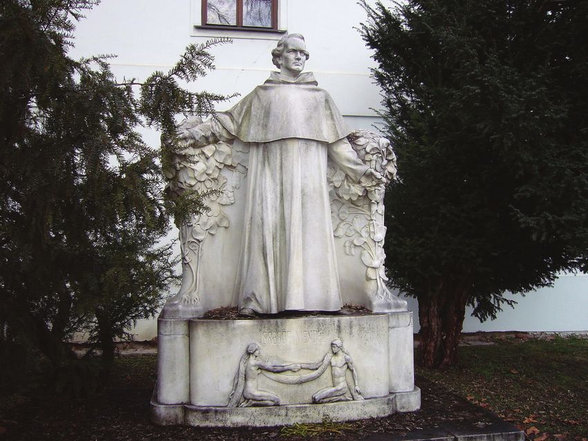
CELLS AND MOLECULES

Molecules of Life

                                                                        Organisms are composed of four basic types of molecules
                                                                        that are essential for cell structure and function: proteins,
                                                                        lipids, carbohydrates, and nucleic acids. Proteins are
                                                                        strings of amino acids that are often folded into complex
                                                                        3-D shapes. The structure of lipids can be described as
                                                                        having    a   hydrophilic     water-loving)     head    and    a
                                                                        hydrophobic      water-repelling) tail     Figure 3.1). When
                                                                        lipids are chained together, they form more-complex
                                                                        molecules called fats and triglycerides. Carbohydrates
                                                                        are composed of carbon and hydrogen atoms that can be
                                                                        broken down to supply energy for an organism. Lastly,
                                                                        nucleic acids carry genetic information about a living
                                                                        organism.
Figure 3.1 Phospholipid molecules forming a bilayer with their
hydrophobic tails and hydrophilic heads.

Probably      the    most      familiar     nucleic     acid     is
deoxyribonucleic acid (DNA). DNA comprises a sugar
phosphate backbone and nucleotides Figure 3.2). More
details on the physical structure of DNA and what
information DNA nucleotides provide will be discussed
later.) Anthropologists can analyze sequences of DNA
nucleotides and determine how different organisms are
related to each other, since they all have their own unique
DNA genetic code. In the case of humans, forensic
scientists can identify individuals by analyzing 20
different short DNA sequences known as “CODIS Core
Loci.” Another nucleic acid is called ribonucleic acid
(RNA). One type of RNA molecule is responsible for
chaining amino acids together in order to build proteins
Figure 3.3 and Figure 3.4). How RNA synthesizes amino
acids into proteins will be reviewed further on in the
chapter.

                                                                      Figure 3.2 Structural components that form double-stranded nucleic
                                                                      acid (DNA) or single-stranded nucleic acid (RNA).

                                                                                                    Molecular Biology and Genetics | 59
Figure 3.3 Chemical elements that characterize an amino acid. C: carbon; N:
                           Nitrogen; O: Oxygen; H: Hydrogen.

            Figure 3.4 Amino acids (20 different types) strung together form a polypeptide chain.

60 | Molecular Biology and Genetics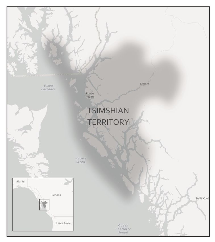
Cells

In 1665, Robert Hooke observed slices of plant cork using a microscope. Hooke noted that the microscopic plant
structures he saw resembled cella, meaning “a small room” in Latin. Approximately two centuries later, biologists
recognized the cell as being the most fundamental unit of life and that all life is composed of cells. Cellular organisms
can be characterized as two main cell types: prokaryotes and eukaryotes.

            Figure 3.5 A representation of the single-celled body of E. coli bacteria.

Prokaryotes include bacteria and archaea, and they are composed of a single cell. Additionally, their DNA and organelles
are not surrounded by individual membranes. Thus, no compartments separate their DNA from the rest of the cell
Figure 3.5). It is well known that some bacteria can cause illness in humans. For instance, Escherichia coli E. coli)
and Salmonella contamination can result in food poisoning symptoms. Pneumonia and strep throat are caused by
Streptococcal bacteria. Neisseria gonorrhoeae is a bacterial sexually transmitted disease. Although bacteria are
commonly associated with illness, not all bacteria are harmful. For example, researchers are studying the relationship
between the microbiome and human health. The bacteria that are part of the healthy human microbiome perform
beneficial roles, such as food digestion, boosting the immune system, and even making vitamins e.g., B12 and K).

                                                                                         Molecular Biology and Genetics | 61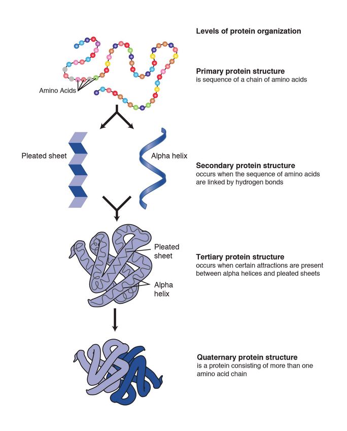
Archaea, the other type of prokaryotic organism, were
once believed to be closely related to bacteria. However,
it was determined through genetic analysis that archaea
have their own distinct evolutionary lineage so biologists
reclassified them into their own taxonomic domain.
Archaea were discovered living in extreme environments
and are therefore known as “extremophiles.” For example,
archaea can be found in high temperatures, such as Old
Faithful Geyser in Yellowstone National Park.

Eukaryotes can be single-celled or multicelled in their
body composition. In contrast to prokaryotes, eukaryotes
possess membranes that surround their DNA and
organelles. An example of a single-celled eukaryote is the
microscopic algae found in ponds phytoplankton), which           Figure 3.6 A microscopic view of plant cell membranes.
can produce oxygen from the sun. Yeasts are also single-
celled, and fungi can be single- or multicellular. Plants and animals are all multicellular.

Although plant and animal cells have a surprising number of similarities, there are some key differences. For example,
plant cells possess a thick outer cell membrane made of a fibrous carbohydrate called cellulose Figure 3.6). Animal and
plant cells also have different tissues. A tissue is an aggregation of cells that are morphologically similar and perform
the same task. For most plants, the outermost layer of cells forms a waxy cuticle that helps to protect the cells and to
prevent water loss. However, humans have skin, the outermost cell layer of which is mostly composed of a tough protein
called keratin. Overall, humans have a diversity of tissue types e.g., cartilage, brain, and heart).

Animal Cell Organelles

Figure 3.7 A phospholipid bilayer with membrane-bound carbohydrates and proteins.

62 | Molecular Biology and Genetics

An animal cell is surrounded by a double membrane called the phospholipid bilayer Figure 3.7). A closer look reveals
that this protective barrier is made of lipids and proteins that provide structure and function for cellular activities. For
example, lipids and proteins embedded in the cell’s membrane work together to regulate the passage of molecules and
ions e.g., H2O and sodium) into and out of the cell. Cytoplasm is the jelly-like matrix inside of the cell membrane. Part
of the cytoplasm comprises organelles, which perform different specialized tasks for the cell Figure 3.8). An example of
an organelle is the nucleus, where the cell’s DNA is located Figure 3.9). The double membrane that encloses the nucleus
is known as the nuclear envelope; its purpose is to regulate molecules into and out of the nucleus and serve as a barrier
to protect DNA integrity.

Figure 3.8 An animal cell with membrane-enclosed organelles.

                                                                                         Molecular Biology and Genetics | 63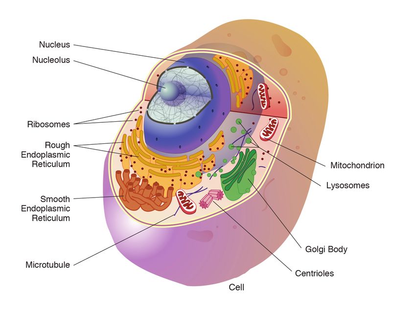
Figure 3.9 A membrane-enclosed nucleus of an animal cell.

Another important organelle is the mitochondrion Figure 3.10). Mitochondria are often referred to as “powerhouse
centers” because they produce energy for the cell in the form of adenosine triphosphate (ATP). Depending on the
species and tissue type, multicellular eukaryotes can have hundreds to thousands of mitochondria in each of their
cells. Scientists have determined that mitochondria played an important role in the evolution of the eukaryotic cell.
Mitochondria were once symbiotic prokaryotic organisms i.e., helpful bacteria) that transformed into cellular organelles
over time. Because mitochondria used to be separate organisms, this explains why mitochondria also have their own
DNA, called mitochondrial DNA (mtDNA). All organelles have important physiological functions, and when they cannot
perform their role optimally, it can result in disease. For example, there are mitochondrial diseases for which cells have
abnormally less mitochondria. In humans, this leads to various neurological symptoms and disorders. Figure 3.11 lists
other organelles found in the cell and their specialized cellular roles.

                      Figure 3.10 Microscopic view of an animal mitochondrion organelle.

64 | Molecular Biology and Genetics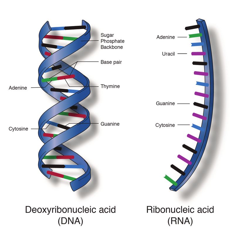
Cell structure         Description

              Cytoplasm              Fluid substance located inside of cell membrane that contains organelles

              Nucleopore             Pores in the nuclear envelope that are selectively permeable

              Nucleus                Contains the cell’s DNA and is surrounded by the nuclear envelope

                                     Resides inside of the nucleus and is the site of ribosomal RNA rRNA)
              Nucleolus
                                     transcription, processing, and assembly

                                     Responsible for cellular respiration, where energy is produced by converting
              Mitochondrion
                                     nutrients into ATP

                                     Located in the cytoplasm and also the membrane of the rough endoplasmic
              Ribosome               reticulum. Messenger RNA mRNA) binds to ribosomes and proteins are
                                     synthesized

                                     Continuous membrane with the nucleus that helps transport, synthesize,
              Endoplasmic
                                     modify, and fold proteins. Rough ER has embedded ribosomes, whereas
              reticulum ER)
                                     smooth ER lacks ribosomes

                                     Layers of flattened sacs that receive and transmit messages from the ER to
              Golgi body
                                     secrete and transport proteins within the cell

                                     Located in the cytoplasm and contains enzymes to degrade cellular
              Lysosome
                                     components

                                     Involved with cellular movement including intracellular transport and cell
              Microtubule
                                     division

                                     Assist with the organization of mitotic spindles which extend and contract for
              Centrioles
                                     the purpose of cellular movement during mitosis and meiosis

Figure 3.11 Names of organelles and their cellular functions.

INTRODUCTION TO GENETICS

Genetics is the study of heredity. Parents pass down their genetic traits to their offspring. Although children resemble
their parents, traits often vary in appearance or molecular function. For example, two parents with normal color vision
can sometimes produce a son with red-green colorblindness. Patterns of genetic inheritance will be discussed in a later
section. Molecular geneticists study the biological mechanisms responsible for creating variation between individuals,
such as DNA mutations see Chapter 4), cell division, and genetic regulation.

Molecular anthropologists use genetic data to test anthropological questions. Although their interests are diverse,
areas of molecular anthropology research include the following: human origins, dispersals, evolution, adaptation,
demography, health, disease, behavior, and animal domestication. In addition to conducting research in a laboratory,

                                                                                              Molecular Biology and Genetics | 65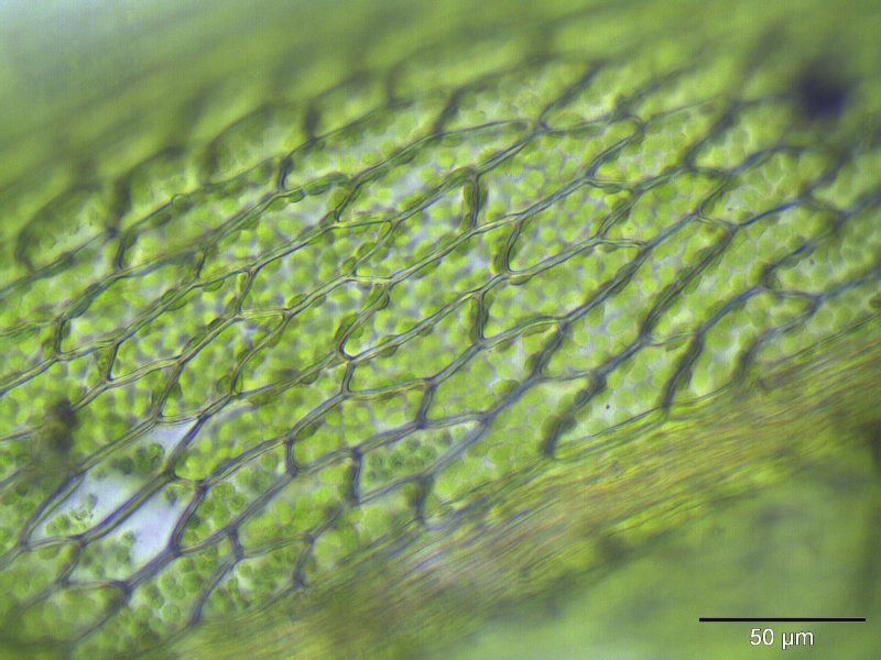
molecular anthropologists also work in the field with different communities of people. Some anthropologists also study
DNA from individuals who have been deceased for decades—even hundreds or thousands of years. The study of ancient
DNA (aDNA) has led to the development of specialized laboratory techniques. Over time, the DNA in skeletons of ancient
individuals becomes degraded i.e., less intact), which is why careful methodological considerations must be taken. A
recent example of an aDNA study is provided in Special Topic: Native American Immunity and European Diseases, and
another will be presented in Chapter 10.

     SPECIAL TOPIC: FOCUS ON NATIVE AMERICAN IMMUNITY
     AND EUROPEAN DISEASES—A STUDY OF ANCIENT DNA

                           Figure 3.12a Tsimshian Native Americans of the Pacific Northwest Coast.

     Beginning in the early 15th century, Native Americans progressively suffered from high mortality rates as
     the result of colonization from foreign powers. European-borne diseases such as measles, tuberculosis,
     influenza, and smallpox are largely responsible for the population collapse of indigenous peoples in the
     Americas. Many Europeans who immigrated to the New World had lived in large sedentary populations,
     which also included coexisting with domestic animals and pests. Although a few prehistoric Native American
     populations can be characterized as large agricultural societies especially in Mesoamerica), their overall
     culture, community lifestyle, and subsistence practices were markedly different from that of Europeans.
     Therefore, because they did not share the same urban living environments as Europeans, it is believed that
     Native Americans were susceptible to old-world diseases.

66 | Molecular Biology and Genetics
In 2016, a Nature article published by John Lindo and
colleagues was the first to investigate whether pre-
contact Native Americans possessed a genetic
susceptibility to European diseases. Their study
included Tsimshians, a First Nation community from
British Columbia Figure 3.12). The DNA from both
present-day and ancient individuals who lived
between 500 and 6,000 years ago) was analyzed.
The research team discovered that a change
occurred in the genetic region HLADQ-1, which is a
member of the major histocompatibility complex
MHC) immune system molecules. These molecules
are responsible for detecting and triggering an
immune response against pathogens. Lindo and
colleagues 2016) concluded that HLADQ-1 helped
Native Americans adapt to their local environmental
ecology. However, when European-borne epidemics
occurred in the Northwest during the 1800s, a
Figure 3.12b Tsimshian territory in present-day British certain HLADQ-1 DNA sequence associated with
Columbia.
ancient Tsimshian immunity was no longer adaptive.
As the result of past selective pressures from
European diseases, present-day Tsimshians have a different frequency of HLADQ-1 sequences. The precise
role that HLADQ-1 plays in immune adaptation still requires further investigation. But overall, this study
serves as an example of how studying ancient DNA from the remains of deceased individuals can help provide
insight into living human populations and historical events.
DNA Carries Hereditary Information
Surprisingly, the study of inheritance preceded the discovery of DNA. For a period of time, it was believed that proteins
carried the hereditary information passed from parents to offspring. Then, in 1944, Oswald Avery, Colin MacLeod, and
Maclyn McCarty discovered an association between extracted nucleic acids and the success of their bacterial genetic
experiments. Specifically, they demonstrated that DNA was the molecule responsible for the genetic transformation
of their pneumonia bacterial strains. Although this was revolutionary work at the time, the field of molecular biology
did not fully embrace their findings it has also been suggested that they were overlooked for a Nobel Prize). It was
eventually accepted by the scientific community that DNA is the hereditary material of an organism, especially after the
chemical structure of DNA was revealed.
Molecular Biology and Genetics | 67DNA Structure
The 1953 discovery of the molecular structure of DNA was one of
the greatest scientific achievements of all time. Using X-ray
crystallography, Rosalind Franklin Figure 3.13) provided the image
that clearly showed the double helix shape of DNA. However, due
to a great deal of controversy, Franklin’s colleague and outside
associates received greater publicity for the discovery. In 1962,
James Watson, Francis Crick, and Maurice Wilkins received a Nobel
Prize for developing a biochemical model of DNA. Unfortunately,
Rosalind Franklin had passed away in 1958 from ovarian cancer. In
current times, Franklin’s important contribution and her
reputation as a skilled scientist are widely acknowledged.
The double helix shape of DNA can be described as a twisted ladder
refer back to Figure 3.2). More specifically, DNA is a double-
stranded molecule with its two strands oriented in opposite
directions i.e., antiparallel). Each strand is composed of
nucleotides with a sugar phosphate backbone. There are four
different types of DNA nucleotides: adenine A), thymine T),
cytosine C), and guanine G). The two DNA strands are held
together by nucleotide base pairs, which have chemical bonding Figure 3.13 Chemist and X-ray crystallographer Rosalind
Franklin.
rules. The complementary base-pairing rules are as follows: A and
T bond with each other, while C and G form a bond. The chemical
bonds between A—T and C—G are formed by “weak” hydrogen atom interactions, which means the two strands can be
easily separated. A DNA sequence is the order of nucleotide bases A, T, G, C) along only one DNA strand. If one DNA
strand has the sequence CATGCT, then the other strand will have a complementary sequence GTACGA. This is an
example of a short DNA sequence. In reality, there are approximately three billion DNA base pairs in human cells.
DNA Is Highly Organized Within the Nucleus
If you removed the DNA from a single human cell and stretched it out completely, it would measure approximately two
meters about 6.5 feet). Therefore, DNA molecules must be compactly organized in the nucleus. To achieve this, the
double helix configuration of DNA undergoes coiling. An analogy would be twisting a string until coils are formed and
then continuing to twist so that secondary coils are formed, and so on. To assist with coiling, DNA is first wrapped
around proteins called histones. This creates a complex called chromatin, which resembles “beads on a string” Figure
3.14). Next, chromatin is further coiled into a chromosome. Another important feature of DNA is that chromosomes can
be altered from tightly coiled chromatin) to loosely coiled euchromatin). Most of the time, chromosomes in the nucleus
remain in a euchromatin state so that DNA sequences are accessible for regulatory processes to occur.
68 | Molecular Biology and GeneticsFigure 3.14 The hierarchical organization of chromosomes.
Human body cells typically have 23 pairs of chromosomes, for a total of 46 chromosomes in each cell’s nucleus Figure
3.15). An interesting fact is that the number of chromosomes an organism possesses varies, and this figure is not
dependent upon the size or complexity of the organism. For instance, chimpanzees have a total of 48 chromosomes,
while hermit crabs have 254. Chromosomes also have a distinct physical structure, including centromeres the “centers”)
and telomeres the ends) Figure 3.16). Because of centromeres, chromosomes are described as having two different
“arms,” where one arm is long and the other is shorter. Centromeres play an important role during cell division, which
will be discussed in the next section. Telomeres are located at the ends of chromosomes and they help protect the
chromosomes from degradation after every round of cell division. However, our telomeres become shorter as we age,
and if chromosome telomeres become too short, then the cell will stop dividing. Therefore, the link between the
regulation of telomere length and cellular aging is of great interest to researchers.
Molecular Biology and Genetics | 69DNA
Figure 3.15 The 23 human chromosome pairs.
Figure 3.16 The regions of a chromosome.
REPLICATION AND CELL DIVISION
For life to continue and flourish, cells must be able to divide. Tissue growth and cellular damage repair are also
necessary to maintain an organism throughout its life. All these rely on the dynamic processes of DNA replication and
the cell cycle. The mechanisms highlighted in this section are tightly regulated and represent only part of the life cycle
of a cell.
DNA Replication
DNA replication is the process by which new DNA is copied from an original DNA template. It is one phase of the
highly coordinated cell cycle and requires a variety of enzymes with special functions. Specifically, enzymes carry
out the structural and high-energy reactions associated with replicating a double helical molecule. The creation of a
complementary DNA strand from a template strand is described as semi-conservative replication. The result of semi-
conservative replication is two separate double-stranded DNA molecules, each of which is composed of an original
“parent” template strand and a newly synthesized “daughter” DNA strand.
DNA replication progresses in three steps referred to as initiation, elongation, and termination. Initiation denotes the
start of DNA replication by recruiting enzymes to specific sites along the DNA sequence. For example, the double helix
of DNA presents structural challenges for replication, so an initiator enzyme, called helicase, “unwinds” DNA by breaking
the hydrogen bonds between the two parent strands. The unraveling of the helix into two separated strands creates a
fork, which is the active site of replication machinery Figure 3.17). Once both strands are separated, the parent template
strands are exposed, meaning they can be read and replicated.
70 | Molecular Biology and GeneticsFigure 3.17 The different enzymes associated with DNA replication.
Elongation describes the assembly of new DNA daughter strands from parent strands. The two parent strands can
further be classified as leading strand or lagging strand and are distinguished by the continuous or discontinuous
direction of replication, respectively. A short fragment of RNA nucleotides acts as a primer, which binds to the parent
DNA strand that will be copied. The leading strand receives one primer and the lagging strand receives several.
Elongation proceeds with help from enzymes called DNA polymerases, which read parent template strands in a specific
direction. Complementary nucleotides are added, and the newly formed daughter strand will grow. The direction in
which replication proceeds depends on whether it is the leading or lagging strand. On the leading parent strand, a DNA
polymerase will create one continuous strand. Because the lagging parent strand requires several primers, disjointed
strands called Okazaki fragments) will be generated. Other enzymes will fill in the missing nucleotide gaps between the
disconnected lagging strand Okazaki fragments.
Finally, termination refers to the end of DNA replication activity. It is signaled by a stop sequence in the DNA, which is
recognized by machinery at the replication fork. The end result of DNA replication is that the number of chromosomes
are doubled so that the cell can divide into two.
DNA Mutations
DNA replication should result in the creation of two molecules with identical DNA nucleotide sequences. Although
DNA polymerases are quite precise during DNA replication, copying mistakes are estimated to occur every 107 DNA
nucleotides. Variation from the original DNA sequence is known as a mutation. The different types of mutations will be
discussed in greater detail in Chapter 4. Briefly, mutations can result in single nucleotide changes as well as the insertion
or deletion of nucleotides and repeated sequences. Depending on where they occur, mutations can be deleterious
harmful). For example, mutations may occur in regions that control cell cycle regulation, which can result in cancer see
Special Topic: The Cell Cycle and Immortality of Cancer Cells). Many other mutations, however, are not harmful to an
organism.
Regardless of their effect, the cell attempts to reduce the frequency of mutations that occur during DNA replication.
Molecular Biology and Genetics | 71To accomplish this, there are polymerases with proofreading capacities that can identify and correct mismatched
nucleotides. These safeguards reduce the frequency of DNA mutations so that they only occur every 109 nucleotides.
SPECIAL TOPIC: THE CELL CYCLE AND IMMORTALITY OF
CANCER CELLS
DNA replication is part of a series of preparatory phases that a cell undergoes prior to cell division,
collectively known as interphase Figure 3.18). During interphase, the cell not only doubles its chromosomes
through DNA replication, but it also increases its metabolic capacity to provide energy for growth and
division. Transition into each phase of the cell cycle is tightly controlled by proteins that serve as
checkpoints. If a cell fails to pass a checkpoint, then DNA replication and/or cell division will not continue.
Some of the reasons why a cell may fail at a checkpoint is DNA damage, lack of nutrients to continue the
process, or insufficient size. In turn, a cell may undergo apoptosis, which is a mechanism for cell death.
Figure 3.18 The phases and checkpoints of the cell cycle.
Unchecked cellular growth is a distinguishing hallmark of cancer. In other words, as cancer cells grow
and proliferate, they acquire the capacity to avoid death and replicate indefinitely. This uncontrolled and
continuous cell division is also known as “immortality.” As previously discussed, most cells lose the ability to
divide due to shortening of telomeres on the ends of chromosomes over time. One way in which cancer cells
retain replicative immortality is that the length of their telomeres is continuously protected. Chemotherapy
is often used to treat cancer by targeting cell division, which halts the propagation of genetically abnormal
72 | Molecular Biology and Geneticscells. Another therapeutic approach that continues to be investigated is targeting telomere activity to stop
the division of cancer cells.
Researchers have exploited the immortality of cancer cells for
molecular research. The oldest immortal cell line is HeLa cells
Figure 3.19), which was harvested from Henrietta Lacks, an
African-American woman diagnosed with cervical cancer in
1955. At that time, extracted cells frequently died during
experiments, but surprisingly, HeLa cells continued to
replicate. Propagation of Lacks’s cell line has significantly
contributed to medical research, including ongoing cancer
research and helping to test the polio vaccine in the 1950s.
Unfortunately, Lacks had not given her consent for her tumor
biopsy to be used in cell culture research. Moreover, her family Figure 3.19 A microscopic slide of HeLa cancer
cells.
was unaware of the extraction and remarkable application of
her cells for two decades. The history of HeLa cell origin was
first revealed in 1976. The controversy voiced by the Lacks family was included in an extensive account of
HeLa cells published in Rebecca Skloot’s 2010 book, The Immortal Life of Henrietta Lacks. A film based on the
book was also released in 2017.
Mitotic Cell Division
The body and its various tissues are comprised of somatic cells. Organisms that contain two sets of chromosomes in
their somatic cells are called diploid organisms. Humans have 46 chromosomes and they are diploid because they inherit
one set of chromosomes n = 23) from each parent. As a result, they have 23 matching pairs of chromosomes, which
are known as homologous chromosomes. These homologous pairs vary in size and are generally numbered from largest
chromosome 1) to smallest chromosome 22), as seen in Figure 3.15, with the exception of the 23rd pair, which is made
up of the sex chromosomes X and Y). Typically, the female sex is XX and the male sex is XY. Individuals inherit an X
chromosome from their mother and an X or Y from their father.
In order to grow and repair tissues, somatic cells must divide. As discussed previously, a cell must first replicate its
genetic material for cell division to occur. During DNA replication, each chromosome produces double the amount of
genetic information. The duplicated arms of chromosomes are known as sister chromatids, and they are attached at the
centromeric region. To elaborate, the number of chromosomes stays the same n = 46); however, the amount of genetic
material is doubled in the cell as the result of replication.
Mitosis is the process of somatic cell division that gives rise to two diploid daughter cells. Figure 3.20 shows a
brief overview of mitosis. Once DNA and other organelles in the cell have finished replication, mitotic spindle fibers
microtubules) assist with chromosomal movement by attaching to the centromeric region of each chromosome.
Specifically, the spindle fibers physically align each chromosome at the center of the cell. Next, the spindle fibers divide
the sister chromatids and move each one to opposite sides of the cell. At this phase, there are 46 chromosomes on each
side of the cell. The cell can now divide into two fully separated daughter cells.
Molecular Biology and Genetics | 73Figure 3.20 Steps of mitotic cell division.
Meiotic Cell Division
Gametogenesis is the production of gametes sperm and egg cells); it involves two rounds of cell division called meiosis.
Similar to mitosis, the parent cell in meiosis is diploid. However, meiosis has a few key differences, including the
number of daughter cells produced four cells, which require two rounds of cell division to produce) and the number
of chromosomes each daughter cell has Figure 3.21). During the first round of division known as meiosis I), each
chromosome n = 46) replicates its DNA so that sister chromatids are formed. Next, with the help of spindle fibers,
homologous chromosomes align near the center of the cell and sister chromatids physically swap genetic material. In
other words, the sister chromatids of matching chromosomes cross over with each other at matching DNA nucleotide
positions. The occurrence of homologous chromosomes crossing over, swapping DNA, and then rejoining segments is
called genetic recombination. The “genetic shuffling” that occurs in gametes increases organismal genetic diversity by
creating new combinations of genes on chromosomes that are different from the parent cell. Genetic mutations can also
arise during recombination. For example, there may be an unequal swapping of genetic material that occurs between the
two sister chromatids, which can result in deletions or duplications of DNA nucleotides. Once genetic recombination is
complete, homologous chromosomes are separated and two daughter cells are formed.
The daughter cells after the first round of meiosis are haploid, meaning they only have one set of chromosomes n =
23). During the second round of cell division known as meiosis II), sister chromatids are separated and two additional
haploid daughter cells are formed. Therefore, the four resulting daughter cells have one set of chromosomes n = 23),
and they also have a genetic composition that is not identical to the parent cells nor to each other.
Figure 3.21 Meiotic cell division.
74 | Molecular Biology and GeneticsAlthough both sperm and egg gamete production undergo meiosis, they differ in the final number of viable daughter
cells. In the case of spermatogenesis, four mature sperm cells are produced. Although four egg cells are also produced in
oogenesis, only one of these egg cells will result in an ovum mature egg). During fertilization, an egg cell and sperm cell
fuse, which creates a diploid cell that develops into an embryo. The ovum also provides the cellular organelles necessary
for embryonic cell division. This includes mitochondria, which is why humans, and most other multicellular eukaryotes,
have the same mtDNA sequence as their mothers.
Chromosomal Disorders
During mitosis or meiosis, entire deletions or duplications of chromosomes can occur due to error. For example,
homologous chromosomes may fail to separate properly, so one daughter cell may end up with an extra chromosome
while the other daughter cell has one less. Cells with an unexpected or abnormal) number of chromosomes are
known as aneuploid. Adult or embryonic cells can be tested for chromosome number karyotyping). Aneuploid cells
are typically detrimental to a dividing cell or developing embryo, which can lead to a loss of pregnancy. However,
the occurrence of individuals being born with three copies of the 21st chromosome is relatively common; this genetic
condition is known as Down Syndrome. Moreover, human males and females can be born with aneuploid sex
chromosome conditions such as XXY, XXX, and XO referring to only one X chromosome).
PROTEIN SYNTHESIS
At the beginning of the chapter, we defined proteins as strings of amino acids that fold into complex 3-D shapes. There
are 20 standard amino acids that can be strung together in different orders in humans, and the result is that proteins
can perform an impressive amount of different functions. For instance, muscle fibers are proteins that help facilitate
movement. A special class of proteins immunoglobulins) help protect the organism by detecting disease-causing
pathogens in the body. Protein hormones, such as insulin, help regulate physiological activity. Blood hemoglobin is a
protein that transports oxygen throughout the body. Enzymes are also proteins, and they are catalysts for biochemical
reactions that occur in the cell e.g., metabolism). Larger-scale protein structures can be visibly seen as physical features
of an organism e.g., hair and nails).
Transcription and Translation
Coding nucleotides in our DNA provide instructions on how to make proteins. Making proteins, also known as protein
synthesis, can be broken down into two main steps referred to as transcription and translation. Protein synthesis relies
on many molecules in the cell including different types of regulatory proteins and RNAs for each step in the process.
Although there are many different types of RNA molecules that have a variety of functions within the cell, we will mainly
focus on messenger RNA (mRNA).
A gene is a segment of DNA that codes for RNA, and genes can vary in length from a few hundred to as many as
two million base pairs in length. The purpose of transcription is to make an RNA copy of that genetic code Figure
3.22). Unlike double-stranded DNA, RNA molecules are single-stranded nucleotide sequences refer back to Figure
3.2). Additionally, while DNA contains the nucleotide thymine T), RNA does not—instead, its fourth nucleotide is uracil
U). Uracil is complementary to or can pair with) adenine A), while cytosine C) and guanine G) continue to be
Molecular Biology and Genetics | 75complementary to each other. For transcription to proceed, a gene must first be turned “on” by the cell see Special Topic: Genetic Regulation of the Lactase LCT) Gene for a more detailed discussion of gene regulation). The double- stranded DNA is then separated, and one side of the DNA strand is used as a template where complementary RNA nucleotides are strung together. For example, if a DNA template is TACGGATGC, then the newly constructed mRNA sequence will be AUGCCUACG. Sometimes the end product needed by the cell is that transcribed RNA, but for protein synthesis constructing the RNA specifically pre-messenger RNA, or pre-mRNA) is just the first step. Figure 3.22 RNA polymerase catalyzing DNA transcription. Genes contain segments called introns and exons. Exons are considered “coding” while introns are considered “noncoding”—meaning the information they contain will not be needed to construct proteins. When a gene is first transcribed into pre-mRNA, introns and exons are both included Figure 3.23). However, once transcription is finished, introns are removed in a process called splicing. During splicing, a protein/RNA complex attaches itself to the pre- mRNA and removes introns and then connects the remaining exons, thus creating a shorter mature mRNA. 76 | Molecular Biology and Genetics
Figure 3.23 RNA processing is the modification of RNA, including the removal of introns, called splicing, between transcription
   and translation.

The process by which mRNA is “read” and amino acids chained together to form new proteins is called translation.
During translation, mature mRNA is transported outside of the nucleus where it is bound to a ribosome Figure 3.24).
The nucleotides in the mRNA are read as triplets, which are called codons. Each codon corresponds to an amino acid,
and this is the basis for building a protein. Continuing with our example from above, the mRNA sequence AUG-CCU-
ACG codes for three amino acids. Using a codon table Figure 3.25), AUG is a codon for methionine Met), CCU is proline
Pro), and ACG is threonine Thr). Therefore, the protein sequence is Met-Pro-Thr. Methionine is the most common
“start codon” AUG) for the initiation of protein translation in eukaryotes. As the ribosome moves along the mRNA, the
growing amino acid chain exits the ribosome and folds into a protein Figure 3.26). When the ribosome reaches a “stop”
codon UAA, UAG, or UGA), the ribosome stops adding new amino acids, detaches from the mRNA, and the protein is
released. Folded proteins can then be used to complete a structural or functional task.

         Figure 3.24 Translation of mRNA into an amino acid.

                                                                                                   Molecular Biology and Genetics | 77
Figure 3.25 This table can be used to identify which mRNA codons (sequence of three nucleotides) correspond
with each of the 20 different amino acids. For example, if the codon is CAU, the first position is “C” and you
would look in that corresponding row, the second position is “A” and you would look in that column. “U’ is the
third position—narrowing the row and indicating that the CAU codon corresponds with the amino acid
“histidine” (abbreviated “His”). The table also indicates the most common “start codon” (AUG) that correlates
with Methionine, and the three “stop” codons (UAA, UAG, or UGA).
78 | Molecular Biology and GeneticsFigure 3.26 Indicates levels of protein organization from the simple amino acid chain that is then folded and organized into more complex
protein structures.
Molecular Biology and Genetics | 79SPECIAL TOPIC: GENETIC REGULATION OF THE LACTASE
(LCT) GENE
The LCT gene codes for a protein called lactase, an enzyme produced in the small intestine. It is responsible
for breaking down the sugar “lactose” found in milk. Lactose intolerance occurs when not enough lactase
enzyme is produced and, in turn, digestive symptoms occur. To avoid this discomfort, individuals may take
lactase supplements, drink lactose-free milk, or avoid milk products altogether.
The LCT gene is a good example of how cells regulate protein synthesis. The promoter region of the LCT gene
helps regulate whether it is transcribed or not transcribed i.e., turned “on” or “off,” respectively). Lactase
production is initiated when a regulatory protein known as a transcription factor binds to a site on the
LCT promoter. RNA polymerases are then recruited; they read DNA and string together nucleotides to make
RNA molecules Figure 3.22). An LCT pre-mRNA is synthesized made) in the nucleus, and further chemical
modifications flank the ends of the mRNA to ensure the molecule will not be degraded in the cell.
Next, RNA processing occurs. A spliceosome complex removes the introns and connects exons to form
the mature mRNA. Once the LCT mRNA is transported outside of the nucleus, it is bound to a ribosome,
which is a multi-protein complex that includes ribosomal RNA (rRNA). The ribosome of eukaryotes has two
main subunits: the smaller bottom subunit that binds to the mRNA and the larger top subunit that contains
transfer RNA (tRNA) binding sites see Figure 3.24). Each tRNA has a nucleotide anticodon that recognizes an
mRNA codon. When a tRNA binds to an mRNA codon in the ribosome, the tRNA transfers the corresponding
amino acid. rRNA ensures the newly added amino acid is linked in the correct order. The growing protein
then folds into the lactase enzyme, which can break down lactose.
Most animals lose their ability to digest milk as they mature due to the decreasing transcriptional “silence” of
the LCT gene over time. However, some humans have the ability to digest lactose into adulthood also known
as “lactase persistence”). This means they have a genetic mutation that leads to continuous transcriptional
activity of LCT. Lactase persistence mutations are common in populations with a long history of pastoral
farming, such as northern European and North African populations. It is believed that lactase persistence
evolved because the ability to digest milk was nutritionally beneficial. More information about lactase
persistence will be covered in Chapter 14.
80 | Molecular Biology and GeneticsMENDELIAN GENETICS AND OTHER PATTERNS OF INHERITANCE
Gregor Johann Mendel 1822–1884) is often described as the “Father of
Genetics.” Mendel was a monk who conducted pea plant breeding
experiments in a monastery located in the present-day Czech Republic
Figure 3.27). After several years of experiments, Mendel presented his
work to a local scientific community in 1865 and published his findings
the following year. Although his meticulous effort was notable, the
importance of his work was not recognized for another 35 years. One
reason for this delay in recognition is that his findings did not agree
with the predominant scientific viewpoints on inheritance at the time.
For example, it was believed that parental physical traits “blended”
together and offspring inherited an intermediate form of that trait. In
contrast, Mendel showed that certain pea plant physical traits e.g., Figure 3.27 Statue of Mendel located at the Mendel
Museum located at Masaryk University in Brno,
flower color) were passed down separately to the next generation in a Czech Republic.
statistically predictable manner. Mendel also observed that some
parental traits disappeared in offspring but then reappeared in later generations. He explained this occurrence by
introducing the concept of “dominant” and “recessive” traits. Mendel established a few fundamental laws of inheritance,
and this section reviews some of these concepts. Moreover, the study of traits and diseases that are controlled by a
single gene is commonly referred to as Mendelian genetics.
Mendelian Genetics
Figure 3.28 Various phenotypic characteristics of pea plants resulting from different genotypes.
The physical appearance of a trait is called an organism’s phenotype. Figure 3.28 shows pea plant Pisum sativum)
phenotypes that were studied by Mendel, and in each of these cases the physical traits are controlled by a single gene.
In the case of Mendelian genetics, a phenotype is determined by an organism’s genotype. A genotype consists of two
gene copies, wherein one copy was inherited from each parent. Gene copies are also known as alleles Figure 3.29),
which means they are found in the same gene location on homologous chromosomes. Alleles have a nonidentical DNA
Molecular Biology and Genetics | 81sequence, which means their phenotypic effect can be different. In other words, although alleles code for the same
trait, different phenotypes can be produced depending on which two alleles i.e., genotypes) an organism possesses. For
example, Mendel’s pea plants all have flowers, but their flower color can be purple or white. Flower color is therefore
dependent upon which two color alleles are present in a genotype.
Figure 3.29 Homologous chromosome pairs showing the different homozygous and heterozygous
combinations that can exist from two different alleles (B and b).
A Punnett square is a diagram that can help visualize Mendelian inheritance patterns. For instance, when parents of
known genotypes mate, a Punnett square can help predict the ratio of Mendelian genotypes and phenotypes that
their offspring would possess. Figure 3.30 is a Punnett square that includes two heterozygous parents for flower color
Bb). A heterozygous genotype means there are two different alleles for the same gene. Therefore, a pea plant that is
heterozygous for flower color has one purple allele and one white allele. When an organism is homozygous for a specific
trait, it means their genotype consists of two copies of the same allele. Using the Punnett square example Figure 3.30),
the two heterozygous pea plant parents can produce offspring with two different homozygous genotypes BB or bb) or
offspring that are heterozygous Bb).
A pea plant with purple flowers could be heterozygous Bb) or homozygous
BB). This is because the purple color allele B) is dominant to the white
color allele b), and therefore it only needs one copy of that allele to
phenotypically express purple flowers. Because the white flower allele is
recessive, a pea plant must be homozygous for the recessive allele in order
to have a white color phenotype bb). As seen by the Punnett square
example Figure 3.30), three of four offspring will have purple flowers and
the other one will have white flowers.
The Law of Segregation was introduced by Mendel to explain why we can
predict the ratio of genotypes and phenotypes in offspring. As discussed
previously, a parent will have two alleles for a certain gene with each copy
on a different homologous chromosome). The Law of Segregation states
Figure 3.30 Punnett square depicting the
possible genetic combinations of offspring from
that the two copies will be segregated from each other and will each be
two heterozygous parents. distributed to their own gamete. We now know that the process where that
occurs is meiosis.
Offspring are the products of two gametes combining, which means the offspring inherits one allele from each gamete
82 | Molecular Biology and Geneticsfor most genes. When multiple offspring are produced like with pea plant breeding), the predicted phenotype ratios are
more clearly observed. The pea plants Mendel studied provide a simplistic model to understand single-gene genetics.
While many traits anthropologists are interested in have a more complicated inheritance e.g., are informed by many
genes), there are a few known Mendelian traits in humans. Additionally, some human diseases also follow a Mendelian
pattern of inheritance Figure 3.31). Because humans do not have as many offspring as other organisms, we may
not recognize Mendelian patterns as easily. However, understanding these principles and being able to calculate the
probability that an offspring will have a Mendelian phenotype is still important.
Mendelian disorder Gene Mendelian disorder Gene
Alpha Thalassemia HBA1 Maple Syrup Urine Disease: Type 1A BCKDHA
Androgen Insensitivity Syndrome AR Mitochondrial DNA Depletion Syndrome TYMP
Bloom Syndrome BLM MTHFR Deficiency MTHFR
Canavan Disease ASPA Oculocutaneous Albinism: Type 1 TYR
Cartilage-Hair Hypoplasia RMRP Oculocutaneous Albinism: Type 3 TYRP1
Persistent Mullerian Duct Syndrome:
Cystic Fibrosis CFTR AMH
Type I
Familial Chloride Diarrhea SLC26A3 Polycystic Kidney Disease PKHD1
Fragile X Syndrome FMR1 Sickle-cell anemia HBB
Glucose-6-Phosphate Dehydrogenase
G6PD Spermatogenic failure USP9Y
Deficiency
Hemophilia A F8 Spinal Muscular Atrophy: SMN1 Linked SMN1
Huntington disease HTT Tay-Sachs Disease HEXA
Hurler Syndrome IDUA Wilson Disease ATP7B
Figure 3.31 Human diseases that follow a Mendelian pattern of inheritance.
Example of Mendelian Inheritance: The ABO Blood Group System
In 1901, Karl Landsteiner at the University of Vienna published his discovery of ABO blood groups. This was a result
of conducting blood immunology experiments in which he combined the blood of individuals who possess different
blood cell types and observed an agglutination clotting) reaction. The presence of agglutination implies there is an
incompatible immunological reaction, whereas no agglutination will occur in individuals with the same blood type. This
work was clearly important because it resulted in a higher survival rate of patients who received blood transfusions.
Molecular Biology and Genetics | 83Blood transfusions from someone with a different type of blood causes agglutinations, and the resulting coagulated
blood can not easily pass through blood vessels, resulting in death. Accordingly, Landsteiner received the Nobel Prize
1930) for explaining the ABO blood group system.
Blood cell surface antigens are proteins that coat the surface of red blood cells, and antibodies are specifically
“against” or “anti” to the antigens from other blood types. Thus, antibodies are responsible for causing agglutination
between incompatible blood types. Understanding the interaction of antigens and antibodies helps to determine
ABO compatibility amongst blood donors and recipients. In order to better understand blood phenotypes and ABO
compatibility, blood cell antigens and plasma antibodies are presented in Figure 3.32. Individuals that are blood type A
have A antigens on the red blood cell surface, and anti-B antibodies, which will bind with B antigens should they come
in contact. Alternatively, individuals with blood type B have B antigens and anti-A antibodies. Individuals with blood type
AB have both A and B antigens but do not produce antibodies for the ABO system. This does not mean type AB does
not have any antibodies, just that anti-A or anti-B antibodies are not produced. Individuals who are blood type O have
nonspecific antigens but produce both anti-A and anti-B antibodies.
Figure 3.32 The different ABO blood types with their associated antibodies and antigens.
Figure 3.33 shows a table of the ABO allele system, which
has a Mendelian pattern of inheritance. Both the A and B
alleles function as dominant alleles, so the A allele always
codes for the A antigen, and the B allele codes for the B
antigen. The O allele differs from A and B, because it codes
for a nonfunctional antigen protein, which means there is
no antigen present on the cell surface of O blood cells. To
have blood type O, two copies of the O allele must be
inherited, one from each parent, thus the O allele is Figure 3.33 The different combinations of ABO blood alleles (A, B,
and O) to form ABO blood genotypes.
considered recessive. Therefore, someone who is a
heterozygous AO genotype is phenotypically blood type A and a genotype of BO is blood type B. The ABO blood system
84 | Molecular Biology and Geneticsalso provides an example of codominance, which is when the effect of both alleles is observed in the phenotype. This is
true for blood type AB: when an individual inherits both the A and B alleles, then both A and B antigens will be present
on the cell surface.
Also found on the surface of red blood cells is the rhesus group antigen, known as “Rh factor.” In reality, there are
several antigens on red blood cells independent from the ABO blood system, however, the Rh factor is the second most
important antigen to consider when determining blood donor and recipient compatibility. Rh antigens must also be
considered when a pregnant mother and her baby have incompatible Rh factors. In such cases, a doctor can administer
necessary treatment steps to prevent pregnancy complications and hemolytic disease, which is when the mother’s
antibodies break down the newborn’s red blood cells.
An individual can possess the Rh antigen be Rh positive) or lack the Rh antigen be Rh negative). The Rh factor is
controlled by a single gene and is inherited independently of the ABO alleles. Therefore, all blood types can either be
positive O+, A+, B+, AB+) or negative O-, A-, B-, AB-).
Individuals with O+ red blood cells can donate blood to A+, B+, AB+, and O+ blood type recipients. Because O- individuals
do not have AB or Rh antigens, they are compatible with all blood cell types and are referred to as “universal donors.”
Individuals that are AB+ are considered to be “universal recipients” because they do not possess antibodies against other
blood types.
Mendelian Patterns of Inheritance and Pedigrees
A pedigree can be used to investigate a family’s medical history by determining if a health issue is inheritable and will
possibly require medical intervention. A pedigree can also help determine if it is a Mendelian recessive or dominant
genetic condition. Figure 3.34 is a pedigree example of a family with Huntington’s disease, which has a Mendelian
dominant pattern of inheritance. In a standard pedigree, males are represented by a square and females are represented
by a circle. When an individual is affected with a certain condition, the square or circle is filled in as a solid color.
With a dominant condition, at least one of the parents will have the disease and an offspring will have a 50% chance of
inheriting the affected chromosome. Therefore, dominant genetic conditions tend to be present in every generation. In
the case of Huntington’s, some individuals may not be diagnosed until later in adulthood, so parents may unknowingly
pass this dominantly inherited disease to their children.
Figure 3.34 A three-generation pedigree depicting an example of dominant Mendelian inheritance like
Huntington’s.
Molecular Biology and Genetics | 85Because the probability of inheriting a disease-causing recessive allele is more rare, recessive medical conditions can
skip generations. Figure 3.35 is an example of a family that carries a recessive cystic fibrosis mutation. A parent that
is heterozygous for the cystic fibrosis allele has a 50% chance of passing down their affected chromosome to the next
generation. If a child has a recessive disease, then it means both of their parents are carriers heterozygous) for that
condition. In most cases, carriers for recessive conditions show no serious medical symptoms. Individuals whose family
have a known medical history for certain conditions sometimes seek family planning services see the Genetic Testing
section).
Figure 3.35 A three-generation pedigree depicting an example of recessive Mendelian inheritance like
cystic fibrosis.
Pedigrees can also help distinguish if a health issue has an autosomal or X-linked pattern of inheritance. As previously
discussed, there are 23 pairs of chromosomes and 22 of these pairs are known as autosomes. The provided pedigree
examples Figure 3.34–35) are autosomally linked genetic diseases. This means the genes that cause the disease are
located on one of the chromosomes numbered 1 to 22. Disease causing genes can also be X-linked, which means they
are located on the X chromosome.
Figure 3.36 depicts a family in which the mother is a carrier for the X-linked recessive disease Duchenne Muscular
Dystrophy DMD). The mother is a carrier for DMD, so daughters and sons will have a 50% chance of inheriting the
pathogenic DMD allele. Because females have two X chromosomes, females will not have the disease although in rare
cases, female carriers may show some symptoms of the disease). On the other hand, males who inherit a copy of an
X-linked pathogenic DMD allele will typically be affected with the condition. Males are more susceptible to X-linked
conditions because they only have one X chromosome. Therefore, when evaluating a pedigree, if a higher proportion of
males are affected with the disease, this could suggest the disease is X-linked recessive. Finally, Y-linked traits are very
rare because compared to other chromosomes, the Y chromosome is smaller and only has a few active transcribed)
genes.
86 | Molecular Biology and GeneticsYou can also read