Management of Snake Bite - STANDARD TREATMENT GUIDELINES Quick Reference Guide January 2016 - National Health Mission
←
→
Page content transcription
If your browser does not render page correctly, please read the page content below
1

        STANDARD TREATMENT
            GUIDELINES

   Management of Snake Bite
                      Quick Reference Guide
                          January 2016

                Ministry of Health & Family Welfare
                        Government of India

QRG Snakebite             Version 4 Final     December 22, 2015
2

                                                      Table of Content
1.      INTRODUCTION ......................................................................................................... 4

2.      INCIDENCE OF SNAKE BITE IN INDIA ........................................................................... 5
3.      WHEN TO SUSPECT/RECOGNIZE ................................................................................. 6
4.     RECOMMENDATIONS ................................................................................................ 8
     4.1 FIRST AID MEASURES ...................................................................................................... 8
     4.2 SIGN & SYMPTOMS .......................................................................................................... 10
     4.3. ASSESSMENT ............................................................................................................... 17
     4.4 LAB INVESTIGATIONS .................................................................................................... 18
     4.5 ANTI SNAKE VENOM (ASV) THERAPY ............................................................................ 22
       4.5.5 ASV dose in pregnancy .......................................................................................... 24
       4.5.6 ASV dose in children .............................................................................................. 24
       4.5.7 ASV dosage in victims requiring life saving surgery .............................................. 25
       4.5.8 Repeat dose of ASV................................................................................................ 25
       4.5.9 Victims who arrive late .......................................................................................... 25
     4.5.10 MONITORING OF PATIENTS ON ASV THERAPY ................................................................... 26
     4.6. ASV REACTION ................................................................................................................ 26
     4.7 MANAGEMENT NEUROTOXIC (NEUROPARALYTIC) ENVENOMATION .......................... 29
     4.8 MANAGEMENT OF VASCULOTOXIC SNAKEBITE: ........................................................... 30
     4.9 MANAGEMENT OF SEVERE LOCAL ENVENOMING ........................................................ 32
     4.10 RECOVERY PHASE OR OBSERVATION OF THE RESPONSE TO ADEQUATE DOSE OF ANTISNAKE VENOM 33
     4.11 OTHER MEASURES ........................................................................................................... 33
     4.12 SURGICAL PROCEDURES IN SNAKEBITE....................................................................... 34
     4.12.1 DEBRIDEMENT OF NECROTIC TISSUE................................................................................. 34
     4.12.2 COMPARTMENTAL SYNDROME ....................................................................................... 34
     4.12.3 CRITERIA FOR FASCIOTOMY IN SNAKEBITE LIMB .................................................................. 34
     4.13 DISCHARGE .................................................................................................................... 36
     4.14 FOLLOW-UP ................................................................................................................... 36
     4.15 REHABILITATION ............................................................................................................. 36
5. LEVEL SPECIFIC MANAGEMENT OF SNAKEBITE ............................................................. 37
   5.1 REFERRAL CRITERIA ...................................................................................................... 37
     5.1.1 Vasculotoxic envenomation................................................................................... 37
     5.1.2 Referral Criteria: Neurotoxic Envenomation.......................................................... 37
     5.1.3 Instructions while referring ................................................................................... 38
   5.2 SNAKE BITE MANGEMENT AT A PRIMARY HEALTH CARE CENTER (PHC) ....................... 39

QRG Snakebite                                          Version 4 Final                           December 22, 2015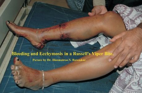
3

     5.3 SNAKE BITE MANGEMENT AT THE DISTRICT HOSPITAL ................................................ 41
     5.4 SNAKE BITE MANGEMENT AT THE TERTIARY CARE OR MEDICAL COLLEGE .................. 42
6. PATIENT INFORMATION SHEET .................................................................................... 43
7.     REFERENCES............................................................................................................. 47

8. SNAKE BITE EXAMINATION PERFORMA ....................................................................... 51

QRG Snakebite                                     Version 4 Final                         December 22, 2015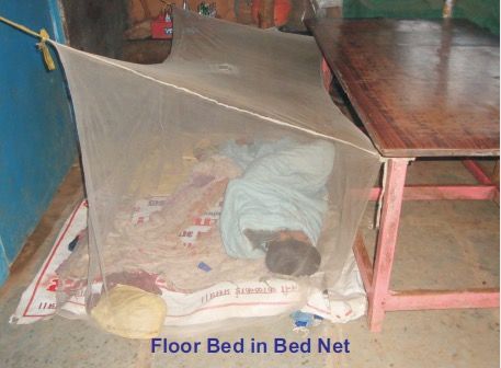
4

1. Introduction

Snakebite is an acute life threatening time limiting medical emergency. It is a
preventable public health hazard often faced by rural population in tropical and
subtropical countries with heavy rainfall and humid climate.

There are more than 2000 species of snakes in the world and about 300 species are
found in India out of which 52 are venomous. The venomous snakes found in India
belong to three families Elapidae, Viperidae and hydrophidae (Sea Snakes). The
most common Indian elapids are Naja naja (Indian Cobra) and Bungarus caeruleus
(Indian Krait), Daboia russalie (Russells’ Viper) and Echis carinatus (Saw scaled viper)
(Alirol et al 2010). Clinical effects of envenoming by same species of snake are
almost similar except a few regional variations. Kraits are active during night hours,
often biting a person sleeping on floor bed. Maximum Viper and Cobra bites occur
during the day or early darkness, while watering the plantation or walking bare foot
in grown grass or soybean crops.

Although total number of bites may be more than 5-6 lakhs but only 30% are
venomous bites. According to Mahapatra et al (on the basis of Million Death Study),
non-fatal bites may be as high as 1.4 million per year. Though snakebite is a life
threatening centuries old condition, it was included in the list of neglected tropical
diseases by World Health Organization in the year 2009 (Warrell and WHO 2009;
Bawaskar HS 2014).

Currently, treatment quality is highly varied, ranging from good quality in some
areas, to very poor quality treatment in others. The high fatality due to Krait bite is
attributed to the non-availability of antisnake venom (ASV), delayed and
inappropriate administration of ASV, lack of standard protocol for management and
inexperienced doctors and non-availability of ventilator or bag and valve (Bawaskar
et al 2008). In India, there has always been a crisis of antivenom supply (Bawaskar
HS and Bawaskar PH 2001). On one hand there is shortage of ASV but on the other
hand scarce ASV is being wasted due to excessive dosage of ASV in the absence of a
Standard Treatment Guideline. Victims are not only misdiagnosed as - abdominal
colic, and vomiting due to indigestion, appendicitis, stroke, head injury, ischemic
heart disease, food poisoning, trismus, hysteria and Guillain-Barre´ syndrome but
also subjected to unnecessary investigations including MRI scans of the brain and
lumbar puncture thus causing undue delay in ASV therapy. Delayed administration

QRG Snakebite                     Version 4 Final            December 22, 2015
5

of ASV or waiting until victim develops systemic manifestations i.e., a 6 h wait
results in systemic envenoming and high fatality (Bawaskar et al 2008).

1. Incidence of Snake Bite in India
There is a huge gap between the number of snakebite deaths reported from direct
survey and official data. Only 7.23% snakebite deaths were officially reported
(Majumdar, 2014 and Mohapatra 2011). Earlier hospital based reports estimated
about 1,300 to 50,000 annual deaths from snakebites per year in India. Mohapatra
et al, 2011, reported direct estimates from a national mortality survey of 1.1 million
homes in 2001–03. The study found 562 deaths (0.47% of total deaths) were
assigned to snakebites, mostly in rural areas, and more commonly among males
than females and peaking at ages 15–29. This proportion represents about 45,900
annual snakebite deaths nationally or an annual age-standardized rate of
4.1/100,000, with higher rates in rural areas (5.4) and with the highest rate in the
state of Andhra Pradesh (6.2). Annual snakebite deaths were greatest in the states
of Uttar Pradesh (8,700), Andhra Pradesh (5,200), and Bihar (4,500). Other Indian
states with high incidence of snakebites cases are Tamil Nadu, West Bengal,
Maharashtra and Kerala. Because a large proportion of global totals of snakebites
arise from India, global snakebite totals might also be underestimated. (Mohapatra
et al 2011).

Only 22.19% of the snakebite victims attended the hospitals. Nearly 65.7% of the
snakebite deaths were due to common krait bite, most of them occurring in the
months of June to September (Majumder et al, 2014). This is because even today
most of the victims initially approach traditional healers for treatment and many are
not even registered in the hospital. Singh et al reported among the snakebite
victims, about 60.76% received first aid at the site of incident, and 20.25% of them
sought hospital care after consulting the traditional healers (ozhas, or mantrik and
tandrik). Time lapsed for seeking hospital treatment was less than 4 h in 55.69% of
the cases and more than 12 h in 7.59% of the cases. Most (41.79%) patients were
frightened, but no local or systemic symptoms had appeared when they reported
the emergency (Singh A et al 2015).

QRG Snakebite                    Version 4 Final           December 22, 2015
6

2. WHEN TO SUSPECT/RECOGNIZE

CLINICAL PRESENTATION:

Clinical presentation of snakebite victim depends upon species of snake, amount of
venom injected, season of the bite, whether snake is fed or unfed, site of bite, area
covered or uncovered, dry or incomplete bite, multiple bites, venom injection in
vessel, weight of the victim and time elapsed between the bite and administration
of ASV. Venom concentration and constitution depends on environmental
conditions as well as snake’s maturity and darkness of colour of snake (Bawaskar HS
et al 2014).
Patient can present in the four clinical syndromes or in combination i.e. progressive
weakness (neuroparalytic/neurotoxic), bleeding (vasculotoxic/haemotoxic),
myotoxic and painful progressive Swelling (Figure 1).

QRG Snakebite                    Version 4 Final           December 22, 2015
7

                                          Suspected snake bite

                                                                                            • Neuroparalytic
                                                                               Krait          symptoms with
        Overt bite                                        Occult bite                         no local signs
        History of bite                                   No history of bite                • Severe
        Nonvenomous (70%) / venomous (30%)                                                    abdominal pain,
                                                                                              vomiting

        Asymptomatic      Dry bite       Symptomatic*                                        1. ASV
                                         Predominant symptom manifestation                   2. AN***
 Anxiety, palpitations,                                                                      3. Ventilation
 tachycardia, Paraesthesia

                   Progressive painful       Neuroparalytic           Vasculotoxic             Myotoxic
                   swelling
                                          Cobra             Russel’s viper             Flat tailed
                                          Krait             Saw Scale viper            Sea snake
           Viper
                                          • Ptosis                                        • Muscle ache
                                                                 • Bleeding               • Muscle swelling
                                          • Diplopia             • DIC
            • Local necrosis                                                              • Involuntary
                                          • Dysarthia            • Shock
            • Ecchymosis                                                                    contractions of
                                          • Dysphonia            • Acute kidney
            • Blistering                                                                    muscles
                                          • Dyspnoea               injury
            • Painful                                                                     • Compartment
                                          • Dysphagia
              swelling                                                                      syndrome
                                          • Paralysis
            • Compartment
              syndrome
                                                                   ASV**¥
                                         1. ASV**                                             ASV**¥
                                                                   Supportive treatment
                                         2. AN***                                             Supportive
             ASV#                                                  Dialysis
                                         3. Ventilation                                       treatment
                                                                   Blood transfusion
                                                                                              Dialysis

    *   Even though present as predominant manifestation but there may be overlap of syndrome as
        well.
    #   ASV indicated in rapidly developing swelling only. Purely localized swelling with or without bite
        marks is not an indication of ASV.
    ** For reaction to antisnake venom (ASV) Dose of Adrenaline 0.5 mg IM (in children 0.01 mg/kg)
    ¥   Specific ASV for sea snake and Pit viper bite is not available in India. However, available ASV
        may have some advantage by cross reaction.
    *** Atropine 0.6 mg followed by neostigmine (1.5mg) to be given IV stat (In children Inj. Atropine
        0.05 mg/kg followed by Inj. Neostigmine 0.04 mg/kg IV.) Repeat neostigmine dose 0.5 mg (in
        children 0.01mg/kg) with atropine every 30 minutes for 5 doses. Thereafter taper dose at 1
        hour, 2 hour, 6 hours and 12 hour. Positive response is measured as 50% or more recovery of
        the ptosis in one hour. If no response after 3rd dose. Stop AN injection.

Figure 1. Four presenting clinical syndromes of snakebite i.e. progressive weakness
(neuroparalytic/neurotoxic), bleeding (vasculotoxic/haemotoxic), myotoxic and
painful progressive Swelling and its management.

QRG Snakebite                                Version 4 Final                      December 22, 2015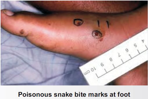
8

3. Recommendations
4.1 FIRST AID MEASURES
4.1.1 - by bystander or victim- Immediately transfer after providing first aid to a
health facility where optimal medical care with antisnake venom (ASV) is available,
close observation can be maintained, facility for laboratory investigation is available,
and definite treatment can be provided.

4.1.2 At The Community or Village Level

–   Check history of snakebite and look for obvious evidence of a bite (fang puncture
    marks, bleeding, swelling of the bitten part etc.). However, in krait bite no local
    marks may be seen. It can be noted by magnifying lens as a pin head bleeding
    spot with surrounding rash.
–   Reassure the patient as around 70% of all snakebites are from non-venomous
    species.
–   Immobilize the limb in the same way as a fractured limb. Use bandages or cloth
    to hold the splints (wooden stick), but do NOT block the blood supply or apply
    pressure. Ideally the patient should lie in the recovery position (prone, on the
    left side) with his/her airway protected to minimize the risk of aspiration of
    vomitus.

–   Nil by mouth till victim reaches a medical health facility.
–   Traditional remedies have NO PROVEN benefit in treating snakebite.
–   Shift the victim to the nearest health facility (PHC or hospital) immediately.
–   Arrange transport of the patient to medical care as quickly, safely and passively
    as possible by vehicle ambulance (toll free no. 102/108/etc.), boat, bicycle,
    motorbike, stretcher etc.

QRG Snakebite                     Version 4 Final            December 22, 2015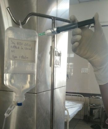
9

–   Victim must not run or drive himself to reach a Health facility. Motorbike
    Ambulance may be a feasible alternative for rural India.
–   If possible PHC medical officer can accompany with patient to know the progress
    and management and facilitate resuscitation on the way.
–   Inform the doctor of any symptoms such as progress of swelling, ptosis or new
    symptoms that manifest on the way to hospital.
–   Remove shoes, rings, watches, jewellary and tight clothing from the bitten area
    as they can act as a tourniquet when swelling occurs.
–   Leave the blisters undisturbed.

Important don’ts
  l Do not attempt to kill or catch the snake as this may be dangerous.

  l Discard traditional first aid methods (black stones, scarification) and

     alternative medical/herbal therapy as they have no role and do more harm
     than good by delaying treatment.
  l Do not wash wound and interfere with the bite wound (incisions, suction,

     rubbing, tattooing, vigorous cleaning, massage, application of herbs or
     chemicals, cryotherapy, cautery) as this may introduce infection, increase
     absorption of the venom and increase local bleeding.
  l Do NOT apply or inject antisnake venom (ASV) locally.

  l Do not tie tourniquets as it may cause gangrenous limbs.

  l If victim is expected to reach the hospital in more than 30 minutes but less

     than 3 hours crepe bandage may be applied by qualified medical personnel till
     the patient is shifted to the hospital. The bandage is wrapped over the bitten
     area as well as the entire limb with the limb placed in a splint. It should be
     capable of admitting a finger beneath it (See Figure 2.)

QRG Snakebite                   Version 4 Final          December 22, 2015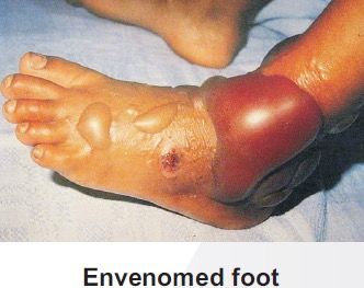
10

                Figure 2: Pressure immobilization (Sutherland method)

4.1.3 At A Health Care Facility

   –   Admit all victims of snakebite confirmed or suspected and keep under
       observation for 24 hours.
   –   Provide first-aid measures, supportive measures immediately. Observe for
       signs of envenomation. Administer ASV therapy as soon as there is evidence
       of envenomation.

4.2 Sign & Symptoms
Examine the bite site and look for fang marks, or any signs of local envenomation.
Fang mark or their patterns have no role to determine whether the biting species
was venomous or non venomous or amount of venom injected, severity of
systemic poisoning and nature of poisoning – Elapidae or viperidae venom etc.
Some species like Krait may leave no bite marks.

See figure 1 for presenting clinical syndromes of venomous snakebite.

QRG Snakebite                     Version 4 Final         December 22, 2015
11
4.2.1 Asymptomatic (i.e., non Venom related symptoms)
Patients many a times present with nonspecific symptoms related to anxiety.
Common symptoms in these patients are:
– Palpitations, sweating, tremoulessness, tachycardia, tachypnoea, elevated
blood pressure, cold extremities and paraesthesia. These patients may have
dilated pupils suggestive of sympathetic over activity.
– Differentiate from symptoms and signs of envenomation listed below.
– Redness, increased temperature, persistent bleeding and tenderness locally.
However, local swelling can be present in these patients due to tight ligature
4.2.2 Dry Bite
– Bites by nonvenomous snakes are common and bites by venomous species
are not always accompanied by the injection of venom (dry bites).
– The percentage of dry bites ranges from 10–80% for various poisonous
snakes.
– Some people who are bitten by snakes (or suspect or imagine that they have
been bitten) or have doubts regarding bite may develop quite striking
symptoms and signs, even when no venom has been injected due to
understandable fear of the consequences of a real venomous bite.
– Even in case of dry bite, symptoms due to anxiety and sympathetic over-
activity (as above) may be present. As symptoms associated with panic or
stress sometimes mimic early envenoming symptoms, clinicians may have
difficulties in determining whether envenoming occurred or not.
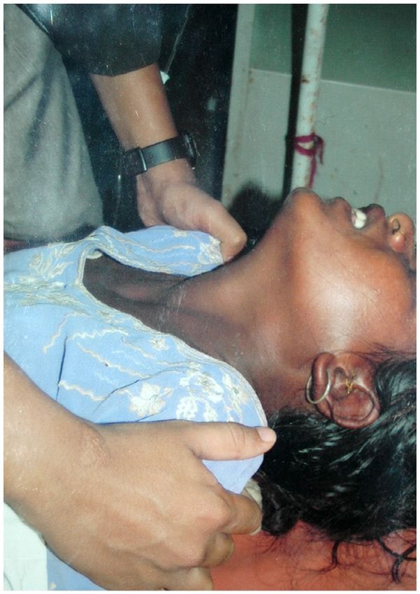
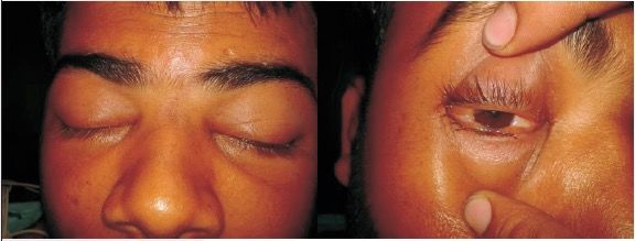
4.2.3- Neuroparalytic (Progressive weakness; Elapid envenomation)
– Neuroparalytic snakebite patients present with typical symptoms within 30
min– 6 hours in case of Cobra bite and 6 – 24 hours for Krait bite; however,
ptosis in Krait bite have been recorded as late as 36 hours after
hospitalization.
– These symptoms can be remembered as 5 Ds and 2 Ps.
• 5 Ds – dyspnea, dysphonia, dysarthria, diplopia, dysphagia
• 2 Ps – ptosis, paralysis
QRG Snakebite Version 4 Final December 22, 201512
– In chronological order of appearance of symptoms – furrowing of forehead,
Ptosis (drooping of eyelids) occurs first (Figure 3), followed by Diplopia
(double vision), then Dysarthria (speech difficulty), then Dysphonia (pitch of
voice becomes less) followed by Dyspnoea (breathlessness) and Dysphagia
(Inability to swallow) occurs. All these symptoms are related to 3rd, 4th, 6th
and lower cranial nerve paralysis. Finally, paralysis of intercostal and skeletal
muscles occurs in descending manner.
– Other signs of impending respiratory failure are diminished or absent deep
tendon reflexes and head lag.
– Additional features like stridor, ataxia may also be seen.
– Associated hypertension and tachycardia may be present due to hypoxia.
Figure 3. Ptosis with neuroparalytic snakebite
– To identify impending respiratory failure bedside lung function test in adults
viz.
• Single breath count – number of digits counted in one exhalation - >30
normal
• Breath holding time – breath held in inspiration – normal > 45 sec
• Ability to complete one sentence in one breath.
– Cry in a child whether loud or husky can help in identifying impending
respiratory failure.
– Bilateral dilated, poorly or a non-reacting pupil is not the sign of brain dead
in elapid envenoming (Figure 3).
– Refer patients presenting with neuroparalytic symptoms immediately to a
higher facility for intensive monitoring after giving Atropine Neostigmine
(AN) injection (schedule of AN injection described below).
QRG Snakebite Version 4 Final December 22, 201513
4.2.4 Vasculotoxic (haemotoxic or Bleeding) - General signs and symptoms of
Viperine envenomation)
Vasculotoxic bites are due to Viper species. They can have local manifestations as
well as systemic manifestations.
– Local manifestations –
these are more prominent in Russel’s viper bite followed by Saw scaled viper
and least in Pit viper bite. Local manifestations are in form of:
• Local swelling, bleeding, blistering, and necrosis.
• Pain at bite site and severe swelling leading to compartment syndrome.
Pain on passive movement. Absence of peripheral pulses and
hypoesthesia over the fuels of nerve passing through the compartment
helps to diagnose compartment syndrome.
• Tender enlargement of local draining lymph node.
– Systemic manifestations –
• Visible systemic bleeding from the action of haemorrhagins (Figure 4) e.g.
gingival bleeding, epistaxis, ecchymotic patches, vomiting, hematemesis,
hemoptysis, bleeding per rectum, subconjunctival hemorrhages, continuous
bleeding from the bite site, bleeding from pre-existing conditions e.g.
haemorrhoids, bleeding from freshly healed wounds.
• Bleeding or ecchymosis at the injection site is a common finding in Viper
bites.
• The skin and mucous membranes may show evidence of petechiae, purpura
ecchymoses, blebs and gangrene.
• Swelling and local pain.
• Acute abdominal tenderness may suggest gastro-intestinal or retro
peritoneal bleeding.
• Lateralizing neurological symptoms such as asymmetrical pupils may be
indicative of intra-cranial bleeding.
• Consumption coagulopathy detectable by 20WBCT, develops as early as
within 30 minutes from time of bite but may be delayed.
QRG Snakebite Version 4 Final December 22, 201514
Figure 4. Local and systemic Vasculotoxic (haemotoxic or Bleeding) manifestations
of Viperine envenomation.
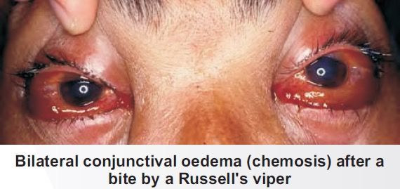
4.2.5 Life threatening complications are due to renal involvement. Patient presents
with hematuria, hemoglobinuria, myoglobinuria followed by oliguria and anuria with
acute kidney injury (AKI).
• Bilateral renal angle tenderness.
• Passage of discolored (reddish or dark brown urine or declining urine output.
• Acute Kidney Injury e.g. declining or no urine output, deteriorating renal signs
such as rising serum creatinine, urea or potassium. Some species e.g. Russell’s
viper (Daboia sp) and Saw scale vipers (Echis sp) frequently cause acute Kidney
Injury.
• Hypotension due to hypovolaemia or direct vasodilatation or direct
cardiotoxicity aggravates acute kidney injury.
• Parotid swelling, conjunctiva oedema, sub-conjunctival haemorrhage, renal
failure, acute respiratory distress syndrome [leaking syndrome] and refractory
shock.
• Long term sequelae e.g. pituitary insufficiency with Russell’s viper (Daboia sp),
Sheehan’s syndrome or amenorrhea in females.
4.2.6 Painful Progressive Swelling (PPS)
QRG Snakebite Version 4 Final December 22, 201515
Progressive painful swelling is indicative of local venom toxicity. It is prominent in
Russel’s viper bite, Saw scaled viper bite and Cobra bite. This is associated with
• Local necrosis which often has a rancid smell. Limb is swollen and the skin is taut
and shiny. Blistering with reddish black fluid at and around the bite site. Skip
lesions around main lesion are also seen. (Figure 5).
• Ecchymoses due to venom action destroying blood vessel wall.
• Significant painful swelling potentially involving the whole limb and
extending onto the trunk.
• Compartment syndrome will present invariably.
• Regional tender enlarged lymphadenopathy.
Figure 5. Snakebite marks and local swelling and necrosis
4.2.7 Myotoxic
This presentation is common in Sea snakebite. Patient presents with:
• Muscle aches, muscle swelling, involuntary contractions of muscles.
• Passage of dark brown urine.
• Compartment syndrome, cardiac arrhythmias due to hyperkalaemia, acute
kidney injury due to myoglobinuria, and subtle neuroparalytic signs.
4.2.8- Occult snakebite
• Krait bite victims often present in the early morning with paralysis with no local
signs. Krait has nocturnal habitat and has fine slender teeth. Hence bite marks
usually cannot be identified even on close examination.
• Typical presenting history is that the patient was healthy at night, in the morning
gets up with severe epigastric/umbilical pain with vomiting persisting for 3 – 4
hours and followed by typical neuroparalytic symptoms within next 4- 6 hours.
There is no history of snakebite.
• Unexplained respiratory distress in children in the presence of ptosis or sudden
onset of acute flaccid paralysis in a child (locked-in syndrome) are highly
suspicious symptoms in endemic areas particularly of Krait bite envenomation.
Sometimes patients may present with throat pain or chest pain also.
QRG Snakebite Version 4 Final December 22, 201516 Early morning symptoms of acute pain abdomen with or without neuroparalysis can be mistaken for a acute appendicitis, acute abdomen, stroke, GB syndrome, myasthenia gravis and hysteria (Bawaskar 2002). Krait bite envenoming is diagnosed by developing descending neuroparalysis while GB syndrome is by ascending paralysis. Strong clinical suspicion and careful examination can avoid not only costly and unnecessary investigations such as CT scan, MRI, nerve conduction studies, CSF studies and many others but also help in avoiding undue delay in initiation of a specific treatment with ASV. Atropine neostigmine (AN) test helps to rule out myasthenia gravis. 4.2.9 Differential identification of type of snakebite based on the symptoms and signs Though to a large extent the manifestation of snakebite depends upon the species of snake, unfortunately, in many cases the biting snake is not seen, and if it is, its description by the victim is often misleading (Harris et al 2010). Therefore identification of the type of snake should not hold the treatment. At times the bite mark might not be visible (e.g., in the case of Krait). The clinical manifestations of the patient may not correlate with the species of snake brought as evidence. However, it is advantageous to know the appearance of the snake so as to recognize the species (Figure 6). The killed snake brought as evidence helps in identification of snake, in which case species-specific monovalent Antisnake venom (ASV) can be administered. However, monovalent ASV is not available in India. Inspection of local site of bite can also help to identify snake’s species. Local swelling, bleeding, blistering, necrosis suggests Cobra bite. Minimum local changes indicate Krait bite. Local bleeding suggests Nilgiri Russel’s viper. Pain in abdomen and hyper peristalsis indicates Krait bite. QRG Snakebite Version 4 Final December 22, 2015
17

Figure 6. Snake identification by the patient

ON PRESENTATION, PATIENTS CAN BE CRITICAL OR NON CRITICAL (See FIGURE 1).

4.3. ASSESSMENT
4.3.1 Critical Arrival: Patient assessment on arrival

–   Vasculotoxic patients presenting with bleeding from multiple orifices with
    hypotension, reduced urine output, obtunted mentation (drowsy, confused),
    cold extremities need urgent attention and ICU care for volume replacement,
    pressor support, dialysis and infusion of blood and blood products (See following
    sections).

–   Neuroparalytic patients presenting with respiratory paralysis, tachypnoea or
    bradypnoea or paradoxical respiration (only moving abdomen), obtunded
    mentation, and peripheral skeletal muscle paralysis need urgent ventilator
    management with endotracheal intubation, ventilation bag or ventilator
    assistance.

–   Other patients can be evaluated to decide severity of their illness.

4.3.2 Patient assessment: Non critical arrival and Critical patients after
stabilization

–   Determine the time elapsed since the snakebite and as to what the victim was
    doing at the time of the bite, history of sleeping on floor bed in previous night.
–   Determine if any traditional medicines have been used.
–   Obtain a brief medical history (e.g., date of last tetanus immunization, use of any
    medication, presence of any systemic disease, and history of allergy)
–   If the victim has brought the snake, identification of the species should be
    carried out carefully, since crotalids can envenomate even when dead. This is
    why bringing the killed snake into the emergency department should be
    discouraged.

4.3.3 Physical examination
– Careful assessment of the site of the bite and signs of local envenomation and
   examination of the patient should be carried out and recorded (Annexure 1).
   Monitor the patient closely and repeat all above, every 1-2 hourly.
– Check for and monitor the following: Pulse rate, respiratory rate, blood pressure
   and 20 minutes Whole Blood clotting test (20 WBCT) every hour for first 3 hours
   and every 4 hours for remaining 24 hours.

QRG Snakebite                     Version 4 Final            December 22, 2015
18
– Check distal pulses and monitor if there is presence of gross swelling. The
presence of a pulse does not rule out compartment syndrome. Pain on passive
movement, pallor, pulseless limb, hypoaesthesia over the sensory nerve
passing through the compartment are suggestive of compartment syndrome.
Measure compartment pressure directly if there is concern that a compartment
syndrome is developing. The diagnosis is established if the compartment
pressure, measured directly by inserting a 16 G IV cannula and connecting it with
manometer, is raised above 40 cm water/saline. Direct measurement is
necessary before resorting to fasciotomy since compartment syndrome is rare in
snakebite victims and fasciotomy done without correction of hemostatic
abnormality may cause the patient to bleed to death
4.3.4 Examination of pregnant women
Monitor uterine contractions and foetal heart rate. Lactating women who have been
bitten by snakes should be encouraged to continue breast feeding .
Clues for severe snake envenomation should be sought are:
• Rapid early extension of local swelling from the site of the bite. In Cobra bite
on finger, necrosis may start in few minutes.
• Early tender enlargement of local lymph nodes, indicating spread of venom
in the lymphatic system
• Visible signs of neurological impairment such as ptosis, muscular weakness,
respiratory distress or respiratory arrest.
• Early spontaneous systemic bleeding especially bleeding from the gums, bite
site, haematuria, haemoptysis, epistaxis or ecchymoses.
• Unconsciousness either with or without respiratory arrest.
• Passage of dark brown urine
• Snake identified as a very venomous one i.e., Cobra, Russel’s viper.
4.4 LAB INVESTIGATIONS
4.4.1 20 minute whole blood clotting test (20 WBCT):
– It is a bedside test.
– Place 2 ml of freshly sampled venous blood in a small glass test tube and
leave undisturbed for 20 minutes at ambient temperature.
– Gently tilt the test tube to see if the blood is still liquid; the patient has
hypofibrinogenaemia (“incoagulable” blood or “not clotted”) as a result of
QRG Snakebite Version 4 Final December 22, 201519
venom-induced consumption coagulopathy (Figure 7).
– If blood clot is formed and signs and symptoms of neurotoxic envenomation
present, classify as neurotoxic envenomation.
– If there is any doubt, repeat the test in duplicate, including a “control”
(blood from a healthy person).
– Caution: If the test tube used for the test is not made of ordinary glass, or if
it has been used before and cleaned with detergent, its wall may not
stimulate clotting of the blood sample in the usual way and test will be
invalid).
– Counsel patient and relatives in the beginning that, 20WBCT may be
repeated several times before giving any medication.
Figure 7. 20 minute whole blood clotting test (20 WBCT).
– If clotted, the test should be carried out every 1 h from admission for three
hours and then 6 hourly for 24 hours. In case test is non-clotting, repeat 6
hour after administration of loading dose of ASV. In case of neurotoxic
envenomation repeat clotting test after 6 hours.
Other investigations that may assist in the management of snake bite at various
levels of healthcare
4.4.2 – Other Lab tests at Primary health centre
– Peak flow meter in patients (adolescents and adults) presenting with
neuroparalytic syndrome.
– If Peak flow meter is not available in PHC then assess respiratory function
using bedside tests - single breath count, breath holding time and ability to
complete one sentence in one health as described earlier.
– Urine examination for albumin and blood by dipstick.
4.4.3 Others lab test at District Hospital
QRG Snakebite Version 4 Final December 22, 201520
In addition to the above
– Prothrombin time
– Platelet count,
– Clot retraction time
– Liver function test (LFT)
– Renal Function test (RFT)
– Serum Amylase
– Blood sugar
– ECG
– Abdominal ultrasound
– 2D Echo (if available)
4.4.4 Others Lab test at Tertiary Health Care Centre
In addition to the above
– In neuroparalytic envenomation
• Arterial blood gases. Caution: Arterial puncture is contraindicated in patients
with haemostatic abnormalities.
• Pulmonary function tests
– In Vasculotoxic venomation
• For coagulopathy- BT, CT, PT, APTT, Platelet, Serum Fibrinogen, FDP D-Dimer
assay, LDH, peripheral blood smear
• Hemolysis -Urine for myoglobin, Urine haemoglobin
• For renal failure- Urine microscopy for RBC, casts, RFT, urinary proteins,
creatinine ratio
• Hepatic injury – LFTs including SGOT, SGPT, Alkalien phosphatase, serum
proteins
• Cardiotoxicity- CPK-MB, 2D Echo, BNP
• Myotoxic – CPK, SGOT, Urine myoglobin, compartment pressure
• Infection- Serum procalcitonin, culture (blood, urine, wound) and sensitivity
– Arterial blood gases and urine examination should be repeated at frequent
intervals during the acute phase to assess progressive systemic toxicity).
4.4.5- Rationale and interpretation of the tests:
4. Hemogram: The hemogram may show transient elevation of
hemoglobin level due to hemoconcentration (because of the increased
capillary leak) or may show anemia (due to hemolysis, especially in
QRG Snakebite Version 4 Final December 22, 201521
viper bites). Presence of neutrophilic leucocytosis signifies systemic
absorption of venom. Thrombocytopenia may be a feature of viper
envenomation.
5. Platelet count: This may be decreased in victims of envenoming by vipers.
White blood cell count: An early neutrophil leucocytosis is evidence of systemic
envenoming from any species.
Blood film: Fragmented red cells (“helmet cell”, schistocytes) are seen when
there is microangiopathic haemolysis.
Plasma/serum: May be pinkish or brownish if there is gross haemoglobinaemia
or myoglobinaemia.
6. Serum creatinine: This is necessary to rule out acute kidney injury after viper and
sea snakebite.
7. Serum creatinine phosphokinase (CPK): Elevated levels of these markers
suggests muscle damage (caution for renal damage) and raised amylase suggests
pancreatic injury
8. Prothrombin time (PT) and activated partial thromboplastin time (aPTT):
Prolongation may be present in viper bite (to be repeated 6 hourly, if abnormal).
9. Fibrinogen and fibrin degradation products (FDPs): Low fibrinogen with elevated
FDP is present when venom interferes with the clotting mechanism.
10. Urine examination for Proteinuria/ RBC/ Haemoglobinuria/ Myoglobinuria: The
colour of the urine (pink, red, brown, black) should be noted and the urine
should be tested by dipsticks for blood or haemoglobin or myoglobin. Standard
dipsticks do not distinguish blood, haemoglobin and myoglobin. Haemoglobin
and myoglobin can be separated by immunoassays but there is no easy or
reliable test. Microscopy will confirm whether there are erythrocytes in the
urine.
11. Electrocardiogram (ECG): Nonspecific ECG changes such as bradycardia and
atrioventricular block with ST-T changes may be seen.
12. Electroencephalogram (EEG): Recently, EEG changes have been noted in up to
96% of patients bitten by snakes. These changes start within hours of the bite
but are not associated with any features of encephalopathy. Sixty-two percent
showed grade I changes, 31% cases manifested grade II changes (moderate to
severe abnormality), and the remaining 4% showed severe abnormality (grade
III). These abnormal EEG patterns were seen mainly in the temporal lobes
(Ramachandran S et al 1995). However, rarely needed for patient management.
13. Pulse oximetry for oxygen in patients with respiratory failure or shock.
14. Electrolyte determinations: These tests are necessary for patients with
respiratory paralysis and systemic symptoms.
15. Arterial blood gases and pH may show evidence of respiratory failure (neurotoxic
QRG Snakebite Version 4 Final December 22, 201522
envenoming) and acidaemia (respiratory or metabolic acidosis).
16. X-Ray/ CT/ Ultrasound (The use of X-Ray and ultrasound are of unproven benefit,
apart from identification of bleeding in Viperine bites).
4.5 ANTI SNAKE VENOM (ASV) THERAPY
– If ASV is indicated i.e. signs and symptoms of envenomation with or without
evidence of laboratory tests, administer full dose.
– There are no absolute contraindications to ASV.
– Do not routinely administer ASV to any patient claiming to have bitten by a
snake as ASV exposes such patients to the risks of ASV reactions unnecessarily;
besides wastage of valuable and scarce stocks of ASV. However, at the same
time do not delay or withhold ASV on the grounds of anaphylactic reaction to
a deserving case. Do NOT give incomplete dose.
– Purely local swelling, even if accompanied by a bite mark from an apparently
venomous snake, is not a ground for administering ASV. Swelling, a number of
hours old is also not a ground for giving ASV. However, rapid development of
swelling indicates bite with envenoming requiring ASV.
4.5.1 Antisnake venom (ASV)
– Antisnake venom treatment is the only specific treatment, should be given as
soon as it is indicated. It may reverse systemic envenomation abnormality even
when this has persisted for several days or, in the case of haemostatic
abnormalities, persisting for two or more weeks. The dosage required varies
with the degree of envenomation.
– In the presence of coagulopathy, Polyvalent ASV freeze-dried (heat stable; to be
stored at cool temperature; shelf life 5 years) or neat liquid ASV (heat labile;
requires reliable cold chain (2-8oC) with a refrigeration shelf life of 2 years)
whichever is available may be used. If integrity of the cold chain is not
guaranteed then use lyophilized ASV.
– ASV supplied in dry powder form has to be reconstituted by diluting in 10 ml of
distilled water/normal saline (Figure 8). Mixing is done by swirling and not by
vigorous shaking. Caution: Do not use, if reconstituted solution is opaque to any
extent.
4.5.2 Precautions during ASV Administration -
– ASV should be given only by the IV route, and should be given slowly, with the
QRG Snakebite Version 4 Final December 22, 201523
physician at the bed side during the initial period to intervene immediately at
the first sign of any reaction. The rate of infusion can be increased gradually in
the absence of a reaction until the full starting dose has been administered
(over a period of ~1 hour).
– Epinephrine (adrenaline) should always be drawn up in readiness before ASV
is administered.
– ASV must NEVER be given by the IM route because of poor bioavailability by this
route. Also do NOT inject the ASV locally at the bite site since it is not effective,
is extremely painful and may increase intra-compartmental pressure.
– Take all aseptic precautions before starting ASV to prevent any pyrogenic
reactions to ASV.
4.5.3- Dose of ASV for neuroparalytic snakebite – ASV 10 vials stat as infusion over
30 minutes followed by 2nd dose of 10 vials after 1 hour if no improvement within 1st
hour.
4.5.4- Dose of ASV for vasculotoxic snakebite - Two regimens low dose infusion
therapy and high dose intermittent bolus therapy can be used. Low dose infusion
therapy is as effective as high dose intermittent bolus therapy and also saves scarce
ASV doses (Expert Consensus).
– Low Dose infusion therapy – 10 vials for Russel’s viper or 6 vials for Saw scaled
viper as stat as infusion over 30 minutes followed by 2 vials every 6 hours as
infusion in 100 ml of normal saline till clotting time normalizes or for 3 days
whichever is earlier.
OR
– High dose intermittent bolus therapy - 10 vials of polyvalent ASV stat over 30
minutes as infusion, followed by 6 vials 6 hourly as bolus therapy till clotting
time normalizes and/or local swelling subsides.
– No ASV for Sea snakebite or pit viper bite as available ASV does not contain
antibodies against them.
– The range of venom injected is 5 mg-147 mg. The total required dose range
between 10 and 30 vials as each vial neutralizes 6 mg of Russell’s Viper venom.
Depending on the patient condition, additional vials can be considered.
QRG Snakebite Version 4 Final December 22, 201524
ASV in
syringe
10 vials of AVS dissolved
in 100 ml of distilled
water and added to
400ml of normal saline
Mention date and
time of starting
infusion
Administer 10 vials of ASV in first hour.
Maintain slow drip for 24 hours
Figure 8. ASV infusion and dosage schedule Each vial of AVS be dissolved in 10 ml
of distilled water and added to an infusion medium such as normal saline (i.e. 10
vials of AVS dissolved in 100 ml of distilled water and added to 400ml of normal
saline). The volume of infusion is reduced according to the body size and the state of
hydration of the patient. In oliguric patients restrict fluids and use infusion pump to
give full dose of ASV over 30 minutes.
4.5.5 ASV dose in pregnancy
Pregnant women are treated in exactly the same way as other victims. The same
dosage of ASV is given. Refer the victim to a gynecologist for assessment of any
impact on the foetus.
4.5.6 ASV dose in children
Children also are given exactly the same dose of ASV as adults as snakes inject the
same amount of venom into children and adult.
Infusion: liquid or reconstituted ASV is diluted in 5-10 ml/kg body weight of normal
saline. However, reduce amount of fluid in running bottle to 200 ml to avoid fluid
over load.
QRG Snakebite Version 4 Final December 22, 201525
4.5.7 ASV dosage in victims requiring life saving surgery
Rarely patient may develop intracranial bleeding for which a life saving surgery is
required. In such cases before surgery coagulation must be restored to avoid
catastrophic bleeding and higher initial dose of ASV (up to 30 vials) can be
administered.
4.5.8 Repeat dose of ASV
– Repeat dose: in Vasculotoxic or haemotoxic envenomation
Repeat clotting test every 6 hours until coagulation is restored. Administer ASV
every 6 h until coagulation is restored. Envenomation by the Hump-nosed Pit viper
does not respond to normal Indian polyvalent ASV and coagulopathy may continue
for up to 3 weeks. If 30 vials of ASV have been administered reconsider whether
continued administration of ASV is serving any purpose, particularly in the absence
of proven systemic bleeding.
– Repeat dose: neuroparalytic or neurotoxic envenomation
Repeat ASV when there is worsening neurotoxic or cardiovascular signs even after
1–2 h. Maximum dose 20 vials of ASV for neurotoxically envenomed patients. If
large doses have been administered and the coagulation abnormality persists, give
fresh frozen plasma (FFP) or cryoprecipitate (fibrinogen, factor VIII), fresh whole
blood, if FFP not available or platelet concentrate.
4.5.9 Victims who arrive late
Sometimes victims arrive late after the bite, often after several days, usually with
acute kidney injury. Determine current venom activity such as bleeding in case of
viperine envenomation. Perform 20WBCT and determine if any coagulopathy is
present then administer ASV. If no coagulopathy is evident, treat kidney injury, if
any.
In patients with neuroparalytic envenomation (ptosis, respiratory failure etc.)
• Continue respiratory support until recovery
• Give 10 vials of ASV on arrival and if no improvement within one hour repeat
10 vials of ASV (No more than 20 vials of ASV).
• No further ASV and Atropine Neostigmine (AN) infusion is required ONLY to
reverse the Ptosis. Ptosis in Common Krait bite is due to presynaptic
blockage, further ASV and Neostigmine dose beyond 3 doses cannot reverse
QRG Snakebite Version 4 Final December 22, 201526
it, since regeneration is a natural process and may take 4-5 days. Both ASV
and AN injection should be stopped when the initial syndrome of pharyngeal
muscle palsy is over.
4.5.10 Monitoring of Patients on ASV therapy
– All patients should be watched carefully every 5 min for first 30 min, then at
15 min for 2 hours for manifestation of a reaction. At the earliest sign of an
adverse reaction suspend temporarily.
– Maintain a strict intake output chart and note colour of urine to detect acute
kidney injury early.
4.6. ASV reaction
– NO ASV TEST DOSE MUST BE ADMINISTERED.
– SKIN/CONJUNCTIVAL HYPERSENSITIVITY TESTING DOES NOT RELIABLY PREDICT
EARLY OR LATE ANTISNAKE VENOM REACTIONS AND IS NOT RECOMMENDED.
– Rarely patients may develop severe life-threatening anaphylaxis characterized by
hypotension, bronchospasm, and angioedema. However, 20%-60% patients
treated with ASV develop either early or late mild reactions.
– Early anaphylactic reactions occurs within 10–180 min of start of therapy and is
characterized by itching, urticaria, dry cough, nausea and vomiting, abdominal
colic, diarrhoea, tachycardia, and fever.
– Pyrogenic reactions usually develop 1–2 h after treatment. Symptoms include
chills and rigors, fever, and hypotension. These reactions are caused by
contamination of the ASV with pyrogens during the manufacturing process.
– Any new sign or symptom after starting the ASV in drip should be suspected as
a reaction to ASV.
– Late (serum sickness–type) reactions develop 1–12 (mean 7) days after
treatment. Clinical features include fever, nausea, vomiting, diarrhoea, itching,
recurrent urticaria, arthralgia, myalgia, lymphadenopathy, immune complex
nephritis and, rarely, encephalopathy.
4.6.1 Treatment of Early ASV reaction
– Stop ASV temporarily.
QRG Snakebite Version 4 Final December 22, 201527
– Oxygen
– Start fresh IV normal saline infusion with a new IV set
– Administer Epinephrine (adrenaline) (1 in 1,000 solution, 0.5 mg (i e 0.5 ml) in
adults intramuscular over deltoid or over thigh; In children 0.01 mg/kg body
weight) for early anaphylactic and pyrogenic ASV reactions.
– Administer Chlorpheniramine maleate (adult dose 10 mg, in children 0.2 mg/kg)
intravenously.
– Role of Hydrocortisone in managing ASV reaction is not proved.
– Once the patient has recovered, re-start ASV slowly for 10-15 minutes keeping
the patient under close observation. Then resume normal drip rate.
– For high risk patients
In patients with history of hypersensitivity or exposure to animal serum such as
equine ASV, tetanus-immune globulin or rabies-immune globulin in past, severe
atopic conditions:
• Give ASV only if they have signs of systemic envenoming.
• Give Inj. Hydrocortisone 200 mg and Chlorpheniramine maleate 22.75 mg
prior to the administration of ASV.
– Epinephrine premedication is not given as routine as it can cause hypertension
and in patients with bleeding tendency can lead to intracranial bleeding (Expert
Consensus). However, epinephrine should be kept handy for adults. No trials
have been done in children and old people. Inj. Adrenaline 0.25 ml of 1:1000 (as
available in one ampoule of 1 ml) Subcutaneously just before adding ASV to the
running IV fluid.
4.6.2 Treatment of Late (serum sickness–type) reactions
– Inj. Chlorpheniramine 2 mg in adults (In children 0.25 mg/kg/day) 6 hourly for 5
days.
– In patients who fail to respond within 24–48 h give a 5-day course of
Prednisolone (5 mg 6 hourly in adults and 0.7 mg/kg/day in divided doses in
children.
4.6.3 Desensitization procedure only in case of severe anaphylaxis reaction to
ASV
– Pre-medication: Administer Inj. Hydrocortisone 100 mg I.V. and Inj. Adrenaline
0.5 ml subcutaneously/ intramuscularly (+/- Promethazine)
Table . Steps of dilution of ASV
Steps Instructions Total Solution Dilution
of Volume
dilution
QRG Snakebite Version 4 Final December 22, 201528
1. Dilute 1 ml of ASV in a vial with 10ml A
10 ml of normal saline
2. 1ml of solution A + 9 ml of 10 ml B 1: 10
saline
3. 1ml of solution B+ 9 ml of 10 ml C 1: 100
saline
4. 1ml of solution C + 9 ml of 10 ml D 1: 1000
saline
5. 1ml of solution D + 9 ml of 10 ml E 1: 10,000
saline
After dilution and preparation of Solution E,
QRG Snakebite Version 4 Final December 22, 201529
4.7 MANAGEMENT NEUROTOXIC (NEUROPARALYTIC) ENVENOMATION
Antisnake venom treatment alone cannot be relied upon to save the life of a
patient with bulbar and respiratory paralysis. Administer following in addition:
– Oxygen
– Assisted ventilation. The duration of mechanical ventilation in snakebite victims
is usually short since neuroparalysis reverses quickly with prompt administration
of ASV. Manual ventilation (self ventilating anaesthetic bag) has been effective
where no mechanical ventilator was available. In case of Guillain-Barre
syndrome and delayed neuropathy following snakebite prolonged assisted
ventilation with room air or oxygen is followed by complete recovery.
– Administer ‘Atropine Neostigmine (AN)’ schedule described as below.
– Refer to a higher facility where ASV is available, in case of no improvement.
4.7.1 Atropine neostigmine (AN) dosage schedule
– Atropine 0.6 mg followed by neostigmine (1.5mg) to be given IV stat and repeat
dose of neostigmine 0.5 mg with atropine every 30 minutes for 5 doses (In
children, Inj. Atropine 0.05 mg/kg followed by Inj. Neostigmine 0.04 mg/kg
Intravenous and repeat dose 0.01 mg/kg every 30 minutes for 5 doses). A fixed
dose combination of Neostigmine and glycopyrolate IV can also be used.
– Thereafter to be given as tapering dose at 1 hour, 2 hour, 6 hours and 12 hour.
Majority of patients improve within first 5 doses. Observe the patient closely
observed for 1 hour to determine if the neostigmine is effective. After 30
minutes, any improvement should be visible by an improvement in ptosis.
Positive response to “AN” trial is measured as 50% or more recovery of the
ptosis in one hour.
– Stop Atropine neostigmine (AN) dosage schedule if:
• Patient has complete recovery from neuroparalysis. Rarely patient can have
recurrence, carefully watch patients for recurrence.
• Patient shows side effects in the form of fasciculations or bradycardia.
• If there is no improvement after 3 doses.
– Improvement by atropine neostigmine indicates Cobra bite. A few Nilgiri Russel’s
viper bites victims also improve with this regimen.
– Give one dose of “AN” injection before transferring to the higher centre. Rapid
deterioration of Cobra bite neurotoxic syndrome may kill the patient on the way
to transfer.
QRG Snakebite Version 4 Final December 22, 201530
– If there is no improvement after 3 doses of atropine neostigmine (within 1
h), this indicates probable Krait bite. Krait affects pre-synaptic fibres where
calcium ion acts as neurotransmitter. Give Inj. Calcium gluconate 10ml IV (in
children 1-2 ml/kg (1:1 dilution) slowly over 5-10 min every 6 hourly and
continue till neuroparalysis recovers which may last for 5-7 days.
4.8 MANAGEMENT OF VASCULOTOXIC SNAKEBITE:
– Strict bed rest to avoid even minor trauma.
– Screen for hematuria, hemoglobinuria, myoglobinuria by Dipstick method.
Dipstick test is positive in all three presentations listed above. Centrifuged urine
showing pink color indicates hemoglobinuria, clear supernatant (RBCs settle
down as deposit) indicates myoglobinuria.
– Closely monitor urine output and maintain 1 ml/kg/h urine output.
4.8.1 Volume Replacement in snake bite:
– If the patient has intravascular volume depletion, indicated by supine or
postural hypotension, or empty neck veins, proceed as follows:
– Establish intravenous access.
– Give fluid challenge: An adult patient can be given two litres of isotonic saline
over one hour or until the jugular venous pressure/central venous pressure
has risen to 8-10 cm above the sterna angle (with the patient propped up at
450).
– Observe the patient closely while this is being done. The fluid challenge must
be stopped immediately if pulmonary oedema develops.
4.8.2 Forced Alkaline Diuresis
– If the patient has oliguria or dipstick positive for blood give a trial of
forced alkaline diuresis (FAD) within first 24 hours of the bite to avoid
pigment nephropathy leading to acute tubular necrosis (ATN).
– Delayed FAD has no role.
– Sequence of FAD in adults is as follows:
• Inj. Frusemide 40 mg IV stat
• Inj. Normal saline 500 ml + 20 ml of NaHCO3 over 20 minutes
• Inj. Ringer’s lactate 500 ml + 20 ml of NaHCO3 over 20 minutes
• Inj. 5% dextrose 500 ml + 10 ml of Potassium Chloride over 90 minutes
• Inj. Mannitol 150 ml over 20 min
– Whole cycle completes in 2 h 30 min and urine output of 3 ml/min is
expected.
QRG Snakebite Version 4 Final December 22, 201531
– If patient responds to first cycle continue for 3 cycles. FAD converts
oliguria into polyuria and avoid ATN and acute kidney injury needing
dialysis in more than 75% patients.
– If there is no response to furosemide discontinue FAD and refer patient
immediately to a higher center for dialysis.
– Indications for dialysis are:
• Absolute value of Blood urea >130 mg/dl (27 mmol/L) (BUN 100 mg/dl),
Sr. Creatinine > 4 mg/dl (500 μmol/L) OR evidence of hypercatabolism in
the form of daily rise in blood urea 30 mg/dL (BUN > 15), Sr. Creatinine >
1 mg/dL, Sr. Potassium > 1 mEq/L and fall in bicarbonate >2 mmol/L
• Fluid overload leading to pulmonary oedema
• Hyperkalaemia (>7 mmol/l (or hyperkalaemic ECG changes)
• unresponsive to conservative management.
• Uremic complications – encephalopathy, pericarditis.
– Haemodialysis is preferable in cases of hypotension or hyperkalaemia.
Peritoneal dialysis can be performed at a secondary health care center.
Continuous renal replacement therapies and intermittent hemodialysis are
equivalent in patients with severe sepsis and acute renal failure because they
achieve similar short-term survival rates.
– Continuous therapies are recommended to facilitate management of fluid
balance in hemodynamically unstable patients. An efficient dose for
continuous renal replacement therapy would be 20 to 25 mL/kg/h of effluent
generation.
4.8.3 In case of Shock, myocardial damage:
– Correct hypovolaemia with colloid/crystalloids, controlled by observation
of the central venous pressure.
– Infusion of isotonic crystalloids or albumin, with boluses of up to 20 ml/kg for
crystalloids (or albumin equivalent) over 5 to 10 mins titrated to reversing
hypotension, increasing urine output, and attaining normal capillary refill,
peripheral pulses and level of consciousness without inducing lung
crepitations or hepatomegaly.
– If hepatomegaly or rales develop, initiate inotropic support with dopamine
or dobutamine. If patient doesn’t respond to fluid resuscitation, inotropic
support must be given.
QRG Snakebite Version 4 Final December 22, 2015You can also read