The skin microbiome facilitates adaptive tetrodotoxin production in poisonous newts - eLife
←
→
Page content transcription
If your browser does not render page correctly, please read the page content below
RESEARCH ARTICLE

                                    The skin microbiome facilitates adaptive
                                    tetrodotoxin production in poisonous
                                    newts
                                    Patric M Vaelli1,2†, Kevin R Theis2,3, Janet E Williams2,4,5, Lauren A O’Connell6,
                                    James A Foster2,5,7, Heather L Eisthen1,2*
                                    1
                                      Department of Integrative Biology, Michigan State University, East Lansing, United
                                    States; 2BEACON Center for the Study of Evolution in Action, Michigan State
                                    University, East Lansing, United States; 3Department of Biochemistry, Microbiology,
                                    and Immunology, Wayne State University, Detroit, United States; 4Department of
                                    Animal and Veterinary Science, University of Idaho, Moscow, United States;
                                    5
                                      Institute for Bioinformatics and Evolutionary Studies, University of Idaho, Moscow,
                                    United States; 6Department of Biology, Stanford University, Stanford, United
                                    States; 7Department of Biological Sciences, University of Idaho, Moscow, United
                                    States

                                    Abstract Rough-skinned newts (Taricha granulosa) use tetrodotoxin (TTX) to block voltage-
                                    gated sodium (Nav) channels as a chemical defense against predation. Interestingly, newts exhibit
                                    extreme population-level variation in toxicity attributed to a coevolutionary arms race with TTX-
                                    resistant predatory snakes, but the source of TTX in newts is unknown. Here, we investigated
                                    whether symbiotic bacteria isolated from toxic newts could produce TTX. We characterized the
                                    skin-associated microbiota from a toxic and non-toxic population of newts and established pure
*For correspondence:                cultures of isolated bacterial symbionts from toxic newts. We then screened bacterial culture media
eisthen@msu.edu
                                    for TTX using LC-MS/MS and identified TTX-producing bacterial strains from four genera, including
Present address: †Department        Aeromonas, Pseudomonas, Shewanella, and Sphingopyxis. Additionally, we sequenced the Nav
of Molecular and Cellular           channel gene family in toxic newts and found that newts expressed Nav channels with modified TTX
Biology, Harvard University,        binding sites, conferring extreme physiological resistance to TTX. This study highlights the complex
Cambridge, United States            interactions among adaptive physiology, animal-bacterial symbiosis, and ecological context.
Competing interest: See
page 23
Funding: See page 24
Received: 23 November 2019          Introduction
Accepted: 26 February 2020          Coevolutionary interactions among species are a central force driving the origin of novel, adaptive
Published: 07 April 2020            phenotypes, yet the traits under selection are often complex and arise from multifaceted interactions
                                    among genetic, physiological, and environmental forces that are not well understood (Ehrlich and
Reviewing editor: Christelle AM
Robert, University of Bern,
                                    Raven, 1964; Futuyma and Agrawal, 2009; Schoener, 2011). Chemical interactions among species
Switzerland                         in the form of defensive compounds have evolved across all domains of life, and these toxins often
                                    target evolutionarily conserved proteins in potential predators (Brodie and Ridenhour, 2003; Hodg-
   Copyright Vaelli et al. This
                                    son, 2012; Whittaker and Feeny, 1971). For example, tetrodotoxin (TTX), the primary neurotoxin
article is distributed under the
                                    found in poisonous pufferfishes (Tsuda and Kawamura, 1952), has been discovered across a broad
terms of the Creative Commons
Attribution License, which          phylogenetic distribution of animals (Chau et al., 2011; Hanifin, 2010). The unusual molecular struc-
permits unrestricted use and        ture of this toxin serves to selectively target voltage-gated sodium (Nav) channels, which are critical
redistribution provided that the    for generating action potentials in neurons, muscles, and other excitable cells (Hille, 2001). Thus,
original author and source are      TTX toxicity can have substantial impacts on eco-evolutionary interactions among species, impacting
credited.                           both the toxic species and potential predators.

Vaelli et al. eLife 2020;9:e53898. DOI: https://doi.org/10.7554/eLife.53898                                                         1 of 29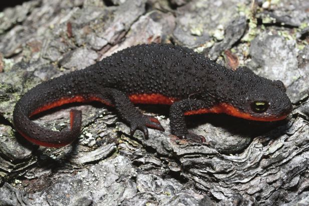
Research article                                                                                               Evolutionary Biology

                                    eLife digest Rough-skinned newts produce tetrodotoxin or TTX, a deadly neurotoxin that is also
                                    present in some pufferfish, octopuses, crabs, starfish, flatworms, frogs, and toads. It remains a
                                    mystery why so many different creatures produce this toxin. One possibility is that TTX did not
                                    evolve in animals at all, but rather it is made by bacteria living on or in these creatures. In fact,
                                    scientists have already shown that TTX-producing bacteria supply pufferfish, octopus, and other
                                    animals with the toxin. However, it was not known where TTX in newts and other amphibians comes
                                    from.
                                        TTX kills animals by blocking specialized ion channels and shutting down the signaling between
                                    neurons, but rough-skinned newts appear insensitive to this blockage, making it likely that they have
                                    evolved defenses against the toxin. Some garter snakes that feed on these newts have also evolved
                                    to become immune to the effects of TTX. If bacteria are the source of TTX in the newts, the
                                    emergence of newt-eating snakes resistant to TTX must be putting evolutionary pressure on both
                                    the newts and the bacteria to boost their anti-snake defenses. Learning more about these complex
                                    relationships will help scientists better understand both evolution and the role of beneficial bacteria.
                                        Vaelli et al. have now shown that bacteria living on rough-skinned newts produce TTX. In the
                                    experiments, bacteria samples were collected from the skin of the newts and grown in the
                                    laboratory. Four different types of bacteria from the samples collected produced TTX. Next, Vaelli
                                    et al. looked at five genes that encode the channels normally affected by TTX in newts and found
                                    that all them have mutations that prevent them from being blocked by this deadly neurotoxin. This
                                    suggests that bacteria living on newts shape the evolution of genes critical to the animals’ own
                                    survival.
                                        Helpful bacteria living on and in animals have important effects on animals’ physiology, health,
                                    and disease. But understanding these complex interactions is challenging. Rough-skinned newts
                                    provide an excellent model system for studying the effects of helpful bacteria living on animals.
                                    Vaelli et al. show that a single chemical produced by bacteria can impact diverse aspects of animal
                                    biology including physiology, the evolution of their genes, and their interactions with other creatures
                                    in their environment.

                                        Rough-skinned newts (Taricha granulosa) are among the most poisonous TTX-producing animals
                                    and serve as an excellent model system for understanding ecological influences on toxin production
                                    and predation (Figure 1A). This species is endemic to the Pacific Northwest of North America,
                                    where certain populations possess high quantities of TTX relative to other TTX-laden species includ-
                                    ing pufferfishes and blue-ringed octopuses (Hanifin, 2010; Williams, 2010). In some populations,
                                    individual newts possess enough TTX to kill several adult humans (Brodie et al., 2005; Hanifin, 2010;
                                    Hanifin et al., 1999). Variation in newt toxicity is driven in part by the evolution of TTX resistance in
                                    predatory garter snakes (Thamnophis sirtalis), as TTX toxicity and resistance in newts and snakes are
                                    strongly correlated geographically, suggesting that these two phenotypes are coevolving
                                    (Brodie et al., 2005; Brodie et al., 2002; Hanifin et al., 2008). Furthermore, TTX resistant Nav chan-
                                    nels have evolved independently across different garter snake populations, suggesting multiple
                                    independent origins of TTX resistance in predatory snakes (Feldman et al., 2009; Geffeney, 2002;
                                    Geffeney et al., 2005).
                                        Despite the central role of TTX toxicity in coevolutionary interactions between newts and snakes,
                                    the origin of TTX in newts and other freshwater animals is unknown (Daly, 2004; Hanifin, 2010). In
                                    TTX-bearing marine species, toxicity is derived either from dietary accumulation from TTX-laden
                                    prey, or from symbiotic interactions with TTX-producing bacteria (Chau et al., 2011; Miyazawa and
                                    Noguchi, 2001). Pufferfishes harbor numerous TTX-producing bacteria symbionts in toxic tissues
                                    including skin, liver, intestines, and ovaries, and cultured non-toxic pufferfishes are able to sequester
                                    dietary-administered TTX under laboratory conditions (Jal and Khora, 2015). TTX-producing bacte-
                                    ria have also been isolated from xanthid crabs, horseshoe crabs, starfish, chaetognaths, nemerteans,
                                    gastropods, and blue-ringed octopuses (Jal and Khora, 2015; Magarlamov et al., 2017). However,
                                    the origin of TTX in rough-skinned newts has been more controversial (Hanifin, 2010). Newts raised
                                    in long-term captivity on artificial diets maintain their TTX toxicity (Hanifin et al., 2002), and captive

Vaelli et al. eLife 2020;9:e53898. DOI: https://doi.org/10.7554/eLife.53898                                                            2 of 29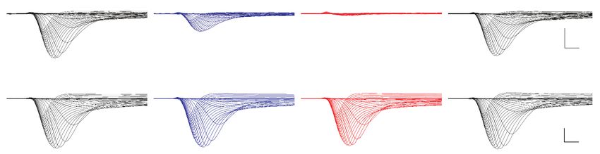
Research article Evolutionary Biology
TTX (ng/ml) per mg skin
A B C
48
latitude
46
44
ï ï ï
Oregon (TTX+) Idaho (TTX-)
longitude newt population
D F &KDR5LFKQHVV H Genus
Arthrobacter
Rhodoferax
&KDR,QGH[
Flavobacteriaceae 8Q
Methylococcaceae 8Q
Proteobacteria 8Q
Methylmonas
B Comamonadaceae 8Q
5HODWLYH$EXQGDQFH
Aeromonas
Pedobacter
Opitutus
Burkholderiales 8Q
Romboutsia
Pseudomonas
Opitutae 8Q
6LPSVRQ ' Flavobacterium
E G
Methylophilus
Burkholderiales 8Q
Fusobacteriaceae 8Q
6LPSVRQ,QGH[
Chromatiaceae 8Q
Thiodictyon
Geobacter
Anaeromyxobacter
Syntrophaceae 8Q
Arcobacter
Ktedonobacteria 8Q
Burkholderia
Ktedonobacter
Peptostreptococcaceae 8Q
Ventral
Soil
Soil
Soil
'RUVDO
'RUVDO
'RUVDO
'RUVDO
Ventral
Ventral
Ventral
Soil
Fluviicola
Acinetobacter
Other genera
OR (TTX+) ID (TTX-) OR (TTX+) ID (TTX-)
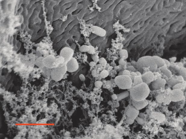
Figure 1. Characterization of the skin-associated microbiota of rough-skinned newts. (A) Rough-skinned newt (Taricha granulosa); photo by Gary Nafis
(CC by ND NC 3.0). (B) Two populations of T. granulosa previously reported to possess either high concentrations of TTX (Lincoln Co, OR; red) or no
TTX (Latah Co, ID; black) were compared in our study. (C) TTX measured from dorsal skin biopsies: newts from Oregon possessed 126.5 ± 42.1 ng mL 1
TTX (n = 5) while Idaho newts possessed no detectable TTX (n = 17). (D–E) Scanning electron micrographs of host-associated bacterial communities
upon the dorsal skin surface and within the ducts of TTX-sequestering granular glands. Scale bars 10 mm and 1 mm for D and E, respectively. (F–G)
Comparison of bacterial community richness and diversity between these two populations, along with soil samples collected contemporaneously from
the ponds in which the newts were caught. Non-toxic Idaho newts possessed higher OTU richness (Chao1 index, t unequal var. = 7.90, p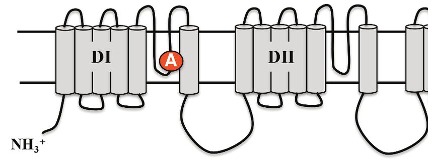
Research article                                                                                             Evolutionary Biology

                                    intestine, which contains low levels of TTX (Lehman et al., 2004). These studies suggest that the
                                    source of TTX in newts is not dietary, but whether newts have acquired the ability to produce TTX
                                    endogenously via convergent evolution or horizontal gene transfer, or from symbiosis with TTX-pro-
                                    ducing bacteria remains unclear.
                                        Furthermore, despite the extreme toxicity of some newt populations, the molecular basis of TTX
                                    resistance in this species is not well understood. A previous study identified amino acid replace-
                                    ments in the highly conserved pore-loop (P-loop) region of the skeletal muscle isoform Nav1.4 and
                                    found that skeletal muscle fibers were considerably resistant to TTX (Hanifin and Gilly, 2015).
                                    Amphibians, however, possess six Nav channel isoforms that are differentially expressed across excit-
                                    able tissues (Zakon et al., 2011). Unlike pufferfishes in which TTX is sequestered in certain tissues,
                                    newts possess TTX throughout their bodies (Mebs et al., 2010; Wakely et al., 1966; Yotsu et al.,
                                    1990), indicating that the central and peripheral nervous systems are exposed to TTX. Thus, the evo-
                                    lution of whole animal resistance should necessarily involve all Nav channel subtypes, providing an
                                    opportunity to examine molecular evolution in response to a specific selective pressure (i.e., TTX)
                                    across an entire gene family.
                                        In this study, we investigated the source of TTX toxicity and the molecular basis for TTX resistance
                                    in rough-skinned newts. We re-examined the hypothesis that newts derive their TTX from symbiosis
                                    with TTX-producing bacteria, focusing on the bacterial communities inhabiting the skin of T. granu-
                                    losa, as this organ possesses specialized granular glands for storing toxins and contains the highest
                                    quantities of TTX in the animal (Daly et al., 1987; Hamning et al., 2000; Hanifin et al., 2004;
                                    Santos et al., 2016; Toledo and Jared, 1995; Tsuruda et al., 2002). We took advantage of the nat-
                                    ural variation in TTX toxicity across newt populations to characterize the skin-associated microbiota
                                    in newts from a highly toxic and a non-toxic population and applied an unbiased cultivation-based
                                    approach to isolate numerous bacterial symbionts from the skin of toxic newts. Subsequent LC-MS/
                                    MS screening of bacterial cultivation media revealed TTX production in eleven bacterial strains from
                                    four genera: Aeromonas, Pseudomonas, Shewanella, and Sphingopyxis. Furthermore, to determine
                                    the molecular and physiological basis of extreme TTX resistance observed in this species, we cloned
                                    and sequenced five uncharacterized Nav channel paralogs (Nav1.1, Nav1.2, Nav1.3, Nav1.5, Nav1.6)
                                    from a highly toxic population of newts in Oregon. We identified amino acid substitutions in all five
                                    genes, many of which have been observed in other TTX-possessing species. To test whether Nav
                                    channel mutations impact TTX resistance in newts, we used site-directed mutagenesis to insert three
                                    newt-specific replacements identified in Nav1.6 into the TTX-sensitive Nav1.6 ortholog from Mus
                                    musculus. We found that each amino acid replacement reduced TTX sensitivity compared to wild-
                                    type M. musculus channels, but these mutations had the greatest effect when combined together.
                                    Overall, our results suggest that host-associated bacteria may underlie the production of a critical
                                    defensive compound in a vertebrate host, impacting predator-prey coevolution and potentially shap-
                                    ing the evolution of TTX resistance in a well-known ecological model system.

                                    Results
                                    Characterization of newt-associated microbiota from toxic and non-
                                    toxic newts
                                    To investigate whether bacterial symbionts produce TTX in newts, we leveraged natural variation in
                                    toxicity across newt populations to characterize skin-associated microbiota and determine whether
                                    highly toxic newts harbored candidate TTX-producing bacteria. We compared two populations pre-
                                    viously reported to differ substantially in TTX levels (Hanifin et al., 2008; Hanifin et al., 1999), a
                                    toxic population in Lincoln County, OR and a non-toxic population in Latah County, ID (Figure 1A–
                                    B). As expected, skin biopsies collected from the dorsal skin of adult newts confirmed that Oregon
                                    newts (n = 5) possessed high TTX concentrations (126.5 ± 42.1 ng mL 1 per mg skin) while Idaho
                                    newts (n = 17) lacked detectable levels of TTX (Figure 1C and Figure 1—source data 1). T. granu-
                                    losa have particularly enlarged granular glands, which amphibians use to store and secrete noxious
                                    or toxic compounds (Hanifin, 2010; Santos et al., 2016; Toledo and Jared, 1995). Interestingly,
                                    scanning electron micrographs of dorsal granular glands revealed the presence of bacteria inhabiting
                                    the surface and outer pore of TTX-sequestering granular glands, and mixed bacterial communities
                                    including rod and coccus-shaped bacterial cells were present within the ducts of these glands

Vaelli et al. eLife 2020;9:e53898. DOI: https://doi.org/10.7554/eLife.53898                                                          4 of 29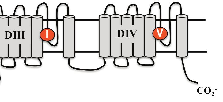
Research article                                                                                            Evolutionary Biology

                                    (Figure 1D–E). We therefore focused our sequencing and cultivation efforts on skin microbiota as a
                                    potential source of TTX in this species.
                                        Bacterial communities inhabiting the dorsal and ventral skin, cloacal gland, and submandibular
                                    gland of T. granulosa were compared by culture-independent 16S rRNA gene sequencing targeting
                                    the V4 hypervariable region. Bacterial samples were collected by swabbing wild newts captured in
                                    the field at each site (Oregon n = 12 and Idaho n = 16). Bacterial samples were collected from the
                                    Oregon and Idaho populations at separate times, in June 2013 and September 2016, respectively.
                                    However, all bacterial DNA samples were extracted, amplified, and sequenced together on the
                                    same run. In total, we identified 4160 operational taxonomic units (OTUs) with an average Good’s
                                    coverage (Good, 1953) of 0.9454 ± 0.0067 (mean ± SEM) across samples from both populations.
                                    614 OTUs were unique to toxic newts, 1943 were unique to non-toxic newts, and 1603 were shared
                                    between the two populations. Among the 20 most abundant OTUs, 8 OTUs were shared between
                                    both populations while 12 were present in only one population (Figure 1—figure supplement 1 and
                                    Figure 1—source data 2). These highly abundant and conserved OTUs may represent core skin
                                    microbiota of T. granulosa. Idaho newts possessed a greater number of distinct bacterial types with
                                    a more even distribution across their microbiota, reflected in a higher number of observed OTUs (t
                                    unequal var. = 7.70, p
Research article Evolutionary Biology
A B
Jaccard Index Bray-Curtis Index
PCo2 (17.8% of total variation)
Dorsal
PCo2 (6% of total variation)
Ventral
Chin
Cloaca
Soil
Idaho (TTX-)
Oregon (TTX+)
PCo1 (17.9% of total variation) PCo1 (29.9% of total variation)
C D
Otu00042 Pseudomonas
0.030
Idaho (TTX-) Oregon (TTX+)
Relative Abundance
0.020
0.010
0.000
Otu00224 Pseudomonas
0.006
Relative Abundance
0.004
0.002
0.000
Otu00445 Pseudomonadaceae
0.0008
Relative Abundance
0.0004
-4.8 -3.6 -2.4 -1.2 0 1.2 2.4 3.6 4.8
0.0000 LDA Score (log10)
Idaho (TTX-) Oregon (TTX+)
Figure 2. Comparison of skin microbiota from toxic and non-toxic newt populations reveal distinct host-associated bacterial communities and
enrichment of TTX-producing bacteria. (A–B) Principal coordinates analysis of bacterial community composition (OTU presence/absence; Jaccard index)
and community structure (OTU relative abundance; Bray-Curtis index) of skin microbiota across four body sites from toxic Oregon and non-toxic Idaho
newts reveal distinct clustering of bacterial communities within each population. Non-parametric multivariate analysis of variance (PERMANOVA) test
indicates a significant effect of location on both the composition (F = 18.12, pResearch article Evolutionary Biology
Figure 2 continued
Figure supplement 1. Principal coordinates analysis highlighting variation in bacterial community structure (Bray-Curtis index) of Oregon (red circles)
and Idaho (black squares) newt skin microbiota.
Figure supplement 2. Relative abundance of all OTUs classified to the same genera as TTX-producing bacteria identified in this study (Aeromonas,
Pseudomonas, Shewanella, and Sphingopyxis).
purification and liquid chromatography tandem mass spectrometry (LC-MS/MS) for molecular
screening (Figure 3A).
Applying this strategy, we detected TTX in cultures from four distinct genera: Aeromonas,
Pseudomonas, Shewanella, and Sphingopyxis (Table 1 and Figure 3B). LC-MS/MS screening con-
firmed the presence of product ions at 162.1 and 302.1 m/z, corresponding to the primary and sec-
ondary product ions formed by TTX fragmentation, respectively (Jen et al., 2008). For
quantification, we ran standard curves of 0.01, 0.05, 0.1, 0.5, 1, 2.5, 5, 10, and 25 ng mL 1 pure TTX
and used linear regression to estimate TTX concentrations in bacterial culture media from the base
peak intensity (BPI) signal in each LC-MS/MS run. Across all TTX-positive samples, TTX concentra-
tions produced were on average 0.236 ± 0.087 ng mL 1 (mean ± SEM, n = 14). TTX was occasionally
detected in samples with signals clearly above background noise and our limit of detection (LOD),
but below our lower limit of quantification (LLOQ; 0.01 ng mL 1). These samples were not included
in our quantitative analyses, but this observation suggests that TTX production could be enhanced
with strain-specific optimization of bacterial culture conditions.
TTX was detected in seven independent isolates of Pseudomonas spp. cultured from newt skin.
Pairwise alignment of 16S rRNA gene sequences suggests that these isolates may represent four
bacterial strains (Figure 3—figure supplement 2). Strains TX111003, TX174011, and TX180010
shared >99% nucleotide identity across homologous bases, and TX135003 and TX135004 also
shared >99% sequence identity, but these two groups appeared to be distinct from each other
(maximum similarity is 96.1%); these two groups were also isolated on different cultivation media,
blood agar and R2A agar, respectively. 16S rRNA gene sequences for the remaining two Pseudomo-
nas spp., strains TX111008 and TX111009, were unique from each other and the other two groups.
Furthermore, we identified two TTX-producing Shewanella spp. strains that were isolated on differ-
ent media and shared 94.2% 16S sequence identity. Thus, it appears several strains of Pseudomonas
and Shewanella can produce TTX in lab culture. We also identified one individual strain each of
Aeromonas and Sphingopyxis that produced TTX under these culture conditions (Table 1). Identifi-
cation of numerous TTX-producing symbionts from distinct genera present on newt skin is consistent
with observations in other toxic animal hosts such as the pufferfish and blue-ringed octopus, from
which numerous distinct TTX-producing strains have been isolated (Magarlamov et al., 2017). How-
ever, we note that the vast majority of bacterial isolates screened in this study did not produce TTX
under our culture conditions.
Molecular basis of TTX resistance in newt Nav channels
Rough-skinned newts are the most toxic of TTX-producing animals (Hanifin, 2010), but the molecu-
lar basis of their TTX resistance has not been characterized. To determine the basis of TTX resistance
in T. granulosa, we sequenced the Nav channel gene family and investigated the TTX-binding site,
the S5-6 pore loop (P-loop), to determine if they possessed adaptive mutations that affect TTX bind-
ing and resistance. We generated transcriptomes from two excitable tissues (brain and nose) from a
toxic newt and obtained partial sequences for five SCN genes that encode Nav1.1, Nav1.2, Nav1.3,
Nav1.5, and Nav1.6 proteins. We then cloned and sequenced the DI-DIV transmembrane sequences
of each gene for verification, including the Nav1.6 channel of both toxic and non-toxic newts.
SCN4A (Nav1.4) was obtained from GenBank for sequence comparison (accession number
KP118969.1).
Although S5-S6 pore-loop (P-loop) sequences are highly conserved across the vertebrate Nav
gene family, several amino acid substitutions were present in the P-loops across all six Nav channels
in T. granulosa (Figure 4). In DI, Tyr-371 was replaced independently across four of the six channels;
this parallel substitution involves a replacement from an aromatic Tyr or Phe to a non-aromatic
amino acid, either Cys, Ser, or Ala. In the mammalian Nav1.5 channel, this site is also replaced with a
Vaelli et al. eLife 2020;9:e53898. DOI: https://doi.org/10.7554/eLife.53898 7 of 29Research article Evolutionary Biology
A
B Sample solvent Pseudomonas (strain ID: TX111008)
100
75 0.53 ng mL-1
50
25
0
TTX standard (1 ng mL-1) Pseudomonas (strain ID: TX111003)
Relative Signal Intensity (%)
100
75 0.14 ng mL-1
50
25
0
Positive control (10% R2B + 100 ng mL-1) Shewanella (strain ID: TX140004)
100
75 0.16 ng mL-1
50
25
0
Negative control (10% R2B media) Sphingopyxis (strain ID: TX150006)
100
75 0.01 ng mL-1
50
25
0
0 1 2 3 4 5 0 1 2 3 4 5
Time (mins)
Figure 3. Bacterial isolates cultured from newt skin produce TTX in vitro. (A) Schematic overview of procedure for isolating and screening newt bacteria
for TTX production. Bacterial samples were collected from dorsal skin of toxic newts, taxonomically identified by 16S rRNA gene sequencing, grown in
liquid culture for 2 weeks, centrifuged, and the supernatant was purified by solid-phase extraction (SPE). Extracts were screened against TTX analytical
standards by LC-MS/MS. (B) Representative extracted ion chromatographs showing peaks corresponding to major product ion transitions 320.1 to 162.1
Figure 3 continued on next page
Vaelli et al. eLife 2020;9:e53898. DOI: https://doi.org/10.7554/eLife.53898 8 of 29Research article Evolutionary Biology
Figure 3 continued
m/z (black) and 320.1 to 302.1 m/z (red) for TTX in cultures from four bacterial isolates. The retention time for each peak was 2.9 min and matched that
of both authentic TTX standards and culture media supplemented with 100 ng mL 1 TTX. Peaks were absent in untreated culture media. Estimated TTX
concentrations in bacterial cultures are shown next to each peak. Multiple strains of Pseudomonas spp. were found to produce TTX and three
additional TTX-producing genera were identified: Aeromonas, Shewanella, and Sphingopyxis.
The online version of this article includes the following figure supplement(s) for figure 3:
Figure supplement 1. Phylogenetic tree of bacteria cultivated from the skin of toxic rough-skinned newts.
Figure supplement 2. Pairwise comparison of 16S rRNA sequences between TTX-producing strains of Pseudomonas spp. identified in this study.
Cys and has been shown to underlie the classic TTX resistance of the cardiac Na+ current
(Satin et al., 1992). An additional DI difference was found at N374T in Nav1.5; this site is also
altered in amniotes, but not in Xenopus frogs, indicating that these mutations may be convergent. In
DII, only one substitution is present at T938S in Nav1.3, which is adjacent to the electronegative Glu-
937 that directly binds the positively charged guanidinium group of TTX (Shen et al., 2018). This
region is otherwise well-conserved across tetrapods, suggesting that the DII P-loop sequence is
under strong purifying selection. Three sites differed in DIII including V1407I, M1414T, and A1419P
in Nav1.6, Nav1.4, and Nav1.1, respectively. The DIII M1414T substitution is present in at least five
Nav channel paralogs in TTX-laden pufferfishes and increases TTX resistance at least 15-fold
(Jost et al., 2008); thus, T. granulosa and pufferfish have converged on the identical molecular solu-
tion to reduce TTX sensitivity. The other two differences have not been previously characterized, but
we subsequently tested the effects of Nav1.6 V1407I on TTX binding (below). Finally, replacements
in DIV occur across four sites, including a substitution of A1703G in the selectivity filter DEKA motif
in Nav1.2, Ile-1699 in Nav1.4 and Nav1.6, D1706S in Nav1.4, and Gly-1707 in Nav1.1, Nav1.2, and
Nav1.4.
To determine whether TTX resistance was conferred by the P-loop mutations in T. granulosa, we
focused on the neural subtype Nav1.6, which is widely expressed in both the central and peripheral
nervous system (Caldwell et al., 2000; Hu et al., 2009; Lorincz and Nusser, 2010; Mercer et al.,
2007). We identified three amino acid replacements in the Nav1.6 channel of both toxic and non-
toxic newts (Figure 5A) and used site-directed mutagenesis to insert each mutation (DI Y371A, DIII
V1407I, and DIV I1699V), as well as all three mutations, into the TTX-sensitive Nav1.6 ortholog from
mouse. We found that TTX sensitivity was greatly reduced in triple mutant channels (Figure 5B–C),
and that each individual substitution contributed to TTX resistance (Figure 5—figure supplement
1). Estimated half-maximal inhibitory concentrations (IC50) confirmed that DIII and DIV mutations
provided 1.5-fold and 3-fold increases in resistance, respectively, while the DI and triple mutant
channels were estimated to provide a > 600 fold increase in resistance (Table 2 and Figure 5D).
Table 1. Summary of TTX-producing bacteria isolated from rough-skinned newts.
Genus-level identification of each TTX-producing isolate was determined by 16S rRNA gene sequencing and taxonomic classification
by the Ribosomal Database Project classifier tool at 80% similarity cut-off (Cole et al., 2014). TTX production was determined by
screening 1 mL culture media using LC-MS/MS. Overall, we cultured 11 TTX-producing isolates, though 16S gene sequence align-
ments suggest that some isolates may represent the same bacterial strain (see text). In most cases, TTX was detected in replicate cul-
tures above the limit of detection (LOD), but not always above the lower limit of quantification (LLOQ). Mean TTX
production ± standard error (SEM) is shown for samples above the LLOQ. Some of these bacterial genera contain TTX-producing
strains that have previously been identified in other toxic animals (reviewed in Chau et al., 2011).
TTX-producing Replicates above Replicates above TTX (ng TTX symbiont in other
Genus Isolation media isolates LOD LLOQ mL 1)± SEM animals
Aeromonas Blood agar 1 1 1 0.12 Pufferfishes, sea snails
Pseudomonas R2A or blood 7 23 9 0.19 ± 0.07 Pufferfishes, Blue-ringed
agar octopus, sea snails
Shewanella R2A or blood 2 4 3 0.49 ± 0.36 Pufferfishes
agar
Sphingopyxis R2A 1 2 1 0.01 None
Vaelli et al. eLife 2020;9:e53898. DOI: https://doi.org/10.7554/eLife.53898 9 of 29Research article Evolutionary Biology
1707
374
938
1407 1706
1414 1699
371 1703
DI DII DIII DIV
371 374 938 1407 1414 1699 1703 1706 1707
Homo sapiens Nav1.1 FRLMTQDYWENLY FRVLCGEWIETMW LQVATFKGWMDIM FQITTSAGWDGLL
Mus musculus Nav1.1 ------D------ ------E------ ------K------ ------A------
Anolis carolinensis Nav1.1 ------D------ ------E------ ------K------ -M----A------
Xenopus tropicalis Nav1.1 ------D------ ------E------ ------K------ ------A------
Taricha granulosa Nav1.1 ------K------ -E----A---R--
Homo sapiens Nav1.2 FRLMTQDYWENLY FRVLCGEWIETMW LQVATFKGWMDIM FQITTSAGWDGLL
Mus musculus Nav1.2 ------D------ ------E------ ------K------ ------A------
Anolis carolinensis Nav1.2 ------D------ ------E------ ------K------ ------A------
Xenopus tropicalis Nav1.2 ------DC----- ------E------ ------K---P-- ------A---L--
Taricha granulosa Nav1.2 ------DC----- ------E------ ------K------ ------G---V--
Homo sapiens Nav1.3 FRLMTQDYWENLY FRVLCGEWIETMW LQVATFKGWMDIM FQITTSAGWDGLL
Mus musculus Nav1.3 ------D------ ------E------ ------K------ ------A------
Anolis carolinensis Nav1.3 ------D------ ------E------ ------K------ ------A------
Xenopus tropicalis Nav1.3 ------D------ ------E------ ------K---E-- ------A------
Taricha granulosa Nav1.3 ------DA----- ------E---S-- ------K------ -M----A------
Homo sapiens Nav1.4 FRLMTQDYWENLF FRILCGEWIETMW LQVATFKGWMDIM FEITTSAGWDGLL
Mus musculus Nav1.4 ------D------ ------E------ ------K------ ------A------
Anolis carolinensis Nav1.4 ------D------ ------E------ ------K------ -M----A------
Xenopus tropicalis Nav1.4 ------D------ ------E------ ------K------ -Q----A------
Taricha granulosa Nav1.4 ------D------ ------E------ ------K--T--- -QS---A--SD--
Homo sapiens Nav1.5 FRLMTQDCWERLY FRILCGEWIETMW LQVATFKGWMDIM FQITTSAGWDGLL
Mus musculus Nav1.5 ------DC--R-- ------E------ ------K------ ------A------
Anolis carolinensis Nav1.5 ------DY--R-- ------E------ ------K---E-- ------A------
Xenopus tropicalis Nav1.5 ------DY--N-- --V---E------ ------K------ ------A------
Taricha granulosa Nav1.5 ------DS--T-- --V---E------ ------K------ ------A------
Homo sapiens Nav1.6 FRLMTQDYWENLY FRVLCGEWIETMW LQVATFKGWMDIM FQITTSAGWDGLL
Mus musculus Nav1.6 ------D------ ------E------ ------K------ ------A------
Anolis carolinensis Nav1.6 ------D------ ------E------ ------K------ ------A------
Xenopus tropicalis Nav1.6 ------D------ ------E------ ------K------ ------A------
Taricha granulosa Nav1.6 ------DA----- ------E------ --I---K------ --V---A------
Figure 4. Protein alignment of Nav channels across representative vertebrates. Sequence alignment of S5-S6 P-loops from newts and other vertebrates
showing amino acid substitutions relative to the P-loop consensus sequence for each Nav channel shown here. Putative TTX resistance mutations are
highlighted in orange; mutations that are not highlighted are either synapomorphic in a gene clade or are present in TTX sensitive channels. Data are
missing for DI and DII of Nav1.1 in newts, which we did not recover in our sequencing efforts. The approximate locations of newt mutations are shown
as orange circles, and the amino acid site of each mutation is numbered based on Nav1.6 from Mus musculus.
The online version of this article includes the following source data and figure supplement(s) for figure 4:
Source data 1. GenBank accession numbers of vertebrate Nav channel protein sequences used in multiple sequence alignments and analysis.
Figure supplement 1. Parallel evolution of DIII and DIV P-loop substitutions in Nav1.6 of toxic newts and TTX resistant garter snakes.
Thus, while all three mutations impact TTX resistance, the DI Y371A replacement provides consider-
able resistance independently. These results show that the three P-loop modifications in newt
Nav1.6 provide resistance to even extremely high concentrations of TTX, and comparison of Nav
sequences from toxic and non-toxic newts revealed identical substitutions in both populations, sug-
gesting that newts are broadly TTX-resistant regardless of toxicity.
Vaelli et al. eLife 2020;9:e53898. DOI: https://doi.org/10.7554/eLife.53898 10 of 29Research article Evolutionary Biology
A
Homo sapiens Nav1.6 FLALFRLMTQDYWENLYQLTL FLIVFRVLCGEWIETMW YLALLQVATFKGWMDIMYA MICLFQITTSAGWDGLLLP
Mus musculus ----------D---------- ----------E------ ----------K-------- ----------A--------
Gallus gallus ----------D---------- ----------E------ ----------K-------- ----------A--------
Anolis carolinensis ----------DF--------- ----------E------ ----------K-------- ----------A--------
Xenopus tropicalis ----------D---------- ----------E------ ----------K-------- ----------A--------
Taricha granulosa (Oregon) ----------DA--------- ----------E------ ------I---K-------- ------V---A--------
Taricha granulosa (Idaho) ----------DA--------- ----------E------ ------I---K-------- ------V---A--------
B
Nav1.6 Wild-type
1 µA
1 ms
Nav1.6 Triple mutant
P9
1 µA
-80 mV 1 ms
Control 1 µM TTX 10 µM TTX Wash
C Nav1.6 Wild-type Nav1.6 Triple Mutant
D
ï ï 0 40 ï ï 0 40
1.0
Ratio of unblocked to total current
0
Normalized peak current
0.8
0.6
ï Wild-type
Treatment 0.4 DI Y371A
Control
DIII V1407I
1uM TTX 0.2 DIV I1699V
10uM TTX Triple mutant
ï Wash
0 100 nM 1 µM 10 µM 100 µM
Membrane potential (mV) Membrane potential (mV) TTX concentration (M)
Figure 5. Newts possess Nav channel mutations that confer physiological resistance to TTX. (A) Predicted topology of Nav1.6 with mutations in
domains I, III, and IV. Sequence alignment of Nav1.6 pore-loop motifs revealed three amino acid differences in newts from Oregon or Idaho
populations. (B) Representative currents from wild-type mouse Nav1.6 or Nav1.6 with newt substitutions Y371A, V1407I, and I1699V treated with 1 mM
(blue) or 10 mM (orange) TTX. (C) Current-voltage (I–V) relationships showing normalized currents for wild-type (n = 21) and mutant Nav1.6 (n = 20)
channels. Wild-type Nav1.6 was blocked by TTX (Tukey’s multiple comparisons test with Bonferroni correction: control vs. 1 mM, pResearch article Evolutionary Biology
Figure 5 continued
Figure supplement 1. Newt Nav1.6 mutations increase TTX resistance in the orthologous mouse Nav1.6.
Discussion
In this study, we found that bacterial isolates from four genera, Aeromonas, Pseudomonas, Shewa-
nella, and Sphingopyxis, cultured from the skin of T. granulosa produce TTX under laboratory condi-
tions. Although TTX-producing symbionts have been identified in marine animals (Chau et al.,
2011), this is the first identification of TTX-producing bacteria associated with a freshwater or terres-
trial animal. The origin of TTX in rough-skinned newts and other amphibians has been controversial:
wild-caught toxic newts maintain their toxicity in long-term laboratory captivity (Hanifin et al.,
2002), and newts forced to secrete their TTX by electric shock regenerate their toxicity after nine
months, despite laboratory conditions that prevented access to dietary sources of TTX
(Cardall et al., 2004). Such results demonstrate that newts do not derive TTX from their natural diet,
but the results do not explicitly rule out a symbiotic origin for TTX toxicity. A subsequent investiga-
tion attempted to amplify 16S rRNA genes from DNA extracted from newt tissues by PCR, but the
authors were unable to amplify bacterial DNA from any tissue except the gut (Lehman et al., 2004).
This result has been widely cited to claim that newts lack symbiotic bacteria altogether, thus sup-
porting an endogenous origin for TTX (Cardall et al., 2004; Gall et al., 2011; Gall et al., 2014;
Hanifin, 2010; Hanifin and Gilly, 2015; Williams, 2010). However, sequencing-based approaches
for characterization of microbial communities were limited at that time, and it is increasingly clear
that most, if not all, animals possess cutaneous bacterial communities on their external epithelium
(McFall-Ngai et al., 2013). Thus, our results strongly suggest that symbiotic bacteria are the ultimate
source TTX toxicity in rough-skinned newts.
Surprisingly, many of the TTX-producing strains isolated from newts are from the same genera as
those previously identified in marine animals. TTX-producing Pseudomonas spp. have been isolated
from toxic pufferfish, blue-ringed octopus, and sea snails (Cheng et al., 1995; Hwang et al., 1989;
Yotsu et al., 1987), and TTX-producing Aeromonas spp. and Shewanella spp. have both been iso-
lated from pufferfish and sea snails (Auawithoothij and Noomhorm, 2012; Cheng et al., 1995;
Simidu et al., 1990; Wang et al., 2008; Yang et al., 2010). TTX-producing Sphingopyxis spp. have
not been identified in host animals or environmental samples, and this strain may be unique to fresh-
water or terrestrial environments. Interestingly, several other newt species from diverse genera are
known to possess TTX, including Notophthalmus, Triturus, Cynops, Paramesotriton, Pachytriton, and
Laotriton (Brodie et al., 1974; Yotsu-Yamashita and Mebs, 2001; Yotsu-Yamashita et al., 2007;
Yotsu-Yamashita et al., 2017). Frogs and toads from the genera Atelopus, Brachycephalus, Coloste-
thus, and Polypedates also possess TTX (Daly et al., 1994; Kim et al., 2003; Mebs et al., 1995;
Tanu et al., 2001; Yotsu-Yamashita and Tateki, 2010), as well as two species of freshwater flat-
worms (Stokes et al., 2014). Thus, the TTX toxicity observed in other amphibians and freshwater
animals could be derived from bacterial sources similar to those identified in this study.
One of the most interesting insights to arise from this work is the possibility that the skin micro-
biome contributes to the predator-prey arms race between toxic newts and TTX-resistant garter
Table 2. Estimated half-maximal inhibitory concentrations (IC50) of TTX for each Nav1.6 construct.
IC50 values are shown as the concentration (mean ± SEM) of TTX (mM) that blocked half of the chan-
nels, estimated from the dose-response curve. The IC50 ratio was taken as the fold increase in TTX
resistance.
Construct N IC50 IC50 Ratio
Mouse Nav1.6 21 1.25 ± 0.09 1
DI (Y371A) 17 763.7 ± 284 609
DIII (V1407I) 13 2.34 ± 0.23 1.2
DIV (I1699V) 15 4.73 ± 0.42 2.0
Triple mutant 20 3551 ± 469 2832.4
Vaelli et al. eLife 2020;9:e53898. DOI: https://doi.org/10.7554/eLife.53898 12 of 29Research article Evolutionary Biology
snakes. Populations of garter snakes sympatric with TTX-laden newts possess several amino acid
replacements in their Nav channels that prevent TTX binding, allowing resistant snakes to prey on
highly toxic newts (Feldman et al., 2009; Geffeney, 2002; Geffeney et al., 2005). As snake popula-
tions accumulate stepwise adaptive mutations in their Nav channels, selection drives increasing levels
of toxicity in newts. Reciprocal selection for elevated toxicity and resistance in newt and snake popu-
lations, respectively, leads to an asymmetric escalation of these two traits, or a ‘coevolutionary arms
race’ (Brodie and Brodie, 1999; Brodie et al., 2005; Dawkins and Krebs, 1979). If selection by
predatory garter snakes favors increasing levels of toxicity in newt populations, selection may be act-
ing not only on genetic variation in the host species, but also potentially on variation across the skin
microbiome.
Selection could also act by increasing the relative abundance of TTX-producing symbionts in the
skin (Bordenstein and Theis, 2015; Theis et al., 2016). Consistent with this hypothesis, we found
that three abundant Pseudomonas OTUs were present in greater relative abundance in the micro-
biota of toxic newts compared to non-toxic newts (Figure 2C–D). Pseudomonas OTU00042 was par-
ticularly abundant in toxic newts and a significant driver of beta diversity between the toxic and non-
toxic populations. Numerous TTX-producing Pseudomonas strains were also isolated in our cultiva-
tion assay, suggesting that this differential abundance may contribute to observed variation in TTX
toxicity across newt populations. However, we did not observe a differential abundance of Aeromo-
nas OTUs, which were abundant in both populations, nor of Shewanella or Sphingopyxis OTUs,
which were found only on toxic newts, but were only present in a few samples and in very low abun-
dance (Figure 2—figure supplement 2). These results may also reflect more favorable culture condi-
tions for TTX-producing Pseudomonas spp. than for the other genera. Thus, further population-level
comparisons across toxic and non-toxic newts are needed to determine whether the composition
and/or structure of the microbiome directly influences newt toxicity.
Additionally, if variation in TTX toxicity is subject to selective forces, TTX-producing symbionts
would need to be heritable, directly or indirectly, across generations. The mechanisms underlying
microbiome heritability vary from environmental acquisition of microbes across each generation to
direct vertical transmission from parent to offspring (Mandel, 2010). The development of skin-asso-
ciated microbial communities in newts, and amphibians more broadly, is not clear, as both host spe-
cies identity and habitat appear to play important roles across different amphibian taxa
(Ellison et al., 2019; Ross et al., 2019). In newts, one possibility is that TTX-producing bacteria are
vertically transferred from females to their eggs, as newt eggs contain TTX and egg toxicity is corre-
lated with the toxicity of the mother (Gall et al., 2012; Hanifin et al., 2003). Another possibility is
that newts possess adaptive traits to facilitate the acquisition and proliferation of TTX-producing
bacteria anew from the environment through each generation. Host factors impacting the micro-
biome may include anti-microbial peptide expression (SanMiguel and Grice, 2015) or the produc-
tion of metabolites that favor TTX-producing microbes. Other traits may influence interspecific
interactions within the microbiome to promote colonization and proliferation of TTX-producing sym-
bionts. These traits may be under selective pressure to ultimately benefit TTX-producing symbionts
and increase TTX toxicity across newt populations (Carroll et al., 2003; Magarlamov et al., 2017).
Further investigations comparing toxic and non-toxic newts through developmental stages in the
wild and in captivity may begin to shed light on this complex process.
Furthermore, because of the challenges of in vitro cultivation and characterization of microbial
physiology in symbiotic microbes isolated from their hosts, it is difficult to determine how the
dynamics of TTX production are regulated within the in vivo host-associated communities
(Magarlamov et al., 2017). Under our culture conditions in the lab, we observed TTX production
that was typically less than 0.5 ng mL 1. However, given that the TTX-producing bacteria identified
in this study and in other toxic animals were grown under artificial lab conditions independent of
host factors and interactions with other host-associated microbes, estimating the true biosynthetic
potential of these TTX-producing bacteria poses a major technical challenge. Identifying the genetic
basis of TTX production may help circumvent this problem and allow future researchers to apply
sequencing-based metagenomic approaches to determine which organisms are capable of produc-
ing TTX (Chau and Ciufolini, 2011; Chau et al., 2011). These efforts may also facilitate the develop-
ment of targeted cultivation strategies to better replicate the host environment and more accurately
measure TTX production in vitro.
Vaelli et al. eLife 2020;9:e53898. DOI: https://doi.org/10.7554/eLife.53898 13 of 29Research article Evolutionary Biology
Our results also show that toxic newts possess adaptations in their Nav channels that confer TTX
resistance. The presence of parallel mutations across the Nav channel family of newts and other TTX-
resistant animals suggests that the evolution of resistance involves a highly constrained walk through
a narrow adaptive landscape. For example, studies of the skeletal muscle isoform Nav1.4 across a
variety of TTX-resistant snake species identify numerous convergent substitutions in the P-loop
regions of DIII and DIV, but never in DI or DII (Feldman et al., 2012). The Nav1.4 subtype of TTX-
resistant newts, including T. granulosa, also possess several mutations in DIV and one in DIII, but
none in DI or DII. Conversely, mutations in the DI Y/F-371 site are often seen in neural subtypes of
TTX-resistant pufferfishes, and we found that this mutation was present in three of the four neural
subtypes of newts. Furthermore, when comparing Nav channel sequences in newts and other TTX-
resistant animals, we found that Nav1.6 sequences in newts and garter snakes share two identical
substitutions in the P-loops of DIII V1407I and DIV 1699V (Figure 4—figure supplement 1). Both
newt and snake Nav sequences were derived from individuals caught in Benton Co., OR, where
newts are highly toxic and snakes are highly resistant. These mutations may reflect convergent
molecular evolution between predators and prey responding to the same selection pressure.
Whether or not these patterns have arisen by chance or through Nav subtype-specific constraints on
P-loop evolution would be interesting to explore in future studies.
Given the potential strength of selection on interactions between newts and their symbiotic
microbiota with regard to TTX toxicity, it may be more appropriate to consider the effects of selec-
tion across the hologenome, the collective genetic variation present in both host and symbionts
(Bordenstein and Theis, 2015; Rosenberg and Zilber-Rosenberg, 2013). Many recent studies
emphasize the critical importance of host-associated microbes in basic animal physiology, develop-
ment, nutrition, nervous system function, and even behavior (Archie and Theis, 2011; Eisthen and
Theis, 2016; McFall-Ngai et al., 2013; Shropshire and Bordenstein, 2016; Theis et al., 2016;
van Opstal and Bordenstein, 2015). In the coevolutionary arms race between toxic newts and resis-
tant snakes, selection may act upon the phenotype that emerges from the collective interactions
between the newt host and bacterial symbionts, termed the holobiont. One prediction of the holo-
genome theory is that adaptive evolution can occur rapidly by increasing the relative abundance of
specific symbionts if the metabolites derived from that symbiont are critical for holobiont fitness
(Theis et al., 2016). This potential evolutionary force would avoid a long and winding road through
a complex adaptive landscape for the host, particularly for epistatic traits such as TTX biosynthesis,
which is predicted to involve a dozen or more enzymes (Chau and Ciufolini, 2011; Chau et al.,
2011). Future studies exploring the relationship between newt host toxicity and the composition of
newt skin microbiota could provide a mechanistic basis for the observed variation in newt toxicity
across different populations, revealing potentially interesting cases of parallel evolution occurring at
the hologenomic level. Overall, chemical defenses such as neurotoxins provide excellent models for
investigating adaptive evolution, as these toxins often target evolutionarily conserved proteins in ani-
mal nervous systems, revealing mechanistic associations among protein sequence, physiology, and
evolution.
Materials and methods
Key resources table
Reagent type
(species) or Source or Additional
resource Designation reference Identifiers information
Strain, strain STBL2 ThermoFisher 10268019
background competent Scientific
(Escherichia coli) cells
Recombinant mSCN8A DOI: 10.1523/ Construct kindly
DNA reagent (Mus musculus) JNEUROSCI. provided by Dr. Al
18-16-06093.1998 Goldin, UC Irvine
Biological Oocytes xenopus1.com
sample
(Xenopus laevis)
Continued on next page
Vaelli et al. eLife 2020;9:e53898. DOI: https://doi.org/10.7554/eLife.53898 14 of 29Research article Evolutionary Biology
Continued
Reagent type
(species) or Source or Additional
resource Designation reference Identifiers information
Sequence- 16S_rRNA_ Integrated DNA 51-01-19-06 AGAGTTTGATCCTGGCTCAG
based 8F Technologies
reagent
Sequence- 16S_rRNA_ DOI: 10.1128/ PCR primer GTGCCAGCMGCCGCGGTAA
based 515F AEM.01043–13
reagent
Sequence- 16S_rRNA_ DOI: 10.1128/ PCR primer TGGACTACHVGGGTWTCTAAT
based 806R AEM.01043-13
reagent
Sequence- 16S_rRNA_ Integrated DNA 51-01-19-07 CGGTTACCTTGTTACGACTT
based 1492R Technologies
reagent
Commercial Q5 Site- New England E0554S
assay or kit directed Biolabs
mutagenesis kit
Commercial T7 mMessage ThermoFisher AM1344
assay or kit mMachine kit Scientific
Chemical Tetrodotoxin Alomone Labs T-550
compound,
drug
Software, Clampfit Molecular
algorithm v10.7 Devices
Software, Geneious geneious.com
algorithm v11.0.5
Software, mothur mothur.org
algorithm v1.39.5
Software, RStudio rstudio.com
algorithm (v3.6.1)
Other Oasis MCX Waters 186000252
cartridge
Other Acquity UPLC Waters 186004801
BEH amide
column
All procedures involving animals were approved by and conducted under the supervision of the Insti-
tutional Animal Care and Use Committee at Michigan State University (approval no. 10/15-154-00),
in accordance with guidelines established by the US Public Health Service.
Laboratory animals
Adult male rough-skinned newts (Taricha granulosa) were collected in Oregon, USA (January Pond;
44˚36’13.8"N 123˚38’12.1"W) under Oregon Department of Fish and Wildlife permit number 104–
15. Animals were housed in glass aquaria containing Holtfreter’s solution (60 mM NaCl, 0.67 mM
KCl, 0.81 mM MgSO4, and 0.68 mM CaCl2; pH 7.2–7.6). Floating platforms in each aquarium pro-
vided terrestrial refuges, and newts were maintained at 20˚C with a 14:10 light-dark cycle and fed
blackworms (Lumbriculus variegatus) 2–3 times weekly.
Cultivation of skin bacteria
To collect bacterial samples, newts were first rinsed in reverse osmosis (RO) H2O for 5 s to remove
transient bacteria and swabbed 10 times (down and back) each on the dorsal and ventral skin surfa-
ces using a sterile cotton swab (Puritan Medical Products, Guilford, ME). The sample swab was then
placed in 1 mL Hank’s Buffered Salt Solution (HBSS; 0.137 M sodium chloride, 5.4 mM potassium
chloride, 0.25 mM disodium phosphate, 0.56 M glucose, 0.44 mM monopotassium phosphate, 1.3
mM calcium chloride, 1.0 mM magnesium sulfate, 4.2 mM sodium bicarbonate) and diluted ten-fold
over four serial dilutions: 10 1, 10 2, 10 3, and 10 4. 100 mL of each dilution was then plated on
Vaelli et al. eLife 2020;9:e53898. DOI: https://doi.org/10.7554/eLife.53898 15 of 29Research article Evolutionary Biology
either R2A agar (0.5 g casein hydrolysate, 0.5 g dextrose, 0.5 g soluble starch, 0.5 g yeast extract,
0.3 g potassium phosphate, 0.3 g sodium pyruvate, 0.25 g casein peptone, 0.25 g meat peptone,
0.024 g magnesium sulfate, 15 g agar, final volume 1 L) or blood agar (10 g peptone, 10 g meat
extract, 5 g sodium chloride, 15 g agar, final volume 1 L) infused with defibrinated sheep’s blood
(10% v/v) (Fisher Scientific, Hampton, NH). Petri dishes containing these mixed community cultures
were wrapped in Parafilm to prevent desiccation and incubated at room temperature (20˚C) for 1–2
weeks. The combination of nutrient-limited media, cool temperatures, and relatively long incubation
periods has been shown to promote microbial diversity and the growth of previously uncultivated
microbes (Sommer, 2015; Stevenson et al., 2004; Stewart, 2012).
Following cultivation of mixed communities, individual bacterial colonies were picked and
streaked onto new plates to establish pure cultures. Plates were then wrapped in Parafilm and
allowed to incubate at 20˚C until colonies appeared. Bacterial stocks were generated by collecting
bacterial samples from each streaked plate and submerging in 0.5 mL HBSS with 10% dimethyl sulf-
oxide (DMSO) for cryoprotection. Samples were then stored at 80˚C.
Taxonomic identification of bacterial isolates
To identify bacterial isolates, we performed colony PCR using the 16S rRNA gene universal primers
8F (5’—AGAGTTTGATCCTGGCTCAG—3’) and 1492R (5’—CGGTTACCTTGTTACGACTT—3’). Bac-
terial colonies were picked with sterile toothpicks and submerged directly into a PCR master mix
(final concentration: 1X PCR buffer, 1.5 mM MgCl2, 0.2 mM dNTPs, 0.25 mM forward and reverse
primer, 0.05% NP-40, 1.25U Taq polymerase, and nuclease-free H2O). PCR reactions were per-
formed using the following conditions: 3 min at 95˚C; 30 s at 95˚C, 30 s at 45˚C, 1.5 min at 72˚C
repeated 30 times; and a final elongation for 5 min at 72˚C. PCR products were analyzed by gel elec-
trophoresis and samples yielding products were cleaned using ExoSAP-IT (Affymetrix, Santa Clara,
CA) following manufacturer’s instructions. DNA samples were submitted to Michigan State Univer-
sity’s Genomics Core (East Lansing, MI) for Sanger sequencing using 16S rRNA 8F universal primer
(5’—AGAGTTTGATCCTGGCTCAG—3’). Sequences were screened for quality using 4Peaks (Nucleo-
bytes, Amsterdam, Netherlands) and sequences with at least 400 bp of unambiguous base calls after
quality trimming were assigned genus-level classifications using the Ribosomal Database Project
(RDP) Classifier tool and an 80% confidence threshold (Cole et al., 2014).
Phylogenetic analysis of 16S rRNA gene sequences
Evolutionary relationships among cultured bacteria were inferred by constructing maximum-likeli-
hood phylogenetic trees. Multiple sequence alignments were generated by aligning 16S rRNA gene
sequences with the SILVA ribosomal RNA reference database (Quast et al., 2013). Gaps and non-
informative sites were trimmed to generate the final alignment. Trees were constructed using ran-
domized axelerated maximum-likelihood (RAxML) with 1000 bootstrap replicates (Stamatakis, 2014)
in Geneious v11.0.5 (Kearse et al., 2012) and edited in FigTree v1.4.3 (https://github.com/rambaut/
figtree/).
Sample collection for TTX quantification
To estimate TTX concentrations in newt skin, we followed the non-lethal sampling technique
described by Bucciarelli and coworkers (Bucciarelli et al., 2014). Animals were first anesthetized in
pH-corrected 0.1% tricaine-S (MS-222) dissolved in Holtfreter’s solution. Two skin biopsies were then
collected from symmetrical sites on the dorsal skin surface, approximately 1 cm laterally from the
vertebrae and 1 cm anterior to the hind limbs, using sterile, disposable 2 mm skin biopsy punches
(Acu-Punch, Acuderm Inc, Fort Lauderdale, FL). The two skin biopsies from each individual were
weighed and then combined in 300 mL 0.1 M acetic acid. Each sample was then placed into a boiling
water bath for 5 min followed by an ice bath for an additional 5 min. Subsequent steps were carried
out at room temperature. To minimize protein and macromolecular debris, samples were centri-
fuged at 13,000 x g for 20 min and the supernatant transferred to an Amicon Ultra 10,000 MWCO
centrifugal filter (Sigma-Aldrich, St. Louis, MO) followed by a second centrifugation at 13,000 x g for
20 min. Finally, 100 mL 0.1 M acetic acid was added to the filter and a third centrifugation at 13,000
x g for 20 min was performed to wash any remaining TTX. The final sample volume was adjusted to
1 mL before proceeding to solid-phase extraction (below).
Vaelli et al. eLife 2020;9:e53898. DOI: https://doi.org/10.7554/eLife.53898 16 of 29Research article Evolutionary Biology
To identify TTX-producing bacteria, isolated bacterial strains were revived from frozen stocks and
inoculated in 5 ml of R2B broth (0.5 g casein hydrolysate, 0.25 g casein peptone, 0.25 g meat pep-
tone, 0.5 g dextrose, 0.5 g soluble starch, 0.5 g yeast extract, 0.3 g potassium phosphate, 0.3 g
sodium pyruvate, 0.024 g magnesium sulfate, final volume 1 L) diluted to either 10% or 50% strength
in reverse osmosis (RO) H2O. The use of dilute broth was intended to encourage the production of
secondary metabolites. Cultures were grown at room temperature 20˚C on a tissue culture rotator
for 1 or 2 weeks. After cultivation, each culture was centrifuged at 13,000 x g for 5 min at room tem-
perature, and 1 mL of supernatant was used in solid-phase extraction.
Solid-phase extraction (SPE)
TTX extractions were performed using a modified solid-phase extraction (SPE) protocol based on
that described by Jen et al. (2008). Each skin or bacterial sample was loaded onto a mixed cation
exchange cartridge (Oasis MCX cartridges, Waters, MA) previously regenerated with 1 mL of metha-
nol and equilibrated with 1 mL RO H2O. Samples were drawn through the cartridge over 30 s using
a Vac-Man laboratory vacuum manifold (Promega, Madison, WI) coupled with VacConnectors (Qia-
gen, Germantown, MD). Each cartridge was then washed with 1 mL acetonitrile, 1 mL methanol, and
1 mL distilled H2O. TTX was eluted twice from the cartridge with 0.125 mL 0.2 M HCl in 20% metha-
nol. Both eluates were combined and dried in a SpeedVac vacuum centrifuge (Savant SpeedVac
SC110, Thermo Fisher Scientific, Waltham, MA), then resuspended in 0.2 mL 0.5% acetic acid in
water. 50 mL aliquots of each sample were prepared for LC-MS/MS analysis.
Liquid chromatography tandem mass spectrometry (LC-MS/MS)
TTX analyses were performed using a Waters TQ-D mass spectrometer coupled to a Waters ACQ-
UITY UPLC system with a binary solvent manager. Chromatographic separations were performed on
a Waters ACQUITY UPLC BEH amide column (2.1 100 mm; 1.7 mm particles; Waters Co., Milford,
MA); column temperature was held at 40˚C. For liquid chromatography, we used 0.1% formic acid in
water (mobile phase A) and acetonitrile (mobile phase B) with a flow rate of 0.4 mL/min. The injec-
tion volume was set to 10 mL. The linear gradient elution program was as follows (A/B): 0–1.0 min (5/
95), 1.0–1.5 min (50/50), 1.5–2.0 min (55/45), 2.0–3.5 min (60/40), 3.5–4.0 min (65/35) before the gra-
dient returned to the initial condition (5/95). TTX was analyzed in positive electrospray ionization
mode using multiple reaction monitoring with a transition of 320.1 > 162.1 (cone voltage: 50 eV; col-
lision energy: 40 eV) as the primary channel for quantification and 320.1 > 302.1 (cone voltage: 50
eV; collision energy: 40 eV) as the secondary channel for confirmation. The capillary voltage was 3.0
kV. Source and desolvation temperatures were 130˚C and 500˚C, respectively; cone gas and desolva-
tion gas flows were 40 and 700 L/hr, respectively. Data were acquired using MassLynx 4.1 software
(Waters Co.). Extracts from bacterial and skin samples were compared with TTX analytical standards
acquired from Sigma-Aldrich (St. Louis, MO). A calibration curve was included in each LC-MS/MS run
with the following concentrations: 0.01, 0.05, 0.1, 0.5, 1, 2.5, 5, 10, and 25 ng/ml. Concentrations of
TTX quantified from skin biopsies were normalized relative to tissue mass. The presence of TTX in
skin samples and bacterial cultures was confirmed by a retention time identical to that of authentic
TTX as well as the presence of both primary and secondary ion transitions. All chromatograms were
plotted in R v3.4.1.
Scanning electron microscopy
3 3 mm skin samples were dissected from the dorsal region of a euthanized newt. Each sample
was fixed in 4% glutaraldehyde in 0.1 M sodium phosphate buffer (pH 7.4) overnight at 4˚C. Follow-
ing fixation, samples were briefly rinsed in 0.1 M sodium phosphate buffer and dehydrated in an eth-
anol gradient (25, 50, 75, 95, 100, 100, 100%) for 10 min each. Any remaining liquid in the samples
was removed by critical point drying in a Balzers Model 010 critical point dryer (Balzers Union Ltd.,
Balzers, Liechtenstein) using carbon dioxide as the transitional fluid. Each skin sample was then
mounted on an aluminum stub using carbon suspension cement (SPI Supplies, West Chester, PA)
and coated with platinum (8 nm thickness) using a Q150T turbo pumped sputter coater (Quorum
Technologies, Laughton, East Sussex, England) purged with argon gas. Samples were examined and
images obtained using a JEOL JSM-7500F cold field emission scanning electron microscope (JEOL
Ltd, Tokyo, Japan).
Vaelli et al. eLife 2020;9:e53898. DOI: https://doi.org/10.7554/eLife.53898 17 of 29You can also read