Sushi domain-containing protein 4 controls synaptic plasticity and motor learning - eLife
←
→
Page content transcription
If your browser does not render page correctly, please read the page content below
RESEARCH ARTICLE

                                         Sushi domain-containing protein 4
                                        controls synaptic plasticity and motor
                                        learning
                                        Inés González-Calvo1,2†‡, Keerthana Iyer1†, Mélanie Carquin1, Anouar Khayachi1,
                                        Fernando A Giuliani2, Séverine M Sigoillot1, Jean Vincent3, Martial Séveno4,
                                        Maxime Veleanu1, Sylvana Tahraoui1, Mélanie Albert1, Oana Vigy5,
                                        Célia Bosso-Lefèvre1, Yann Nadjar6, Andréa Dumoulin6, Antoine Triller6,
                                        Jean-Louis Bessereau7, Laure Rondi-Reig3, Philippe Isope2, Fekrije Selimi1*
                                        1
                                          Center for Interdisciplinary Research in Biology (CIRB), Collège de France, CNRS,
                                        INSERM, PSL Research University, Paris, France; 2Institut des Neurosciences
                                        Cellulaires et Intégratives (INCI), CNRS, Université de Strasbourg, Strasbourg,
                                        France; 3Institut Biology Paris Seine (IBPS), Neuroscience Paris Seine (NPS),
                                        CeZaMe, CNRS, Sorbonne University, INSERM, Paris, France; 4BioCampus
                                        Montpellier, CNRS, INSERM, Université de Montpellier, Montpellier, France;
                                        5
                                          Institut de Génomique Fonctionnelle, CNRS, INSERM, Université de Montpellier,
                                        Montpellier, France; 6École Normale Supérieure, Institut de Biologie de l’ENS,
                                        INSERM, CNRS, PSL Research University, Paris, France; 7Université de Lyon,
                                        Université Claude Bernard Lyon 1, CNRS UMR 5310, INSERM U 1217, Institut
*For correspondence:                    Neuromyogène, Lyon, France
fekrije.selimi@college-de-france.fr
†
 These authors contributed
equally to this work
                                        Abstract Fine control of protein stoichiometry at synapses underlies brain function and
Present address: ‡Univ.                 plasticity. How proteostasis is controlled independently for each type of synaptic protein in a
Bordeaux, CNRS,                         synapse-specific and activity-dependent manner remains unclear. Here, we show that Susd4, a gene
Interdisciplinary Institute for         coding for a complement-related transmembrane protein, is expressed by many neuronal
Neuroscience, IINS, UMR 5297,           populations starting at the time of synapse formation. Constitutive loss-of-function of Susd4 in the
F-33000, Bordeaux, France
                                        mouse impairs motor coordination adaptation and learning, prevents long-term depression at
Competing interests: The                cerebellar synapses, and leads to misregulation of activity-dependent AMPA receptor subunit
authors declare that no                 GluA2 degradation. We identified several proteins with known roles in the regulation of AMPA
competing interests exist.              receptor turnover, in particular ubiquitin ligases of the NEDD4 subfamily, as SUSD4 binding
Funding: See page 28                    partners. Our findings shed light on the potential role of SUSD4 mutations in neurodevelopmental
                                        diseases.
Received: 13 December 2020
Accepted: 03 March 2021
Published: 04 March 2021

Reviewing editor: Megan R
Carey, Champalimaud
                                        Introduction
Foundation, Portugal                    Proteostasis is at the core of many cellular processes and its dynamics need to be finely regulated
                                        for each protein in each organelle. In neurons, additional challenges are imposed by their spatial
    Copyright González-Calvo et
                                        complexity. In particular, during long-term synaptic plasticity, the proposed substrate for learning
al. This article is distributed under
                                        and memory (Collingridge et al., 2010; Nicoll, 2017), the number of neurotransmitter receptors
the terms of the Creative
Commons Attribution License,            needs to be regulated independently in a synapse-specific and activity-dependent manner. At excit-
which permits unrestricted use          atory synapses, the modification of AMPA receptor numbers is a highly dynamic process, involving
and redistribution provided that        regulation of receptor diffusion (Choquet and Triller, 2013; Penn et al., 2017), their insertion in the
the original author and source are      plasma membrane, anchoring at the postsynaptic density and endocytosis (Anggono and Huganir,
credited.                               2012). After activity-dependent endocytosis, AMPA receptors are either recycled to the plasma

González-Calvo, Iyer, et al. eLife 2021;10:e65712. DOI: https://doi.org/10.7554/eLife.65712                                            1 of 34
Research article Neuroscience
membrane or targeted to the endolysosomal compartment for degradation (Ehlers, 2000;
Lee et al., 2004; Park et al., 2004). The decision between these two fates, recycling or degradation,
regulates the direction of synaptic plasticity. Recycling promotes long-term potentiation (LTP) and
relies on many molecules such as GRASP1, GRIP1, PICK1, and NSF (Anggono and Huganir, 2012).
Targeting to the endolysosomal compartment and degradation promote long-term depression (LTD;
Fernández-Monreal et al., 2012; Kim et al., 2017; Matsuda et al., 2013), but the regulation of the
targeting and degradation process remains poorly understood.
The Complement Control Protein (CCP) domain, an evolutionarily conserved module also known
as Sushi domain, was first characterized in proteins with role in immunity, in particular in the comple-
ment system. In the past few years, proteins with CCP domains have been increasingly recognized
for their role at neuronal synapses. Acetylcholine receptor clustering is regulated by CCP domain-
containing proteins in Caenorhabditis elegans (Gendrel et al., 2009) and in Drosophila mela-
nogaster (Nakayama et al., 2016). In humans, mutations in the CCP domain-containing secreted
protein SRPX2 are associated with epilepsy and speech dysfunction, and SRPX2 knockdown leads to
decreased synapse number and vocalization in mice (Sia et al., 2013). Recently SRPX2 has been
involved in the regulation of synapse elimination in the visual and somatosensory systems
(Cong et al., 2020). Despite the increase in the diversity of CCP domain-containing proteins in evo-
lution (11 CCP domain-containing in C. elegans and 56 in humans; smart.embl.de), the function of
many CCP domain-containing proteins remains unknown.
The mammalian SUSD4 gene codes for a transmembrane protein with four extracellular CCP
domains (Figure 1A) and is highly expressed in the central nervous system (Holmquist et al., 2013).
The SUSD4 gene is located in a genomic region deleted in patients with the 1q41q42 syndrome that
includes developmental delays and intellectual deficiency (ID; Rosenfeld et al., 2011). SUSD4 is also
among the 124 genes enriched in de novo missense mutations in a large cohort of individuals with
autism spectrum disorders (ASDs) or IDs (Coe et al., 2019). A copy number variation and several de
novo mutations with a high CADD score, which indicates the deleteriousness of the mutations, have
been described in the SUSD4 gene in patients with ASDs (Cuscó et al., 2009; denovo-db, Seattle,
WA [denovo-db.gs.washington.edu] 10, 2019). The SUSD4 protein has been described to regulate
complement system activation in erythrocytes by binding the C1Q globular domain
(Holmquist et al., 2013). Interestingly, this domain is found in major synaptic regulators such as
C1QA (Stevens et al., 2007), CBLNs (Matsuda et al., 2010; Uemura et al., 2010), and C1Q-like
proteins (Bolliger et al., 2011; Kakegawa et al., 2015; Sigoillot et al., 2015). Altogether these
studies point to a potential role of SUSD4 in synapse formation and/or function and in the etiology
of neurodevelopmental disorders.
Proper development and function of the cerebellar circuitry is central for motor coordination and
adaptation, and a range of cognitive tasks (Badura et al., 2018; Hirai et al., 2005; Ichise et al.,
2000; Lefort et al., 2019; Rochefort et al., 2011; Tsai et al., 2012). Cerebellar dysfunction is asso-
ciated with several neurodevelopmental disorders including ASDs (Stoodley, 2016; Stoodley et al.,
2018; Wang et al., 2014). In this circuit, cerebellar Purkinje cells (PCs) receive more than a hundred
thousand parallel fiber (PF) synapses whose formation, maintenance, and plasticity are essential for
cerebellar-dependent learning (Gutierrez-Castellanos et al., 2017; Hirai et al., 2005; Ito, 2006;
Kashiwabuchi et al., 1995). Postsynaptic LTD was first described at synapses between PFs and cere-
bellar PCs (Gao et al., 2012; Hirano, 2018; Ito, 2001; Ito and Kano, 1982), where it can be
induced by conjunctive stimulation of PFs with the other excitatory input received by PCs, the climb-
ing fiber (CF; Coesmans et al., 2004; Ito, 2001; Suvrathan et al., 2016). The function of members
of the C1Q family, such as CBLN1 and C1QL1, is essential for excitatory synapse formation and LTD
in cerebellar PCs (Hirai et al., 2005; Kakegawa et al., 2015; Matsuda et al., 2010; Sigoillot et al.,
2015; Uemura et al., 2010), suggesting that proteins such as SUSD4, that interact with the C1Q
globular domain, could regulate these processes.
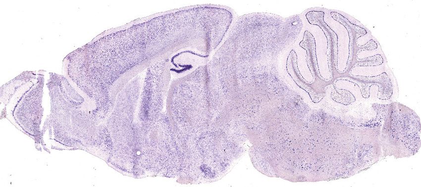
Gene expression studies from our laboratory revealed that Susd4 is highly expressed in the olivo-
cerebellar system of the mouse. In order to uncover the potential link between SUSD4 and neurode-
velopmental disorders, we sought to identify the role of SUSD4 in brain development and function,
by analyzing the phenotype of a Susd4 constitutive loss-of-function mouse model. Here, we show
that knockout (KO) of the Susd4 gene leads to deficits in motor coordination adaptation and learn-
ing, misregulation of synaptic plasticity in cerebellar PCs, as well as an impairment in the degrada-
tion of GluA2 AMPA receptor subunits after chemical induction of LTD. Proteomic analysis of SUSD4
González-Calvo, Iyer, et al. eLife 2021;10:e65712. DOI: https://doi.org/10.7554/eLife.65712 2 of 34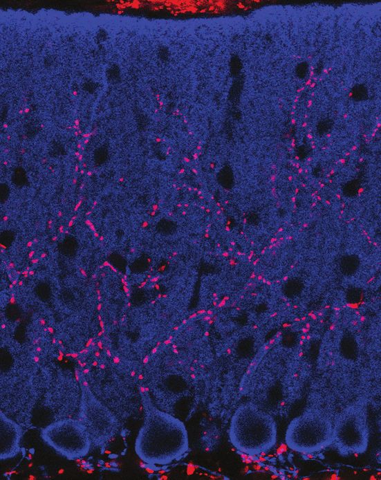
Research article                                                                                                                      Neuroscience

   A Sushi domain-containing protein 4 C
                               NT                                 CCP CCPCCP CCP                             CT
                                                                                         TM
                                                                   Extracellular             Intracellular                                                                   SUSD4
   B                                                         40
                                                                       Cerebellum
                                                                       Brainstem
                                                             30
           (relative to Rpl13a)
           Susd4 expression

                                                             20                                                                                                              GFP

                                                             10

                                                                                                                          GFP                                                GFP
                                                             0
                                                                       P0   P3      P7   P14       P21    3mo
                                                                                                                          SUSD4                                              SUSD4

   D
      AAV2-hSYN-DIO-HA-SUSD4-2A-eGFP

                                                                                                                                           AAV2-hSYN-DIO-eGFP

                                                                                                                                 GFP                                                             GFP
                                                                                                      GluD2                     GluD2                                         GluD2             GluD2
                                             SUSD4                                                   SUSD4                     SUSD4                            CTL             CTL               CTL

   E                               Susd4 131.7 kb                                                                                                                G
                                                                                                                                                                      CTX                       WT
                                                              UTR                                                            UTR
                 5’                                                                                                                   3’
                                                              1                     2    3     4         5 6      7   8
                                                                                                                                                                                           CB
                                                        Deletion

   F                               Accelerating Rotarod
                                                                                                                                                                                      BS
                                                              250             WT (n=11)
                                                                                                   p=0.0079
                                                                              KO (n=7)
                                                              200
                                                                                                                                                                       CTX
                                                                                                                                                                                                KO
                                       Time to fall (sec.)

                                                              150

                                                                                                                                                                                           CB
                                                              100

                                                                  50
                                                                                                                                                                                       BS
                                                                            123          123          123             123      123
                                                                  0
                                                                            DAY1         DAY2        DAY3             DAY4     DAY5

Figure 1. SUSD4 is necessary for motor coordination adaptation and learning. (A) Diagram of the protein SUSD4 showing its domain organization with
four extracellular Complement Control Protein (CCP) domains, one transmembrane (TM) domain and a cytoplasmic domain (CT). (B) Quantitative RT-
PCR shows an increase in Susd4 mRNA expression (relative to the housekeeping gene Rpl13a) during postnatal development in the cerebellum and in
the brainstem. Extracts were prepared from tissue samples of mice aged from 0 to 21 days (P0-21) and 3 months (3mo). Mean ± s.e.m. (n = 3
independent experiments). (C) HA-tagged SUSD4 is found in dendrites (left panel, single plane) and in some of the distal dendritic spines (right panel,
arrowheads, projection of a 1.95 mm z-stack) in adult cerebellar Purkinje cells. Anti-HA and anti-GFP immunolabeling was performed on parasagittal
cerebellar sections obtained from adult L7Cre mice after stereotaxic injection of AAV particles driving the expression of HA-SUSD4 and soluble GFP.
Figure 1 continued on next page

González-Calvo, Iyer, et al. eLife 2021;10:e65712. DOI: https://doi.org/10.7554/eLife.65712                                                                                                            3 of 34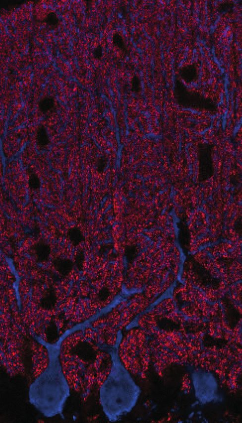
Research article                                                                                                               Neuroscience

Figure 1 continued
Scale bars: 10 mm (left panel) and 2 mm (right panel). (D) Purkinje cells from primary mixed cerebellar cultures of L7Cre mice were transduced at 3 days
in vitro (DIV3) with an HA-tagged SUSD4 expressing virus (AAV2-hSYN-DIO-HA-SUSD4-2A-eGFP) or with a control virus expressing GFP alone (AAV2-
hSYN-DIO-eGFP), and immunostained in non-permeabilizing conditions at DIV17 for HA to localize surface SUSD4 (anti-HA, red), and in permeabilizing
conditions to detect the green fluorescent protein (anti-GFP, green) and the endogenous GluD2 subunit (anti-GRID2, blue). Scale bar 5 mm. (E)
Genomic structure of the Susd4 gene. White boxes represent exons. Exon 1 is deleted in the Susd4 loss-of-function mouse model. See also Figure 1—
figure supplement 2. (F) Motor coordination and learning is deficient in adult male Susd4 -/- (knockout [KO]) mice compared to age-matched Susd4+/+
(wild-type [WT]) littermates. Each mouse was tested three times per day during 5 consecutive days on an accelerating rotarod (4-40 r.p.m. in 10 min)
and the time spent on the rotarod was measured. Mean ± s.e.m. (WT n = 11 and KO n = 7 mice, two-way ANOVA with repeated measures, interaction
(time and genotype): **p=0.0079, F(14, 224)=2.22; time: ****p
Research article                                                                                                      Neuroscience

                                    These spines are the postsynaptic compartments of PF synapses in PCs. Immunofluorescence analy-
                                    sis of transduced cultured PCs showed that HA-tagged SUSD4 could be immunolabeled in non-per-
                                    meabilizing conditions and located at the surface of dendrites and spines (Figure 1D). Double
                                    labeling with the postsynaptic marker GluD2 (GRID2) further showed partial colocalization at the sur-
                                    face of some, but not all, spines. Therefore, the timing of Susd4 mRNA expression during postnatal
                                    development and the subcellular localization of the SUSD4 protein in cerebellar PCs are in agree-
                                    ment with a potential role for SUSD4 in excitatory synapse formation and/or function.

                                    Susd4 loss-of-function leads to deficits in motor coordination and
                                    learning
                                    To determine the synaptic function of SUSD4, we analyzed the phenotype of Susd4-/- constitutive
                                    KO mice with a deletion of exon 1 (Figure 1E and G and Figure 1—figure supplement 2). RT-PCR
                                    using primers encompassing the last exons and the 3’UTR show the complete absence of Susd4
                                    mRNA in the brain of these Susd4 KO mice (Figure 1—figure supplement 2). No obvious alterations
                                    of mouse development and behavior were detected in those mutants, an observation that was con-
                                    firmed by assessment of their physical characteristics (weight, piloerection), basic behavioral abilities
                                    such as sensorimotor reflexes (whisker responses, eye blinking), and motor responses (open-field
                                    locomotion; see Supplementary file 1). We further assessed the behavior of Susd4 KO mice for
                                    motor coordination and motor learning (Kayakabe et al., 2014; Lalonde and Strazielle, 2001;
                                    Rondi-Reig et al., 1997). Using a footprint test, a slightly larger print separation of the front and
                                    hind paws in the Susd4 KO mice was detected, but no differences in the stride length and stance
                                    width were found (Figure 1—figure supplement 3). In the accelerated rotarod assay, a classical test
                                    of motor adaptation and learning (Buitrago et al., 2004), the mice were tested three times per day
                                    at 1 hr interval during 5 consecutive days. The Susd4 KO mice performed as well as the Susd4+/+
                                    (wild-type [WT]) littermate controls on the first trial (Figure 1F, day 1, trial 1). This indicates that
                                    there is no deficit in their balance function, despite the slight change in fine motor coordination
                                    found in the footprint test. However, while the control mice improved their performance as early as
                                    the third trial on the first day, and further improved with several days of training, no learning could
                                    be observed for the Susd4 KO mice either during the first day, or in the following days (Figure 1F).
                                    These results show that Susd4 loss-of-function leads to impaired motor coordination and learning in
                                    adult mice.

                                    Susd4 loss-of-function prevents LTD at cerebellar PF/PC synapses
                                    Because of the high expression of Susd4 in cerebellar PCs (Figure 1G and Figure 1—figure supple-
                                    ment 1), we focused on this neuronal type to identify the morphological and functional consequen-
                                    ces of Susd4 loss-of-function. No deficits in the global cytoarchitecture of the cerebellum and
                                    morphology of PCs were found in Susd4 KO mice (Figure 1—figure supplement 4). Using high-den-
                                    sity microelectrode array (MEA), we assessed the spontaneous activity of PCs in acute cerebellar sli-
                                    ces from Susd4 KO mice and compared to Susd4 WT mice (Figure 1—figure supplement 5). No
                                    differences were detected in either the mean spiking frequency, the coefficient of variation (CV) of
                                    interspike intervals (ISIs), or the intrinsic variability of spike trains (CV2, Holt and Douglas, 1996),
                                    indicating that the firing properties of PCs are not affected by Susd4 loss-of-function.
                                        Co-immunolabeling of PF presynaptic boutons using an anti-VGLUT1 antibody and of PCs using
                                    an anti-calbindin antibody in cerebellar sections from juvenile WT mice revealed an extremely dense
                                    staining in the molecular layer corresponding to the highly numerous PFs contacting PC distal den-
                                    dritic spines (Figure 2A). The labeling pattern appeared to be similar in Susd4 KO. High-resolution
                                    microscopy and quantitative analysis confirmed that there are no significant changes in the mean
                                    density and volume of VGLUT1 clusters following Susd4 loss-of-function (Figure 2A). Electric stimula-
                                    tion of increasing intensity in the molecular layer allows the progressive recruitment of PFs
                                    (Konnerth et al., 1990) and can be used to assess the number of synapses and basic PF/PC trans-
                                    mission using whole-cell patch-clamp recordings of PCs on acute cerebellar slices (Figure 2B). No
                                    difference was observed in the amplitude and the kinetics of the responses to PF stimulation in PCs
                                    from Susd4 KO and control littermate mice (Figure 2C and Figure 2—figure supplement 1). Fur-
                                    thermore, the probability of vesicular release in the presynaptic PF boutons, as assessed by measure-
                                    ments of paired-pulse facilitation (Atluri and Regehr, 1996; Konnerth et al., 1990; Valera et al.,

González-Calvo, Iyer, et al. eLife 2021;10:e65712. DOI: https://doi.org/10.7554/eLife.65712                                          5 of 34
Research article Neuroscience
CABP
A VGLUT1
(No. of clusters/µm2) Norm. to WT
WT
1.5 0.20
Density of VGLUT1 puncta
0.15
Area of VGLUT1 (µm2)
1.0
0.10
0.5
KO
0.05
WT KO WT KO
0.0 0.00
Susd4 WT Susd4 KO
B RECORDING SET-UP C Parallel fiber/Purkinje cell synapse EPSC
Purkinje Cell (PC) -10mV
PF-EPSC Amplitude (|pA|)
Parallel Fiber (PF) STIM Paired-pulse ratio
200
WT 21/8 2.0
ML
STIM 150 KO 16/6
1.5
PPR 20Hz (A2/A1)
REC 100 pA
100 150
REC 25 ms 100 1.0
50 50
PCL 0
STIM 20 40 60 80 100
0.5 WT KO
Climbing Fiber (CF) 0 18/8 16/6
GCL 0 20 40 60 80 100
STIM 0.0
Stimulus Intensity (µA)
D Climbing fiber-dependent Parallel fiber synapse E Parallel fiber synapse Long Term Potentiation
Long Term Depression
150 150 p=0.0199
relative to baseline (%)
relative to baseline (%)
p=0.0476 200
LTD induction
LTP induction
200
PF-EPSC Amplitude
PF-EPSC Amplitude
PF-EPSC Amplitude (%)
PF-EPSC Amplitude (%)
150 150
100 100
100 100
50
50 50 50
WT 13/9 0
WT 16/11 0
KO 14/10 Time (min) WT KO KO 8/6 Time (min)
WT KO
0 16/11 14/10 0 13/9 8/6
0 10 20 30 40 50 0 10 20 30 40 50
*** p=0.0005 with two-way ANOVA RM **** p0.9999; area VGLUT1 clusters:
unpaired Student’s t-test, p=0.3089). Scale bars 30 mm (left) and 10 mm (right). (B) Diagram of the setup for patch-clamp recordings (REC) of Purkinje
cells in 300-mm-thick parasagittal cerebellar slices. PF and climbing fiber responses were elicited by electrical stimulation (STIM). ML: molecular layer;
PCL: Purkinje cell layer; GCL: granule cell layer. (C) Input-output curve of the PF/Purkinje cell transmission. The amplitude of the elicited
excitatory postsynaptic currents (EPSCs) increases with the intensity of the stimulus and is not significantly different between Susd4 KO and WT
littermates. The fitted curves for each genotype are presented in the inset. Representative sample traces are presented. Mean ± s.e.m. (WT n = 18 cells
from eight mice and KO n = 16 cells from six mice; Kolmogorov-Smirnov test, p=0.8793). Short-term plasticity of PF/Purkinje cell synapses is not
affected by Susd4 loss-of-function. PFs were stimulated twice at 50 ms interval and the paired-pulse ratio (PPR) was calculated by dividing the
amplitude of the second peak by the amplitude of the first peak. Mean ± s.e.m. (WT n = 21 cells from eight mice and KO n = 16 cells from six mice;
Figure 2 continued on next page
González-Calvo, Iyer, et al. eLife 2021;10:e65712. DOI: https://doi.org/10.7554/eLife.65712 6 of 34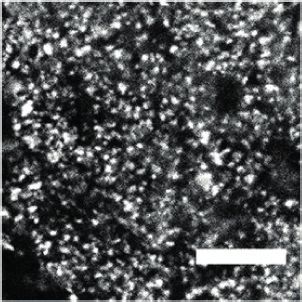
Research article                                                                                                                  Neuroscience

Figure 2 continued
Mann-Whitney test, p=0.9052). (D) Climbing fiber-dependent PF/Purkinje cell synapse long-term depression (LTD) is impaired in the absence of Susd4
expression. LTD was induced by pairing stimulations of PFs and climbing fibers at 100 ms interval during 10 min at 0.5 Hz (see also Figure 2—figure
supplement 1). The amplitude of the PF EPSC was measured using two consecutive PF stimulations at 50 ms interval. Representative sample traces are
presented. Right: EPSC amplitudes from the last 10 min (purple) of recordings were used to calculate the LTD ratio relative to baseline. Mean ± s.e.m.
(WT n = 16 cells from eleven mice and KO n = 14 cells from ten mice; two-tailed Wilcoxon signed rank test with null hypothesis of 100: WT **p=0.0063;
KO p=0.2676; Mann-Whitney test, WT vs. KO *p=0.0476). (E) Loss-of-function of Susd4 facilitates PF/Purkinje cell synapse long-term potentiation (LTP).
Tetanic stimulation of only PFs at 0.3 Hz for 100 times (see also Figure 2—figure supplement 1) induced LTP in Susd4 KO Purkinje cells while inducing
only a transient increase in PF transmission in WT Purkinje cells. Representative sample traces are presented. Right: EPSC amplitudes from the last 7
min (purple) were used to calculate the LTP ratio relative to baseline. Mean ± s.e.m. (WT n = 13 cells from nine mice and KO n = 8 cells from six mice;
two-tailed Wilcoxon signed rank test with null hypothesis of 100: WT p=0.5879; KO *p=0.0234; Mann-Whitney test, WT vs. KO: *p=0.0199).
The online version of this article includes the following source data and figure supplement(s) for figure 2:
Source data 1. Numerical data to support graphs in Figure 2.
Figure supplement 1. Parallel fiber (PF)/Purkinje cell (PC) synapse excitatory postsynaptic currents (EPSCs) kinetics, long-term plasticity induction
protocols, paired-pulse facilitation ratio, and delayed EPSC quanta.

                                      2012), was not changed at PF/PC synapses (Figure 2C). Finally, no differences in the frequency and
                                      amplitude of PF/PC-evoked quantal events were detected (Figure 2—figure supplement 1). Thus,
                                      in accordance with the morphological analysis, Susd4 invalidation has no major effect on the number
                                      and basal transmission of PF/PC synapses in the mouse.
                                          Long-term synaptic plasticity of PF/PC synapses is involved in proper motor coordination and
                                      adaptation learning (Gutierrez-Castellanos et al., 2017; Hirano, 2018; Kakegawa et al., 2018). We
                                      first assessed LTD in PF/PC synapses using conjunctive stimulation of PFs and CFs and whole-cell
                                      patch-clamp recordings of PCs in acute cerebellar slices from juvenile mice. The LTD induction pro-
                                      tocol produced a 42% average decrease in the amplitude of PF excitatory postsynaptic currents
                                      (EPSCs) in PCs from WT mice while the paired-pulse facilitation ratio was not changed during the
                                      course of our recordings (Figure 2D and Figure 2—figure supplement 1). In Susd4 KO PCs, the
                                      same LTD induction protocol did not induce any significant change in PF EPSCs during the 30 min
                                      recording period, showing that LTD induction and maintenance are greatly impaired in the absence
                                      of SUSD4 (Figure 2D). We then assessed LTP induction using high-frequency stimulation of PF in the
                                      absence of inhibition blockade as in Binda et al., 2016. In slices from Susd4 WT mice, tetanic stimu-
                                      lation every 3 s during 5 min induced only a transient increase in transmission of about 20% and the
                                      amplitude of the response returned to baseline after only 15 min (Figure 2E and Figure 2—figure
                                      supplement 1). This result suggests that under our experimental conditions and in this particular
                                      genetic background, LTD might be favored in contrast to previously obtained results (Binda et al.,
                                      2016; Titley et al., 2019). In the case of Susd4 KO PCs, the same protocol induced LTP with a 27%
                                      increase in transmission that was maintained after 35 min (Figure 2E). These results indicate that the
                                      absence of Susd4 expression promoted LTP induction at PF/PC synapses.
                                          Lack of LTD of PF/PC synapses could arise from deficient CF/PC transmission. To test this possi-
                                      bility, we first crossed the Susd4 KO mice with the Htr5b-GFP BAC transgenic line (http://gensat.
                                      org/MMRRC_report.jsp?founder_id=17735) expressing soluble GFP specifically in inferior olivary
                                      neurons in the olivocerebellar system to visualize CFs. We found that CFs had a normal morphology
                                      and translocated along the proximal dendrites of their PC target in Susd4 KO mice (Figure 3—fig-
                                      ure supplement 1). We then assessed whether developmental elimination of supernumerary CFs
                                      was affected by Susd4 invalidation using whole-cell patch-clamp recordings of PCs on cerebellar
                                      acute slices (Crepel et al., 1976; Hashimoto and Kano, 2003). No difference was found in the per-
                                      centage of remaining multiply-innervated PCs in the absence of Susd4 (Figure 3—figure supple-
                                      ment 1). We next used VGLUT2 immunostaining to label CF presynaptic boutons and analyze their
                                      morphology using high-resolution confocal microscopy and quantitative image analysis. VGLUT2
                                      immunostaining revealed the typical CF innervation territory on PC proximal dendrites, extending
                                      up to about 80% of the molecular layer height both in control Susd4 WT and in Susd4 KO mice
                                      (Figure 3A). Furthermore, the number and density of VGLUT2 clusters were not significantly differ-
                                      ent between Susd4 WT and Susd4 KO mice. To test whether the lack of CF-dependent PF LTD was
                                      due to deficient CF transmission, we used whole-cell patch-clamp recordings of PCs in acute cere-
                                      bellar slices. Contrary to what could have been expected, the typical all-or-none CF-evoked EPSC

González-Calvo, Iyer, et al. eLife 2021;10:e65712. DOI: https://doi.org/10.7554/eLife.65712                                                        7 of 34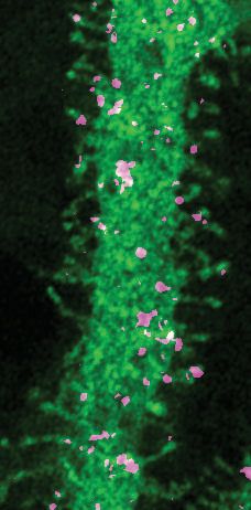
Research article Neuroscience
A CABP
VGLUT2
WT
Susd4 WT Susd4 KO
KO
100 0.6 800
VGLUT2 area (µm2)
VGLUT2 extension
VGLUT2 density
80
(% of PC length)
(puncta/105µm2)
600
0.4
60
400
40
0.2
20 200
WT KO WT KO WT KO
0 0.0 0
B Climbing fiber/Purkinje cell synapse EPSC
WT KO WT KO
-10mV CF EPSC Paired Pulse Ratio
0 26/9 26/7
EPSC amplitude (nA)
1.0
WT(12/3)
KO (17/5)
-1 0.9
PPR (A2/A1)
0.8
500 pA -2
0.7
50 ms Interstimulus interval (ms)
-3 0.6
0 200 400 600 800
p=0.0066
C Climbing Fiber Synapse: Delayed EPSC quanta
CF-Stimuli 0mM Ca++
1.0
Cumulative frequency
-60mV 10mM Sr++
WT 0.8
150 ms
60
* * ** * * * * * *
200 pA
100 ms 0.6
KO 40
0.4
IEI (ms)
* * * * * * * ** * 20
WT KO 0.2 WT (10/6)
KO (8/3) WT KO
0
0.0
20 pA 0 50 100 150 200
5 ms mEPSC amplitude (|pA|)
Figure 3. Transmission at the climbing fiber (CF)/Purkinje cell synapses is increased in Susd4 knockout (KO) mice.
(A) Climbing fiber presynaptic boutons were immunostained with an anti-VGLUT2 antibody in cerebellar sections
from P30 Susd4 wild-type (WT) and Susd4 KO mice. The extension of the CF synaptic territory was calculated by
measuring the extent of the VGLUT2 (red) labeling relative to the height of the Purkinje cell dendritic tree
(immunostained using an anti-CABP antibody, blue). Quantification of the mean density of VGLUT2 puncta and
their mean area showed no differences between Susd4 KO mice and their control littermates. Mean ± s.e.m. (WT
n = 5 and KO n = 7 mice; VGLUT2 extension: Mann-Whitney test, p=0.6389; VGLUT2 area: unpaired Student’s
t-test, p=0.4311; VGLUT2 density: unpaired Student’s t-test, p=0.8925). Scale bars 30 mm (left) and 10 mm (right).
(B) Short-term synaptic plasticity of CF/Purkinje cell synapses was elicited by two consecutive stimulations at
Figure 3 continued on next page
González-Calvo, Iyer, et al. eLife 2021;10:e65712. DOI: https://doi.org/10.7554/eLife.65712 8 of 34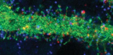
Research article                                                                                                              Neuroscience

                                    Figure 3 continued
                                    various intervals. The amplitude of the CF-elicited excitatory postsynaptic current (EPSC) was increased in Susd4
                                    KO mice compared to WT littermates. (WT n = 26 cells, nine mice and KO n = 26 cells, seven mice, Mann-Whitney
                                    test, **p=0.0066). No difference in the paired-pulse ratios (PPRs) was detected at any interval between Susd4 KO
                                    mice and WT mice. Representative sample traces are presented. See also Figure 3—figure supplement 1. Mean
                                    ± s.e.m. (WT n = 12 cells from three mice and KO n = 17 cells from five mice; Kolmogorov-Smirnov test, p=0.4740).
                                    (C) Delayed CF-EPSC quanta were evoked by CF stimulation in the presence of Sr++ instead of Ca++ to induce
                                    desynchronization of fusion events. Representative sample traces are presented. The cumulative probability for the
                                    amplitude of the events together with the individual amplitude values for each event show an increased amplitude
                                    associated with Susd4 loss-of-function. The individual frequency values for each cell (measured as interevent
                                    interval, IEI) present no differences between the genotypes. See also Figure 3—figure supplement 1. Mean ± s.
                                    e.m. (WT n = 10 cells from six mice and KO n = 8 cells from three mice; amplitude: Kolmogorov-Smirnov
                                    distribution test, ***p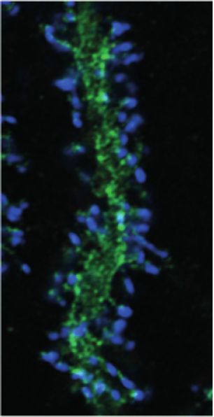
Research article                                                                                                                                                                                                                                                                                         Neuroscience

                            VGLUT2
    A                        GluA2
                                                                                                            GluA2 clusters around climbing fiber boutons

                                                                                                                                                                                                                                           Fraction of total number
                                                                                                                                                                                                                                                                      0.4                        WT
                                                                                                                                                                                                                                                                                                      p
Research article Neuroscience
Figure 4 continued
vitro (DIV3) with AAV particles driving the expression of HA-SUSD4 and soluble GFP (AAV2-hSYN-DIO-HA-SUSD4-2A-eGFP) and immunolabeled at
DIV17 in non-permeabilizing conditions to localize surface SUSD4 (anti-HA, red) and surface GluA2 subunits (anti-GluA2, blue). Direct green fluorescent
protein is shown (GFP, green). Right panels are binarized images of the anti-HA and anti-GluA2 immunolabelings and of the colocalization of these
signals (maximum projection of a 1.8 mm z-stack). Scale bar 5 mm.
The online version of this article includes the following source data and figure supplement(s) for figure 4:
Source data 1. Numerical data to support graphs in Figure 4.
Figure supplement 1. Basal surface GluA2 levels and total GluA2 and GluD2 levels in SUSD4 knockout (KO) mice.
Figure supplement 2. Interaction and colocalization of HA-SUSD4 and the AMPA receptor subunit GluA2.
assay in which we induced chemical LTD (cLTD) in acute cerebellar slices (Kim et al., 2017) and per-
formed surface biotinylation of GluA2 subunits followed by immunoblot quantification. In control
conditions, the mean baseline levels of surface GluA2 were not significantly different between Susd4
WT and Susd4 KO mice (Figure 4—figure supplement 1). As expected, after cLTD a 35% mean
reduction of surface GluA2 receptors was measured in slices from WT mice (Figure 4B; p=0.0212,
two-tailed Student’s t test with a null hypothesis of 1). In acute slices from Susd4 KO mice, a similar,
but not statistically significant, mean reduction of surface GluA2 receptors was detected after cLTD
(28%; p=0.0538, two-tailed Student’s t test with a null hypothesis of 1). Thus, SUSD4 loss-of-function
does not lead on average to a major change in the activity-dependent regulation of the number of
surface GluA2 subunits.
Another parameter that needs to be controlled for proper LTD in PCs is the total number of
AMPA receptors in the recycling pool and the targeting of AMPA receptors to late endosomes and
lysosomes (Kim et al., 2017). Lack of LTD and facilitation of LTP in Susd4 KO mice (Figure 2D and
E) suggest that GluA2 activity-dependent targeting to the endolysosomal compartment and its deg-
radation is affected by Susd4 loss-of-function. Using our cLTD assay in cerebellar slices, we measured
the total GluA2 levels either in control conditions or in the presence of inhibitors of the proteasome
(MG132) and of lysosomal degradation (leupeptin). The comparison of the GluA2 levels in the pres-
ence of both inhibitors and in control conditions allowed us to estimate the GluA2 degraded pool,
regardless of the mechanism behind this degradation. On average, total GluA2 levels were not sig-
nificantly different between Susd4 WT and Susd4 KO cerebellar slices in basal conditions (Figure 4—
figure supplement 1), in accordance with our morphological and electrophysiological analysis of PF/
PC synapses (Figure 2A and C). In slices from WT mice, chemical induction of LTD induced a signifi-
cant reduction of 13% in total GluA2 protein levels (Figure 4C). This reduction was prevented by
incubation with the mixture of degradation inhibitors, MG132 and leupeptin, showing that it corre-
sponds to the pool of GluA2 degraded in an activity-dependent manner (Figure 4C). In slices from
Susd4 KO mice, this activity-dependent degradation of GluA2 was completely absent. Additionally,
the chemical induction of LTD had no effect on the total protein levels of GluD2, another synaptic
receptor highly present at PF/PC postsynaptic densities, either in slices from WT or from Susd4 KO
mice (Figure 4—figure supplement 1). Thus, SUSD4 specifically controls the activity-dependent
degradation of GluA2-containing AMPA receptors during LTD.
Finally, co-immunoprecipitation experiments were performed using extracts from heterologous
HEK293 cells transfected with SEP-tagged GluA2 and HA-tagged SUSD4 or the transmembrane pro-
tein PVRL3a as a control. After affinity purification of SEP-GluA2, HA-SUSD4 was detected in affin-
ity-purified extracts while PVRL3a was not, showing the specific interaction of SEP-GluA2 and HA-
SUSD4 in transfected HEK293 cells (Figure 4—figure supplement 2). In order to assess the poten-
tial colocalization of SUSD4 and GluA2 in neurons, we used a Cre-dependent AAV construct to
express HA-tagged SUSD4 in cultured PCs (Figure 4D) and performed immunolabeling of surface
GluA2 subunits. Clusters of HA-tagged SUSD4 partially colocalize with GluA2 clusters at the surface
of some dendritic spines (Figure 4D). Partial colocalization of GluA2 and SUSD4 in neurons was also
confirmed in transfection experiments in hippocampal neurons (Figure 4—figure supplement 2).
Thus, SUSD4 could regulate activity-dependent degradation of GluA2-containing AMPA receptors
through a direct interaction.
González-Calvo, Iyer, et al. eLife 2021;10:e65712. DOI: https://doi.org/10.7554/eLife.65712 11 of 34Research article Neuroscience
SUSD4 interacts with NEDD4 ubiquitin ligases
To better understand how SUSD4 regulates the number of GluA2-containing AMPA receptors at
synapses, we searched for SUSD4 molecular partners by affinity purification of cerebellar synapto-
some extracts using GFP-tagged SUSD4 as a bait (Figure 5A). Interacting partners were identified
by proteomic analysis using liquid chromatography with tandem mass spectrometry (LC-MS/MS;
Savas et al., 2014). Twenty-eight candidates were identified including proteins with known function
in the regulation of AMPA receptor turnover (Figure 5E). Several candidates were functionally linked
to ubiquitin ligase activity by gene ontology (GO) term analysis (Figure 5A and Table 1). In particu-
lar, five members of the NEDD4 subfamily of HECT E3 ubiquitin ligases were found as potential
interacting partners, three of them (Nedd4l, Wwp1, and Itch) exhibiting the highest enrichment fac-
tors among the 28 candidates. Ubiquitination is a post-translational modification essential for the
regulation of protein turnover and trafficking in cells (Tai and Schuman, 2008). A survey of the
expression of HECT-ubiquitin ligases shows that different members of the NEDD4 subfamily are
broadly expressed in the mouse brain, however with only partially overlapping patterns (Figure 5—
figure supplement 1, http://mouse.brain-map.org, Allen Brain Atlas). Nedd4 and Wwp1 are the
most broadly expressed, including in neurons that also express Susd4, such as hippocampal neurons,
inferior olivary neurons in the brainstem, and cerebellar PCs. Immunoblot analysis of affinity-purified
synaptosome extracts confirmed the interaction of SUSD4 with NEDD4, ITCH, and WWP1
(Figure 5B). Removal of the intracellular domain of SUSD4 (SUSD4DCT mutant) prevented this inter-
action demonstrating the specificity of SUSD4 binding to NEDD4 ubiquitin ligases (Figure 5B).
The NEDD4 subfamily of HECT ubiquitin ligases is known to ubiquitinate and target for degrada-
tion many key signaling molecules, including GluA1- and GluA2-containing AMPA receptors
(Schwarz et al., 2010; Widagdo et al., 2017). Ubiquitin ligases of the NEDD4 family bind variants
of PY motifs on target substrates and adaptors (Chen et al., 2017). However, GluA1 and GluA2 sub-
units lack any obvious motif of this type. In contrast, two potential PY binding sites are present in
the intracellular domain of SUSD4 (Figure 5C). To test whether SUSD4 and GluA2 interaction is
affected by SUSD4 binding to NEDD4 ubiquitin ligases, co-immunoprecipitation experiments were
performed on extracts from heterologous HEK293 cells transfected with SEP-tagged GluA2 and vari-
ous HA-tagged SUSD4 constructs (Figure 5C and D). In addition to several deletion constructs of
SUSD4, we generated single- and double-point mutants of the two PY motifs in its intracellular tail
(Figure 5C). Lack of the cytoplasmic domain completely abrogated binding of NEDD4 to SUSD4,
confirming the results obtained using synaptosome extracts (Figure 5D). Deletion of the N-terminus
domain of SUSD4 did not affect NEDD4 binding. Furthermore, while the mutation of the PPxY site
in the intracellular tail (SUSD4-DPY mutant) abrogated binding of NEDD4 only partially, mutation of
the LPxY site (SUSD4-DLY mutant) or of both sites (SUSD4-DPY/LY mutant) completely prevented
the binding to NEDD4 ubiquitin ligases (Figure 5C and D). These mutations did not change signifi-
cantly the level of HA-SUSD4 protein in transfected HEK293 cells suggesting that the degradation of
SUSD4 itself is not regulated by binding of NEDD4 ubiquitin ligases (Figure 5—figure supplement
2). In accordance with our results obtained using SEP-GluA2 as a bait (Figure 4—figure supplement
2), GluA2 was detected in extracts obtained by affinity purification of the HA-tagged full-length
SUSD4 (HA-SUSD4), while it was absent if HA-SUSD4 was replaced by a control transmembrane pro-
tein, PVRL3a (Figure 5D and Figure 5—figure supplement 2). Deletion of the extracellular domain
(HA-SUSD4DNT) or the cytoplasmic domain (HA-SUSD4DCT) did not reduce significantly the ability
to interact with SEP-GluA2 when compared to HA-SUSD4 (Figure 5D and Figure 5—figure supple-
ment 2). Strong co-immunoprecipitation of GluA2 was detected in anti-HA affinity-purified extracts
from cells expressing the HA-tagged extracellular domain of SUSD4 alone (HA-SUSD4-NT construct),
showing that this domain is sufficient for GluA2 interaction (Figure 5D and Figure 5—figure supple-
ment 2). Finally using the SUSD4-DLY mutant or SUSD4-DPY/LY mutant as a bait did not significantly
modify the levels of co-immunoprecipitated GluA2 compared to HA-SUSD4, showing that binding of
NEDD4 ubiquitin ligases does not affect SUSD4’s ability to interact with GluA2.
Discussion
Our study shows that the CCP domain-containing protein SUSD4 starts to be expressed in various
neurons of the mammalian central nervous system when synapses are formed and mature. Susd4
loss-of-function in mice leads to impaired motor coordination adaptation and learning, misregulation
González-Calvo, Iyer, et al. eLife 2021;10:e65712. DOI: https://doi.org/10.7554/eLife.65712 12 of 34Research article Neuroscience
A B ct
es
es
tra
om
om
ex
os
os
pt
e
es
pt
Sy s
CT
na
e
m
na
4 om
4 om
so
Sy
4∆
SD tos
4
SD s
to
U pto
+
SD SD
+
-S nap
ubiquitin p
na
-S na
ubiquitin-like
SU -SU
FP Sy
FP y
Sy P -
U
G +S
protein ligase protein ligase F A
G HA H
+
FP
FP
activity activity
G
G
G
225
G protein-coupled NEDD4
150 receptor binding
GFP-SUSD4
102
ITCH
76
WWP1
52 NEDD4L WWP1 WWP2 NEDD4 ITCH DNAJA1 ARRB1 PSMC5
76
C2
38
HA
GFP
31
ubiquitin-like ubiquitin 52
protein protein pV 0.005-0.05
Coomassie
transferase transferase pVResearch article Neuroscience
Figure 5 continued
enrichment analysis network (Molecular Function category) of the 28 candidate proteins (Cytoscape plugin
ClueGO) identified in affinity purified samples (A) by liquid chromatography with tandem mass spectrometry (LC
MS/MS). The ubiquitin ligase activity term is significantly enriched in particular due to the identification of several
members of the NEDD4 family of HECT ubiquitin ligases. See also Table 1 (n = 3 independent experiments). (B)
Immunoblot confirmation of SUSD4 interaction with NEDD4 ubiquitin ligases. Affinity purification from cerebellar
synaptosomes was performed using full-length SUSD4 (HA-tagged, HA-SUSD4), a mutant lacking the C-terminal
tail (HA-SUSD4DCT), or GFP as a bait. Proteins were then resolved using SDS-PAGE followed by immunoblot for
NEDD4, ITCH, WWP1, or HA-SUSD4 (anti-HA). HA-SUSD4 interacts with all three members of the NEDD4 family.
This interaction is lost when the C-terminal tail of SUSD4 is deleted or when GFP is used instead of SUSD4 as a
control. (C) Schematic representation of HA-tagged SUSD4 and different mutant constructs: SUSD4DCT (lacking
the cytoplasmic tail), SUSD4DNT (lacking the extracellular domain), SUSD4NT (lacking the transmembrane and
intracellular domains), SUSD4DPY (point mutation of the PPxY site), SUSD4DLY (point mutation of the LPxY), and
SUSD4DPY/LY (double mutant at both PPxY and LPxY). (D) SUSD4 interaction with GluA2 and NEDD4 was
assessed by co-immunoprecipitation using HEK293 cells transfected with SEP-GluA2 together with PVRL3a as a
control or one of the HA-SUSD4 constructs represented in (C). Affinity purification was performed with an anti-HA
antibody and extracts were probed for co-immunoprecipitation of GluA2 (with an anti-GluA2 antibody) and of the
HECT ubiquitin ligase NEDD4 (anti-NEDD4 antibody). Co-immunoprecipitated GluA2 levels are normalized to
input GluA2 and then represented as relative to the immunoprecipitated levels for each SUSD4 construct. N = 3
independent experiments. (E) Potential interactors of SUSD4 control several parameters of AMPA receptor
turnover. Three different pools of AMPA receptors are found in dendrites and spines: synaptic, extrasynaptic, and
intracellular. AMPA receptors are synthetized and delivered close to the synaptic spine to reach the synaptic
surface. At the surface, AMPA receptors can move laterally (lateral diffusion) or vertically by endocytosis and
exocytosis. Endocytosis can be mediated by clathrin (CM-endocytosis) or be clathrin-independent (CI-
endocytosis). CM-endocytosis is often related to activity-dependent processes. After endocytosis, AMPA receptors
can choose between two different pathways from the early endosomes, one for recycling and the other for
degradation. Potential molecular partners of SUSD4 identified by our proteomic analysis could regulate AMPA
receptor turnover at several levels of this cycle (in red).
The online version of this article includes the following source data and figure supplement(s) for figure 5:
Source data 1. Numerical data to support graphs in Figure 5.
Figure supplement 1. Expression of HECT ubiquitin ligases in adult mouse brain.
Figure supplement 2. Total protein levels in HEK293 cells transfected with SEP-GluA2 and different SUSD4
mutant constructs (related to Figure 5C and D).
of synaptic plasticity in cerebellar PCs, and perturbed degradation of GluA2-containing AMPA
receptors after chemically induced LTD. SUSD4 and the GluA2 AMPA receptor subunit interact in
transfected heterologous cells and colocalize partially in transduced cultured neurons. Finally, we
show that SUSD4 directly binds to ubiquitin ligases of the NEDD4 family, which have been previously
shown to regulate GluA2 degradation.
SUSD4 promotes long-term synaptic depression
The choice between recycling of AMPA receptors to the membrane or targeting to the endolysoso-
mal compartment for degradation is key for the regulation of the number of AMPA receptors at syn-
apses, as well as for the direction and degree of activity-dependent synaptic plasticity (Ehlers, 2000;
Lee et al., 2002). Blocking the trafficking of AMPA receptors through recycling endosomes, for
example, using a RAB11 mutant, prevents LTP in neurons (Park et al., 2004). Conversely, blocking
the sorting of AMPA receptors to the endolysosomal compartment, for example, using a RAB7
mutant, impairs LTD in hippocampal CA1 pyramidal neurons and cerebellar PCs (Fernández-
Monreal et al., 2012; Kim et al., 2017). Further support for the role of receptor degradation comes
from mathematical modeling showing that in cerebellar PCs LTD depends on the regulation of the
total pool of glutamate receptors (Kim et al., 2017). The GluA2 AMPA receptor subunit, and its reg-
ulation, is of particular importance for LTD (Diering and Huganir, 2018). Phosphorylation in its
C-terminal tail and the binding of molecular partners such as PICK1 and GRIP1/2 are known to regu-
late endocytosis and recycling (Bassani et al., 2012; Chiu et al., 2017; Fiuza et al., 2017), and
mutations in some of the phosphorylation sites lead to impaired LTD (Chung et al., 2003). The
molecular partners regulating the targeting for degradation of GluA2 subunits in an activity-
González-Calvo, Iyer, et al. eLife 2021;10:e65712. DOI: https://doi.org/10.7554/eLife.65712 14 of 34Research article Neuroscience
Table 1. List of SUSD4 interactors.
Proteomic identification of SUSD4 interacting partners affinity-purified from synaptosomes extracts using GFP-SUSD4 as a bait (2
unique peptides; enrichment factor 4).
UniProtKB accession Gene Mol. weight Unique MS/MS Enrichment
num. Protein name name (kDa) peptides count factor
Q8CFI0 E3 ubiquitin-protein ligase NEDD4-like Nedd4l 115,42 28 319 159.5
Q8BH32 Sushi domain-containing protein 4 Susd4 53,796 4 97 48.5
Q8BZZ3 NEDD4-like E3 ubiquitin-protein ligase WWP1 Wwp1 104,69 13 90 45
Q8C863 E3 ubiquitin-protein ligase Itchy Itch 98,992 24 83 41.5
Q3TXU5 Deoxyhypusine synthase Dhps 40,642 9 81 40.5
Q9DBG3 AP-2 complex subunit beta Ap2b1 104,58 9 47 23.5
P50171 Estradiol 17-beta-dehydrogenase 8 Hsd17b8 26,588 2 32 16
Q9DBH0 NEDD4-like E3 ubiquitin-protein ligase WWP2 Wwp2 98,76 14 31 15.5
Q922R8 Protein disulfide-isomerase A6 Pdia6 48,1 8 26 13
P27773 Protein disulfide-isomerase A3 Pdia3 56,678 12 24 12
P17427 AP-2 complex subunit alpha-2 Ap2a2 104,02 7 23 11.5
Q8BWG8 Beta-arrestin-1 Arrb1 46,972 4 23 11.5
Q91WC3 Long-chain fatty acid – CoA ligase 6 Acsl6 78,016 11 22 11
P27546 Microtubule-associated protein 4 Map4 117,43 9 18 9
Q505F5 Leucine-rich repeat-containing protein 47 Lrrc47 63,589 9 17 8.5
Q9Z2H5 Band 4.1-like protein 1 Epb41l1 98,314 8 17 8.5
P46935 E3 ubiquitin-protein ligase NEDD4 Nedd4 102,71 7 17 8.5
Q8BMK4 Cytoskeleton-associated protein 4 Ckap4 63,691 11 16 8
P47708 Rabphilin-3A Rph3a 75,488 7 15 7.5
P42128 Forkhead box protein K1 Foxk1 74,919 6 15 7.5
P62812 Gamma-aminobutyric acid receptor subunit Gabra1 51,753 7 14 7
alpha-1
Q60737 Casein kinase II subunit alpha Csnk2a1 45,133 7 13 6.5
Q99KV1 DnaJ homolog subfamily B member 11 Dnajb11 40,555 5 10 5
P63037 DnaJ homolog subfamily A member 1 Dnaja1 44,868 4 10 5
Q9QY76 Septin-11 Sept11 49,694 5 9 4.5
O70318 Band 4.1-like protein 2 Epb41l2 109,94 6 8 4
P62196 26S protease regulatory subunit 8 Psmc5 45,626 5 8 4
Q9Z2Q6 Septin-5 Sept5 42,747 4 8 4
dependent manner during LTD remain to be identified. Our study shows that loss-of-function of
Susd4 leads both to loss of LTD and loss of activity-dependent degradation of GluA2 subunits. Loss-
of-function of Susd4 does not affect degradation of another postsynaptic receptor, GluD2, showing
the specificity of SUSD4 action. Furthermore, loss-of-function of Susd4 facilitates LTP of PF/PC syn-
apses. Overall our results suggest a role for SUSD4 in the targeting of GluA2-containing AMPA
receptors to the degradation compartment during synaptic plasticity.
SUSD4 interacts with regulators of AMPA receptor turnover
The degradation of specific targets such as neurotransmitter receptors must be regulated in a stimu-
lus-dependent and synapse-specific manner in neurons, to ensure proper long-term synaptic plastic-
ity, learning, and memory (Tai and Schuman, 2008). How is this level of specificity achieved?
González-Calvo, Iyer, et al. eLife 2021;10:e65712. DOI: https://doi.org/10.7554/eLife.65712 15 of 34Research article Neuroscience
Adaptor proteins, such as GRASP1, GRIP1, PICK1, and NSF, are known to promote AMPA receptor
recycling and LTP (Anggono and Huganir, 2012). Such adaptors for the promotion of LTD remain
to be found.
Our results show that SUSD4 directly binds to HECT E3 ubiquitin ligases of the NEDD4 fam-
ily. The family of HECT E3 ubiquitin ligases contains 28 enzymes including the NEDD4 subfamily
that is characterized by an N-terminal C2 domain, several WW domains, and the catalytic HECT
domain (Weber et al., 2019). This subgroup of E3 ligases adds K63 ubiquitin chains to their
substrate, a modification that promotes sorting to the endolysosomal compartment for degrada-
tion (Boase and Kumar, 2015). NEDD4 E3 ligases are highly expressed in neurons in the mam-
malian brain and have many known substrates with various functions, including ion channels and
the GluA1 AMPA receptor subunit. Accordingly, KO mice for the Nedd4-1 gene die during late
gestation (Kawabe et al., 2010). The activity and substrate selectivity of NEDD4 E3 ligases thus
need to be finely tuned. Both GluA1 and GluA2 AMPA receptor subunits are ubiquitinated on
lysine residues in their intracellular tails in an activity-dependent manner (Lin et al., 2011;
Lussier et al., 2011; Schwarz et al., 2010; Widagdo et al., 2015). Mutation of these lysine res-
idues decreases localization of GluA1 and GluA2 AMPA receptor subunits in the endolysosomal
compartment in neurons (Widagdo et al., 2015). However, GluA1 and GluA2 subunits lack any
obvious intracellular direct binding motif to the WW domain of NEDD4 ubiquitin ligases, raising
questions about the precise mechanism allowing regulation of AMPA subunits trafficking and
degradation by these enzymes. We showed that SUSD4 and GluA2 AMPA receptor subunits
interact in cells and partially colocalize in neurons. SUSD4 could thus regulate the targeting of
NEDD4 ubiquitin ligases to AMPA receptors in an activity-dependent manner in neurons. Alter-
natively, the interaction of SUSD4 with NEDD4 ubiquitin ligases might regulate the trafficking of
the SUSD4/GluA2 complex to the degradation pathway. Furthermore, among the potential part-
ners of SUSD4 identified by our proteomics analysis, several other candidates have functions
that are relevant for the regulation of synaptic plasticity, such as receptor anchoring, clathrin-
mediated endocytosis, and proteasome function (Figure 5E). Further work is needed to deter-
mine the precise mechanism of action of SUSD4 in neurons in the context of synaptic plasticity.
SUSD4 and neurodevelopmental disorders
Susd4 loss-of-function leads to motor impairments, a symptom that is also found in ASD patients
(Fournier et al., 2010). Deficits in LTD such as the one found in the Susd4 KO mice are a common
feature of several mouse models of ASDs (Auerbach et al., 2011; Baudouin et al., 2012;
Piochon et al., 2014). Because of the broad expression of SUSD4 and of ubiquitin ligases of the
NEDD4 subfamily in the mammalian central nervous system, whether motor impairments in the
Susd4 KO mice are directly the results of synaptic deficits in cerebellar PCs remain to be demon-
strated. Very recently, a reduction in exploratory behavior, in addition to impairments of motor coor-
dination, was reported after Susd4 loss-of-function (Zhu et al., 2020). Thus, mutations in the Susd4
gene might contribute to the etiology of neurodevelopmental disorders by impairing synaptic plas-
ticity at many synapse types.
In humans, the 1q41-42 deletion syndrome is characterized by many symptoms including IDs
and seizures, and in a high majority of the cases, the microdeletion encompasses the SUSD4
gene (Rosenfeld et al., 2011). A SUSD4 copy number variation has been identified in a patient
with ASD (Cuscó et al., 2009). SUSD4 was recently identified among the 124 genes with
genome-wide significance for de novo mutations in a cohort of more than 10,000 patients with
ASD or IDs (Coe et al., 2019). The GRIA2 gene (coding for the GluA2 subunit) has been found
as an ASD susceptibility gene (Salpietro et al., 2019; Satterstrom et al., 2020), and mutations
or misregulation of ubiquitin ligases have been found in many models of ASDs or IDs
(Cheon et al., 2018; Lee et al., 2018; Satterstrom et al., 2020). For example, ubiquitination of
GluA1 by NEDD4-2 is impaired in neurons from a model of Fragile X syndrome (Lee et al.,
2018). Understanding the molecular mechanism linking activity-dependent degradation of GluA2
and the SUSD4/NEDD4 complex will thus be of particular importance for our understanding of
the etiology of these neurodevelopmental disorders.
González-Calvo, Iyer, et al. eLife 2021;10:e65712. DOI: https://doi.org/10.7554/eLife.65712 16 of 34Research article Neuroscience
Materials and methods
Key resources table
Reagent type
(species) or
resource Designation Source or reference Identifiers Additional information
Gene (Mus Susd4 NCBI Gene ID: 96935 chr1:182,764,895–182,896,591
musculus)
Strain (Mus Susd4 knockout Lexicon Genetics B6:129S5-Susd4tm1Lex
musculus) mice Incorporated,
Tang et al., 2010
Strain (Mus Htr5b-GFP Gene Expression STOCK Tg
musculus) mouse line Nervous System (Htr5b-EGFP)BZ265Gsat/Mmmh
Atlas (GENSAT)
Project
Strain (Mus L7Cre mouse Jackson Laboratories B6.129-Tg(Pcp2-cre)2Mpin/J Stock
musculus) line number 004146
Cell line (Homo HEK293H Gibco Cat# 11631–017
sapiens)
Cell line (Homo HeLA Sigma Cat# 93021013
sapiens)
Antibody Mouse Swant Cat# 300 (1:1000)
monoclonal anti-
CABP
Antibody Rabbit Swant Cat# CB38 (1:1000)
polyclonal anti-
CABP
Antibody Mouse Abcam Cat# ab1218 (1:1000)
monoclonal anti-
GFP
Antibody Rabbit Abcam Cat# ab6556 (1:1000)
polyclonal anti-
GFP
Antibody Mouse Millipore and Cat# MAB397 (1:500)
monoclonal anti- BD and
GLUA2, clone Cat# 556341
6C4
Antibody Rabbit Abcam Cat# ab206293 (1:1000)
monoclonal anti-
GLUA2
Antibody Rabbit Millipore Cat# AB2285 (1:1000)
polyclonal anti-
GLURd1/2
Antibody Rat monoclonal Roche Life Cat# 11867423001 (1:1000)
anti-HA
Antibody Rabbit Cell Signaling Cat# 12117 (1:1000)
monoclonal anti- Technology
ITCH
Antibody Rabbit Millipore Cat# 07–049 (1:100,000)
polyclonal anti-
NEDD4
Antibody Guinea pig Millipore Cat# AB5905 (1:5000)
polyclonal anti-
VGLUT1
Antibody Guinea pig Millipore Cat# AB2251 (1:5000)
polyclonal anti-
VGLUT2
Antibody Rabbit Proteintech Cat# 13587–1-AP (1:2000)
polyclonal anti-
WWP1
Continued on next page
González-Calvo, Iyer, et al. eLife 2021;10:e65712. DOI: https://doi.org/10.7554/eLife.65712 17 of 34Research article Neuroscience
Continued
Reagent type
(species) or
resource Designation Source or reference Identifiers Additional information
Antibody Donkey Invitrogen Cat# A11057 (1:1000)
polyclonal anti-
goat Alexa Fluor
568
Antibody Donkey anti- Invitrogen Cat# R37114 (1:1000)
mouse Alexa
Fluor 488
Antibody Donkey Invitrogen #A10037 (1:1000)
polyclonal anti-
mouse Alexa
Fluor 568
Antibody Donkey Invitrogen Cat# A21206 (1:1000)
polyclonal anti-
rabbit Alexa
Fluor 488
Antibody Donkey Invitrogen #A21209 (1:1000)
polyclonal anti-
rat Alexa Fluor
594
Antibody Donkey Abcam Cat# 175475 (1:1000)
polyclonal anti-
rat Alexa Fluor
568
Antibody Goat polyclonal Invitrogen Cat# A110-73 (1:1000)
anti-guinea Pig
Alexa Fluor 488
Antibody Goat polyclonal Invitrogen Cat# A21450 (1:1000)
anti-guinea Pig
Alexa Fluor 647
Antibody Goat polyclonal Jackson Immune Cat# 115-035-174 (1:10,000)
anti-mouse HRP Research Laboratories
Antibody Goat polyclonal Jackson Immune #112-035-175 (1:10,000)
anti-rat HRP Research Laboratories
Antibody Sheep Roche Life Cat# 11093274910 (1:2000 - 1:5000)
polyclonal anti- Science
digoxigenin
alkaline
phosphatase
Antibody Mouse Abcam Cat# ab49900 (1:25,000)
monoclonal anti-
bACTIN HRP,
clone AC-15
Recombinant pHA-SUSD4- This paper From pEGFP-N1
DNA reagent GFP (Addgene,
Cat# 6085–1)
Recombinant pHA-SUSD4 This paper
DNA reagent
Recombinant pHA-SUSD4-D This paper
DNA reagent NT
Recombinant pHA-SUSD4-NT This paper
DNA reagent
Recombinant HA-SUSD4-DPY This paper
DNA reagent
Recombinant HA-SUSD4-DLY This paper
DNA reagent
Continued on next page
González-Calvo, Iyer, et al. eLife 2021;10:e65712. DOI: https://doi.org/10.7554/eLife.65712 18 of 34Research article Neuroscience
Continued
Reagent type
(species) or
resource Designation Source or reference Identifiers Additional information
Recombinant HA-SUSD4-DPY/ This paper
DNA reagent LY
Recombinant pIRES2-eGFP Addgene Cat# 6029–1
DNA reagent
Recombinant pCAG-PVRL3a This paper From pCAG-mGFP
DNA reagent (Addgene,
Cat# 14757)
Sequenced- Susd4_WT_F This paper PCR primers CTG TGG TTT CAA
based reagent CTG GCG CTG TG
Sequenced- Susd4_WT_R This paper PCR primers GCT GCC GGT GGG
based reagent TGT GCG AAC CTA
Sequenced- Susd4_KO_F This paper PCR primers TTG GCG GTT
based reagent TCG CTA AAT AC
Sequenced- Susd4_KO_R This paper PCR primers GGA GCT CGT
based reagent TAT CGC TAT GAC
Sequenced- Htr5b-GFP_F PCR primers TTG GCG CGC CTC CAA
based reagent CAG GAT GTT AAC AAC
Sequenced- Htr5b-GFP_R PCR primers CGC CCT CGC CGG
based reagent ACA CGC TGA AC
Sequenced- L7cre_1 PCR primers GGT GAC GGT CAG
based reagent TAA ATT GGA C
Sequenced- L7cre_2 PCR primers CAC TTC TGA CTT
based reagent GCA CTT TCC TTG G
Sequenced- L7cre_3 PCR primers TTC TTC AAG CTG
based reagent CCC AGC AGA GAG C
Chemical Picrotoxin Sigma-Aldrich Cat# P1675
compound, drug
Chemical D-AP5 Tocris Cat# 0106
compound, drug
Chemical CGP52432 Tocris Cat# 1246
compound, drug
Chemical JNJ16259685 Tocris Cat# 2333
compound, drug
Chemical DPCPX Tocris Cat# 0439
compound, drug
Chemical AM251 Tocris Cat# 1117
compound, drug
Software, Sinaptiqs Antoine Valera Software written in Python http://synaptiqs.
algorithm wixsite.com/synaptiqs
Other Hoechst 33342 Sigma Cat# 14533
Recombinant hSYN-DIO-HA- Vector biolabs AAV2 particles
viral particles SUSD4-2A-
eGFP-WPRE
Animals
Susd4 KO mice were generated using 129S5/SvEvBrd ES microinjected in C57BL/6J blastocysts and
maintained on the C57BL/6J background (generated by Lexicon Genetics Incorporated, The Wood-
lands, TX) (Tang et al., 2010). Out of the eight Susd4 exons, coding exon 1 (NCBI accession
NM_144796.2) and the 5’UTR (NCBI accession BM944003) were targeted by homologous recombi-
nation. This resulted in the deletion of a 1.3 kb sequence spanning the transcription initiation site
and exon 1 (Figure 1E and Figure 1—figure supplement 2). Subsequent genotyping of mice was
performed using PCR to detect the WT allele (forward primer: 5’ CTG TGG TTT CAA CTG GCG C
González-Calvo, Iyer, et al. eLife 2021;10:e65712. DOI: https://doi.org/10.7554/eLife.65712 19 of 34You can also read