Smart Wound Dressings for Diabetic Chronic Wounds - MDPI
←
→
Page content transcription
If your browser does not render page correctly, please read the page content below
bioengineering

Review
Smart Wound Dressings for Diabetic Chronic Wounds
Elizabeth Gianino, Craig Miller and Jordon Gilmore *
 Bioengineering Department, Clemson University, Clemson, SC 29632, USA; egianin@g.clemson.edu (E.G.);
 clm6@g.clemson.edu (C.M.)
 * Correspondence: jagilmo@g.clemson.edu; Tel.: +1-864-656-4262

 Received: 26 May 2018; Accepted: 19 June 2018; Published: 26 June 2018

 Abstract: Given their severity and non-healing nature, diabetic chronic wounds are a significant
 concern to the 30.3 million Americans diagnosed with diabetes mellitus (2015). Peripheral arterial
 diseases, neuropathy, and infection contribute to the development of these wounds, which lead to
 an increased incidence of lower extremity amputations. Early recognition, debridement, offloading,
 and controlling infection are imperative for timely treatment. However, wound characterization
 and treatment are highly subjective and based largely on the experience of the treating clinician.
 Many wound dressings have been designed to address particular clinical presentations, but a
 prescriptive method is lacking for identifying the particular state of chronic, non-healing wounds.
 The authors suggest that recent developments in wound dressings and biosensing may allow for the
 quantitative, real-time representation of the wound environment, including exudate levels, pathogen
 concentrations, and tissue regeneration. Development of such sensing capability could enable more
 strategic, personalized care at the onset of ulceration and limit the infection leading to amputation.
 This review presents an overview of the pathophysiology of diabetic chronic wounds, a brief summary
 of biomaterial wound dressing treatment options, and biosensor development for biomarker sensing
 in the wound environment.

 Keywords: diabetes; chronic wounds; smart wound dressing; biochemical sensor

1. Background
      Diabetes mellitus, an increasing health concern that affects more than 9% of the population over
the age 18, is the seventh leading cause of death in North America [1,2]. A number of severe health
concerns are associated with diabetes, such as peripheral arterial disease, neuropathy, limited joint
mobility, abnormal foot pressures, minor trauma, and foot deformity. Improved treatment of diabetes
could significantly decrease associated healthcare costs, given that the cost of healing a single ulcer,
infected ulcer, and amputation are estimated at $8000, $17,000, and $45,000, respectively [3]. Patients
who suffer from diabetes have a 15–25% chance of developing a chronic wound. Chronic wounds
associated with diabetes include foot, venous and pressure ulcers [4]. Diabetic foot ulcers (DFUs) are
classified as chronic, non-healing wounds that create a disruption in the skin with a frustrated and
extended healing process. With a global prevalence of 6.3%, DFUs place a heavy burden on public
health [5]. Peripheral neuropathy is one of the most frequent precursors of diabetic ulceration and is
manifested when the peripheral nerves in the limbs become damaged. The loss of sensation impairs
the ability to sense excessive pressure or pain from minor injuries. The lack of response to what may
initially be minor incidents, combined with the poor blood circulation and impaired healing capacity of
diabetic patients eventually leads to ulceration [6]. Those affected by DFUs and other chronic wounds
are at increased risk for lower extremity amputation due to the threat of osteomyelitis and/or sepsis
resulting from wound infection. Several studies have concluded that 85% of amputations are preceded
by ulcers [7], and the incidence of new ulcer formation at a collateral wound site may be up to 50% [8].

Bioengineering 2018, 5, 51; doi:10.3390/bioengineering5030051             www.mdpi.com/journal/bioengineering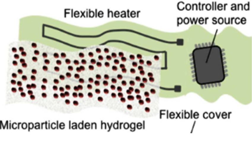
Bioengineering 2018, 5, 51                                                                         2 of 26

2. Chronic Inflammation in Diabetic Wounds
      The complete pathophysiology of ulceration is still unclear; however, clinicians and wound care
specialists widely assert that delayed healing is due to complications of peripheral arterial diseases,
neuropathy, inflammation, and ischemia [9]. A combination of impaired growth factor production,
angiogenic response, collagen accumulation, fibrosis, and abnormal pressure may result in ulceration
and chronic cracks in the feet. Ultimately, the wound healing response at the ulceration site can be
characterized by symptoms of a frustrated and prolonged inflammatory response.
      Inflammation becomes activated within a day of ulcer formation and can last up to two
weeks or longer. Inflammatory cells, such as neutrophils, macrophages, T-lymphocytes, fibroblasts,
and prostaglandin E2 (PGE2) secrete enzymes that result in pain, redness, warmth, and swelling
necessary for the healing cascade [10,11].
      Neutrophils activate upon responding to chemotactic signals that allow for cell localization in the
infected area. In a process called margination, neutrophils roll about the vasculature and migrate into
the wound site with the help of cell adhesion molecules (CAMs) through diapedesis [11]. The lack
of functional adhesion molecules at this point in the inflammatory response delays healing [11].
Neutrophils release elastase and collagenase for the purpose of destroying and removing damaged
structural proteins. They also produce tumor necrosis factor alpha (TNF-α) and interleukins 1 (IL-1),
to allow for fibroblast and epithelial cell homing [11].
      Macrophages are the second key inflammatory cells needed for phagocytosis and releasing
cytokines and growth factors, such as platelet derived growth factors (PDGF), transforming growth
factors beta (TGF-β), beta fibroblast growth factors (β-FGF), TNF-α, IL-1, and IL-6 for promoting
fibroblast proliferation and reepithelialization. Finally, lymphocytes, enter the wound and produce
IL-2, needed to aid in fibroblast recruitment [11].
      The role of fibroblasts in the healing process is significant because they produce matrix
metalloproteinases (MMPs), which destroy impaired structural protein. They also release proteins that
provide structural support for the new extracellular matrix [11]. In chronic wounds, fibroblasts are
non-receptive to cytokines and growth factors; as a result, their activity is impaired and there remains
a distorted extracellular matrix [11]. Fibroblasts release tissue inhibitor metalloproteinases (TIMPs)
in order to regulate the effects of MMPs. Unregulated MMPs are overwhelming and destroy the old
ECM in addition to fresh structural proteins [11].
      Prostaglandin E2 (PGE2) is a hormone that is produced by blood vessels and promotes
vasodilation and angiogenesis by inducing vascular endothelial growth factors (VEGFs). Thus, PGE2
is a vasodilator needed to prevent hypoxia. Individuals with DFUs suffer from vascular disease and
peripheral arterial disease due to lower levels of PGE2 [10].
      Primarily, ulcers occur through minor trauma in the presence of sensory neuropathy. Often,
neuropathy is undiagnosed until ulcer formation or pain develops. Diabetes is associated with nerve
damage, which results from diseased vasa nervorum and interacting metabolic abnormalities [12].
Hyperglycemia may inhibit the production of nitric oxide, ultimately creating an environment that
is more susceptible to reactive oxygen species, such as superoxide and hydrogen peroxide. This can
disrupt the productivity of endothelium-derived vasodilator and can lead to a cascade of platelet
aggregation, inflammation, thrombosis formation, and atherosclerosis of small vessels neighboring
peripheral nerves, eventually leading to peripheral neuropathy. Although damage from neuropathy is
irreversible, controlling blood glucose levels can prevent further ulceration. Other behavioral changes
such as monitoring hyperglycemia, hypertension, smoking, cholesterol levels, and heavy alcohol use
can prevent injury while promoting a better healing environment.
      Patients with diabetes may also have vascular disease and ischemia, which can contribute to
delayed healing. In fact, ischemia is a major contributor in 90% of DFU patients who undergo
amputation [13]. Therefore, there is a high correlation between ischemia and atherosclerosis and
highly infected DFUs. Capillary thickening can both interfere with normal flow of inflammatory cells
Bioengineering 2018, 5, 51                                                                                              3 of 26

and also create inelasticity, ultimately making it difficult for vasodilation needed for responding to
local injury [13].
     Because DFUs are chronic, treating infection can be challenging. Once an ulcer forms, infection
can build and spread due to open air wounds, lack of sterility and a loss of innate barrier function [13].
Dysfunctional leukocytes are common among the inflammatory response of patients with diabetes [14].
Additionally, phagocytosis is reduced due to hyperglycemia, fibroblast migration is hindered, and the
protective barrier and repair mechanism slows down. Normal wound exudate levels are optimized
for healing, while in chronic wounds, exudate levels contain higher concentrations of MMP [11].
MMP-2 and MMP-9 protein concentrations were 10 times and 25 times higher in pressure ulcer fluids
than surgical fluids, respectively [15]. The majority of pathogens found in DFUs are Staphylococcus
aureus, Enterococcus faecalis, Pseudomonas aeruginosa, and anaerobic bacteria [4]. Pathogen concentration
levels above or equal to 105 Colony Forming Units (CFU) per gram are capable of interfering with the
wound healing process [4]. Bacteria proliferate toward this critical concentration level, and a biofilm
forms as bacteria encase themselves within extracellular matrix substances of polysaccharides and
lipids. Resistance to immunological responses becomes a significant problem for patients with infected
chronic wounds.

3. Current Treatment and Challenges
      DFUs can be diagnosed into four different depth ischemic classifications. Table 1 displays the
major classification methods and their respective stages for evaluation of ulceration. Clinicians
responsible for diagnosis may subscribe to any of the classification methods listed below or none of
them, making treatment decisions increasingly subjective in nature, and highly based on the level of
clinician experience.

                                            Table 1. Major classification methods.

                        Wagner-Meggitt              University of Texas                         PEDIS
     Grade 0        Pain only, no open ulcer           Pre-ulceration
     Grade 1            Superficial ulcer            Superficial wound       Skin intact, no infection or loss of sensation
                                                   Wound penetrating to         Superficial ulcer with infection at the
     Grade 2                 Deep ulcer
                                                    tendon or capsule              surface and loss of sensation
                     Deep ulceration with        Wound penetrating to bone      Ulcer reaching the fascia, muscle, and
     Grade 3
                        osteomyelitis                    or joint             tendon, fasciitis and septic arthritis likely
     Grade 4          Localized Gangrene                                     Ulcer depth reaching the bone or joint, SIRS
                      Extensive Gangrene,
     Grade 5
                       Amputation likely
   References                [13,16,17]                     [18]                                  [19]

     Once a chronic wound is properly categorized, management and treatment can start. Historically,
earlier detection and diagnosis have yielded higher rates of healing [13]. If infection is present,
the first priority of treatment is to stop progression to severe osteomyelitis or sepsis. Antibiotics
for treating both the ulcer bed and osteomyelitis should be chosen based on the spectrum of
infecting organisms. Infections typically contain a combination of S. aureus and Escherichia coli,
which can be killed by applying aminopenicillin and penicillinase inhibitor as well as quinolone,
metronidazole or clindamycin [19,20]. Other studies have indicated that intravenous options, such as
imipenem, gentamicin, vancomycin, teicoplanin, rifampicin, or lenozoid can be effective [19,21]. Silver
nanoparticles and many other hard metals, such as zinc, copper, and arsenic also contain antimicrobial
properties, but must be carefully monitored for the development of a metal toxicity [22,23].
Revascularization can be made possible through angioplasty, thrombolysis, and most commonly,
bypass surgery [6]. Off-loading is important since pressure beneath the ulcer bed can significantly
increase build over time, leading to subsequent ulceration. Custom-made orthotic devices can assist in
Bioengineering 2018, 5, 51                                                                        4 of 26

lowering plantar pressure. However, removable orthotics are only as helpful as a patient’s willingness
to comply with clinician instructions.
      Ulcers heal more quickly if the surface is clean [24]; physicians must debride impediments to
healing, such as necrotic tissue and bacteria. Dressings can provide a warm, moist environment
required for healing after debridement [25]. Some engineered dressings include hydrogels,
hydrocolloid films, foams, alginates, etc. [26–28]. Common problems associated with some of these
dressings have been dehydrating the ulcer bed, saturation with exudate, and/or the failure to properly
apply antibiotics and growth factors needed to promote angiogenesis and granulation tissue.
      Currently, there are a wide variety of commercially available polymeric wound dressings that
have proven to enhance healing. Since treatment strategies depend on a unique combination of
comfort, promotion of reepithelialization, prevention of further trauma, moisture, exudate wicking,
antimicrobial properties, etc., the variance of these dressings are extreme.
      Wound contact materials are placed over the wound in the evidence of minor exudation.
Non-medicated dressings include paraffin gauze, while medicated include Xeroform® (Covidien,
Dublin, Ireland) [29]. Hydrogel, foam, and other absorbent dressings are primarily used in managing
highly exudated wounds. However, hydrogels can also rehydrate a wound if moisture level is too low.
Common absorbent dressings include Primapore® (Smith & Nephew, London/Hull, UK), Mepore®
(Mölnlycke, Gothenburg, Sweden) and absorbent cotton gauze (BP 1988). Hydrogel dressings include
ActiformCool® (Activa) and Aquaflo® (Covidien, Dublin, Ireland) [29]. Promgran Prisma® (Systagenix)
and Dermol/Ag™ (DermaRite Industries, North Bergen, NJ, USA) are two collagen matrix dressings
that can transform into a biodegradable gel if exudate levels remain high within the wound [30].
Aquacel Hydrofiber® (ConvaTec, Reading, UK) creates a soft gel-like material as it absorbs wound
fluid, while maintaining a moist environment. Meliplex Ag (Molnlych Health Care, Gothenburg,
Sweden) is a vapor-permeable, waterproof film, that regulates wound moisture while protecting the
environment from bacterial invasion. Tegaderm™ (3 M Health Care) is another popular dressing that
not only absorbs exudate, but is comfortable and easy to remove from fragile and sensitive tissue [30].
      Film dressings are important to consider when a membrane layer is needed to allow for the
passage of oxygen and vapor, while preventing the invasion of water, exudate, or bacteria [29]. Many
polyurethane materials can be used for this purpose and is mentioned in Section 4.2.2. GranuDerm™
and Sentry™ (Acute Care Sollutions, LLC, Canton, OH, USA) are breathable films that rid the wound
of water, dirt, and microbes, while prohibiting leakage. Similarly, Silverlon® (Argentum Medical,
LLC, Geneva, IL, USA) permits the passage of exudate, while preventing microbial invasion and
contamination [30].
      As mentioned before, silver has been used frequently to treat infected wounds. Allevyn
(Smith & Nephew, London/Hull, UK) is a polyurethane film combined with foam containing silver
sulphadiazine [26]. The release of antibacterial action was observed to last approximately 7 days.
Dermacol/Ag™ (DermaRite Industries, North Bergen, NJ, USA) is a collagen matrix wound dressing
that contains silver chloride in order to prevent bacterial colonization. Many of the foam or hydrogel
dressings such as Promgran Prisma® (Systagenix, Skipton, UK), Meliplex Ag (Molnlych Health Care,
Gothenburg, Sweden), and Aqucel Hydrofiber® (ConvaTec, Reading, UK) contain silver within their
matrix to provide a protective barrier for not only fighting infection, but also allowing for an optimal
healing environment [30].
      Many of the commercially available wound dressings have shown potential for healing chronic
DFUs, however, they lack a complete holistic approach. Wound dressings vary on the level of
absorbency. One wet, absorbent dressing might be only practical for highly exudated wounds. On the
other hand, a type of occlusive film might only be advantageous for wounds that need to maintain
moisture [29]. A combination of layers of different material types needs to be developed to potentially
fit the needs of all varieties of DFUs.
      The treatment of chronic diabetic wounds requires the proper balance between experience-based
intuition and science [31]. Modern management of ulcers is inefficient because preliminary assessment
Bioengineering 2018, 5, 51 5 of 26
and diagnosis are often subjectively performed by clinicians. There is a need for an integral system
that combines the use of the therapeutic components such as wound dressings and antibiotics with
diagnostic components such as quantitative sensors to create a holistic treatment strategy for diabetic
chronic wounds. Not only will the chances of wound healing rise, but also, the risk of new ulcer
formation will decrease. Major developments in treatment strategies are needed for wound dressing
design and quantitative diagnosis in order to decrease subjectivity and improve patient compliance.
The subsequent sections highlight the state of the art in wound dressings, sensors, and composite
smart wound dressings for the treatment of chronic diabetic wounds.
4. Current Wound Dressings
Wound dressings are needed to provide a barrier between the ulcer and the external environment.
DFUs excrete wound fluids and have a prolonged healing process. Thus, the quintessential dressing is
made of an antimicrobial material, maintains a moist environment, is permeable to oxygen, removes
wound exudate, and allows the release of needed growth factors or drugs for the wound to facilitate
proper proliferation and tissue remodeling [32]. There are myriad dressings for use in ulcer and
chronic wound care, and the type of dressing used depends on the physiological parameters of the
ulcer. Combinations of natural and synthetic polymers give rise to a more ideal material fit to not only
heal the ulcer bed, but also to provide a foundation for reepithelialization.
While orthotics and skin grafts can play a crucial role in removing excess pressure and
regenerating skin, wound dressings play a significant role in providing not only protection but
also a factor for promoting natural healing. Recently, dressings can facilitate healing through additives
that allow for a moist environment, removal of exudate, antibacterial effects, and the stimulation
and proliferation of fibroblasts and keratinocytes at the site of injury [25,33]. None of these factors
can be applied without patient compliance to facilitate application and removal without aggravating
the symptoms [34]. A holistic approach should also consider minimizing cost. Thus far, no single
dressing has met all the needs for proper wound healing, and therefore, further research is needed
to explore a unique combination. The following section focuses on recent progress in natural and
synthetic polymer-based wound dressings (Table 2) and clinical outcomes and the need to improve
micro- and macro-scale geometry and overall architecture.
4.1. Natural Polymers
Natural polymers have been popular in research and clinical settings because their properties are
biocompatible and closely related to the extracellular matrix. They mimic many biological systems to
prevent the immunologic reactions caused by many synthetic polymers [35]. Additionally, natural
polymers can be synthesized into polysaccharides, proteins, and polyesters by living organisms, or
more recently, by fermentation of microorganisms [35].
4.1.1. Cellulose
Cellulose is the most abundant organic polymer on Earth. It has been used in several
wound healing applications since it releases phosphodiesterase growth factor, epidermal growth
factor (EGF) and basic fibroblast growth factor (bFGF), all of which stimulate fibroblast growth
and anti-inflammatory effects [36]. Beneficial properties of specifically bacterial cellulose include:
hydrophilic surfaces, water uptake capacity, permeability, and tensile strength, all of which are
comparable to the fibrous structure of collagen. Brassolatti et al., designed a bacterial cellulose dressing
on third degree burn wounds in rats and noticed optimized healing response. Their results indicated
that the use of bacterial cellulose dressings with and without lidocaine had comparable advanced repair
outcomes, with both being more effective than the untreated control group. Skin appendages, mild
inflammatory cell influx, collagen fiber organization and mild immunostaining were observed [37].Bioengineering
Bioengineering2018,
2018,5,
5,x51FOR PEER REVIEW 6 6ofof2625
4.1.2. Chitosan
4.1.2. Chitosan
Chitosan, a copolymer derived from chitin, is prominent in the exoskeletons of arthropods and
Chitosan,
the cell a copolymer
wall of fungi derived
[38,39]. It can be from chitin,toisform
fabricated prominent
a gelatin inor
thefilm-like
exoskeletons of arthropods
material, and
and its versatile
the cell wall of fungi [38,39]. It can be fabricated to form a gelatin
effects promote strong adhesion to wound beds. Chitosan is a widely popular wound dressing or film-like material, and its
versatile due
material effects
to promote strong adhesion
its multifunctional to wound
properties suchbeds. Chitosan is a widely
as nonantigenicity, popular
inertness, wound
nontoxicity,
dressing material due to its multifunctional properties such as nonantigenicity,
biodegradability, biocompatibility, bioadhesiveness, antimicrobial properties, and hemostatic effects inertness, nontoxicity,
biodegradability,
[40–42]. A chitosanbiocompatibility, bioadhesiveness,
based Opticell dressing antimicrobial
(Medline Industries, properties,
Chicago, IL, USA) and
hashemostatic
hemostatic
effects [40–42]. A chitosan based Opticell dressing (Medline Industries, Chicago,
efficacy. Stricker-Kongrad et al., examined the Opticell dressing on heparinized rats with excisional IL, USA) has
hemostatic efficacy. Stricker-Kongrad et al., examined the Opticell dressing
wounds that mimicked debridement. Researchers noted that after removing the dressings, the total on heparinized rats
with excisional
blood wounds thatless
loss was significantly mimicked
than that debridement.
of a typical gauze Researchers noted
dressing. Thisthat after removing
indicated the
that Opticell
dressings,with
dressings the total bloodhave
chitosan loss hemostatic
was significantly
effectsless than
that couldthatbeofused
a typical gauzebleeding
to control dressing.associated
This indicated
with
that Opticell dressings with chitosan have hemostatic effects that could
wound debridement [43]. Unlike cellulose, chitosan has beneficial antimicrobial effects. be used to control bleeding
When
associated
crossed with
with wound the
cellulose, debridement
composite [43]. Unlike
in the form cellulose,
of film or chitosan
hydrogelhas beneficial
inhibited antimicrobial
the growth ofeffects.
E. coli
When crossed with cellulose, the composite in the form of
and S. aureus, while enhancing wound repair and epithelial regeneration in woundfilm or hydrogel inhibited theand
growth
burn
of E. coli and S. aureus, while enhancing wound repair and epithelial regeneration in wound and
infections [44,45].
burn infections [44,45].
4.1.3. Collagen and Gelatin
4.1.3. Collagen and Gelatin
Another abundant natural polymer found in the ECM is collagen. Collagen is present in most
Another abundant natural polymer found in the ECM is collagen. Collagen is present in most
epithelial
epithelialand and connective
connective tissues,
tissues, such
such asas bone,
bone, cartilage,
cartilage, ligaments,
ligaments,tendon,
tendon,andandskin.
skin.ItItprovides
provides
strength and integrity and is an essential component in cell-cell interactions that regulate
strength and integrity and is an essential component in cell-cell interactions that regulate anchorage, anchorage,
migration,
migration, proliferation, differentiation,and
proliferation, differentiation, andsurvival
survival[22,46,47].
[22,46,47]. Collagen
Collagen hashas been
been a key
a key ingredient
ingredient in
in promoting
promoting tissue
tissue granulation
granulation and angiogenesis
and angiogenesis and inand in inhibiting
inhibiting bacterialbacterial
growth in growth
chronicinwounds.
chronic
wounds.
Collagen hasCollagen has asbeen
been used usedofas
a carrier a carrier
reactive oxygen of species,
reactivegrowth
oxygen species,
factors, growth factors,
and antibiotics. Wiegandand
antibiotics. Wiegand
et al., fabricated et al.,and
a collagen fabricated
celluloseacomposite
collagen that
and demonstrated
cellulose composite thatofdemonstrated
a reduction cytokines anda
reduction of cytokines and proteolytic enzymes, indicating less inflammation
proteolytic enzymes, indicating less inflammation in the wound bed [48]. A similar study assessed in the wound bed [48].
the
Aeffect
similar study assessed
of collagen theoneffect
dressings of collagen
the size, dressings
granulation tissue,on
andthebacterial
size, granulation
inhibition tissue, andwounds.
of chronic bacterial
inhibition
Patients withof chronic wounds. dressings
collagen-based Patients with collagen-based
could dressings
avoid skin grafting could
since avoid skin
the presence ofgrafting since
granulation
the presence
tissue was a of granulation
suitable tissue
alternative wasFigure
[49]. a suitable alternative
1 depicts a type of[49]. Figuremesh
collagen 1 depicts a type
and the of collagen
generation of
mesh and the generation
fibroblasts around the scaffold.of fibroblasts around the scaffold.
Collagenthread
Figure1.1.Collagen
Figure thread mesh
mesh imaged
imaged using optical microscopy
microscopy (a);
(a); reinforced
reinforcedcollagen
collagensponge
spongewith
with
collagen mesh (b); Collagen sponge reinforced by collagen mesh using SEM micrographs (c); andthe
collagen mesh (b); Collagen sponge reinforced by collagen mesh using SEM micrographs (c); and the
incorporationofoffibroblasts
incorporation fibroblastsgrowing
growingwithin
withincollagen
collagenmesh
meshusing
usingSEM
SEMmicrographs
micrographs(d).
(d).Reprinted
Reprinted by
by permission
permission fromfrom Springer
Springer Nature:Biotechnology
Nature: BiotechnologyandandBioprocess
Bioprocess Engineering
Engineering reference
reference [50].
[50].
Copyright2008.
Copyright 2008.Bioengineering 2018, 5, 51 7 of 26
Additionally, the dressing provided comfort, essential for patient compliance, as opposed to other
conventional methods. Gelatin is a collagen derivative and is applicable to wounds that need a more
hydrogel-like material [38]. It is biocompatible and degradable in physiological medium and can
mimic many of the characteristics of the dermis. A research group that used bFGF-impregnated gelatin
microspheres found that gelatin stimulates angiogenesis and fibroblast proliferation [51].
4.1.4. Hyaluronic Acid
Hyaluronic acid (HA) presents a wide range of natural healing properties such as tissue integrity,
lubrication, and water absorption. Similar to collagen, HA promotes mesenchymal and epithelial
cell migration and differentiation, ultimately improving collagen deposition and angiogenesis for
repair [38]. HA’s properties are sensitive to molecular weight, however. Campo et al., suggested
that only medium molecular weight HA (MMWHA) could enhance wound repair; lower molecular
weights contribute to further inflammation, and higher molecular weights might inhibit nutrient
supply for tissue regeneration by blocking capillary formation [52,53]. Despite the sensitive functional
aspect of HA, there have been promising results in the treatment of lesions where the loss of ECM was
analyzed. Simman and colleagues noticed that during a clinical case series involving 12 patients with
serious surgical wounds treated with HA-type wound dressings, all wounds developed granulation
tissue [54].
4.2. Synthetic Polymers
Synthetic polymers are commercially available to overcome some of the limitations associated
with natural polymers, such as inconsistent and non-reproducible chemical and physical composition.
Synthetic polymers can be fabricated into various shapes, allowing for versatile function. Unlike natural
polymers, most synthetics are insensitive to enzymatic and biological degradation and therefore have
relatively more stable properties [55].
4.2.1. Poly(lactide-co-glycolide)
Poly(lactide-co-glycolide) (PLGA) is a copolymer of polylactic acid (PLA) and polyglycolic acid
(PGA). It is a degradable and biocompatible polymer, and its clinical use in drug delivery, suture
applications in humans has been approved by the FDA. Although PLGA has yet to be approved for
wound healing applications, significant research has been performed due to its efficacy in healing [56].
It exhibits mechanical strength and can conform into various shapes allowing for a plethora of
processing types [57]. The ratio of lactide to glycolide units can greatly affect the release of bioactive
substances and other pharmaceuticals. The larger the lactide units, the longer the polymer lasts
before degrading [58]. Zheng et al., demonstrated that a PLGA and cellulose nanocrystal nanofiber
membrane not only showed advantageous cytocompatibility, but also stimulated fibroblast adhesion,
spreading, and proliferation. The release of neurotensin (NT), an inflammatory moderator, was also
observed and researchers noted that the composite membrane allowed for sustained delivery of NT,
ultimately promoting reepithelialization for the treatment of DFUs [59]. Another study created PLGA
microspheres with high encapsulation of recombinant human epidermal growth factor (rhEGF) by
solvent-evaporation. This research team observed an optimal growth rate of fibroblasts in and around
the wound bed. Thus, the PLGA microsphere provided a compatible environment for the repair
process as well as an optimal delivery route of rhEGF [60].
4.2.2. Polyurethanes
Polyurethanes (PU) possess not only a delivery route for growth factors and antibiotics, but also
create a barrier that prevents bacteria from entering and further infecting the wound bed. PU is a
suitable alternative synthetic polymer that provides a semi-permeable membrane. It imparts a moist
environment and delivers bioactive substances for fighting infection and repair while protecting the
wound from bacterial entry [57]. Additionally, PU-based nanofibers contain fluid drainage propertiesBioengineering
Bioengineering 5, x 5,
2018,2018, FOR51 PEER REVIEW 8 of 268 of 25
proposed that a PU wound dressing could incorporate a combination of antibiotics, pain relievers,
that decrease the risk of swelling, increased exudate, and wound desiccation [61]. Varma et al.,
and proposed
protease inhibitors. Whiledressing
that a PU wound releasing these
could bioactiveasubstances,
incorporate combinationthe barrier layer
of antibiotics, can
pain degrade in
relievers,
contact
andwith the wound.
protease Many
inhibitors. Whiletimes, PUsthese
releasing are used as the
bioactive absorbentthe
substances, material, sandwiched
barrier layer between
can degrade in a
contact layer
contact and
with thea wound.
waterproof
Manyfilm. Meliplex
times, Ag (Molnlycke
PUs are used Health
as the absorbent Care,sandwiched
material, Gothenburg, Sweden)
between a is
an example tri-layered
contact layer wound dressing,
and a waterproof and itAg
film. Meliplex has(Molnlycke
shown effective healing
Health Care, of many types
Gothenburg, Sweden) of is
ulcers
an example
and burns tri-layered wound
[62]. Additionally, PU dressing, and it hastoshown
was electrospun form aeffective
poroushealing of many
membrane thattypes
could ofwick
ulcersaway
fluid from the wound, while preventing fluid buildup and wound desiccation. Figure away
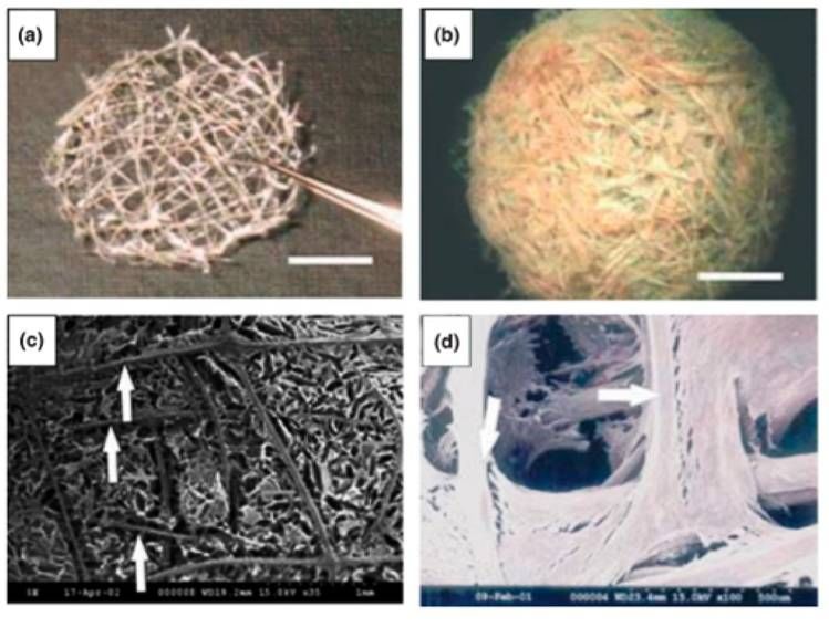
and burns [62]. Additionally, PU was electrospun to form a porous membrane that could wick 2 depicts
fluid from the wound, while preventing fluid buildup and wound desiccation.
electrospun PU fibers and a wound dressing prepared Tegaderm™, made out of a thin layer of PU Figure 2 depicts
electrospun PU fibers and a wound dressing prepared Tegaderm™, made out of a thin layer of PU and
and acrylic adhesives.
acrylic adhesives.
Figure
Figure 2. SEM
2. SEM image
image of PU
of PU electrospunfibers
electrospun fibers (a)
(a) and
and the
theTegaderm™
Tegaderm™ wound
wound dressing (b). (b).
dressing Reprinted
Reprinted
by permission
by permission fromfrom John
John Wileyand
Wiley andSons:
Sons: Journal
Journal ofof Biomedical
BiomedicalMaterials
MaterialsResearch reference
Research [61] [61]
reference
Copyright 2003.
Copyright 2003.
4.2.3. Polyethylene Glycol
4.2.3. Polyethylene Glycol
Polyethylene glycol (PEG) is another synthetic polymer desirable for wound dressings. It is a
Polyethylene glycol (PEG) is another synthetic polymer desirable for wound dressings. It is a
unique material, possessing hydrophilic, flexible, and compatible qualities. Often it can be blended
uniquewithmaterial,
PLGA orpossessing
chitosan for hydrophilic, flexible, and
increased mechanical compatible
stiffness qualities.
and stability [57]. Often
It canitalso
canbebeused
blended
withasPLGA or chitosan
a surface modifierfor
for increased
composite mechanical stiffness
wound dressings and stability
allowing for better[57].
gripItincan
the also be layer.
contact used as a
surface modifier for composite wound dressings allowing for better grip in the
Lee et al., fabricated a tri-bloc polymer of PLGA-PEG-PLGA and observed wound healing of DFUs contact layer. Lee et
al., fabricated a tri-bloc
in mice. Their resultspolymer of PLGA-PEG-PLGA
showed improved and observed
epithelium migration wounddeposition,
and collagen healing ofinDFUs in mice.
addition
Theirtoresults
a higher woundimproved
showed closure rate [63]. A similar
epithelium studyand
migration used varyingdeposition,
collagen amounts ofinPEG interspersed
addition to a higher
with PLGA nanoparticles used to deliver recombinant human insulin as a
wound closure rate [63]. A similar study used varying amounts of PEG interspersed with PLGApotential wound healing
agent. Researchers
nanoparticles used toconcluded
deliver that the sustained
recombinant insulininsulin
human deliveryasfora6 days by thewound
potential PEG/PLGA vehicle
healing agent.
led to enhanced cell proliferation [64]. Growth factors have a high affinity for PEG, and can be
Researchers concluded that the sustained insulin delivery for 6 days by the PEG/PLGA vehicle led to
easily manipulated for local delivery [65]. Huang et al., analyzed the PEGylation of recombinant
enhanced cell proliferation [64]. Growth factors have a high affinity for PEG, and can be easily
human acid fibroblast growth factor (rhaFGF) on diabetic wound healing. Higher expression of
manipulated for local delivery [65]. Huang et al., analyzed the PEGylation of recombinant human
keratinocyte-specific genes was observed, and the rate of wound healing significantly increased [66].
acid fibroblast growth factor (rhaFGF) on diabetic wound healing. Higher expression of keratinocyte-
specific genes was observed, and the rate of wound healing significantly increased [66].
4.2.4. Polycaprolactone
Polycaprolactone (PCL), a biocompatible, biodegradable polymer that is resistant to many
solvents, is similar to PLGA and it can be fabricated into many shapes and forms allowing for multi-
functional properties. PCL has been FDA approved for the design and use of sutures in surgeries,
and therefore, its slow degradation and compatibility warrants use as a potential wound dressing
[67]. Specifically, PCL fibers are used to treat wounds because the fibrous structure is similar to that
of the ECM. PCL is especially advantageous because it has excellent water retention capacities that
capture wound exudate [68]. Because of its lack of antimicrobial effects, PCL is often integrated withBioengineering 2018, 5, 51 9 of 26
Table 2. Summary of Polymer-based Wound Dressings.
Polymer Advantages Disadvantages Reference
1. Readily available with low cost 1. Requires additional
2. Fiber and foam materials antimicrobial substances
3. Creates a gel-like material, forming 2. Resorption in tissues does not
Cellulose [36]
a moist environment occur, which could cause further
4. Releases GFs to stimulate tissue damage or become
fibroblast proliferation overwhelmed by excess exudate
1. Fabricated in a gelatin of
film-like material
1. Extensive swelling in water
2. Antimicrobial and
2. Unable to dissolve in organic
Chitosan hemostatic properties [38,43–45,70,71]
solvents because of its rigid
3. Functional derivatives allowing for
crystalline structure
modified and versatile effects
4. Ability to deliver drugs
1. Promotes tissue granulation
1. May not be absorptive in
and angiogenesis
gelatin form, especially for
2. Inhibits bacterial growth and
Collagen and Gelatin wounds with excessive exudate [22,38,46,48]
prolonged inflammatory response
2. Might require
3. Gelatin derivative forming a
secondary dressing
hydrogel material
1. Lubricative and water absorptive
2. Bi-products promote epithelial
cell migration 1. Only MMWHA enhances
Hyaluronic Acid [52,72–74]
3. Improves collagen deposition and wound repair
angiogenesis4. Popular drug delivery
system and vehicle for growth factors
1. FDA approved for drug delivery,
suture applications
2. Ratio of lactide to glycolide units
1. Requires additional
can modify release of drugs and
antimicrobial substances
Poly(lactide-co-glycolide) growth factors [38,56–59]
2. Properties fail to match ECM
3. Cytocompatible and stimulates
or collagen
fibroblast adhesion, spreading,
and proliferation
4. Fabricated into various shapes
1. Semipermeable membrane that
1. Need composite dressings in
prevents bacteria from entering
order to provide contact layer
2. Provides a moist environment
and waterproof properties
Polyurethanes 3. Delivers bioactive substances for [57,61,62]
2. Wound healing effects are only
fighting infection
associated with
4. Drainage properties that decrease
nanofiber structure
the risk of swelling
1. Hydrophilic, flexible and
1. Adhesiveness might damage
compatible qualities
granulation tissue
2. Surface modifier allowing for better
Poly(ethylene glycol) 2. Does not incorporate [38,57,64]
grip in the contact layer
antibiotics and other drugs so
3. Growth factors have higher affinity
composite materials are needed
for PEG
1. FDA approved for
suture applications
2. Fibrous structure similar to
ECM architecture 1. Lack of
Polycaprolactone [67–69]
3. Water retention capacities used to antimicrobial properties
capture wound exudate
4. Resistant to many solvents allowing
for slow and controlled degradation
4.2.4. Polycaprolactone
Polycaprolactone (PCL), a biocompatible, biodegradable polymer that is resistant to many
solvents, is similar to PLGA and it can be fabricated into many shapes and forms allowing forBioengineering 2018, 5, 51 10 of 26
multi-functional properties. PCL has been FDA approved for the design and use of sutures in
surgeries, and therefore, its slow degradation and compatibility warrants use as a potential wound
dressing [67]. Specifically, PCL fibers are used to treat wounds because the fibrous structure is similar
to that of the ECM. PCL is especially advantageous because it has excellent water retention capacities
that capture wound exudate [68]. Because of its lack of antimicrobial effects, PCL is often integrated
with other polymers and/or antibiotics. Silver nanoparticles incorporated in a PCL matrix have shown
inhibited bacterial invasion. One study also increased the hydrophilicity and diffusion properties by
enriching PCL with nanochitosin (NC). NC provided antibacterial activity as well as sustained release
of curcumin, a supplement used to reduce inflammation and pain, while providing better skin health
and controlling cholesterol and blood sugar [69]. The combination of PCL and chitosan provided a
tunable wound dressing for enhanced drug delivery and ultimately healing.
4.3. Smart Polymers
In recent decades, smart polymers have been heavily researched due to their important
characteristics pertaining to thermal, chemical, and physical responses needed to modify the healing
process. Unlike traditional synthetic or natural polymer wound dressings, smart polymers can control
material properties in response to external cues [75]. The following studies highlight recent work in
the analysis of smart wound dressings’ properties and the mechanism used to create responses.
A stimulus-responsive hydrogel is a soft, hydrophilic material that swells upon the absorption of
water. They can be fabricated using copolymers, blends, or interpenetrating networks (IPNs) and made
to respond to various changes in their environment, such as pH, temperature, chemicals, light, electric
field, and shear stress [76]. Since pH values change overtime in chronic wounds [76], a pH responsible
hydrogel can swell or contract when exposed to an overwhelming pH and ionic strength in the wound.
Additionally, mechanical perturbations can be monitored by shear-responsive hydrogels. Hydrogels
exhibit viscoelastic mechanical behavior upon deformation and will strain in a time-dependent manner
upon application of stress, such as loading and pressure beneath the ulcer bed. Once an external stress
is removed, the hydrogel can recover its original structure [76].
Dermal patches can be engineered by constructing a thermoresponsive drug microcarrier
encapsulated within a hydrogel layer. N-Isopropylacrylamid (NIPAM) is a type of thermoresponsive
material commonly used as a drug vehicle. The hydrophilicity of the material is dependent on
temperature; it is hydrophilic below its critical temperature, 32 ◦ C, and hydrophobic above. It can be
tuned through copolymerization, allowing for optimal drug release [77]. Bagherifard et al., designed
a smart, hydrogel-based, dermal patch with integrated heating elements for on-demand drug and
growth factor delivery. The patch included a Ca-alginate hydrogel sheet, micropatterned gold heating
elements, and the thermoresponsive NIPAM particles as seen in Figure 3 [78].
In addition to proper conformability with the skin, the ability to maintain skin moisture and
protecting the wound from pathogens, the thermoresponsive drug carriers within the chosen hydrogel
controlled for drug release [79].
Shape memory polyurethanes (SMPUs) are being incorporated into composite materials to provide
more ideal mechanical properties and stretching capabilities [80]. The material has shape memory
function that can control deformation and force, particularly in suture applications [81]. Tan et al.,
proposed to design a composite nanofibrous mat that consisted of chitosan, gelatin, and SMPU through
electrospinning. The gelatin and chitosan contributed to wound healing as the incorporation obtained
surface wettability, cytocompatibility, hemostatic properties, and water vapor transmission. Whereas,
the SMPU allowed for controllability upon tensile force under different strains. In wound healing
applications, the SMPU can eventually assist in the closure of cracked wounds [82].
An ideal smart wound dressing can impose the idea of an individualized healing function fit to a
patient’s need, while responding to biological, chemical, and physical responses in the chronic wound
environment. By doing such, drug delivery can be controlled in response to environmental cues and
ultimately, prevent delayed healing.material commonly used as a drug vehicle. The hydrophilicity of the material is dependent on
temperature; it is hydrophilic below its critical temperature, 32 °C, and hydrophobic above. It can be
tuned through copolymerization, allowing for optimal drug release [77]. Bagherifard et al., designed
a smart, hydrogel-based, dermal patch with integrated heating elements for on-demand drug and
growth factor2018,
Bioengineering delivery.
5, 51 The patch included a Ca-alginate hydrogel sheet, micropatterned 11 ofgold
26
heating elements, and the thermoresponsive NIPAM particles as seen in Figure 3 [78].
Figure
Figure3. 3.Schematic
Schematicofofthe
thesmart
smart wound
wound dressing depicting the
dressing depicting themajor
majorelements.
elements.Reprinted
Reprintedbyby
permission from John Wiley and Sons: Advanced Healthcare Materials reference [78] Copyright 2016.
permission from John Wiley and Sons: Advanced Healthcare Materials reference [78] Copyright 2016.
4.4.InFiber
addition to proper
Geometry conformability
and Scaffold Architecture with the skin, the ability to maintain skin moisture and
protecting the wound from pathogens, the thermoresponsive drug carriers within the chosen
In addition to material type, geometry and conformation of the wound dressing can have just
hydrogel controlled for drug release [79].
as big of an impact on successful healing. Important parameters to consider are porosity, dimension,
and strength of the graft as well as macrostructure to ensure orderly extracellular matrix deposition.
Typically, one material does not fit all criteria. We propose that a multilayered wound dressing is
necessary to ensure all advantageous effects. A porous, hydrophobic contact layer is needed to create a
barrier between the outside environment and wound, while promoting sterility and reepithelialization.
A hydrogel or nonstick material is best to prevent further damage to the granulated tissue. A second
and third layer is needed and relies on fibers oriented specifically to provide channels that can wick
away wound exudate both vertically and horizontally. Fiber cross-section is an important variable
to consider as it can alter capillary movement of fluid. In a previous study, permeability and fluid
wicking were tested as a function of weave configuration and fiber geometry of various combinations
of poly-l-lactide and poly-l-lactide-co-ε-caprolactone. The outcomes suggested that grooved wicking
geometry, as opposed to a round cross-section, may be used in scaffold development to regulate fluid
transport toward the area of interest [83].
A second parameter to consider is a single or multi-filament architecture as a component of the
scaffold, especially for antibiotic delivery. A second experiment investigated the elution profile of
gentamicin in a bundle of small wicking fibers and a single large fiber in order to test the effect of
cross-sectional diameter on diffusion. The burst release of the large single fiber was substantially
greater than the bundle of fibers, and the cumulative release was higher at each time point. Researchers
concluded that the bundle had a slower release profile due to a reduced surface area in contact with
the buffer solution when compared to the single large fiber. Although it has a slower release, the fiber
bundle exhibits a stable and consistent elution profile for a larger duration.
Finally, fibers can orient themselves to form a specific type of porous fabric to allow tissue
ingrowth and permeation of metabolites. Fabrics can be classified as woven, knitted, and non-woven,
each having their own unique properties. Woven fabrics can be used where mechanical strength
is required due to their low elongation, high breaking strength, and more mechanical stability.
Knitted fabrics can provide good wrinkle and crush resistance, higher elongation, good elastic
recovery and permeability, allowing for optimal tissue ingrowth. Wang et al., designed a PLGA
knitted mesh-reinforced with collagen-chitosan scaffold and analyzed the geometry’s role in tissue
regeneration. Researchers noted that the knitted-mesh provided mechanical strength, while inhibiting
wound contraction and promoting neotissue formation and blood vessel ingrowth [70]. Non-woven
fabrics are typically fabricated using techniques such as electrostatic or solution blow spinning.Bioengineering 2018, 5, 51 12 of 26
The fibers are staple-length and usually good for low strength applications. Electrospinning can create
continuous fibers with diameters as small as a few nanometers, which possess a similar architecture
to the structure of the ECM. Also, the porous feature of electrospun scaffolds allows for a high
surface-to-volume ratio necessary for optimal cell attachment and oxygen and nutrient transport.
Yang et al., discovered a higher wound recovery rate with complete skin regeneration. Electrospun
fibrous mats were fabricated to allow gaseous and fluid exchanges, while absorbing exudate and
wound odor. With the release of bFGF, enhanced collagen deposition and ECM remodeling was similar
to normal tissue [84].
5. Biosensing in the Chronic Wound Environment
In addition to smart, adaptive wound dressings, researchers and clinicians have recently realized
the need for quantitative assessment of the chronic wound environment through biosensing. There is
a need for a smart dressing for chronic wounds that is composed of sensing elements that can: (1) be
integrated with current biological dressings; (2) sense changes at the wound microenvironment;
(3) provide data to the health care professional; and (4) serve as a theragnostic approach by releasing
antimicrobials when needed [85,86]. This need stems from current assessment procedures (Table 1),
which are mainly subjective and require a healthcare professional to analyze and classify wounds
without any readily quantifiable data [87]. Much of the work in biosensing of chronic wounds
is focused on the ability to discern the probability of wound closure and finding the quantitative
thresholds of various biomarkers related to wound healing [11]. A summary of these developments in
sensing the chronic wound environment will be discussed in the following sections.
5.1. Biomarkers for Wound Healing
Several biomarkers in chronic, non-healing wounds have been identified as potential indicators
for the management and treatment of chronic wounds. These biomarkers can be divided into two
major categories, (1) biochemical and (2) physical. Each can be quantified through electrochemical
or electrophysical transduction, where some biochemical marker (i.e., cytokine, enzyme, metabolic
byproduct, pH, etc.) or some physical marker (i.e., temperature, pressure, moisture level, etc.) is
converted to a measurable electrical signal. Researchers have also explored different methods for
qualitative measures and visualization of wound healing. Various spectroscopy methods, such as
absorption, fluorescence, phosphorescence, Raman, SERS, refraction, and dispersion spectrometry
are used to detect changes in energy, polarization, amplitude, decay time, and/or phase. Amplitude
analysis is one of the most widely used methods for correlation of analyte concentration with signal
amplitude [88].
5.1.1. Biochemical Markers
There are many metabolic pathways in the wound healing process and further research is
needed to identify key biomarkers and their respective roles in each pathway. Cytokines, proteases,
bacteria, oxygen, nitric oxide (NO), etc., are potential biochemical markers that could predict
non-healing wounds [11,89]. Cytokines are cell signaling molecules secreted by platelets, fibroblasts
and inflammatory cells during inflammation. IL-1, IL-6, and TNF-α are cytokines that have been
measured in higher concentrations in chronic non-healing wounds when compared to normal healing
wounds [11]. Beila and coworkers have demonstrated that the overall pro-inflammatory level of
16 tested cytokines were higher in ulcer tissues than normal tissues before treatment of chronic venous
insufficiency ulcers [90]. Proteases are the enzymes responsible for breaking down proteins. Protease
levels have also been shown to be higher in non-healing wounds as compared to normal or acute
healing wounds [11]. The major proteases of interest are MMP-2 and -7. MMPs, are involved in
the inflammatory stage of wound healing and are released by inflammatory and connective tissue
cells. They are responsible for breaking down necrotic tissue and the extracellular matrix prior to
the proliferation stage of the normal wound healing cycle. Utz et al., have demonstrated with highBioengineering 2018, 5, 51 13 of 26
probability (p < 0.001) that MMP-2 and -7 levels were significantly higher in chronic wounds than
in acute wounds [91]. Bacteria concentration levels are abnormally higher in chronic wounds due
to the fact that the wound is stuck in the inflammatory stage. Some of the most commonly found
bacteria in chronic wounds are S. aureus, E. coli, and P. aeruginosa [92]. NO has been well established
as a biomarker in wound healing and is known to have lower concentrations in chronic non-healing
wounds [89]. However, NO has a half-life less than 10 s making it difficult to measure. Investigators
have tried to measure wound fluid NO levels indirectly through wound fluid nitrate, induced NO
response and fasting urine [89,93]. Boykin concluded that deprivation of NO activity contributes
to impaired healing, and a comprehensive method to monitor NO, MMP, and bacterial load could
accelerate healing in chronic wounds [89]. Wound pH and uric acid play a vital role in cell to cell
interaction in wound healing. Change in pH is a good indicator of wound healing whereas a sudden
alkalotic pH followed by a gradual decrease in pH to a steady-state value around 5–6 indicates proper
wound healing [86]. UA concentration tends to be decreased in chronic wounds due to the increase of
infection and bacterial consumption of UA [94]. The fabrication of biochemical sensors for some or all
of these biomarkers would exploit a specific method of transduction, therefore enabling quantification.
5.1.2. Physical Biomarkers
Physical biomarkers that have been explored for chronic wound healing are bioimpedance,
pressure, and ambient temperature. Bioimpedance includes measurement of resistance, reactance and
the associated phase angle. Resistance is related to the amount of extracellular fluid (ECF) in a given
sample of tissue; reactance is related to the cell mass and is a good indicator of cell accumulation and
proliferation; and phase angle corresponds to the vitality of the tissue and is a good prognosis of tissue
nutrition [87]. The resistance to current arises from the fact that less ECF decreases the number of
ions and lowers the conductivity of the matter. While the reactance value arises from the capacitive
nature of cell membranes, lower values at a specified frequency value indicate decreased wound
mass. Increased pressure hinders proper healing in foot ulcers; relieving pressure before the onset of
an ulcer may prevent its genesis [95]. Increase in temperature is associated with increased bacteria
levels, which often indicates complications in wound healing [96]. Table 3 lists the biomarkers that are
associated with chronic non-healing wounds.
Table 3. Biomarkers associated with chronic wound healing.
Biochemical Biomarkers
Wound Biomarker Significance in Chronic Wounds vs. Acute Wounds Reference
Cytokines (IL-1, IL-6, TNF-α) Elevated levels of Cytokine [11,90]
Nitric Oxide Decreased levels of NO [89,93]
Matrix Metalloproteinase Increased protease activity [11,91]
Oxygen Higher probability for ischemia due to decreased oxygen levels [9]
Bacteria concentration levels are higher indicating extent
Bacteria [9,92]
of infection.
Wound pH Remains more alkalotic for extended period of time [86]
Uric Acid Decreased levels due to bacteria [94]
Reactive Oxygen Species Increased levels due to oxidative stress [11]
Increase in bacterial housekeeping genes; decrease in ulcer
Gene Expression [11]
housekeeping genes.
Growth Factors Decreased level (i.e., PDGF) [11]
Physical Biomarkers
Wound Biomarker Significance in Chronic Wounds vs. Acute Wounds Reference
Bioelectrical Impedance Phase angle, resistance, and reactance are all decreased [87]
Pressure Increased pressure [95]
Temperature Increased temperature [96]Bioengineering 2018, 5, 51 14 of 26
5.2. Biochemical Sensors
Biochemical sensors are designed for a specific analyte which makes them highly selective and
sensitive. Ideally, a biosensor has a biorecognition element where the specified analyte binds to a
transducer to change the chemical reaction or physical signal into an electrical signal, and a signal
amplification and processing component [88]. These sensors are very complex, but very specific
and promising in sensing the ulcer environment. The use of a sensor that could detect one of the
biochemical markers listed in Table 3 above would be useful where the identification of a specific
wound condition is needed. Table 4 summarizes the sensors discussed in this section.
5.2.1. Matrix Metalloproteinases
MMP sensors require the use of enzymes that are capable of catalyzing a specific biochemical
reaction under a desired condition. Signal reduction from bio-fouling is a major concern as unwanted
biological molecules interfere with the reaction [97]. One of the most widely used types of protein
assays is the enzyme-linked immunosorbent assay (ELISA). The ELISA consists of the immobilization
of an antigen on a substrate, addition of a buffer solution, detection of a target analyte, and observation
of the optical density via absorbance spectroscopy [98]. Milne and colleagues created a device that
was able to detect wound pH, moisture content and the MMP activity [99]. The investigators created
an ELISA sandwich assay to electrochemically detect the MMP activity in the wound site where they
were able to detect MMP-9 concentrations between 0.1–100 ng/mL [99]. Biela et al., were able to make
a disposable MMP-9 sensor that relied on the degradation of a peptide cross-linker [90]. The team
used a microfabrication technique in order to fabricate 1.5 mm interdigitated gold electrodes with
0.1 mm thick gate channel [90]. Their design models a standard semiconductor field effect transistor.
They used electrochemical impedance spectroscopy to analyze the presence of MMP-9 where they
were able to detect 200 ng/mL of MMP-9 within 5 min [90].
5.2.2. Uric Acid
The sensing of UA is a promising methodology that uses enzymatic sensing techniques. Kassal
and colleagues designed a wireless UA sensor by the use of screen printing and immobilization of
uricase on a working electrode with a −0.3 V operating voltage [94]. The sensitivity coefficient of
the sensor to 100–800 uM of UA was 2.4 nA/uM UA with an on board potentiostat compared to an
electrochemical analyzer [94]. The sensor remained highly selective for UA when compared to a control
400 µM UA solution. In the presence of common biological interferences and ascorbic acid, the values
of UA concentration decreased by 3% and 10%, respectively [94]. Carbon fiber-based sensor electrodes
have been developed by Sharp et al., to detect uric acid levels electrochemically. The electrodes
were modified with the application of a cellulose acetate permselective barrier to avoid biofouling.
The team reported a sensitivity range of 0–500 µM for a linear fit (R2 = 0.97) [100]. Choudhury and
coworkers developed an enzymatic electrochemical sensor to detect uric acid changes in real-time
by the immobilization of uricase and detecting the byproduct H2 O2 . The team was able to achieve a
sensitivity of 0.14 µ/M-cm2 and a limit of detection of 14 µM [101].
5.2.3. pH
Most pH sensors utilize fluorescent or colorimetric agents to measure the pH level of solutions.
This could be achieved by inserting a pH sensitive dye into the fiber matrix of a desired dressing.
The pH range of the sensor depends on the receptor type where a hydroxyl groups or an amine group
are the main receptors of choice [102]. McLister and Davis demonstrated the use of a poly-tryptophan
modified carbon fiber composite to detect wound pH [103]. They utilized square wave voltammetry
to detect the pH of electrogenerated indolic quinone moieties and horse blood. The voltammograms
varied with pH and were fit to a linear model over a range of pH from 3 to 8 (R2 = 0.993) [103]. Tamayol
et al., fabricated a composite smart sensor that utilized alginate-based fibers that responded to changesYou can also read