GLORI 2020 Wednesday 19th February Glasgow University Student Union 32 University Avenue, G12 8LX - Centre for the Cellular Microenvironment
←
→
Page content transcription
If your browser does not render page correctly, please read the page content below
GLORI 2020
 Wednesday 19th February
 Glasgow University Student Union
 32 University Avenue, G12 8LX
 • For more information visit:
 https://glasgow.thecemi.org/events/glori/
 •
The Glasgow Orthopaedic Research Initiative (GLORI) has been established to
encourage collaboration between the basic sciences, applied sciences,
engineering and clinic. This has the aim of bringing the latest ideas in basic
materials research into use to deliver the next-generation of orthopaedic care.
It combines expertise from orthopaedic surgeons, plastic surgeons, biologists,
engineers and chemists.
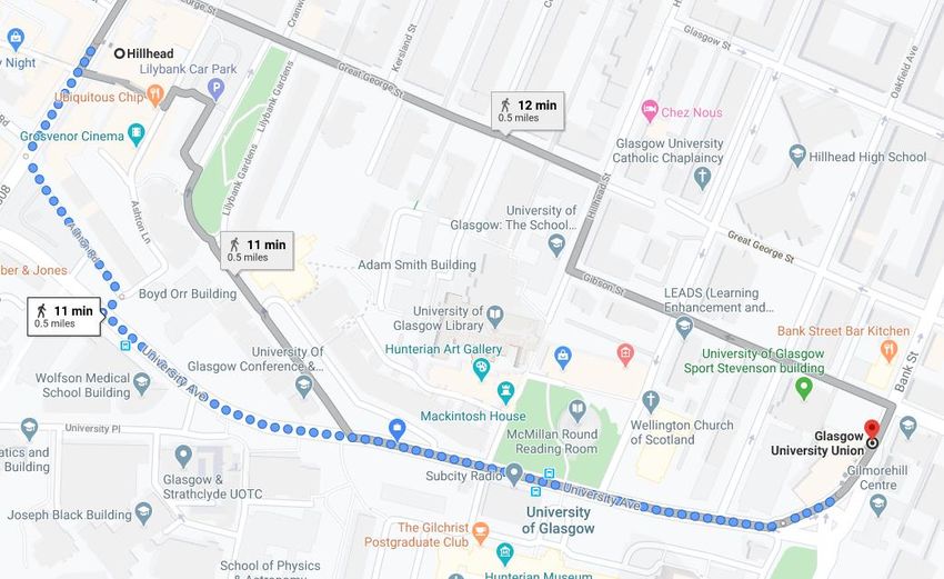
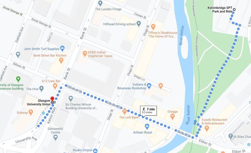
Directions from Hillhead Subway:

Directions from Kelvinbridge Subway:
GLORI 2020
Meeting of the Glasgow Orthopaedic Research Initiative on Wednesday 19th
 February
 Glasgow University Union, 32 University Avenue, G12 8QQ

 PROGRAMME
09:00 Registration/coffee

Session 1 – Chairs: Mark Sprott & Simon Clark

09:30 Welcome from (Dominic/Matt)

09:35 Speaker: Dr Sylvie Coupaud (35+5)
 Talk title: What do astronauts and paraplegics have in common?
 Affiliation: Biomedical Engineering, University of Strathclyde
10:15 Speaker: Mr William Marshall (15+5)
 Talk title: Clinical application of healiOst in dogs and cats (2018-2020): selected
 short stories
 Affiliation: Small Animal Clinical Sciences, University of Glasgow
10:35 Speaker: Ms Laila Damiati (7+3)
 Talk title: Biofunctional and antibacterial titanium nanotopography for
 orthopaedic implants
 Affiliation: Centre for the Cellular Microenvironment, University of Glasgow
10:45 Tea/coffee

Session 2 – Chairs: Virginia Llopis-Hernandez & Matt Walker

11:00 Speaker: Professor Richie Gill (35+5)
 Talk title: In Silico Trials and Machine Learning for Orthopaedics and Trauma
 Affiliation: Department of Mechanical Engineering, University of Bath
11:40 Speaker: Mr Robert Silverwood (15+5)
 Talk title: MicroRNA expression and function in fragility fractures
 Affiliation: NHS, Glasgow
12:00 Speaker: Mr Ian Kennedy (15+5)
 Title: Nanoscale vibration to modulate osteoclastogenesis
 Affiliation: Department of Trauma and Orthopaedics, Queen Elizabeth University
 Hospital
12:20 Speaker: Ms Caroline Busch (7+3)
 Talk title: 3D modelling of the bone marrow niche as a potential platform for
 testing new therapeutic approaches in Chronic Myeloid Leukaemia
 Affiliation: Centre for the Cellular Microenvironment, University of Glasgow
12:30 LI-COR presentation, Shula Dawson

12:35 Biocomposites presentation, Bethany Loughlin
12:40 PromoCell presentation, Andrew Woods
12:45 Posters and lunch

Session 3 – Robert Silverwood & Monica Tsimbouri
13:45 Speaker: Dr Donal Wall (35+5)
 Talk title: Microbiome-derived carnitine mimics as novel mediators of gut-brain
 axis communication
 Affiliation: Institute of Infection, Immunity and Inflammation, University of
 Glasgow
14:25 Speaker: Mr David Shields (15+5)
 Title: Bone Defects: Opportunities and Obstacles
 Affiliation: Orthopaedic Registrar, NHS Greater Glasgow and Clyde
14:45 Speaker: Dr Mark Williams (15+5)
 Talk title: Establishing a unique 3D co-culture system to model and
 therapeutically target leukaemia-bone marrow microenvironment interactions
 in Acute Myeloid Leukaemia
 Affiliation: Glasgow Caledonian University
15:05 Speaker: Dr Jennifer Z Paxton (15+5)
 Talk title: Making Connections: Using anatomy to guide tissue-engineered
 design
 Affiliation: The University of Edinburgh
15:25 Speaker: Ms Gillian Higgins (15+5)
 Talk title: Superparamagnetic Iron oxide Nanoparticle labelling of
 therapeutic cells
 Affiliation: Canniesburn Plastic and Reconstructive Surgery Department,
 Glasgow Royal Infirmary
15:45 Speaker: Dr Hannah Donnelly (15+5)
 Talk title: Mechanistic and metabolic insights into bioengineering the bone
 marrow niche in vitro
 Affiliation: Centre for the Cellular Microenvironment, University of Glasgow
16:05 Tea/coffee
 Posters and prizes
16:30 Meeting ends
Oral presentations

 WHAT DO ASTRONAUTS & PARAPLEGICS HAVE IN COMMON?

 Sylvie Coupaud 1,2 & Mariel Purcell 2

 1 Department of Biomedical Engineering, Wolfson Centre, Strathclyde University, UK
 2 Scottish Centre for Innovation in Spinal Cord Injury, Queen Elizabeth National Spinal
 Injuries Unit, Glasgow

Why should anyone worry about their bone health if they have been living in space for less
than a year, especially if they are young and fit? The same question can be directed at
paraplegics who have suffered a complete spinal cord injury for this relatively short period
in the skeleton’s lifetime. And yet, there are astronauts and paraplegics for whom 11
months of disuse from microgravity exposure or muscle paralysis, respectively, are sufficient
to lead to significant decreases in bone mineral density (BMD) in the lower limb long bones.
Although there is considerable inter-subject variability in patterns of bone loss, those
individuals with the most rapid and extensive decreases in BMD are of particular concern, as
low BMD is associated with an increased risk of fragility fractures in the lower-limb long
bones. We present detailed longitudinal imaging data quantifying the extent of that bone
loss in the femur and tibia in patients with motor-complete spinal cord injury, and compare
these with published data from astronauts who have returned from long-term space
missions. To complement imaging outcome measures, data from biomarkers of bone
formation and resorption in serum/plasma are available from studies in: i) patients with
spinal cord injuries, ii) subjects on bedrest studies, and iii) astronauts returning from long-
term space missions. The rapid decline in BMD in astronauts and paraplegics – compared to
the slower time-course seen in post-menopausal osteoporosis – is not easily mitigated.
Those at risk of rapid and significant bone loss may be offered early treatment interventions
(physical or pharmacological). However, exercise/rehabilitation interventions have shown
limited efficacy in attenuating bone loss in the early stages of disuse osteoporosis, let alone
reversing it once it has already occurred. The effects of available interventions and their
limitations will be discussed, as well as avenues for current and further research.

CLINICAL APPLICATION OF HEALIOST IN DOGS AND CATS (2018-2020):
 SELECTED SHORT STORIES

 William Marshall 1, Cristina Gonzalez-Garcia2, Sara Trujillo Munoz2, Peter Childs2, David
 Shields2,3, Elena Addison1, Sandra Corr1, Matt Dalby 2, Manuel Salmeron-Sanchez 2

 1 Small Animal Hospital, School of Veterinary Medicine, University of Glasgow, UK
 2 Centre for the Cellular Microenvironment, University of Glasgow, UK
 3 Queen Elizabeth University Hospital, Glasgow, UK

HealiOst is a novel system that delivers bone morphogenetic protein-2 (BMP-2) to sites of
problematic bone healing. It binds BMP-2 to material surfaces by exploiting a unique
interaction between the polymer poly (ethyl acrylate) and fibronectin. The system allows
efficient delivery of an ultra-low dose of BMP-2, potentially improving safety compared with
other systems currently in clinical use. In 2017, the first ever clinical application saw HealiOst
successfully unite an infected and non-healing humeral fracture in a dog (Cheng et al, 2019).
Subsequently we have treated a further seven dogs and two cats: three fracture non-unions,
two delayed unions and one arthrodesis that failed to fuse, implant failure in a femoral
fracture, a distal radial peri-implant fracture, persistent intertarsal subluxation following
talocrural fusion and a radial valgus deformity caused by fracture malunion. Outcomes were
initially assessed using standard clinical examination and radiography. Six of the nine cases
have achieved bony union. There have been no instances of excessive bone formation, or any
other significant complications caused by HealiOst. We have seen good or excellent limb
function in five of the nine cases at short-term follow-up, and satisfactory function in one.
One case was euthanased due to an unrelated co-morbidity, and the fracture site containing
HealiOst was then examined histologically. At the time of writing (January 2020), follow-up
of two cases is ongoing. We are also now seeking objective, long term follow-up and blinded
scoring of the radiographs using a modified Radiographic Union Score for Tibial fractures
(RUST) system.
References
Cheng ZA, Alba-Perez A, Gonzalez-Garcia C, et al. Nanoscale coatings for ultralow dose BMP-
2-driven regeneration of critical-sized bone defects. Adv Sci 2019, 6, 1800361
https://doi.org/10.1002/advs.201970009
Funding gratefully received from
European Research Council Proof of Concept Grant
The Vet Fund, University of Glasgow
Find a Better Way
BIOFUNCTIONAL AND ANTIBACTERIAL TITANIUM NANOTOPOGRAPHY FOR
 ORTHOPAEDIC IMPLANTS
 Laila Damiati1, Virginia Llopis-Hernández1, Pefing Li2, Richard Oreffo3,
 Gordon Ramage4, Penelope M. Tsimbouri1, Bo Su5;
 Manuel Salmeron-Sanchez1, Matthew J. Dalby1

 1Centre for the Cellular Microenvironment, University of Glasgow, Glasgow, UK;
 2Schoolof Engineering, University of Glasgow, UK; 3Bone and Joint Research Group,
 University of Southampton, UK; 4Institute of Infection Immunity and Inflammation,
 University of Glasgow, UK. 5Biomaterials Engineering Group, University of Bristol, UK.
 L.damiati.1@research.gla.ac.uk

Orthopaedic device-related infection (ODRI) is one of the major complications in orthopaedic
surgery. Infections caused by Gram-negative bacilli, including Pseudomonas aeruginosa
account for approximately 6%– 17% of ODRI. Furthermore, as the antibiotic treatment starts
to lose effectiveness new approaches become required. Such approaches can include
bioactive coating and implants with nanotopography. Here, we applied these two approaches
on titanium (Ti) substrates to aim to reduce bacterial attachment while enhancing
osteogenesis. Ti nanostructures were produced using an alkaline hydrothermal method to
produce antibacterial high aspect ratio features followed by coating with poly-ethyl acrylate
(PEA) using plasma polymerisation to create a thin layer of polymer. PEA causes spontaneous
revealing of fibronectin (FN) upon contact which helps to expose the cell (e.g. osteoblast)
adhesion and growth factor binding domains that allow the bone morphogenic protein-2
(BMP2) to adsorb on the surface; this coating is to promote osteogenesis. The optimised
surface showed a reduction in the P. aeruginosa attachment and change in bacterial
metabolite pathways. Enhanced MSC osteogenesis with the PEA/FM/BMP2 coating on the
nanotopography was confirmand using different techniques, such as Raman spectroscopy,
qPCR, Calcine blue, and Giemsa staining. Furthermore, MSCs cultured on the coated
nanotopography substrate in the presence of P. aeruginosa or with their quorum sensing
molecules (C12-HSL and C4-HSL) were more resistant to infection compared to the MSCs
growth on the flat surface. These aspects suggest that the developed novel nanotopography/
bioactive coating could promote bone regeneration and reduce the biofilm formation.
IN SILICO TRIALS AND MACHINE LEARNING FOR ORTHOPAEDICS AND
 TRAUMA

 Richie Gill 1,2

 1Department of Mechanical Engineering, University of Bath, UK,
 2Centre for Therapeutic Innovation, University of Bath, UK,

 Abstract
This talk will cover the use of two cutting-edge computer methods for advancing
orthopaedic and trauma. The first is the use of in silico trials for establishing safety
equivalence for new devices. An in silico trial allows a new paradigm, cohorts under
investigation can be duplicated and given treatments from both arms of a study, allowing
paired statistics and eliminating bias between arms. Hip fracture diagnosis and classification
is difficult, and there is a relatively low level of agreement in terms of classification amongst
human observers. Machine learning offers a possibly more reliable and repeatable method
for classification.
MICRORNA EXPRESSION AND FUNCTION IN FRAGILITY FRACTURES

 R.K. Silverwood1, R.M.D. Meek2, C.C. Berry1, M.J. Dalby1

1. Centre for the Cellular Microenvironment, Institute of Molecular, Cell and Systems Biology,
 University of Glasgow
2. Department of Trauma & Orthopaedics, Queen Elizabeth University Hospital, NHS Greater
 Glasgow & Clyde

Osteoporosis represents an ever increasing burden to health care services world-wide. Within the UK
alone, over 500 000 fragility fractures secondary to osteoporosis occur each year, resulting in
significant morbidity and mortality. Furthermore, the management of these injuries cost health care
services billions of pounds. A revolution in the diagnosis and treatment of the disease is required to
prevent significant suffering of future generations.
MicroRNAs are known to regulate many key physiological processes, including mesenchymal stromal
cell (MSC) differentiation. They have been demonstrated to be abnormally expressed in many
musculoskeletal conditions, including osteoporosis. Patients who have suffered a neck of femur
fracture are known to be at risk of, or have undiagnosed, osteoporosis. Dysregulated microRNAs have
been identified in bone and serum samples of patients with this injury type. Great hope has been
placed on the development of targeted therapies to manipulate microRNA expression and improve
bone quality.
MicroRNA expression was analysed, and key microRNAs, microRNA-143 and microRNA-31, were
manipulated using functionalised gold nanoparticles to further understand their role in MSC
differentiation. Furthermore, the metabolomic effect of inhibiting microRNA-31 was explored to
further understand its role in osteogenesis.
These results provide further insight to the impaired MSC function of patients who have suffered a
fragility fracture. Furthermore, the potential of microRNAs to be utilised as a biomarker or therapeutic
target for osteoporosis has been reinforced.
NANOSCALE VIBRATION TO MODULATE OSTEOCLASTOGENESIS

Ian Kennedy 1, P. Monica Tsimbouri 1, Dominic R.M. Meek 2, Carl S. Goodyear 3, Matthew J. Dalby 1

1
 Centre for the Cellular Microenvironment, Institute of Molecular, Cell and Systems Biology, College
of Medical, Veterinary and Life Sciences, Joseph Black Building, University of Glasgow, Glasgow, G12
8QQ. 2 Department of Trauma and Orthopaedics, Queen Elizabeth University Hospital, Glasgow, G54
 4TF. 3 Institute of Infection, Immunity and Inflammation, Glasgow Biomedical Research Centre,
 University Place, Glasgow, G12 8TA.

Introduction: Mechanical factors have been shown to significantly influence stem cell
differentiation and fate. Researchers have demonstrated that nanoscale vibration can
promote osteogenesis in isolated mesenchymal stem cell (MSC) cultures. In the bone marrow
niche, there is a co-dependent existence between MSCs and cells from the haematopoietic
lineage (HSCs), particularly osteoclasts. While MSC derived osteoblasts stimulate new bone
formation, osteoclasts break down bone. Given the close overlap between these two cells
types, an investigation in to the effect of nanoscale vibration on osteoclasts was required.
Methods: Two culture methods were used: an isolated culture of osteoclasts and osteoclast-
precursors, and a co-culture of bone marrow derived MSCs and HSCs. Vibration was produced
with the nanokick bioreactor – a recently developed technology that facilitates the delivery
of accurate and reproducible nanoscale vertical displacements. This bioreactor allows
otherwise standard cell culture techniques to be used. A range of experiments was used to
investigate the effect of nanoscale vibration, including immunostaining, resorption analysis,
RT-PCR, ELISA and metabolomics.
Results: Nanoscale vibration was found to influence osteoclast differentiation and function.
A reduction in osteoclast numbers was observed in both culture conditions. Furthermore, less
resorption occurred in the nanokick group. There was no significant impairment in osteoblast
development or function when osteoclasts were present, with evidence of increased
cytoskeleton tension and mineralisation following stimulation. A number of gene, protein and
metabolome changes were observed, suggesting a state of lower inflammation in the
nanokick group.
Conclusion: It is hoped that these results will provide further evidence to validate the use of
the nanokick bioreactor as a method of producing tissue-engineered bone graft for clinical
applications.
3D modelling of the bone marrow niche as a potential platform for testing
 new therapeutic approaches in Chronic Myeloid Leukaemia.

 Caroline Busch 1,2, Theresa Mulholland 3, Matthew J. Dalby 1, Michele Zagnoni 3, Helen
 Wheadon 2, Catherine C. Berry 1

1 Centre for the Cellular Microenvironment, Institute of Molecular, Cell and Systems Biology,
 University of Glasgow, Glasgow, UK,
 2 Paul O'Gorman Leukaemia Research Centre, Institute Cancer Sciences, University of
 Glasgow, Glasgow, UK
 3 Department of Electronic and Electrical Engineering, University of Strathclyde, Glasgow,
 UK

Chronic myeloid leukaemia (CML) is characterised by the excessive proliferation of leukaemic
stem cells (LSCs) in the bone marrow (BM). LSCs achieve this via modification of the BM
microenvironment -the niche- to their advantage, whilst impairing normal haematopoiesis.
To date it has proved difficult to both understand how LSCs dominate and alter the niche and
to target LSCs with current therapies. In the healthy BM, mesenchymal stem cells (MSCs) and
haematopoietic stem cells (HSCs) reside together in the niche, where they interact closely,
maintaining their stem cell properties via self-renewal. Most in vitro systems representing the
niche are basic, relying on 2D-models consisting of a stromal monolayer in co-culture with
HSCs. However, such systems overlook many niche factors, including the BM 3D architecture.
3D-culture systems provide a more realistic reflection of the BM microenvironment in vitro
and can better predict in vivo responses of chemotherapy in disease modelling.

We are working on 3D BM niche-models, comprising MSC spheroids embedded in medical-
grade collagen type I, mimicking the BM biological and mechanical microenvironment. We
have extended this model to include LSCs and use our model to study MSC-LSC interactions
within a niche-like environment and LSC-mediated remodelling. Initially, we investigated LSC
invasion using a collagen type I coated Transwell/MSC culture, demonstrating migration into
the collagen network and homing to the MSCs beneath. Subsequently, we introduced the
LSCs to our BM-model and noted migration into the gel scaffold. Our artificial 3D BM model
enables us to study the cell-cell interaction within a niche-like environment, and the
processes the LSCs undertake in order to remodel the bone marrow to their advantage.
In parallel, LSC monoculture experiments have revealed new potential CML treatment
approaches, a combination of tyrosine kinase and bone morphogenetic protein inhibitors. We
observed a synergistic mode of action displaying changes in cell cycle, increase in apoptosis
and fewer cell divisions. Furthermore, we investigated MSC spheroid drug sensitivity, using
microfluidic devices. Microfluidic devices are a powerful tool to study MSC spheroid
formation and LSC-MSC interactions, and can be used as a platform for testing new
therapeutic approaches. We observed no changes in MSC spheroid growth or viability upon
treatment which ensures specific targeting of leukaemic cells with our inhibitors.
Future experiments will aim to assess inhibitor effect on LSCs in the presence of MSC
spheroids, firstly in a microfluidic system and secondly using our collagen model.
MICROBIOME-DERIVED CARNITINE MIMICS AS NOVEL MEDIATORS OF GUT-
 BRAIN AXIS COMMUNICATION

 Heather Hulme1, Lynsey M. Meikle1, Nicole Strittmatter2, Justin J.J. van der Hooft3,
RuAngelie Edrada-Ebel4, Victor H. Villar5, Saverio Tardito5, Richard J. A. Goodwin2, Richard
 Burchmore1, Dónal (Daniel) Wall1

 1Institute of Infection, Immunity and Inflammation, University of Glasgow, UK
 2Pathology, Clinical Pharmacology & Safety Sciences, AstraZeneca, UK
 3Bioinformatics Group, Wageningen University, The Netherlands
 4Natural Products Metabolomics Group, University of Strathclyde, UK
 5Cancer Research UK Beatson Institute, Glasgow

Alterations to the gut microbiome are associated with various human diseases, yet evidence
of causality and identity of microbiome-derived compounds that mediate host-microbe
communication remain elusive. Focusing on the gut-brain axis we identified two novel
bacterial metabolites that are structural analogues of carnitine and are present in both gut
and brain of specific pathogen free mice, while completely absent in germ free mice. We
demonstrate that as well as being structurally similar to carnitine, these compounds co-
localize with carnitine in brain white matter, and inhibit carnitine mediated fatty acid
oxidation in a murine cell culture model of white matter. The Lachnospiraceae producing
these metabolites are a relatively poorly understood family of bacteria, although recently
their isolation in reproducibly high numbers from the human gut microbiome in specific
diseases drew attention to their potential role in certain conditions. Our work is the first
description of direct molecular inter-kingdom exchange between gut prokaryotes and the
mammalian brain, leading to inhibition of brain cell function and we believe that there is
compelling evidence for a more widespread role of these molecules in human health and
disease.
CRITICAL BONE DEFECTS: OPPORTUNITIES AND OBSTACLES

 David Shields1 , Virginia Llopis-Hernandez2, Cristina Gonzalez-Garcia3, Vineetha
 Jayawarna3, Matt Dalby2, Manuel Salmeron-Sanchez3

 1.Department of Trauma and Orthopaedics, Queen Elizabeth University Hospital, Glasgow
 2.Centre for Cell Engineering, Institute of Molecular Cell and Systems Biology,
 University of Glasgow
 3.Division of Biomedical Engineering, University of Glasgow

Introduction. Critical bone defects, are loss of bone which will not spontaneously heal, due
to the amount of bone loss. They occur clinically most commonly as a result of trauma,
infection, tumour resection or non-union. The present strategies available to clinicians are
limited by either unpredictable graft performance or unacceptable functional/symptomatic
consequences for patients. This work outlines the need for an engineered bone inducting
(osteoinductive) graft and demonstrates some of the limitations to its implementation in
clinical practice. The osteoinductive system explored in particular is a polymer-protein
nanocoating (poly ethylacrylate & fibronectin) able of presenting growth factors, in particular
bone morphogenetic protein 2 (BMP-2), in ultra-low doses.
Methods. Resilience of the nanocoating to ethylene oxide (EO) was carried out by comparison
of EO sterilised samples to freshly prepared controls for topography (atomic force
microscopy), functionality (ELISA) and biocompatibility (C2C12 differentiation). The use of
canine bone chips as a carrier scaffold for the nanocoating was evaluated in a murine in vivo
critical defect model. Finally, the prospects of a fully synthetic 3D printed scaffold (poly
caprolactone) was explored for mechanical and biological compatibility using compression
testing in vitro cell culture.
Results. Following EO sterilisation, the topographic appearances of the nanocoating
remained consistent with their non-sterilised counterparts. Functionality remained the same
for the protein coating, however the addition of BMP-2 was not found to resist the 1 week
long sterilisation process. Cells were able to differentiate at a statistically similar rate whether
materials were sterilised or not. The use of bone chips as carrier graft for the nanocoating
was found to subjectively promote mature bone formation histologically, however
quantitative analysis was confounded by the persistent present of mineralised bone chips.
Fully synthetic scaffolds were mechanically inferior to currently used load bearing materials.
The reduction of porosity was associated with improved mechanical properties, however not
to a required load bearing degree. The addition of HA into the PCL matrix did not improve
mechanical properties, however all scaffolds remained capable of inducing bone formation
using mesenchymal stem cell cultures.
Conclusions. The use of this polymer-protein nanocoating to present BMP-2 is
topographically and functionally resilient to sterilisation and demonstrates early translational
promise. The optimal scaffold carrier is yet to be established and further work on industrial
limitations is required.
ESTABLISHING A UNIQUE 3D CO-CULTURE SYSTEM TO MODEL AND
 THERAPEUTICALLY TARGET LEUKAEMIA-BONE MARROW
 MICROENVIRONMENT INTERACTIONS IN ACUTE MYELOID LEUKAEMIA.
 Aikaterini Miari1, Helen Wheadon2, Monica Guzman3, Mark Williams1

 1Department of Biological and Biomedical Sciences, School of Health and Life Sciences,
 Glasgow Caledonian University, Glasgow, UK.
 2Institute of Cancer Sciences, Paul O’Gorman Leukaemia Research Centre, University of
 Glasgow, UK.
 3Division of Hematology and Medical Oncology, Weill Cornell Medicine, Cornell University,
 New York, USA.

Background: Clinical outcomes for the majority of Acute Myeloid Leukaemia (AML) patients
remain sub-optimal. Chemoresistance mediated by cross-talk between the leukeamic cells
and the bone marrow microenvironment (BMME), is a key factor responsible for inferior
survival in AML [1]. An enhanced understanding of leukemic cell–BMME interactions will drive
novel drug development and/or instruct drug repositioning to enhance AML patient survival.
Rationale: Approaches for modelling and therapeutically targeting leukaemia–BMME cross-
talk (i.e. 2D co-culture models) fail to recapitulate key features of the BMME, including:
hypoxia; leukaemic cell clustering and the complex interplay between AML cells and BM-
resident cells, including fibroblasts and macrophages. This has led to 2D culture systems
significantly overestimating drug efficacy compared to 3D model systems. As a result, agents
identified from studies using existing 2D in vitro model systems, have yielded suboptimal
clinical outcomes in the treatment of resistant/relapsed AML [2]. Aim: To establish a unique
in vitro 3D (spheroid) co-culture system that originally reflects internal conditions within the
BMME. Methods: U937 and THP-1 (CD45+) AML cell lines were cultured alone or co-cultured
with a human fibroblast cell line expressing green fluorescent protein (HS5-GFP[CD45-]) or
primary human monocyte derived macrophages in agarose-coated or ultra-low adherent
(ULA) 96 well round bottom plates. Spheroid formation, leukaemic cell clustering and
establishment of hypoxic conditions were assessed via the EVOS Cell Imaging System together
with the Image-iT Green Hypoxia Reagent. Leukaemic cell separation from GFP+ stromal
counterparts were undertaken using CD45 magnetic activated cell sorting (MACS), with AML
cell purity determined by the MACSQuant Analyzer 10 flow cytometry, based on differential
GFP expression. Results: HS5-GFP+ cells formed compact and defined spheroids, with primary
macrophages forming clusters within 24 hours of cultivation. U937 and THP-1 cells formed
clusters in monocultures, and around the HS5-GFP+ cells and primary macrophages in the co-
cultures within 24 hours. Hypoxic conditions were detected within the U937 and THP-1
clusters and in the stromal core of the co-cultures within 24 hours post culture. Leukaemic
cell purities of ~95% were achieved with U937 cells after 24 hours of co-culture with HS5-
GFP+ cells, via CD45 MACS. Discussion/conclusion: This system will now be used to investigate
targetable chemoresistant mechanisms in AML.
[1] Burnett, A.K. Hematology Am Soc Hematol EducProgram 2012, 1–6 (2012).
[2] Lapusan, S. Invest New Drugs 30, 1121–1131 (2012).
MAKING CONNECTIONS: USING ANATOMY TO GUIDE TISSUE-ENGINEERED
 DESIGN
 Jennifer Z Paxton

 Anatomy, Edinburgh Medical School: Biomedical Sciences, University of Edinburgh,
 Edinburgh, EH8 9AG

Tissue engineering is an emerging field of research focussed on the manufacture of body
tissues and organs in the laboratory. It combines the basic principles of biology with
engineering and materials science to produce tissue-like structures with the potential for
implantation. While research within tissue engineering is making significant advances, a major
drawback regarding the implantation of tissue-engineered structures remains the creation of
suitable tissue interfaces. This is extremely relevant within the musculoskeletal field, where
the main function of the tissue is to generate or transmit force. Without suitable mechanical
and biochemically graded transitions between the tissues of muscle, tendon, ligament and
bone, stress and strain concentrations would cause tissue injury and in the case of an
implanted tissue, certain implant failure. For example, clinically, bone to tendon/ligament
healing results in a weak interface, formed mainly of scar tissue, which does not possess the
graded structure present in native tissue. This means that, even if advances in biomaterials
research and tissue engineering reach the point of creating tissue analogues possessing all
the desired native tissue characteristics, adequate fixation and interface generation on
implantation will always remain the biggest challenge for a successful clinical outcome.

This talk will describe the current progress in engineering tissue interfaces in the laboratory
and how we use anatomy to guide the design and development of structurally relevant
models and implants. It will also examine the methods currently under investigation to
achieve the ultimate goal of functional tissue replacement.
NANOPARTICLE LABELLING OF THERAPEUTIC CELLS

 GC Higgins,1,2 SE Thomson,1,2 M Kearns,1,2 E Ross,1 C Berry,1 JF Riddell,1 R Wallace,2 MO
 Riehle,1 AM Hart.1,2

 University of Glasgow,1 Canniesburn Plastic Surgery Unit, Glasgow Royal Infirmary,2
 University of Edinburgh.3

Introduction: Peripheral nerve injury is common and in an effort to improve outcomes
alternatives to nerve grafting are sought. The literature suggests human Adipose Derived
Stem Cells (ADSC) and ADSC differentiated to Schwann cell phenotype (dADSC) may improve
nerve regeneration, however, a lack of in vivo cell tracking and use of animal derivatives in
cell culture pose barriers to clinical translation. This study examines Super Paramagnetic
Iron Oxide Nanoparticle (SPION) labelling of therapeutic cells and compares traditional
animal serum culture medium supplement to human Platelet Lysate (hPL).
Materials and Methods: The ADSC were extracted from human adipose, as per Human
Tissue Act (2004) and Biobank approval (314). Cells were cultured in medium supplemented
with Foetal Bovine Serum (FBS) or hPL and an established differentiation protocol (Kingham
et al. 2007) was implemented. Cells were labelled with fluorescent, 200nm SPIONs at
0.01mg/ml and 0.1mg/ml concentration. ADSC stemness was assessed by flow cytometry,
viability by Live Dead assay, proliferation by growth curves, and phenotype by morphology
and immunohistochemistry analysis.
Results: ADSC retained markers of stemness (CD90, CD73 and CD105) irrespective of
medium supplement and SPION labelling. 98% and 96.4% of ADSC and dADSC exhibited
uptake of nanoparticles, respectively. Cell viability was SPION dose dependent and was
better for cells cultured in FBS compared to hPL supplemented medium (p=
MECHANISTIC AND METABOLIC INSIGHTS INTO BIOENGINEERING THE BONE MARROW NICHE IN VITRO Hannah Donnelly1, Ewan Ross1, Christopher West2, Bruno Peault2, Manuel Salmeron- Sanchez1 & Matthew J Dalby1 1 Centre for the Cellular Microenvironment, University of Glasgow, Scotland, UK. 2MRC Centre for Regenerative Medicine, University of Edinburgh, UK. Stem cells lose their regenerative capacity when they are removed from their regulatory microenvironment, termed the stem cell niche. Perivascular stem cells, or pericytes, are key bone marrow niche cells, they act to support haematopoietic stem cells (HSCs) in the bone marrow (BM). However, upon ex vivo culture self-renewal their capacity is lost. In order to maintain ‘stemness’ in culture, aspects of the BM microenvironment should be recapitulated in vitro. Here, we aim to create a system supporting a niche-like pericyte phenotype, investigate mechanisms important for self- renewal and assess HSC maintenance. Noting that soft gels can support nestin1 expression, a key niche marker, we have shown stiffness related support of other key markers important for a niche-like phenotype, including surface markers and HSC maintenance cytokine expression. Here, poly(ethyl acrylate) (PEA) was used to assemble fibronectin (FN) into physiological-like networks, thus allowing growth factor tethering and presentation in synergy with integrin binding sites2. Then, a low-stiffness gel or hypoxia was used to investigate the metabolic mechanisms required to support these phenotypes in this niche-like microenvironment. Transcriptomic analysis and flow cytometry identified changes in key niche and differentiation related genes supported by the different systems, such as increased expression of nestin when the low-stiffness gel is added. Addition of low-stiffness gels also revealed an increase of sustained activate HIF1 (hypoxia-inducible factor 1) compared to hypoxic conditions and controls. Metabolomic screening highlighted strong agreement in down- regulation of metabolites involved in oxidative phosphorylation with the low-stiffness gel and hypoxic system, pointing to maintenance of a hypoxic-like metabolism suggestive of a quiescent HSC- regulatory phenotype. However, downstream analysis of HIF1a-driven VEGF production and lactate dehydrogenase levels did not increase with gels, suggesting differing mechanisms of action. Upon co- culture with HSCs significant maintenance of long-term repopulating HSCs was observed only when in the presence of low-stiffness gels. We have demonstrated the PEA system can be utilised to probe aspects of the BM niche microenvironment, allowing investigation into fundamental mechanisms important in stem cell regulation. The addition of low-stiffness gels leads to a ‘hypoxic-like’ mechanistic response that could be important in maintaining this HSC-supportive phenotype. This system demonstrated that low- stiffness was required to maintain the most naïve and clinically useful HSC population. Systems such as these can have large implications for ultimately the production of clinically relevant HSCs in the clinic, but also provide a platform on which to investigate important niche mechanisms in both stromal cells and HSCs. 1 Engler AJ, Sen S, Sweeney HL, et al. Cell 2006; 126: 677-89. 2 Llopis-Hernandez V, Cantini M, Gonzalez-Garcia C, et al. Sci Adv 2016; 1-11. This work was supported by grant BB/N018419/1 (BBSRC) & an EPSRC studentship. We thank Carol- Anne Smith for technical support.
Sponsor Presentations

 NEAR INFRARED DETECTION TECHNOLOGY – A ROBUST SOLUTION FOR
 QUANTITATIVE BIOASSAY IMAGING

 Shula Dawson1

1 Biotechnology Scientific Solutions and Support Group, LI-COR Biosciences Ltd, Cambridge,
 UK

Near Infrared (NIR) fluorophores provide exceptional sensitivity and high signal-to-noise
ratios for detection and quantification in a region where light absorption and
autofluorescence of cellular and tissue components is low. Imaging agents, cells, antibodies
and molecules labelled with these fluorophores allow biochemical activity measurements
of in vitro and in vivo assays with high sensitivity and reproducibility. Using high resolution
imaging and scanning technology in the NIR region of the spectrum allows a fuller
understanding of the details of protein expression, ligands, molecular targets, pathways, and
cellular effects across a range of different bioassays including membrane, gel, cell, and tissue
formats, as well as small animal in vivo applications. This allows an integrated approach to
pre-clinical investigations spanning multiple stages from initial characterisation of biological
markers, through functional studies and therapeutic development all the way to clinical
translation.

 Biocomposites presentation
 TREATMENT OF INFECTION WITH STIMULAN
 Bethany Loughlin

 PromoCell presentation
 Andrew Woods
Poster abstracts
 1. ENGINEERED BACTERIAL BIOFILMS TO CONTROL STEM CELL
 DIFFERENTIATION
 Aleixandre Rodrigo-Navarro 1, Jake Hay1, Michaela Petaroudi1, Matt Dalby 2, Manuel
 Salmeron-Sanchez 1,2
 1Divison of Biomedical Engineering, University of Glasgow, UK,
 2Centre for Cell Engineering, Institute of Molecular Cell and Systems Biology,

 University of Glasgow, UK

Introduction. The use of materials for stem cell culture is attractive since it can be easily
upscaled using techniques such as 3D printing or fibrous hydrogel scaffolds. These materials
can be engineered to deliver specific and/or dynamic cues to the stem cells. However, these
systems lack the ability to deliver the precise cues required for stem cell differentiation in a
controlled manner, since stem cells usually reside in a highly complex niche and their fate is
tightly regulated by a plethora of factors.
We present a novel approach using bacteria as a substrate to influence mesenchymal stem
cells in a facile and temporal manner. Lactococcus lactis spontaneously develops biofilms on
a variety of surfaces (e.g polymers, metals) and can be genetically modified to produce a
variety of probiotic and efficacious proteins1,2. Here we show that controlled expression of
fibronectin fragments supports growth and temporal regulation of secreted bone
morphogenetic protein 2 drives osteogenesis in an on-demand manner.
Methods. Lactococus lactis NZ9020 has been genetically modified to express the III7-10
fragment of human fibronectin and human bone morphogenetic protein 2 (BMP-2) in an
inducible fashion. Protein expression was characterised with ELISA and Western blot.
Human MSCs were co-cultured with L. lactis biofilms for up to 28 days and assessed for
osteogenic differentiation using von Kossa for mineralization, osteocalcin
immunofluorescence and ALP expression.
Results and discussion. Lactococcus lactis develops stable biofilms on a variety of substrates.
These biofilms contain viable bacteria and are stable for up to 28 days, supporting hMSC
adhesion, proliferation and differentiation.
Lactococcus lactis expresses biologically active III7-10 fibronectin and BMP-2. The expression
levels of BMP-2 can be adjusted and induce osteogenic differentiation, as assessed with the
techniques described in the previous section. The behaviour of hMSCs cultured on
engineered L. lactis biofilms is equivalent to the use of a fibronectin coated substrate with
the addition of 100 ng/mL of BMP-2, the standard for osteogenic differentiation of hMSCs.
Conclusion
Engineered L. lactis biofilms expressing FN III7-10 and BMP-2 in an inducible manner can be
as a functional, living biointerface between a wide range of substrates and hMSCs to drive its
differentiation to the osteogenic lineage in a predictable and controlled way.
References
1. Rodrigo-Navarro, A., Rico, P. Scientific Reports, 4, 5849 (2014)
2. Hay, J. J., Rodrigo-Navarro, A. Scientific Reports, 6, 21809 (2016)
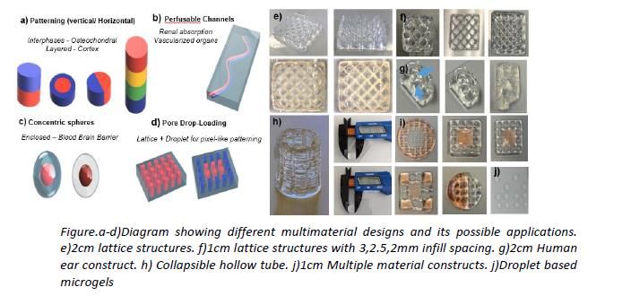
2. DEVELOPMENT OF A MULTIPURPOSE ALG/GEL BASED PLATFORM FOR 3D
 BIOPRINTING AND ITS APPLICATION FOR BONE ENGINEERING

 A. Sanchez-Rubio1, V. Jayawarna1, M. Dalby1, M. Salmeron-Sanchez1

 1Center for the Cellular Microenvironment, University of Glasgow
 2Centre for Cell Engineering, Institute of Molecular Cell and Systems Biology,
 University of Glasgow

Introduction. 3D Bioprinting has arisen as a key enabling technology which by combining
biomaterials, cells and biomolecules is capable of fabricating structures that emulate tissues and
organs present in the human body. This summed up to the fact that it allows the possibility of
tailoring the fabricated constructs, via. patient based geometries or specific cell lading,
represents a paradigm shift in medicine, allowing for extensive personalized medicine
approaches. In this work we present a 3D Bioprinting platform, including a set of designs,
workflows and a family of alginate-gelatine based bioinks that could be used for many different
end applications due to granting control over their physicochemical properties and the
possibility of further tuning with biomolecules tailored for specific tissue types. We also used
the proposed platform for bone engineering with the creation of vascularized bone disks that
could either be used as bone in vitro models or stacked to be used as bone defect fillers
implants.
Methods.The family of Alg/Gel-based bioinks was developed by dissolving the given polymers at
different ratios in various solvents (MiliQ water, PBS and cell culture media). The printed Alg/Gel
structures were exposed to different concentrations of 2 solutions for different times to
crosslink after printing.

HTERT cells previously expanded in regular cell culture conditions, were laden into the hydrogels
and then loaded into printing cartridges. The cartridges were then placed in the printheads and
layered into different pre-designed structures by using a commercially available bioprinter from
RegenHu. Cellular viability was assessed using Live/ Dead assays. Several printed structures
were designed having various end applications in mind. Some of these included multiple
materials, belonging to the Alg/Gel family but also Gelatine-Methylacryloyl (GelMA)
photocrosslinkable bioinks, which allowed for the creation of perfussable channels, by
incorporating sacrificial geometries.
Results. Rheology data showed control over the mechanical properties amongst different
Alg/Gel bioinks, being able to obtain materials with Young’s Modulus ranging from 4 KPa to
more than 40 KPa. Cellular viability inside the printed constructs was also found to be over 90%
after printing and even distributions of the cells throughout the construct was ensured.
The proposed printing platform was used to print different structures spanning from standard
lattices, commonly used for bone engineering to human-size ears that could represent future
implants for cartilaginous body parts, collapsible hollow tubes that could be used for vessel
modelling in vitro and high-throughput droplet-based microgels. Printed constructs were
composed by up to 80 layers, sizes varying from 1 cm to 4 cm depending on the construct.
Additionally, vascularized chips were designed and fabricated, which included multiple
materials, a main GelMA hydrogel enclosing a sacrificed Alg/Gel structure. These gel chips have
an inner channel network enclosed in a solid structure, serving potentially as co-culture models
for in vitro applications, allowing the perfusion of solutions that will better mimick and ensure
appropriate nutrient distribution within the main structure.
Multimaterial approaches were also used to design and print 7 mm disks with differentiated
areas. Congo red was use to visually assess the separation, while NeutrAvidin’s conjugates were
used to fluorescently tag the different areas and estimate protein diffusion within the structure.
Structures with different stiffnesses distributions were also obtained via this method.
Conclusions. The developed platform showed great versatility, firstly due to the tunability of the
bioink’s mechanical properties facilitating the utilization of a variety of cell types, as well as per
the different structures obtained using multiple printing approaches. Potentially, this platform
could be employed for the creation of complex in vitro models used to study biological
processes, systems or diseases, which could also include enhanced biomimicry, e.g.
incorporating inner channels. Specific cell types obtained from patients could be added to these
models to assess drug toxicity or efficacy, highlighting the platform’s potential in the
development of future personalized medicine approaches.
3. SEEKING THE LINK BETWEEN MECHANICAL AND METABOLIC ACTIVITIES IN
 CELL DIFFERENTIATION

Ana San Felix Garcia-Obregon1, Sara Trujillo1, Matthew Dalby1, Manuel Salmeron-Sanchez1
 1Centre for the Cellular Microenvironment (CeMi), University of Glasgow, UK

INTRODUCTION. Cells sense the environment stablishing focal adhesions (FAs) formed by
the attachment of
integrins with proteins from the extracellular matrix (ECM) (e.g. fibronectin (FN)). Every
information gathered by cells from these links will play an important role in their fate [1].
Therefore, it is important to understand cell-ECM interactions and what intracellular
changes occur upon these interactions. We hypothesise that to create an adaptive response
to ECM signals, cells exert forces in response to the surrounding matrix, and this requires a
mechanical energy that will have an implication in the biology of the cell [2]. This work aims
to understand how cells test the environment from a mechanobiological perspective during
their attachment, proliferation, migration and differentiation. This will provide a better
understanding on how microenvironmental cues can be used to control cell fate, enhancing
studies in multiple fields e.g. tissue regeneration or drug testing.
METHODS. The proposed system consists on a 2D Full-length Fibronectin- based PEG
hydrogels model developed in our group [3]. This system allows stiffness control by varying
the amount of PEG and also, control over the degradability of the matrix by adding
protease-sensitive crosslinkers. Murine L929 fibroblasts were used for optimisation of the
system. Forces exerted by cells were analysed using traction force microscopy (TFM). In
order to do this, fluorescent microspheres were embedded in the gels. Stacks of the
position of the beads after and before cells trypsinisation were analysed to calculate
displacements, which yields gel’s stress and hence, the forces exerted. Also, a myosin II
inhibitor (blebbistatin), was added to the media to study the implication of cytoskeletal
tension in force generation.
RESULTS AND DISCUSSION. Cells pull less on non-degradable hydrogels compare with
degradable ones when maintaining same bulk stiffness. As expected, the inhibition of
myosin II resulted in a decrease in the forces exerted by cells in both conditions. However,
forces were still higher in the degradable condition compared to the non-degradable one.
This could mean that the degradability of the system is an important parameter in cell
mechanosensing, allowing cells to apply higher forces. Moreover, cells tend to acquire a
more spread shape when blebbistatin is added, which could be related to the lack of
cytoskeletal tension. In the future, all these results will be compared with the metabolic
activity of the cell to understand the implication of the mechanical forces in the biology of
the cell.
CONCLUSIONS. From these early experiments we can conclude that cells respond
differently with changes in matrix stiffness and degradability. A direct evidence of how cells
respond to these stimuli is through morphological changes, which indicates that further
adaptive response might be taking place inside the cell. Furthermore, these results show the
great importance of the actin cytoskeleton contraction in force loading. Changes in the
biophysical properties of the matrix alter how cells sense their surroundings and might have
an implication in their metabolic activity, determining cell fate. Consequently, cell metabolic
activity will be studied in detail in further experiments.

REFERENCES
[1] A. J. Engler, S. Sen, H. L. Sweeney, and D. E. Discher, “Matrix Elasticity Directs Stem Cell
Lineage Specification,” Cell, vol. 126, no. 4, pp. 677–689, Aug. 2006.
[2] A. Elosegui-Artola et al., “Rigidity sensing and adaptation through regulation of integrin
types,” Nat. Mater., vol. 13, no. 6, pp. 631–637, Jun. 2014.
[3] S. Trujillo et al., “Engineered full-length Fibronectin-based hydrogels sequester and
present growth factors to promote regenerative responses in vitro and in vivo,” bioRxiv.

ACKNOWLEDGMENTS
The authors acknowledge funding from EPSRC through their Scholarship program 2018.
4. AN IN-VITRO ASSESSMENT OF THE EXTENDED KILLING OF BIOFILMS
 BEYOND THE SPACER BY ANTIBIOTIC-LOADED CALCIUM SULFATE BEADS

Craig Delury 1, Devendra H Dusane 2, Casey W Peters 2, Sean Aiken 1, Phillip Laycock 1, Ed
 McPherson 3, Anne Sullivan 2, Jeffrey Granger 2, Paul Stoodley 2

 1Biocomposites Ltd, Keele Science Park, Keele, UK
 2The Ohio State University, Columbus, OH, USA
 3UCLA, California, USA

Introduction: Antibiotic-loaded spacers and absorbable beads are used in the management
of orthopaedic infection to provide prolonged high local concentrations of antibiotics
required to better kill bacterial biofilms. In quiescent areas of the joint space, diffusion will
control the spread of antibiotics; therefore, the distribution of beads may be important to
ensure adequate antibiotic coverage. Do antibiotic-loaded absorbable calcium sulfate beads
(ALCSB*) provide a greater zone of coverage and killing of biofilms than antibiotic-loaded
PMMA spacers alone?
Method: Biofilms of bioluminescent strains of Staphylococcus aureus and Pseudomonas
aeruginosa were formed on stainless steel, hydroxyapatite, titanium and polyethylene
coupons for 3 days. Diffusion experiments were performed in large glass plates as (i) control
PMMA spacer with no antibiotics, (ii) a PMMA spacer with vancomycin and tobramycin (2000
mg of each per 40 g pack) and (iii) an antibiotic loaded PMMA spacer plus ALCSB containing
vancomycin and tobramycin (1000 mg and 240 mg per 10cc pack respectively). The spacer
was placed in the centre of the glass plate and the coupons placed radiating from the spacer.
The ALCSB were spread evenly and the whole plate was overlaid with agar. The plates were
incubated and killing of biofilms was analysed using luminescence and white light imaging.
Results: Growth and spread of in-vitro biofilms from the coupons was observed for the
control unloaded spacer. The antibiotic-loaded spacer showed a localized zone of inhibition
radiating a few mm from the spacer. The addition of ALCSB beads demonstrated a much
greater zone of clearance, killing in-vitro biofilms on the coupons and preventing bacterial
spread from the coupons.
Conclusions: In quiescent areas where the spread of antibiotics is dominated by diffusion, the
distribution from an antibiotic -loaded spacer alone might be limited. The addition of Stimulan
Rapid Cure beads (ALCSB) in addition to a loaded PMMA spacer can increase the area of
coverage. To achieve the antibiotic concentration and exposure times required to kill or
reduce biofilm bacteria, placing other reservoirs such as ALCSB may be necessary to cover as
much exposed joint space as possible. Further work is required to confirm this clinically.
This study was funded by Biocomposites Ltd.
*Stimulan Rapid Cure, Biocomposites Ltd.
5. ERADICATION OF PRE-FORMED BIOFIMS USING AN ANTIBIOTIC-LOADED
 CALCIUM SULFATE: AN IN-VITRO STUDY

 Craig Delury 1, Sean Aiken 1, Leanne Cornes 1, Phillip Laycock 1, Hannah Thomas 2, Liam E
 Purcell 2, Cate Winstanley 2, Samantha J Westgate 2

 1Biocomposites Ltd, Keele Science Park, Keele, UK
 2Perfectus Biomed, Daresbury, UK

Introduction: Formation of biofilms following periprosthetic joint surgery is a severe
complication due to difficulties with diagnosis and eradication. Treatment for periprosthetic
joint infection (PJI) often includes procedures such as surgical intervention and antibiotic
therapy, whereby the most effective antimicrobial treatments are administered both locally
to the infection and systemically. There is also evidence that releasing antibiotics directly at
surgical sites can be used effectively in preventing implant colonisation and eradicating
established biofilms. This investigation assesses the ability of synthetic recrystallised calcium
sulfate (SRCS*) mixed to contain a combination of vancomycin and gentamicin (VG), or
vancomycin and tobramycin (VT) to eradicate pre-formed biofilms in vitro.
Method: Biofilms of Pseudomonas aeruginosa (NCIMB 10434) or Staphylococcus aureus
(NCTC 8325) were established on polycarbonate coupons within a CDC biofilm reactor. Biofilm
coupons were exposed to plates containing suspended SRCS with VG/VT beads at
concentrations of 500mg/240mg per 10cc and 1g/240mg per 10cc respectively. Control
coupons were tested concurrently. Challenge plates were incubated for 24 hours at 37°C ±
2°C. All testing was performed in triplicate. Data was analysed by Students T-Test to
determine statistical significance
Results: An average of 6.78 ± 0.23 Log10CFUmL-1 and of 6.60 ± 0.23 Log10CFUmL-1 of
Pseudomonas aeruginosa and Staphylococcus aureus were recovered from the negative
control biofilms respectively. No viable organisms were recovered from biofilms exposed to
the positive control or those exposed to SRCS beads containing VG or VT, within the
detectable limits. This equated to an average log reduction in P. aeruginosa of >5.78
Log10CFUmL-1 and >5.60 Log10CFUmL-1 in S. aureus (p < 0.001).
Conclusions: Exposure of P.aeruginosa and S.aureus biofilms to SRCS containing a mixture of
vancomycin and gentamicin or vancomycin and tobramycin resulted in eradication of pre-
formed biofilms in the method described. This may have uses in management of biofilm
infections, however further assessments are required to confirm clinical performance.
This study was funded by Biocomposites Ltd.
*Stimulan Rapid Cure, Biocomposites Ltd.
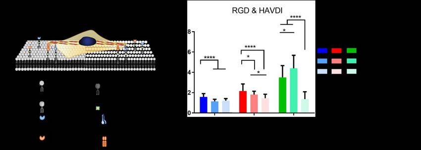
6. ENGINEERING LIGAND MOBILITY IN THE ADHESIVE CROSSTALK TO
 CONTROL STEM CELL DIFFERENTIATION
 Eva Barcelona 1, Marco Cantini 1, Matt Dalby 2, Manuel Salmeron-Sanchez 1

 1Divison of Biomedical Engineering, University of Glasgow, UK,
 2Centre for Cell Engineering, Institute of Molecular Cell and Systems Biology,
 University of Glasgow,

Introduction. Cell behaviour is influenced by biochemical cues mediated by interactions
with the extracellular matrix (ECM) through integrins, and by interactions between cells via
cadherins. On the other hand, the tissues in which the cells live are dynamic being the cells
equally sensitive to the physical properties of the environment, such as stifness or viscosity,
which has been traditionally ignored. In our group, we demonstrated that cells sense this
property using the same mechanotransductive mechanism as with stiffness (1).
In this work, we address the role of ligand mobility (viscosity) in cell fate, and how it affects
the adhesive crosstalk between integrins (RGD receptors) and cadherins (HAVDI-containing
proteins) to elucidate stem cell mechanosensing of viscosity. To do this, we use supported
lipid bilayers (SLBs) with varying mobility, functionalised with RGD and HAVDI peptides
(Figure 1a).
Materials and Methods. Supported lipid bilayers with varying viscosities were prepared by
following the vesicle fusion method. To determine how this property affects human
mesenchymal stem cells (hMSCs) behaviour, two kind of bilayers were used: one of them
made of DOPC, which present a fluid phase at cell culture conditions, and the other one
made of DPPC, which is a gel phase bilayer. Also, glass was used as a non-mobile control
surface. All the surfaces were functionalised with different ratios of RGD and HAVDI.
hMSCs are then seeded on these surfaces and parameters such as cell adhesion, protein
translocation or expression of transcription factors are investigated via AFM
immunostraining and in-cell western.
Results and Discussion. An increase in cell area and cell adhesion, in particular its strength,
is observed when the viscosity of the surface and the concentration of RGD increase. On the
other hand, when HAVDI is included in the bilayers, cell spreading is reduced and changes in
the location of mechanosensitive proteins (i.e. YAP) are observed, showing an altered
sensing of viscosity (Figure 1b).
Figure 1: A) Scheme of the cells seeded on SLBs. B) YAP/TAZ ratio of h MSCs cultured on surfaces with different viscosities and
amounts of HAVDI. C and D) ICW showing the expression of early differentiation markers (SOX9 for chondrogenesis and PPARϒ
for adipogenesis) of hMSCs seeded on surfaces with different viscosities and ligands.

These changes in viscosity and ligand concentration provoke not only differences in
adhesion and mechanosensing, but also in the expression of nuclear lamina and early
differentiation markers (Figure 1c and d), indicating the influence of viscosity in cell fate.
Conclusions. These findings reveal the influence of viscosity in the adhesive crosstalk
between integrins and cadherins as well as how this mobility affects stem cell
differentiation. Our results show that when cell-cell interactions are involved, hMSCs have a
different perception of the mechanical properties of their surroundings compared to when
there are only ECM-cell interactions. All these changes in the sensing of the environment
(viscosity, in this case) provoke changes in cell behaviour and cell fate. Further investigations
on these changes will allow to finally establish a paradigm to understand and exploit cell
response to viscous interactions.
References
1. Bennett M. et al. P. Natl. Acad. Sci. USA. 115(6), 1192-1197, 2018
Acknowledgements
The authors acknowledge funding from EPSRC (EP/P001114/1) and MRC (MR/S005412/1).
This work was also funded by a grant from the UK Regenerative Medicine Platform.
You can also read