UNIVERSITY OF LEEDS 27th Northern Cardiovascular Research Group Meeting - Tuesday 30th April 2019
←
→
Page content transcription
If your browser does not render page correctly, please read the page content below
27th Northern Cardiovascular Research Group Meeting
                UNIVERSITY OF LEEDS

            Committee Organiser: Dr. Hannah M. Kirton

                 Tuesday 30th April 2019
              The Great Hall and Parkinson Court

                      University of Leeds

                   Woodhouse Lane, LS2 9JT

                                1
27th Annual Northern Cardiovascular Research Group Meeting

                               Tuesday 30th April 2019

  Welcome to the 27th Annual Northern Cardiovascular Research Group Meeting, which is
   being held at the University of Leeds. We hope you find the day both beneficial and
                                       stimulating.

The Northern Cardiovascular Research Group meeting provides a stimulating arena for high
calibre scientific content, informative and enjoyable discussions, collaborative opportunities
 and a relaxed atmosphere within a cardiovascular research community. This annual event
 has brought together those with an interest in the cardiovascular system since 1991 at the
University of Leeds, but has moved a long way from its initial roots in the North of England,
          and now includes researchers from Bristol, Northern Ireland and Scotland.

  The NCRG has also proudly recognised and supported students, early career researchers,
 and more senior researchers, to present their work to a friendly environment, providing a
stimulating arena for discussion; and welcomed a New&Notable platform at the Leeds 2016
                                          meeting.

This year we also highlight the cardiovascular research of Professor Clive Orchard (formally a
   University of Leeds academic member and founding organiser of this conference) with
                          research recognition throughout the day.

                                   Dr Hannah M. Kirton

                   Cardiovascular and Ion channel Postdoctorate Researcher

                                              2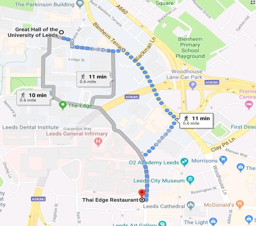
Sponsors

We are very grateful and honoured to receive generous support and funding from the
British Heart Foundation, University of Leeds, University of Bristol and Badrilla to allow
us to hold this honorary 2019 research meeting. Many thanks also to MeetInLeeds who
have helped organise this meeting.

We are proud to receive support from the following:

Badrilla Mini Symposium Sponsor
We are grateful for the generous support provided by Badrilla, for today’s plenary and
Guest speakers.

Cairn Research New and Notable Sponsor
We are grateful for the continued and generous sponsorship from Cairn Research for the
New and Notable platform sessions.

Exhibitor Sponsors
We are grateful for the generous and continued sponsorship from: ADInstruments,
GeneFlow, Sarstedt, StarLab and WPI.

Gifts and Prizes
Thanks to the generous support of our local companies we are able to honour prizes and
gifts at this event. Thanks to Everyman and Vue cinema, Tesco and Café Nero.

We highly encourage you to take some time to visit the exhibitors during your visit at
                                     Leeds.

                                           3
The NCRG 2019 congress is kindly sponsored by

                      4
5
Meeting Programme 2019

                        Parkinson Building and The Great Hall

09.30 - 10.20   Registration and Congress Meal deposit at The Parkinson Court front entrance
                Exhibitors/Posters
                Fresh Tea/Coffee with Mini Continental Breakfast Buffet

10.20 - 10.30   Welcome Dr. Hannah Kirton (Leeds Organising Committee)

                       Session 1 The Great Hall: 10.30 – 12:00
Chairs: Dr. Matt Hardy (University of Bradford) and Ms Natasha Hadgraft (University of Salford)

10:30 – 11:00   Cairn Research Sponsored New & Notable: Dr Izzy Jayasinghe (University of
                Leeds. Presented by Martin Thomas, Author at Cairn Research

                The new frontiers of optical microscopy tools for studying molecular-scale cardiac
                structure and function

11:00 – 11:15   Oral Communication: Prof. Kenichi Hongo (Jikei University School of Medicine)
                Thrombin can be a novel target of the treatment of dilated cardiomyopathy

11:15 – 11:30   Oral Communication: Dr. Parveen Sharma (University of Liverpool)
                Attenuation of doxorubicin-induced cardiotoxicity in a human in vitro cardiac model by
                the induction of the NRF-2 pathway

11:30 – 11:45   Oral Communication: Dr. Olivia Robertson-Gray (University of Glasgow)
                Small molecules activating Nrf2 as a Therapeutic Approach to Prevent Cardiac
                Ischaemia/Reperfusion Injury

11:45 – 12:00   Oral Communication: Dr. Riaz Akhtar (University of Liverpool)
                Unique patterns of elastin degradation in ascending aortic aneurysms in bicuspid aortic
                valve patients

12:00 – 13:00   Parkinson Building: Buffet lunch with time for informal posters and exhibitors

                                                  6
Session 2 The Great Hall: 13:00 – 16:00
Prof. Clive Orchard Mini Symposium
Introduction to Professor Clive Orchard
Chair: Prof. John Colyer (University of Leeds, CEO and Founder of Badrilla)
Academic Career and Research Areas
Clive Orchard BSc, PhD, DSc, is an eminent physiologist with a career-long interest in cardiac physiology. He trained at the University
of London (BSc, PhD), performed post-doctoral research at University College London and the National Institutes of Health, Baltimore,
and enjoyed a successful academic career at the Universities of Leeds (1986-2005) and Bristol (2005-2018). His research explored the
physiology and pathophysiology of cardiac muscle contraction: calcium-signalling, acidosis, metabolic inhibition, novel therapeutics,
and the role of t-tubules in excitation-contraction coupling. He created methodologies that enabled new discoveries, most notably
in the area of t-tubular function.
Clive is an excellent strategist and collaborator. He is generous, collaborative, and good counsel. One of his particular strengths is the
development of academic strategy for projects, groups and departments: strategies that are based fairly in the available evidence,
the input from those concerned and his vision for a worthy goal. With these strengths, Clive has led the School of Biomedical Science
(University of Leeds; 1998-2001), the Department of Physiology (University of Bristol; 2005-8), the Faculty of Medical and Veterinary
Sciences (University of Bristol; Dean 2009-13) and the Physiological Society (President 2008-10).
Today we mark Clive’s contribution to science. Beyond H-factors, money raised and papers published, Clive’s contribution has been
the advancement of knowledge and the development of researchers. A number of the fellows he has trained have returned to Leeds
today to celebrate with Clive: some continue their interest in cardiac biology, others have branched out into new areas, but all have
built upon the foundation created in Clive’s lab.
Badrilla sponsors this event with delight: the company was founded on antibodies created, and first used in cardiac myocytes in
collaboration with Clive in the mid-1990s. We look forward to welcoming you all to Leeds, and to celebrating together the
contributions Clive Orchard has made to physiological science.
Recent Publications
Bryant, S.M., Kong, C.H.T., Cannell, M.B., Orchard, C.H. and James, A.F. Loss of caveolin-3-dependent regulation of ICa in rat
ventricular myocytes in heart failure. American Journal of Physiology 314, H521-H529. 2018.
Bryant, S.M., Kong, C.H.T., Watson, J.J., Gadeberg, H.C., James, A.F., Cannell, M.B. and Orchard, C.H. Caveolin 3-dependent loss
of t-tubular ICa during hypertrophy and heart failure in mice. Experimental Physiology 103, 652-665. 2018.
Kong, C.H.T., Rog-Zielinska, E.A., Kohl, P., Orchard, C.H. and Cannell, M.B. Solute movement in the t-tubule system of rabbit and
mouse cardiomyocytes. PNAS 115, E7073-E7080. 2018.
Bryant, S.M., Kong, C.H., Watson, J.J., Gadeberg, H.C., Roth, D.M., Patel, H.H., Cannell, M.B., James, A.F. and Orchard, C.H.
Caveolin-3 KO disrupts t-tubule structure and decreases t-tubular Ica density in mouse ventricular myocytes. American Journal
of Physiology 315, H1101–H1111. 2018.
Kong, C.H., Bryant, S.M., Watson, J.J., Roth, D.M., Patel, H.H., Cannell, M.B., James, A.F. and Orchard, C.H. Cardiac-specific
overexpression of caveolin-3 preserves t-tubular ICa during heart failure in mice. Experimental Physiology (ePub ahead of print).
2019
7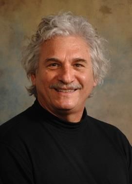
13:10 – 14:10
                             Badrilla Sponsored Plenary Communication

 The University of Leeds and Badrilla welcome our plenary speaker Dr. Edward G. Lakatta, from
     the National Institutes of Health and National Institute on Aging; to the 27th Northern
                            Cardiovascular Research Group meeting.

                                        Dr. Edward G. Lakatta, M.D., Chief

                        “The Smart Heart Operates on the Edge of Criticality”
Each heart beat is initiated by spontaneous rhythmic action potentials (APs) that emanate from pacemaker cells within
the sinoatrial node (SAN). Spontaneous APs in single, isolated cardiac SAN pacemaker cells are driven by a coupled-
clock system of chemical and current oscillators: spatio-temporal self-organization of local Ca2+ releases (LCR)
generates an ensemble Ca2+ signal during diastolic depolarization that ignites electrogenic surface membrane
molecules, resulting in rhythmic oscillatory electrochemical gradient oscillations that underlie rhythmic AP cycles that
determine the heart rate (HR), which varies widely across species. The coupling kinetics of the interaction of
pacemaker clocks within single, isolated SAN pacemaker cells are self-similar across species from mouse, guinea-pig,
rabbit to human. This pan-species self-similarity of clock-coupling kinetics in isolated SAN cells extends not only to EKG
derived heartbeat intervals in vivo, but also to body mass. Thus, self-similar kinetic functions that drive rhythmic AP
firing in individual pacemaker cells, to the HR in vivo, and to BM in species from mouse to human reveal novel universal
scales that link microscopic subcellular mechanisms to macroscopic structural properties among diverse organisms.

                                                            8
Prof. Clive Orchard Mini Symposium
                                               PART II

14:10 – 14:30   Honorary Communication by Prof. David Eisner (University of Manchester)
                Seeing the Orchard for the trees

14:30 – 15:00   Parkinson Building - Coffee break with informal poster session and exhibitors

15:00 – 15:20   Honorary Communication by Prof. Mark Boyett (University of Manchester)
                Debunking myths concerning the vagus: from 1987 to now

15:20 – 15:40   Honorary Communication by Dr. Fabien Brette (INSERM: University of
                Bordeaux)
                A Calcium Odyssey

15:40 – 16:00   Honorary Communication by Dr. Andy James (University of Bristol)
                Of mice and men: insights into the role of caveolin-3 in cardiac muscle in health and
                disease from genetically manipulated models.

                                              Session 3
                             Parkinson Building: 16:00 – 17:00
                          Formal Poster Session and exhibitions
                        (Bespoke wine and Black Sheep beer reception)

                                                   9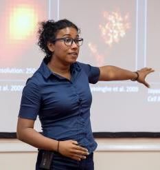
Session 4 The Great Hall: 17:00 – 18:00
Chairs: Dr. Sandra Jones (University of Hull) and Dr. Charlotte Smith (University of Manchester)

17:00 – 17:30   Cairn Sponsored New & Notable: Dr. Tom Claydon (SFU, Canada)
                Presented by Martin Thomas, Author at Cairn Research

                Targeting hERG channel gating steps to provide cardio-protective therapies for LQTS

17:30 – 17:45   Oral Communication: Prof. Andrew Trafford (University of Manchester)
                The role of protein S-nitrosylation in regulating mitochondrial function

17:45 – 18:00   Oral Communication: Ms Florah Tshepo Moshapa (University of Bradford)
                Stabilising SOCS3 to inhibit smooth muscle cell dysfunction responsible for neointimal
                hyperplasia

18:00 – 18:15   Closing remarks, prizes and announcements

18.30 – 20:30   Conference Three Course Buffet Meal at Thai Edge
                (with complimentary Jasmine tea or Filtered Coffee on departure)
                Directions: New Portland Place, 7 Calverley Street, LS1 3DB

                                                  10
Abstracts (Honorary Communications)
HC1: Seeing the Orchard for the trees
Professor David Eisner, University of Manchester
Once upon a time, I arrived at University College London intending to study sodium regulation in the heart. As luck would
have it, however, I found myself in the laboratory next door to where Clive Orchard was working and, together with David
Allen, we studied the regulation of Ca2+ ions. One of our projects focused on the control of resting cytoplasmic Ca2+
concentration ([Ca2+]i ). In particular, we investigated the effects of membrane potential, metabolism and extracellular
Ca2+ concentration.
In this talk, I will begin with a self-indulgent review of some of my work with Clive and then fast forward 35 years to explain
why I am still fascinated by the control of resting and diastolic [Ca2+]i . As far as I am concerned, the control of diastolic
[Ca2+]i and its implications for heart failure is one of the major unanswered questions in the field.
Finally, we all owe Clive an enormous debt. Today’s attendees might reflect on his role in setting up the NCRG. More
broadly, we are indebted to him, not only for his contributions to science but his leadership roles, both at Leeds and Bristol
Universities and, nationally, as President of The Physiological Society.
HC2: Debunking myths concerning the vagus: from 1988 to now
Professor Mark Boyett, Division of Cardiovascular Sciences, University of Manchester
In 1986, Clive Orchard was recruited to the Department of Physiology at the University of Leeds. I was a young lecturer at
the time, and Clive and I became close colleagues and friends. One of my best memories was in 1995 when Clive and I were
awarded Personal Chairs on the same day and we celebrated with pints of beer at the Faversham pub! Clive had been working
in Baltimore with Ed Lakatta and he had shown that the vagal nerve transmitter, acetylcholine (ACh), affected myofibrillar
Ca2+ sensitivity and twitch relaxation in ferret ventricular muscle1 and yet the dogma at the time was that the vagus only
acted on supraventricular tissues in the heart. At the University of Leeds, using the jelly fish protein aequorin to measure the
Ca2+ transient, we investigated the underlying mechanisms and showed it to be the result of the ACh-activated current and
shortening of the action potential. We published the work in the Journal of Physiology,2 the journal we all aspired to publish
in (not Nature), and I was the first author, not because I did all the work, but because at this time authors were always in
alphabetical order! In my career, I have continued to question the role of the vagus. We have shown that ACh affects the
ventricles of various species.e.g.3 Heart rate variability is widely used to measure cardiac vagal activity (there are >20,000
papers concerning heart rate variability), but we have shown that heart rate variability is primarily determined by heart rate
and cannot be used in this way.4 The failure of heart rate variability means that we have to reinvestigate phenomena
attributed to the vagus: athletes have a low resting heart rate (~30 beats/min in elite cyclists) and this is always attributed
to high vagal tone based on heart rate variability, but we reinvestigated it and found it to be dependent on a downregulation
of the pacemaker channel, HCN4, in the pacemaker of the heart, the sinus node. 5,6 Based on heart rate variability, the
circadian rhythm in heart rate and the low heart rate at night is attributed to high vagal tone at night, but we have shown
that instead it may involve a circadian rhythm in HCN4 expression.7
References
1. McIvor ME, Orchard CH and Lakatta EG. Dissociation of changes in apparent myofibrillar Ca2+ sensitivity and twitch relaxation
induced by adrenergic and cholinergic stimulation in isolated ferret cardiac muscle. Journal of General Physiology. 1988;92:509-
529.
2. Boyett MR, Kirby MS, Orchard CH and Roberts A. The negative inotropic effect of acetylcholine on ferret ventricular myocardium.
Journal of Physiology. 1988;404:613-635.
3. Yang ZK, Boyett MR, Janvier NC, McMorn SO, Shui Z and Karim F. Regional differences in the negative inotropic effect of
acetylcholine within the canine ventricle. Journal of Physiology. 1996;492:789-806.
4. Monfredi O, Lyashkov AE, Johnsen AB, Inada S, Schneider H, Wang R, Nirmalan M, Wisloff U, Maltsev VA, Lakatta EG, Zhang H
and Boyett MR. Biophysical characterisation of the under-appreciated and important relationship between heart rate variability
and heart rate. Hypertension. 2014;64:1334-1343.
5. D'Souza A, Bucchi A, Johnsen AB, Logantha SJ, Monfredi O, Yanni J, Prehar S, Hart G, Cartwright E, Wisloff U, Dobryznski H,
DiFrancesco D, Morris GM and Boyett MR. Exercise training reduces resting heart rate via downregulation of the funny channel
HCN4. Nature Communications. 2014;5:3775.
6. D'Souza A, Pearman CM, Wang Y, Nakao S, Logantha S, Cox C, Bennett H, Zhang Y, Johnsen AB, Linscheid N, Poulsen PC, Elliott J,
Coulson J, McPhee J, Robertson A, da Costa Martins PA, Kitmitto A, Wisloff U, Cartwright EJ, Monfredi O, Lundby A, Dobrzynski
H, Oceandy D, Morris GM and Boyett MR. Targeting miR-423-5p reverses exercise training-induced HCN4 channel remodeling
and sinus bradycardia. Circulation Research. 2017;121:1058-1068.
7. Wang Y, D'Souza A, Johnsen A, Olieslagers S, Cox C, Bucchi A, Wegner S, Gill E, Cartwright E, Wisloff U, Martins PDC, Difrancesco
D, Dobrzynski H, Piggins H and Boyett M. Circadian rhythm in heart rate is due to an intrinsic circadian clock in the sinus node.
European Heart Journal. 2016;37:618.
11HC3: A Calcium Odyssey
Dr. Fabien Brette, INSERM, University Hospital Bordeaux
Excitation-contraction coupling is the link between electrical activity and cell contraction. In cardiac field, the contribution
from Prof. Clive Orchard is enormous. I was lucky enough to have had the opportunity to be a post-doc in his lab. This
presentation highlights how working with him improved scientific career (and life). I will show some early data on the
detubulation technique, which allowed me to transform from an electrophysiologist to an accomplish cardiac physiologist I
will also show what happened when I became independent which lead me to a permanent position, thanks to prof. Clive
Orchard. Clive is a great scientist, but also a tremendous teacher, natural leader and of course an exceptional mentor.
HC4: Of mice and men: insights into the role of caveolin-3 in cardiac muscle in health and disease
from genetically manipulated models.
Authors: Andrew F. James1, Simon M. Bryant1, Cherrie H.T. Kong1, Hanne C. Gadeberg1, Judy J.
Watson1, David M. Roth2, Hemal H. Patel2, Mark B. Cannell1 & Clive H. Orchard1.
1
School of Physiology, Pharmacology & Neuroscience, Faculty of Life Sciences, University of Bristol, UK, 2VA
San Diego Healthcare and Department of Anesthesiology, University of California, San Diego, USA.
It is becoming increasingly apparent that cell architecture plays an important role in the regulation of contraction in the
heart. Invaginations of the sarcolemma called t-tubules conduct the action potential into the cell, both ensuring co-ordinated
release of Ca2+ from the sarcoplasmic reticulum and providing the basis for the local regulation of excitation-contraction
coupling. Caveolin-3 is an ~18 kDa cholesterol-binding protein that plays an essential role in the formation of caveolae in
skeletal and cardiac muscle. Caveolae are ~100 nm diameter invaginations of the sarcolemma enriched in cholesterol and
sphingolipid that play important roles in cell signalling, and in membrane trafficking and composition in a wide variety of cell-
types. In cardiac muscle, caveolae and caveolin-3 have been proposed to contribute to the genesis and maintenance of t-
tubules and to the compartmentation of cyclic AMP signalling and regulation of L-type Ca2+ current at the t-tubule. Mutations
in the caveolin-3 gene have been associated with familial hypertrophic cardiomyopathy and sudden cardiac death
syndromes, demonstrating the importance of caveolin-3 to cardiac myocyte development and function. However, the role
of caveolin-3 in the regulation of cardiac function remains unclear. This presentation summarises a series of studies that
employed two genetically manipulated mouse strains, one with global knockout of caveolin-3 and the other with cardiac-
specific transgenic overexpression of caveolin-3, to examine the role of caveolin-3 in the regulation of signalling at the t-
tubule in health and in heart failure. The data provide evidence that loss of caveolin-3 contributes to remodelling in heart
failure and ageing.
12Abstracts (New & Notable)
Cairn Research Sponsored New & Notable
Dr. Izzy Jayasinghe - University of Leeds
The new frontiers of optical microscopy tools for studying molecular-scale cardiac
structure and function
Izzy Jayasinghe, Miriam E. Hurley, Thomas T.M. Sheard, Kaarjel K. Narayanasamy, Alexander H. Clowsley,
Michael Colman and Christian Soeller
Over the last decade, super-resolution methods known as PALM, STORM, STED and SIM have enabled better
visualisation of structures and cellular signalling mechanisms in cardiomyocytes. Structures which are better-
resolved include protein machineries in intracellular calcium release sites, plasmalemmal topologies (e.g. t-
tubules, caveolae) and intracellular compartments. This capability is owed to an improved imaging resolution
(from 250 nm to ~40 nm), albeit compromises in imaging speed (> 30 min per image), repeatability and depth
imaging.
Here, we introduce a new generation of super-resolution methods which we have adapted or developed to
visualise molecular-scale structures in the heart. These include DNA-PAINT which has allowed the repeated
visualisation and ‘counting’ of single proteins at an unprecedented resolution of 10 nm. We developed
“Enhanced Expansion Microscopy” (EExM) which allowed in-situ identification of the three-dimensional position
and phosphorylation state of individual ryanodine receptors (RyRs) within cardiomyocytes. A new imaging
method developed by us, called ‘sandSTORM’, uses a new generation of label probes to both accelerate (by a
factor of ~ 5) and sustain long-term imaging (>10 hours continuously) of proteins such as RyRs.
We have used these image data to characterise the nanometre-scale reorganisation and spatially-
heterogeneous phosphorylation of RyRs in ventricular myocytes in a model of right ventricular failure. By
incorporating the experimentally-mapped positions and chemical signatures of RyRs into computational models,
we are now able to predict (i) the likely mechanisms of RyR cluster self-assembly in situ and (ii) the nanometre-
scale evolution of calcium signalling patterns resulting from the structural and chemical remodelling.
13Cairn Research Sponsored New & Notable
Dr. Tom Claydon – Simon Fraser University, Canada
Targeting hERG channel gating steps to provide cardio-protective therapies for LQTS
Patrick Shi, Samrat Thouta, Christina Hull, May Cheng, Jacob Kemp, Kyle Simpson, Ravichandra V, Shoaib Faizi,
Zhaokai Pang, Raj Johal, Tom Claydon
Cardiac potassium channels contribute to repolarization of the cardiomyocyte action potential. Their functional
diversity produces heterogeneous repolarization in different regions of the heart, which contributes to the
orchestration of rhythmic excitation of the myocardium. This diversity arises from variations in biophysical gating
mechanisms, which produces markedly different channel behaviours. Cardiac Kv11.1 potassium channels are of
particular interest, because they have unique and unusual gating, their dysfunction predisposes sudden cardiac
death, and their high affinity for a diverse range of pharmacological compounds presents a significant challenge to
pharmaceutical drug development. We have used conventional and fluorescence-based electrophysiological
approaches to study potassium channel gating mechanisms. Measuring ion current flow through the channel pore
and dynamics of the voltage sensing domain in Kv11.1 channels, we have characterized important gating
mechanisms and explored their molecular determinants. Using optical mapping of ex vivo whole zebrafish hearts,
we highlight the role of these biophysical events in the protection against cardiac arrhythmias and suggest potential
novel therapeutic strategies for ameliorating the effects of inherited channel dysfunction.
14Abstracts (Oral Communications)
OC1: Thrombin can be a novel target of the treatment of dilated cardiomyopathy
Authors: Kenichi Hongo, Makoto Kawai, Kimiaki Komukai
The Jikei University School of Medicine
Introduction: Hypercoagulability state has been observed in patients with dilated cardiomyopathy (DCM) compared
to healthy subjects. In addition to being found in blood, thrombin is also expressed in the heart. Therefore, thrombin
in the heart tissue may contribute to the pathophysiology of DCM.
Purpose: This study aimed to investigate whether tissue thrombin expression is associated with the pathogenesis of
DCM.
Methods: We evaluated the expression of thrombin by immunohistochemical analysis in the left ventricle of 5
patients with DCM undergoing Batista operation and that of 4 patients without heart disease serving as control. The
immunohistochemical staining was scored subjectively on a semi-quantitative scale of 0–4. We investigated the
effects of the direct thrombin inhibitor, dabigatran, in the development of DCM in a mouse model carrying a deletion
mutant of cardiac troponin T (DCM mouse) which causes human DCM, by using echocardiography and Kaplan-Meier
method. We also estimated the apoptotic index using the terminal dUTP nick-end labeling (TUNEL) assay using the
heart tissue from DCM mouse.
Results: Immunohistochemical analysis showed strong thrombin expression in DCM patients compared to patients
without heart disease. Dabigatran significantly improved impaired cardiac function of DCM mouse by
echocardiographic examination. Dabigatran also rescued poor outcome of DCM mouse. The apoptotic index in DCM
mouse heart was significantly reduced by dabigatran treatment.
Conclusion: Upregulation of tissue thrombin might be involved in the pathogenesis of DCM. The direct thrombin
inhibitor, dabigatran, possibly be a treatment option against DCM.
OC2: Attenuation of doxorubicin-induced cardiotoxicity in a human in vitro cardiac model by the
induction of the NRF-2 pathway
Authors: Lauren Tomlinson, Zhen Qi Lu, Robert A Bentley, Helen E. Colley, Craig Murdoch, Steven D.
Webb, Michael J. Cross, Ian M. Copple, Parveen Sharma
University of Liverpool
Dose-dependent cardiotoxicity is the leading adverse reaction seen in cancer patients treated with doxorubicin.
Currently, dexrazoxane is the only approved drug that can partially protect against this toxicity in patients, however,
its administration is restricted to those patients receiving a high cumulative dose of anthracyclines. Investigations
into the mechanisms of cardiotoxicity and efforts to improve cardioprotective strategies have been hindered by the
limited availability of a phenotypically relevant in vitro adult human cardiac model system. In this study, we adapted
a readily reproducible, functional 3D human multi-cell type cardiac system to emulate patient responses seen with
doxorubicin and dexrazoxane. We show that administration of two NRF2 gene inducers namely the semi-synthetic
triterpenoid Bardoxolone methyl (CDDO-me), and the isothiocyanate sulfurophane, result in cardioprotection
against doxorubicin toxicity comparable to dexrazoxane as evidenced by an increase in cell viability and a decrease
in the production of reactive oxygen species. We further show a synergistic attenuation of cardiotoxicity when the
NRF2 inducers and dexrazoxane are used in tandem. Taken together, our data indicate that the 3D spheroid is a
suitable model to investigate drug induced cardiotoxicity and we reveal an essential role of the NRF2 pathway in
cardioprotection providing a novel pharmacological mechanism and intervention route towards the alleviation of
doxorubicin-induced toxicity.
15OC3: Small molecules activating Nrf2 as a Therapeutic Approach to Prevent Cardiac Ischaemia/Reperfusion Injury Authors: Olivia Robertson-Gray, Alexandra Riddell, Albena Dinkova-Kostova, William Fuller. University of Glasgow Myocardial ischaemia/reperfusion (I/R) injury occurs via several mechanisms including the production of reactive oxygen species (ROS) upon reperfusion. The transcription factor, Nrf2, regulates cytoprotective processes associated with detoxification of ROS. Under homeostatic conditions, Nrf2 is targeted for ubiquitination and proteosomal degradation by Keap1, keeping cytoplasmic levels low. In response to electrophiles which chemically modify Keap1, Nrf2 ubiquitination is inhibited, causing it to accumulate and increase transcription of its protective target genes. RTA dh404 is an established activator of Nrf2 target genes and may represent a clinically useful strategy to reduce I/R injury. Methods: Mice were dosed with dh404 (10-50mg/kg; oral gavage) once daily for 3 days with experiments taking place 24 hours after the final dose. RNA was extracted from pulverised hearts, reverse transcribed, and cytoprotective target gene / housekeeping gene abundance assessed by qPCR. In separate experiments, hearts were perfused in Langendorff mode, subjected to a 30-minute ischaemia/40-minute reperfusion protocol where indices of cardiac function were measured. Hearts were then frozen, sectioned and stained with TTC to delineate infarcted tissue, the area of which was measured via computerised planimetry. Results: dh404 (30-50mg/kg) increased the expression of cytoprotective target genes within the heart (P
OC5: The role of protein S-nitrosylation in regulating mitochondrial function
Authors: E. J Radliffe1, J Sun2, E Murphy2, M. Murphy3, G Galli1, A Trafford1*
1.                               2.
     University of Manchester.        National Institutes of Health. 3. University of Cambridge.

Introduction: Heart failure is associated with a loss of cardiac contractility and an increase in nitric oxide production.
The electron transport chain is the primary source of ATP for cardiac contraction and has previously been shown to
be inhibited by S-nitrosylation following ischemia. However, relatively little is understood about the role of this
modification in chronic heart failure.
Methods: This study used an ovine model of heart failure. S-nitrosylation was enriched for using resin assisted
capture. Mitochondria were isolated for oxygraph experiments.
Results: Heart failure resulted in loss of left ventricular contractility and an increase in the number of S-nitrosylated
proteins. Several electron transport chain subunits had increased levels of S-nitrosylation without corresponding
changes in abundance. The addition of a mitochondrially targeted nitic oxide donor (mitoSNO) to control
mitochondria inhibited pyruvate, malate and glutamate driven state 3 respiration. MitoSNO inhibition was reversible
on the application of 1mM DTT but not by oxyhaemoglobin suggesting that this was an effect of S-nitrosylation as
opposed to free nitric oxide.
Conclusions: This study demonstrates that heart failure is associated with an increase in myocardial and
mitochondrial S-nitrosylation. S-nitrosylation of the electron transport chain inhibits respiration and thereby ATP
production and thus contributes to the progressive loss of function observed in heart failure.

OC6: Stabilising SOCS3 to inhibit smooth muscle cell dysfunction responsible for neointimal hyperplasia
Authors: Florah T. Moshapa1, Jamie J.J.L. Williams2, Jacobo Ellies1, Kirsten Riches-Suman3, Timothy M.
Palmer4.
School of Pharmacy and Medical Sciences, University of Bradford, Bradford, BD7 1DP, UK 1. Institute of Molecular,
Cell and Systems Biology, University of Glasgow, Glasgow, G12 8QQ, UK 2. School of Chemistry and Biosciences,
University of Bradford, Bradford, BD7 1DP, UK3. Centre for Atherothrombosis and Metabolic Disease, Hull York
Medical School, University of Hull, Hull, HU6 7RX, UK4.

Suppressor of cytokine signalling 3 (SOCS3) limits JAK/STAT pathways involved in vascular inflammation and
remodelling responsible for vein graft failure. However, SOCS3 is limited by its short biological half-life. We
hypothesise that a stabilised “Lys-less” SOCS3 may have greater therapeutic potential than wild type (WT) in limiting
JAK/STAT-mediated processes responsible for neointimal hyperplasia in type 2 diabetes mellitus (T2DM).

Smooth muscle cells (SMCs) and endothelial cells (ECs) isolated from human saphenous vein (HSV) were transduced
with recombinant lentiviruses, MOI=3.6 (WT), 22.2 (Lys-less SOCS3) and 5.6 (GFP) tu/cell. Successful transduction
was confirmed by immunofluorescence and immunoblotting. Ubiquitylation was tested by immunoprecipitation and
immunoblotting. SOCS3 stability was determined by emetine chase. HSV-SMC proliferation (cell counting and
CyQuant assay) and migration (Boyden chamber) were also assessed. Finally, SOCS3 effects on signalling were
assessed by measuring phosphorylation of STAT3 (Tyr705) and ERK1/2 (Thr202/Tyr204) by immunoblotting.

Lentivirus transduction of WT and Lys-less SOCS3 in HSV-SMCs and ECs was highly efficient after 48h (n=4) and
sustained for at least 2 weeks. Lys-less SOCS3 was resistant to ubiquitylation contrary to WT transduced HSV-ECs
(n=3). Lys-less SOCS3 was also more stable (t1/2=4h) than WT (t1/2
Abstracts (Posters)
Arranged in order of presenting author’s surname
P1: Identification of key transcription factors in the adult human sinus node
Authors: Abimbola J Akerele, Maria Petkova, Joseph Yanni, Andrew J. Atkinson, Peter Molenaar, Filip
Perde, Delvac Oceandy, Alicia D’Souza, Halina Dobrzynski
University of Manchester
The sinus node (SN) is the heart’s pacemaker. Its myocytes are small, embedded in connective tissue. The SN expresses ion
channels (e.g., HCN4, Cav3.1) important for its function. Developmental transcription factors (TFs) such as Tbx3, Tbx5, Tbx18,
Shox2, Nkx2-5 and Isl1 regulate these molecules during embryogenesis. It is known that developmental TFs are re-employed in
diseased adult heart (e.g., Nkx2-5). Therefore, our aim was to identify TFs in the adult human SN.
Three SN/right atrium (RA) specimens were obtained from healthy human hearts with appropriate ethical approval. Histology
and immunofluorescence identified the SN by abundance of connective tissue and HCN4 expression. RNA was isolated from the
SN and its surrounding RA. Next generation sequencing was performed and Ingenuity Pathway Analysis was used to identify TFs
in our datasets. Mean data (SN, n=3 and RA, n=3) revealed 68 significantly more (e.g., Isl1, Shox2, Tbx3 and Tbx18; log2 fold
change>1, pP4: Variation in cardiac long non-coding RNAs in congenital heart disease patients
Authors: Stephanie Baross, Simon Williams, Kathryn Hentges, Andrew Sharrocks, Bernard Keavney
University of Manchester
Congenital heart disease (CHD) is the most common birth defect affecting 1% of live births. CHD shows a high degree of heritability
with many of the known causative genes having roles in gene regulation. However, the genetic causes of CHD are still poorly
understood overall. One possible explanation for the “missing heritability” of CHD is long non-coding RNAs (lncRNAs), which often
act as regulators of gene expression and are excluded from exome-focussed studies. Previous studies have identified multiple
lncRNAs with roles in regulation of heart development but these lncRNAs have not been studied in CHD patients to determine if
they have a causal role. We have used whole genome sequencing data from 660 CHD patients in the 100,000 Genomes Project
to identify variants in six cardiac lncRNAs. Variants in all six tested lncRNAs were significantly enriched (p20). We have now investigated
the role of this gene in heart development further. Firstly we have studied KMT2C expression using RT-PCR and in situ
hybridisation. Kmt2c is expressed throughout the mouse embryonic heart from embryonic day 11.5 to 14.5 which is the key
developmental time period for heart development with respect to the defects observed in tetralogy of Fallot. In human
embryonic hearts expression of KMT2C was also found at equivalent stages (between Carnegie stages 13 and 20). Using a mouse
model where the SET domain of KMT2C has been deleted, we have investigated the role of KMT2C in heart development. In
mouse embryos homozygous for the deletion, all embryos appear to have abnormal heart development. Ventricular septal
defects with or without an over-riding aorta are the most common defect indicating similarities to tetralogy of Fallot. This work
demonstrates that KMT2C is a good candidate gene for tetralogy of Fallot, both due to its cardiac expression and the defects
identified in mouse studies.
19P7: Disordered yet functional atrial t-tubules on recovery from heart failure.
Authors: Jessica L. Caldwell, Jessica D. Clarke, Christian Pinali, David A. Eisner, Andrew W. Trafford & Katharine M.
Dibb.
University of Manchester
Transverse (T)-tubules are vital for the synchronous rise of systolic calcium. Heart failure (HF) is commonly associated with t-
tubule loss leading to dyssynchronous calcium release. Recovery from HF is associated with t-tubule restoration and
normalisation of systolic calcium. The mechanisms that control t-tubules remain unknown, thus we aim to determine; i) if atrial
t-tubule loss in HF can be recovered, ii) the effect of t-tubule restoration on systolic calcium, iii) proteins involved in t-tubule
recovery.
HF was induced in sheep by rapid ventricular pacing. Rapid pacing lead to loss of virtually all atrial t-tubules and the initial rise of
calcium was restricted to the surface sarcolemma. Cessation of pacing resulted in recovery of cardiac function and recovery of
atrial tubule density to control values. Furthermore, when loaded with Fluo-8AM, calcium was firstly released along the tubules
in the recovered myocytes, followed by propagation to the rest of the cell. Expression of BIN1, Tcap and MTM1 correlated with
t-tubule density. Vectors encoding BIN1, Tcap and MTM1 were transiently expressed in neonatal rat ventricular myocytes. After
48hrs, overexpression of BIN1 led to the development of tubules, the structure of which was altered by coexpression with MTM1
and Tcap.
In conclusion, atrial t-tubules are lost in a sheep model of HF; this is associated with dyssynchronous calcium release. T-tubule
associated proteins BIN1, MTM1 and Tcap also decrease during heart failure. Following recovery from HF t-tubule density,
alongside calcium transient amplitude, is restored which could be due to up regulation of these proteins.
P8: Nanomechanical and nanostructural properties of collagen in ascending aortic aneurysm
Authors: Chim, Y.H.1, Davies, H.2, Field, M.3, Madine, J.2, Akhtar, R.1
1
Dept. of Mechanical, Materials and Aerospace Engineering, University of Liverpool, UK; 2 Institute of Integrative
Biology, University of Liverpool, Liverpool, UK; 3 Department of Cardiac Surgery, Liverpool Heart and Chest Hospital,
Liverpool UK
Introduction: Degradation of collagen is an important pathway related to aortic aneurysms. However, it is unclear whether
collagen properties differ in different aneurysm aetiologies. Here, we measured the nanomechanical properties and
characterised collagen fibres in the aortic tissue of two specific groups; bicuspid aortic valve with associated aneurysm (BAV-A)
and idiopathic degenerative aneurysm (DA). Methods: Aortic tissue was retrieved from 14 age-matched patients undergoing
either BAV-A or DA aneurysmal repair. Atomic force microscopy (AFM) was used to characterise the collagen fibres within the
medial layer; the elastic modulus (E) and deformation was obtained. Captured AFM images (n=4 per patient) were used to
quantify the collagen fibres; the fibre diameter and collagen d-periodicity was measured. Results: E of BAV-A was found to be
significantly higher than DA (p=0.007). Expectedly, deformation was significantly lower in BAV-A relative to DA (p=0.01). DA was
found to have larger collagen fibre diameter and d-periodicity compared to BAV-A. When the collagen properties were compared
with E, a positive correction was found in both aneurysmal tissues. However, when collagen diameter was correlated with
deformation, opposite trends were observed for BAV-A and DA; negative and positive respectively. Discussion: BAV-A tissue was
significantly different to DA tissue, having shorter fibre properties and higher tissue stiffness. Interestingly, depending on the
type of aneurysm the collagen fibre diameter correlates differently with its nanomechanical properties. These initial observations
provide new insight to ascending aortic aneurysms.
P9: Investigating the role of Pak2 in the heart following myocardial infarction
Authors: Lucy Collins, Pablo Binder, Wei Liu, Xin Wang
University of Manchester
Myocardial infarction (MI) and hypertension related disease, such as cardiac hypertrophy, are two principal causes of heart failure
(HF), for which the prognosis has not improved in 20 years. Evidently, therapies to prevent and treat HF are required. Pak2 has
been shown to localise near to the ER membrane and promote a cardioprotective ER stress response during cardiac hypertrophy,
resulting in reduced cardiomyocyte death and improved cardiac function. This project aims to assess whether Pak2 has a similar
role in the heart following MI. Pak2 cardiac knockout mice (Pak2cko) are used to investigate Pak2’s role in vivo. Due to the high
mortality of Pak2cko and their Pak2f/f littermates following MI, a less severe MI model, which involves ligating the lateral anterior
descending artery closer to the apex of the heart, has been verified and will be used as the MI model for this project. Preliminary
data has shown that Pak2 becomes activated in the acute MI response in vivo, as well as following hydrogen peroxide-induced
oxidative stress in vitro. To assess Pak2’s mechanism of action, an adenovirus overexpressing constitutively active Pak2 has been
generated and tested on adult rat cardiomyocytes. This will be used alongside an adenovirus expressing short-hairpin Pak2,
leading to Pak2 knockdown, to investigate Pak2’s mechanism in vitro.
20P10: Arrhythmia from dyad to whole-heart: bi-directional coupling between re-entry and spontaneous calcium
release
Authors: Michael A. Colman
University of Leeds
The mechanisms underlying the initiation and perpetuation of cardiac arrhythmias are inherently multi-scale: whereas
arrhythmias are intrinsically tissue-level phenomena, they have a significant dependence cellular electrophysiological factors.
Spontaneous sub-cellular calcium release events (SCRE), such as calcium waves, are exemplars of the multi-scale nature of cardiac
arrhythmias: stochastic dynamics at the nanometre-scale can influence tissue excitation patterns at the centimetre scale, as
triggered action potentials elicit focal excitations. This has been long proposed as a mechanism underlying the initiation of rapid
arrhythmias such as tachycardia and fibrillation, yet systematic analysis of these multi-scale interactions is lacking. Moreover,
potential bi-directional coupling has been seldom explored even in concept.
A major challenge of dissecting the role and importance of SCRE in cardiac arrhythmias is that of experimentally simultaneously
exploring sub-cellular and tissue function. Computational modelling provides a potential approach to perform such analysis, but
requires new techniques to be employed to practically simulate sub-cellular stochastic events in tissue-scale models comprising
thousands or millions of coupled cells.
This presentation will outline the novel techniques developed to achieve this aim, and explore preliminary studies investigating
the mechanisms and importance of SCRE in tissue-scale arrhythmia: How do independent, small-scale sub-cellular events
overcome electrotonic load and manifest as a focal excitation? How can SCRE focal (and non-focal) dynamics lead to re-entrant
excitation? How does long-term re-entrant excitation interact with SCRE to perpetuate and degenerate arrhythmia?
P11: Palmitoylation and the regulation of the “funny” current HCN411
Authors: Samitha Congreve, Dr Fiona Plain, Dr Will Fuller, Professor Jules Hancox
University of Glasgow
S-palmitoylation regulates key cardiac Na+ and Ca2+ handling proteins, influencing their membrane microdomain localisation
and function. The cardiac conduction system involving the sinoatrial node (SAN) generates coordinated rhythmic action potentials
propagating to the rest of the heart, and is essential for the generation of the heartbeat. Various ion channels contribute to the
spontaneous activity of the SAN, including the hyperpolarisation-activated cyclic nucleotide-gated channel HCN4. The HCN4 is
responsible for the “funny” pacemaker current (If), a key component of the “membrane clock”. HCN4 channels localise to lipid
rafts and disorganisation of rafts results in redistribution of the channels, altering its kinetic properties. S-palmitoylation as the
only dynamic reversible lipid modification, acts as a mechanism of targeting transmembrane proteins and ion channels into lipid
rafts. This project will adopt an in vitro approach to determine the biochemical and biophysical characterisation of HCN4
palmitoylation and its functional consequences. We have successfully mapped the primary palmitoylation sites on HCN4,
established the impact of thioesterase inhibitors and expressing caveolar coat proteins on HCN4 palmitoylation. Furthermore,
we will identify DHHC-PAT enzyme isoforms that mediate HCN4 palmitoylation, quantify effects of palmitoylation on HCN4
function and explore whether SAN HCN4 palmitoylation is altered in a heart failure model. The results will provide extensive new
information on hitherto unstudied post-translational regulation of a key cardiac pacemaker ion channel.
P12: Diet induced obesity leads to impaired mitochondrial dynamics in the heart
Authors: Hussam Daghistani, Sophie Saxton, Sukhpal Prehar, Min Zi, Elizabeth Cartwright and Ashraf Kitmitto
University of Manchester
Background: The development of cardiac mitochondrial dysfunction occurs in the early stages of type 2 diabetes, T2DM.
However, there is a gap in knowledge surrounding the mechanisms underpinning changes to mitochondrial function, particularly
dynamics, as a result of obesity and/or T2DM. This project aims to i) characterise cardiac function and investigate mitochondrial
fission/fusion mechanisms in a diet-induced murine model of obesity and ii) determine whether introducing an exercise regimen
can improve cardiac and mitochondrial function. Methods: Eight weeks old C57BL/6J male mice were fed with either 60% HFD
or chow (control) for 12 weeks. A second HFD group was established incorporating exercise training starting at week 12 for 5
weeks (whilst still maintained on a HFD) to determine the impact upon cardiac and mitochondrial function. Results: HF feeding
led to weight gain, hyperglycaemia and insulin resistance with indications of early LV dysfunction. Molecular analyses showed
increases to the expression of the inner mitochondrial fusion protein Opa1 and fission related proteins Drp1 and Fis1 (p ≤ 0.05).
While exercising training led to weight loss the mice remained insulin resistant with alterations to the fission-fusion protein axis.
Conclusion: Our results showed an imbalance to mitochondrial dynamics occurs in the murine model of diet-induced obesity.
Opa1 may be cardioprotective to stabilise cristae structure since there was no change to the proteins regulating fusion of the
outer mitochondrial membrane. The increase in Drp1 and Fis1 would indicate mitochondrial fragmentation. While exercise led
to a reduction in heart rate and improvement to cardiac function, at the cellular level mitochondrial dynamics remained
perturbed.
21P13: Idiopathic degenerative thoracic aneurysms are associated with increased aortic medial amyloid Authors: aHannah A. Davies, bEva Caamaño-Gutiérrez, cYa Hua Chim, dMark Field, dOmar Nawaytou, cRiaz Akhtar, a* Jillian Madine aInstituteof Integrative Biology, University of Liverpool, Liverpool, UK, bComputational Biology Facility, Technology Directorate, University of Liverpool, Liverpool, UK, cDepartment of Mechanical, Materials and Aerospace Engineering, School of Engineering, University of Liverpool, Liverpool, UK, dLiverpool Heart and Chest Hospital, Liverpool, UK Objective: To explore the relationship of aortic medial amyloid with biochemical and micromechanical properties of the aortic wall in aneurysm patients. Methods: Human aortic tissues removed during aneurysm surgery from tricuspid (idiopathic degenerative aneurysm, DA) and bicuspid valve (BAV) patients were subjected to oscillatory nanoindentation experiments to determine localised mechanical properties of the tissue (shear storage modulus, G´ and shear loss modulus, G˝). Collagen, elastin, matrix metalloproteinase 2 and glycosaminoglycans concentrations were determined, along with relative levels of aortic medial amyloid-related factors (medin, milk fat globule-EGF factor 8, oligomers and fibrils). Measurements were combined with clinical data and statistical analyses performed. Results: The DA cohort can be divided based on their phenotype. One group shared similar characteristics with BAV patients, termed bicuspid like phenotype-tricuspid valve. The second group had high amyloid oligomer species present with a significantly lower G´ (p=0.01), indicative of reduced elastic response of the tissue, termed amyloid-rich. Conclusions: We identified a group of DA patients with high amyloid oligomers and altered micromechanical properties of the vessel wall. We propose these findings as a cause for aneurysm formation in these patients. Amyloid is not found in BAV patients, suggesting at least two distinct mechanisms for pathogenesis. P14: A shark from Napoleonic wars: 3D segmentations of the organelles from the Greenland shark (Somniosus microcephalus) cardiac myocytes provide insights on extreme longevity Authors: Pierre Delaroche, Christian Pinali, Holly Shiels University of Manchester Abstract: The Greenland shark (Somniosus microcephalus) live up to 392 ± 120 years, making it the world’s oldest-living vertebrate. Because cardiovascular diseases are synonymous with age in humans, we aimed to understand how the heart of this vertebrate can beat since Shakespearian times without failing. Our objective was to elucidate morphological characteristics of organelles associated with natural aging, the mitochondria and the nuclei. Heart tissue samples from the compact region of the Greenland shark ventricle were collected from a ~200 year old female Greenland shark and processed for serial block-face scanning electron microscopy according to the Ellisman protocol. Serial images were collected using Gatan 3View and analysed with IMOD. Heart tissue samples from female Greenland shark (aged 108-220 years-old) preserved in formalin were processed following immunohistochemistry procedures. Image analysis was performed using ImageJ. Approximately 1,200 mitochondria were reconstructed providing a mitochondrial volume density of 69% which is higher than that found in other polar fishes, and similar to that found in highly aerobic muscles such as billfish heater cells, which may reflect aerobic need relative to its cold environment. It can be a consequence of mitochondrial biogenesis which is known to contribute to longevity in a variety of species. Clues for mitochondrial fusion, the shape of the cardiomyocyte nuclei and the heterochromatin arrangement further support a phenotype resilient to age. In the future, our dataset will be complemented with an increased sampling size, comparisons with juvenile Greenland shark cardiac myocytes and molecular assessments investigating mitochondrial dynamics. P15: Prostanoid-mediated inhibition of IL-6 trans-signalling in pulmonary arterial hypertension: a role for “suppressor of cytokine 3” (SOCS3)? Authors: Gillian A. Durham, Jacobo Elies-Gomez, M. Talat Nasim, Timothy M. Palmer. University of Bradford Inflammation has been highlighted as a key factor in pulmonary arterial hypertension (PAH) development1, in particular interleukin-6 (IL-6)2. IL-6 trans-signalling activates JAK/STAT signalling to induce transcription of pro-inflammatory and pro- angiogenic genes, enabling PAH progression, as well as the transcription of suppressor of cytokine signalling 3 (SOCS3) which limits IL-6 signalling3. Current PAH therapies include prostanoid drugs which induce vasodilation via stimulating intracellular cyclic adenosine monophosphate (cAMP) levels. cAMP is also an inhibitor of endothelial dysfunction via induction of SOCS34. Thus, my studies are testing the hypothesis that an important mechanism by which cAMP-mobilising prostanoid drugs limit PAH is by inhibiting IL-6-mediated pulmonary inflammation and remodelling via SOCS3 inhibition of IL-6 induced JAK/STAT signalling. We have demonstrated that prostanoid drugs beraprost and treprostinil both induce SOCS3 mRNA and protein in pulmonary arterial ECs to inhibit IL-6 mediated Tyr705 phosphorylation of STAT3 by 30% ±8 (P
P16: PROTAC-mediated degradation of phospholamban as a novel therapeutic strategy for heart failure
Authors: Miss Emily Kathleen Gallen, Ms Sarah Memarzadeh, Dr David France & Dr Will Fuller
University of Glasgow
Reduced SERCA activity leading to impaired Ca2+ storage in the sarcoplasmic reticulum (SR) of ventricular myocytes is a hallmark
of heart failure, and strategies to enhance Ca2+ reuptake by the SR are established to improve contractility in the failing heart. In
its dephosphorylated state, phospholamban (PLB) inhibits cardiac muscle sarcoplasmic reticulum Ca 2+-ATPase (SERCA2) which
reduces the size of the SR Ca2+ store, and in turn decreases contractility and muscle relaxation. This research aims to disrupt the
inhibitory PLB:SERCA complex through proteolysis-targeting chimera (PROTAC)-mediated degradation of PLB. PROTACs are
bifunctional molecules that recruit E3 ubiquitin ligases to a target protein for their ubiquitination and subsequent degradation by
the proteasome. To date, the use of PROTACs in cardiovascular disease has not been investigated. We tested three compounds
with functional ligands to recruit a Von-Hippel Lindau (VHL)/Cereblon (CRBN) ligase or the molecular chaperone Hsp70 to a Halo-
tagged PLB (Halo-PLB). In an engineered HEK cell line expressing Halo-PLB, PROTAC-mediated recruitment of a VHL ligase
successfully degraded the protein by 60 %. Functional studies of contractility in transfected neonatal ventricular rat myocytes
expressing unphosphorylatable Halo-PLB are underway. Data collected thus far supports the development of ligands to target
endogenous PLB for recognition by PROTACs as a novel therapeutic strategy to promote Ca2+ uptake into the SR and enhance
inotropy and lusitropy in ventricular myocytes.

P17: Do the protein kinases CDK12 and CDK13 interact in the developing heart?
Authors: Tushar K Ghosh, Qazi W Ullah, José J. Aparicio-Sánchez, Sarah Buxton, Sophie Rochette, Siobhan Loughna
and J. David Brook
University of Nottingham
CDK12 and CDK13 are cyclin-dependent protein kinases that regulate cell cycle progression, transcription and splicing. In humans,
only CDK13 is associated with syndromic congenital heart disease (s-CHD). Mutations in CDK13 result in abnormal heart and other
development defects in the brain and skeletal muscles. Given the role of CDK13 in the developmental processes of heart, brain
and skeletal muscle formation, this study focuses on the tissue specific expression profile of Cdk13 both in embryonic and adult
mice and seeks to understand how functional deficiency of Cdk13 results in s-CHD. We have analysed Cdk12+/- and Cdk13+/- mice
which are viable and do not show any obvious phenotype. We have studied the changes in the protein level of Cdk13 and the
closely related Cdk12 in these heterozygous mice. Cdk12 shares 43% sequence identity with Cdk13 and they have a similar central
kinase domain, suggesting the possibility of functional complementarity. In order to test this hypothesis we are generating double
heterozygous mice (Cdk12+/- Cdk13+/-) and we propose to analyse the changes in protein levels and study the phenotypic
consequences in these heterozygous mice employing high resolution episcopic microscopy (HREM).

P18: DHHC3-mediated palmitoylation in NCX1 trafficking and function
Authors: Caglar Gok, Fiona Plain, Ana Costa, Will Fuller
University of Glasgow
The cardiac Na/Ca Exchangers (NCX1) is an antiporter membrane protein that regulates cytoplasmic Ca2+ by facilitating its
electrogenic exchange for 3 Na+. NCX1 is implicated in the pathogenesis of heart failure and number of arrhythmias. NCX1 is
palmitoylated at position 739 in its regulatory intracellular loop and loss of palmitoylation in NCX1 altered its inactivation. Despite
the importance of palmitoylation on NCX1 mediated current, the palmitoylation mechanism of NCX1 still remains unclear. An
enzyme family called zDHHC palmitoyl acyl-transferases (zDHHC-PATs) governs palmitoylation. Herein, we screened for the
candidate zDHHC-PAT(s) interacting with NCX1 using affinity purification with an NCX1 peptide representing residues 740-756
which forms an amphipathic α-helix and is required for palmitoylation of NCX1. zDHHC 3, 7, 14, 15, 22 and 24 were shortlisted as
candidate zDHHC-PATs. Strikingly, we found that only overexpression of zDHHC3 significantly enhanced the palmitoylation of
NCX1. Furthermore, we probed NCX1 activity in Neonatal Rat Ventricular Myocytes (NRVM) transiently co-transfected with YFP
and CFP tagged NCX1 at position 266 of the intracellular loop using Forster Resonance Energy Transfer (FRET). Overexpression of
DHHC3 drastically increased FRET activity of NCX1 in NRVM cells in contrast to cells transfected with NCX1 only and NCX1 co-
transfected with DHHS3; catalytically non-functional DHHC3. We, next, asked if NCX1- zDHHC3 interaction affects spatial
organization of NCX1 in the cell. Overexpression of DHHC3 enhanced Golgi localization of NCX1 by increasing its palmitoylation.
Currently we are studying calcium transients and contractility in NRVMs for further insights into functional aspect of zDHHC3
mediated palmitoylation.

                                                                   23
You can also read