Ex Vivo Lung Perfusion - Experimental and Clinical Studies - Tobias Nilsson Department of Anaesthesiology and Intensive Care Institute of ...
←
→
Page content transcription
If your browser does not render page correctly, please read the page content below
Ex Vivo Lung Perfusion
- Experimental and Clinical Studies

Tobias Nilsson

Department of Anaesthesiology
and Intensive Care
Institute of Clinical Sciences
Sahlgrenska Academy at
University of Gothenburg
Gothenburg, 2018
Cover photo: Tobias Nilsson

Ex Vivo Lung Perfusion – Experimental and Clinical Studies
© Tobias Nilsson 2018
drtobiasnilsson@gmail.com

ISBN 978-91-629-0466-1 (Printed edition)
ISBN 978-91-629-0467-8 (Electronic edition)
http://hdl.handle.net/2077/55384

Printed in Gothenburg, Sweden 2018
Printed by BrandFactory
Till Arvid och Ella
ABSTRACT

Background: Ex vivo lung perfusion (EVLP) has since its introduction in clinical practice
experienced a rapid expansion and made more organs available for transplantation. Different
protocols and strategies have been implemented at transplantation centres around the world.
Aims: In an experimental setup in pigs, the effect of haemofiltration during EVLP on lung function,
perfusate oncotic pressure and lung weight (paper I), was evaluated, and two clinically used
strategies for EVLP were compared, with respect to lung function, metabolism, inflammatory
response, oxidative stress, and cell viability (paper II). To assess the clinical outcome of patients in
Gothenburg and Copenhagen undergoing lung transplantation after EVLP they were compared to
a contemporary control group (paper III). Correlations between lung physiologic variables during
EVLP and short-term clinical outcome in lung transplant recipients were assessed, with the
intention to identify variables during EVLP predicting post-transplantation outcome.
Methods: In paper I, pulmonary oedema was induced in pigs, and lungs randomized to EVLP with
or without haemofiltration. Oncotic pressure, lung performance and weight were measured before
and after EVLP. In paper II porcine lungs were harvested and randomized to EVLP according to
either of two clinically used protocols. The groups were compared before and after four hours of
EVLP. In paper III lungs not accepted for donation, but with potential for improvement,
underwent EVLP and were transplanted if predefined criteria were met. Outcome was compared
to a control group of patients transplanted with conventional donor lungs. Variables during EVLP
were examined for correlation with short-term outcome after lung transplantation in paper IV.
Results: Haemofiltration during EVLP increased oncotic pressure and decreased lung weight
compared to EVLP without haemofiltration, but without effect on lung oxygenation capacity in
either group (paper I). There was a trend towards more lung oedema formation in the acellular,
open left atrium group, but otherwise there were no differences between groups (paper II). Patients
receiving lungs after EVLP had a lower PaO2/FiO2 ratio at arrival in the intensive care unit (ICU),
longer time to extubation and spent longer time in ICU, however without difference in lung
function at one year or survival at intermediate follow-up (paper III). No correlations could be
found between variables measured during clinical EVLP and short-term outcome in lung transplant
recipients (paper IV).
Conclusions: Haemofiltration during EVLP may decrease pulmonary oedema. No major
differences in effect could be established between the two clinically most used methods for EVLP.
Outcome in patients transplanted with lungs after EVLP was comparable to patients receiving
conventional lungs at intermediate-term follow-up. There were no clear correlations between
commonly measured variables during EVLP and short-term outcome.
Keywords:
Ex vivo lung perfusion, EVLP, lung transplantation

                               Ex Vivo Lung Perfusion – Experimental and Clinical Studies             V
VI
LIST OF PAPERS

   This thesis is based on the following studies, referred to in the text by their Roman
   numerals:

  I.I.     T. Nilsson, C. Hansson, A. Wallinder, C-J Malm, M. Silverborn, S-E Ricksten,
           G. Dellgren.
           Hemofiltration in ex vivo lung perfusion - a study in experimentally
           induced pulmonary edema.
           The Journal of Thoracic and Cardiovascular Surgery. 2016;151(2):570-575.

 II.II.    T. Nilsson, J. F. Gielis, A. Slama, C. Hansson, A. Wallinder, S-E. Ricksten,
           G. Dellgren.
           Comparison of two strategies for ex vivo lung perfusion.
           The Journal of Heart and Lung Transplantation. 2018;37(2):292-298.

III.III.   T. Nilsson, A. Wallinder, I. Henriksen, J. C. Nilsson, S-E. Ricksten,
           H. Møller-Sørensen, G. Riise, M. Perch, G. Dellgren.
           Lung transplantation after ex-vivo lung perfusion in two Scandinavian
           centres.
           Submitted manuscript.

IV.IV.     T. Nilsson, A. Wallinder, I. Henriksen, J. C. Nilsson, S-E. Ricksten,
           H. Møller-Sørensen, G. Riise, M. Perch, G. Dellgren.
           Correlation of factors during ex vivo lung perfusion with short-term
           outcome post transplantation.
           Manuscript.

                              Ex Vivo Lung Perfusion – Experimental and Clinical Studies   VII
VIII
SUMMARY IN SWEDISH
SAMMANFATTNING PÅ SVENSKA

Lungtransplantation är en etablerad slutgiltig behandling för patienter med terminal
andningssvikt av olika genes. Långtidsresultaten är goda, och patienter transplanterade
vid Sahlgrenska Universitetssjukhuset har en femårsöverlevnad på omkring 80%, vilket i
en internationell jämförelse är mycket bra.

Tillgången på organ är dock en begränsande faktor och trots ett långsiktigt arbete för att
öka tillgången på donerade organ, genom bättre urvalsprocesser och användande av
marginella organ från fler donatorer, är det fortsatt kö till lungtransplantation. Andelen
multiorgandonatorer som donerar lungor varierar stort mellan olika centra, och det
föreligger ett behov av att öka andelen organ som kan tas tillvara.

Ex-vivo lungperfusion (EVLP) utvecklades som en metod för att utvärdera lungor
utanför kroppen, initialt beskrivet av Stig Steen och kollegor 2001. De beskrev i en studie
hur lungor transplanterades från en hjärtdöd donator till en recipient efter att lungorna
utvärderats med hjälp av EVLP. EVLP har sedan dess genomgått en snabb utveckling
och används nu kliniskt på ett stort antal lungtransplantationscentra världen över. Man
kan, när man tror att patologin är reversibel, utvärdera, bedöma och optimera marginella
organ, för att därefter ta beslutet att transplantera. Lundprotokollet som perfunderar med
blodkroppsinnehållande perfusat (cellulärt perfusat) och öppet vänster förmak, samt
evaluerar under fullt flöde, var det första protokollet beskrivet av Stig Steen.
Torontoprotokollet som perfunderar med acellulärt perfusat i en sluten krets och
evaluerar vid lägre tryck och flöden, beskrevs 2008, och har därefter blivit tongivande.
Ytterligare ett protokoll, det s.k. OCS-protokollet, som möjliggör EVLP under transport,
har utvecklats.

Utöver möjligheterna att noggrant utvärdera funktionen hos lungorna, erbjuder EVLP
vissa terapeutiska möjligheter. Man kan ”lufta upp” sammanfallna lungdelar under ögats
överinseende, inspektera (bronkoskopi) och suga rent i luftvägarna, samt genom att
perfundera med en särskild, för ändamålet framtagen lösning, med hyperonkotiska
egenskaper, reducera vätskeansamling i lungorna (lungödem).

I arbete I visades i en djurexperimentell studie på grislungor, i vilka lungödem
framkallades genom att strypa det venösa avflödet från lungorna och ge stora mängder
kristalloid vätska samtidigt, att man genom att koppla ett hemofilter till perfusionskretsen
under EVLP, och hemofiltrera perfusatet, kunde öka det kolloidosmotiska trycket i
perfusatet och därmed öka den ödemreducerande effekten av EVLP.

                          Ex Vivo Lung Perfusion – Experimental and Clinical Studies     IX
I arbete II jämfördes, i en djurexperimentell studie, de två förhärskande strategierna för EVLP, Lund- och Torontoprotokollen, så som de kliniskt appliceras i Göteborg respektive Wien. Hälften av lungparen randomiserades till vardera gruppen och genomgick 4 timmar EVLP, enligt respektive protokoll. Organen utvärderades avseende vikt och ödemutveckling, lungfunktion, inflammatoriskt svar, ischemi-reperfusionsskada och histopatologiskt utseende. Några avgörande skillnader mellan metoderna kunde inte påvisas. I arbete III, studerades utfallet på kort och lång sikt efter transplantation med lungor som genomgått EVLP, hos 54 recipienter, jämfört med en samtida grupp patienter som transplanterats med konventionella lungor. Tid till extubation och tid på IVA var signifikant längre hos patienter som fått lungor som genomgått EVLP, men det var ingen skillnad i tid till utskrivning från sjukhus, lungfunktion efter ett år eller överlevnad utan re-transplantation. I arbete VI, studerades, som ett led i att bättre förstå vilka faktorer under EVLP som påverkar lungornas funktion i recipienten efter transplantation, korrelationen mellan ett antal parametrar under EVLP med tre utfallsmått efter transplantation, första PaO2/FiO2 vid ankomst till IVA, tid i ventilator och tid på IVA. Vi kunde inte finna några signifikanta korrelationer, och i uni- och multivariat analys fanns inte några samband mellan det vi vanligen mäter under EVLP och korttidsresultaten efter transplantation. X
Ex Vivo Lung Perfusion – Experimental and Clinical Studies XI
TABLE OF CONTENTS

Abstract                                                                V
List of papers                                                         VII
Summary in Swedish - Sammanfattning på svenska                         IX
Table of contents                                                      XII
Abbreviations                                                          XV
Introduction                                                            1
  Lung transplantation                                                  1
  Ex vivo lung perfusion                                                3
    Stig Steen and the early days                                       3
    Rational and indications for clinical EVLP                          4
    The EVLP circuit and the clinical protocols                         5
    Clinical results - the Lund protocol                                7
    Clinical results - the Toronto protocol                             8
    Clinical results - Hannover/Madrid – OCS                           10
    EVLP in special cases                                              10
    Establishing the baseline in human EVLP and predicting outcome     11
    EVLP as an experimental platform in animal studies                 11
    EVLP in the context of DCD                                         14
Aims                                                                   15
  General aims                                                         15
  Study aims                                                           15
Methods                                                                17
  Ethical considerations                                               17
    Papers I and II – experimental animal studies                      17
    Papers III and IV – clinical studies                               17
  Animal studies (papers I and II)                                     17
    Animals                                                            17
    Anaesthesia and preparation                                        18
    Lung harvesting                                                    18
    Induction of pulmonary oedema (paper I)                            19
    Haemofiltration during EVLP (paper I)                              20
    EVLP protocol comparison (paper II)                                24
        Ex vivo lung perfusion – COA group                             25
        Ex vivo lung perfusion – ACA group                             25
      Quantification of reactive oxygen species in tissue (paper II)   26
      Tissue gene expression (paper II)                                27

XII
Histopathology (paper II) 27
Tryphan blue staining to assess cell viability (paper II) 27
Clinical Studies (Papers III and IV) 27
Clinical EVLP 28
Statistics 29
Experimental animal studies (papers I and II) 29
Short- and medium-term outcome after EVLP (paper III) 29
Correlation of physiological variables during EVLP with short-term
outcome post transplantation (paper IV) 29
Results 31
Animal studies (papers I and II) 31
Induction of pulmonary oedema (paper I) 31
Haemofiltration during EVLP (paper I) 32
EVLP protocol comparison (paper II) 32
Quantification of reactive oxygen species in lung tissue (paper II) 33
Tissue gene expression (paper II) 33
Histopathology (paper II) 33
Tryphan blue staining to assess cell viability (paper II) 33
Clinical studies (papers III and IV) 35
Short- and medium-term outcome after EVLP compared to
conventional lungs (paper III) 35
Correlation of physiological variables during EVLP with short-term
outcome after transplantation (paper IV) 36
Discussion 39
Ethical considerations 40
Experimental studies (paper I and II) 40
Lung oedema model (paper I) 40
Reduction of pulmonary oedema by haemofiltration during EVLP (paper I) 41
Impact of prolonged EVLP on haemodynamics and compliance 41
PaO2/FiO2 for the evaluation of lung function 42
Comparison of EVLP protocols (paper II) 43
Limitations 45
Clinical studies (papers III and IV) 45
Outcome in patients transplanted after EVLP 45
Variables during EVLP predicting good organ function in the donor 48
Limitations 48
Conclusions 51
Future perspectives 53
Acknowledgements 55
References 57
Appendix (Papers I-IV) 71
Ex Vivo Lung Perfusion – Experimental and Clinical Studies XIIIXIV
ABBREVIATIONS

ABG           Arterial blood gases
ACA group     Acellular perfusate and closed left atrium, study group in paper II with
ARDS          Acute respiratory distress syndrome
BOS           Bronchiolitis obliterans syndrome
BW            Body weight
CLAD          Chronic lung allograft dysfunction
CO            Cardiac output
COA group     Cellular perfusate and open left atrium, study group in paper II with
COPD          Chronic obstructive pulmonary disease
DBD           Donation after brain death
DCD           Donation after cardiac/circulatory death
ΔPO2/FIO2     Transpulmonary oxygen gradient ratio
EtCO2         End-tidal carbon dioxide
ESR           Electron spin resonance
EVLP          Ex vivo lung perfusion
FEV1          Forced expiratory volume in the first second
FiO2          Inspired fraction of oxygen
GAPDH         Glyceraldehyde-3-phosphatedehydrogenase
HF            Haemofiltration
HF group      Haemofiltration group in paper I
HIF-1α        Hypoxia induciblefactor-1 alpha
ICU           Intensive care unit
IL-x          Interleukin-x
ISHLT         International Society for Heart and Lung Transplantation
LA            Left atrium
LTx           Lung transplantation
MAC           Minimal alveolar concentration
MAP           Mean arterial pressure
mRNA          Messenger ribonucleic acid
nonHF group   Non-haemofiltration group in paper I
P-F/ratio     PaO2/FiO2
PA            Pulmonary artery
PAs/PAm/PAd   Systolic/mean/diastolic pulmonary artery pressure
PaO2          Pulmonary Arterial Oxygen Tension
PCWP          Pulmonary capillary wedge pressure
PEEP          Positive end-expiratory pressure
PGD           Primary graft dysfunction
PIP           Peak inspiratory pressure
PO2           Oxygen partial pressure

                     Ex Vivo Lung Perfusion – Experimental and Clinical Studies          XV
PVR Pulmonary vascular resistance PVRI Pulmonary vascular resistance index Qp Perfusate flow rate qPCR Quantitative polymerase chain reaction ROS Reactive oxygen species SO2 Oxygen saturation TNF-α Tumour necrosis factor alpha Vt Tidal volume XVI
INTRODUCTION

Lung transplantation
In 1963, Hardy and colleagues in the USA, reported the first human lung transplantation,
with lungs retrieved from a donor after cardiac death, on a convict with lung cancer and
obstruction of the distal airways with recurring pneumonias.2,3 This first patient survived
18 days after transplantation, but subsequently died due to renal failure. Over the coming
years, few transplantations were performed worldwide, with disappointing outcomes.4 In
1970 a review of 23 lung transplantations performed up until then, reported only one
patient surviving longer than a month.5 In that single case, the patient died after 10
months in what was described as chronic rejection.6 Poor healing of the airway
anastomosis, secondary to the prevailing immunosuppressive regimes with high doses of
prednisolone, impeded anastomotic site healing and resulted in poor survival.7

The introduction of cyclosporine to the immunosuppressive arsenal, with significant
improvements in survival after kidney and liver transplantation,8 triggered new interest
into heart-lung and lung transplantation. The advent of cyclosporine enabled steroid
doses to be kept considerably lower, and this, together with improved surgical techniques,
promoted anastomotic site healing.9

Successful combined heart-lung transplantation performed in 1981 indicated that
transplanted lungs could function in a recipient.10 In 1983 the Toronto Lung Transplant
group performed the first successful single lung transplantation, in a 58-year-old man with
pulmonary fibrosis, still alive at the time of publication of the results in 1986.11 At the
same centre, the first double lung transplantation was performed in 1986 in a 46-year-old
woman with emphysema. She survived until 2001, for almost 15 years, and succumbed to
unrelated illness.12

These events laid the foundation for a rapid development and increasing numbers of lung
transplantations were performed. From about 45 worldwide in 1987, increasing gradually
to over 1400 yearly in the mid-nineties, to over 4000 reported yearly to the International
Society for Heart and Lung Transplantation (ISHLT) Registry in recent years. A total of
over 60000 transplantations had been reported to the registry up until 2016. Over time,
the proportion of double lung to single lung transplantations has increased steadily.
Numbers of double lung transplantations are increasing and contributing to the upward
trend over time in total yearly numbers of performed transplantations, while numbers of
single lung transplantations are remaining fairly stable over the years. The continuous
upward trend in total yearly numbers of transplantations has plateaued over the last few
years.13

                                                                       Introduction      1
The most frequent indication for lung transplantation is chronic obstructive pulmonary disease (COPD), with or without α-1-anti-trypsin deficiency, contributing to 36% of all cases, followed by the broad category of interstitial lung disease or pulmonary fibrosis, contributing to 30% of the cases. The third most common indication, in 18% of the cases, is bronchiectasis, most often related to cystic fibrosis. A range of other less common disorders make up the remainder, including pulmonary arterial hypertension, sarcoidosis, lymphangioleiomyomatosis, obliterative bronchiolitis, connective tissue disease and cancer.13 Over the years, outcome has improved, due to a multitude of factors; better surgical technique, improvements in anaesthesia and intensive care, refined immunosuppressive therapy, more adequate donor and recipient selection. However, outcome in modern days are far from ideal, with chronic rejection now being the main obstacle to better long-term survival.14,15 Although short-term and 1-year survival has steadily increased, median survival times have plateaued, with the 5-year survival being similar between 1999-2008 (55%), compared to 2009-2015 (57%). Median unconditional survival time between 1999- 2008 was 6.1 years after transplantation.13 In certain groups however, long-term median survival over 10 years has been achieved.16 Lungs, as opposed to other solid organs, have to function more or less directly after transplantation, despite the trauma inflicted during cold ischemia and transportation. The extent of ischemia-reperfusion injury during this process may led to short-term failure expressed as primary graft dysfunction (PGD) with the development of tissue oedema, pulmonary infiltrates, and reduced oxygenation capacity.17-20 Improvements in lung preservation techniques have come a long way in minimizing these effects.21 Shortage of suitable donor organs still remains a major limitation to extending the possibility of lung transplantation to even more patients. Patients are still waiting in vain for new organs, there is a substantial waiting-list mortality, and there are many more patients with co-morbidities that would benefit from lung transplantation but that, due to shortage of donor lungs, currently are not even considered for it. Lung transplantation compared to other solid organs has a relatively low organ procurement rate with only as little as 15-20% of organs being harvested in multi-organ brain dead donors, in the USA and United Kingdom.22-24 Numbers are however higher in certain centres; in Gothenburg data from the time period of 2008-2017 indicates that 36% of multi-organ donors donate lungs. Kotecha et al published a study in 2017 achieving an overall donor use of 41% using extended criteria donor lungs.25 Despite this, there is still a demand-supply mismatch, and lungs from multi-organ-donors are used for transplantation much less often compared to other solid organs such as the liver and the kidneys. Ideal lung transplant donor acceptability criteria according to the ISHLT were published in 2003.26 The donor should be ≤ 55 years of age; ABO compatible; have a clear chest radiograph; PaO2/FiO2 mmHg > 300 at a FiO2 of 1.0; a tobacco history of less than 20 pack-years; no chest trauma; no evidence of aspiration or sepsis; no prior cardiopulmonary surgery; a sputum Gram stain without organisms; and absence of 2
purulent secretions at bronchoscopy. These rather strict criteria together with the lungs’
inherent susceptibility to injury, may in many instances lead to a too conservative
approach and low utilization rate. Extended criteria accepting organs not fulfilling all
above mentioned criteria have been implemented and shown to not result in inferior
clinical outcome, thereby increasing numbers of potential donors.25,27-29 However,
accepting more marginal organs increases risk and may lead to a more complicated post-
operative course.30,31

Donation after brain death is currently the largest pool of donors. Several factors during
the death-process and the subsequent management of the brain-dead donor can
contribute to lung injury. Trauma, aspiration, ventilator-associated pneumonia and sepsis,
ventilator-induced lung injury through volume- and barotrauma, atelectasis, oxygen
toxicity and volume overload are all prevalent causes of injury.

Inherent to the brain-death process is a cytokine storm, inducing a systemic inflammatory
response and lung injury similar to that of acute respiratory distress syndrome (ARDS).32
A catecholamine surge during the brain death process, in an attempt by the body to
maintain cerebral perfusion, induces severe hypertension with concomitant increased left-
sided heart pressures, leading to neurogenic pulmonary oedema.33

To expand the possible donor pool further, there has been resurgence of interest in
donation after cardiac death (DCD).34 In 1995 D’Alessandro et al reported the first
successful DCD donor lung transplantations as part of a program with controlled
(withdrawal of life support in the ICU) DCD.35 Since then several centres worldwide have
introduced DCD programs, primarily in the setting of controlled donation after
withdrawal from life support, with encouraging results, as a way of increasing donor
numbers.34,36 In Sweden DBD has been the only option up until now, however a DCD-
programme is under way of being implemented.

Ex vivo lung perfusion
Stig Steen and the early days

During the twentieth century, in conjunction with the development of cardiopulmonary
bypass and growing interest in transplantation, perfusion and preservation of organs,
perfusion of thoracic organs was studied. Early attempts of perfusing lungs resulted in
deteriorating function and oedema formation.37 This era has been thoroughly described
by Sanchez et al.38 Normothermic ex vivo lung perfusion (EVLP) in the setting of auto
perfusion of the heart and lungs for preservation during distant procurement was studied
in a clinical setting by Hardesty et al. in the 80’s, but was abandoned due to lower
outcomes.39

Stig Steen and colleagues developed EVLP in the latter part of the 1990’s in the context
of donation from non-heart-beating donors.40-43 In 2001 they reported, after careful
ethical consideration, the successful single lung transplantation of a right lung from a non-

                                                                         Introduction      3
heart-beating donor to a 54-year-old recipient with COPD, after EVLP of the organs.44 Crucial to this success was the development of Steen Solution™, a buffered perfusate containing a high albumin concentration with optimal colloid osmotic pressure, which allowed for perfusion at physiologic pressures without the development of pulmonary oedema, later also having been attributed antioxidative and cytoprotective effects.45-47 Steen solution™ have since then been widely used in all three prevailing EVLP protocols, although subsequently, in the OCS Lung protocol Steen solution™ has been substituted. In part due to its high cost, alternative solutions have been investigated.48 In 2005 the Lund group performed the first transplantation of an initially rejected lung after seventeen hours of EVLP reconditioning. The donor was brain-dead following a traffic accident and the lung subjected to EVLP was oedematous, with bleeding spots and atelectasis. However transplantation was successful and computed tomography (CT) after three months showed a normal lung.49 A following study by the group in 2006 reported the ex vivo evaluation of six donor lungs deemed non-acceptable for transplantation.50 In 2009, Ingemansson et al. followed up on these results with a series of 6 patients receiving double lung transplantation after EVLP, from nine marginal donor lung pairs evaluated with EVLP.51 A second study evaluating EVLP as a method for assessing transplantability was published 2006 by Egan et al.52 They evaluated lungs from six brain-dead donors, using a similar setup as the Lund group. In a porcine experimental setup Erasmus et al. used EVLP to evaluate two different clinical non-heart-beating donor protocols, and whether EVLP could be used for 6 hours of ex vivo preservation. They experienced stable gas exchange during this period of EVLP, but pulmonary artery pressures and ventilation pressures were increased.53 In 2008 Cypel et al. in Toronto published a paper on extended EVLP assessment of lung function using a new protocol, the “Toronto protocol”,54 differing in several aspects from the one developed earlier by Steen et al., being the starting point for one of the more influential EVLP strategies. It put a stronger emphasis on the reconditioning properties of EVLP, aiming to extend the timeframe of EVLP, and in 2009 they published a study comparing extended cold preservation with prolonged (12 hours) EVLP.55 Rational and indications for clinical EVLP The rationale behind EVLP is to acquire a window of time to evaluate and assess lungs of marginal quality, outside the body of the donor, when it is believed that organ pathology could be reversible, using this time to “recondition” the organs.50,56 In this way, it is possible to evaluate, and in many cases subsequently transplant many organs that previously would have been discarded, thereby substantially increasing the availability of transplantable organs.57,58 Neurogenic pulmonary oedema developing in DBD donors is reversible,33,59 however measures taken to minimize it in vivo, using diuretics and fluid restriction, may not always 4
be successful. During EVLP, the lungs are re-warmed to body temperature and perfused
with a hyperoncotic solution, believed to promote the clearing of oedema during the
EVLP procedure.50 However, experiences from Gothenburg somewhat questions this
hypothesis, as lungs may both gain or lose weight during EVLP.60 In an effort to further
enhance the oedema-reducing properties of EVLP, Wallinder et al. published a case-
report on haemofiltration during EVLP.61

Further, during EVLP, it is possible to directly examine the tissues for any signs of trauma,
tumour, infection or atelectasis, as well as to perform a collapse-test assessing the
compliant properties of the lungs. It is possible to under direct vision re-expand any
atelectatic tissue, atelectasis that in vivo often is not being responsive to recruitment
manoeuvres. Bronchoscopy can be performed, the bronchial tree inspected for evidence
of inflammation, infection and secretions, and bronchial secretions can be cleared and
sent for further analysis. The lungs can be weighed and conclusions drawn from weights
before and after EVLP.38

Pulmonary vascular resistance can be calculated from pulmonary artery pressures and
perfusate flow measurements and followed over time. Ventilatory variables such as airway
pressures and calculated lung compliance can be recorded and followed over time.
Perfusate samples can be taken for blood-gas analysis and further testing.

Criteria for EVLP differ between institutions. However, gross trauma and established
irreversible lung damage is considered a contraindication. In the study in 2012 by Cypel
et al., lungs without evidence of irreversible lung damage, with an oxygenation index
(PaO2/FiO2) less than
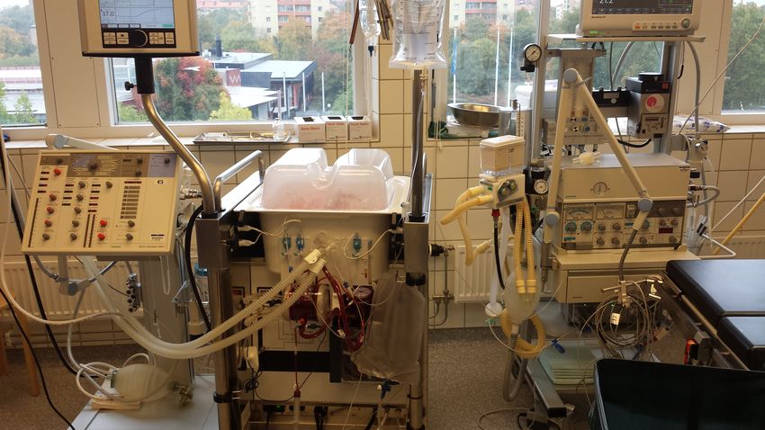
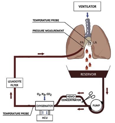
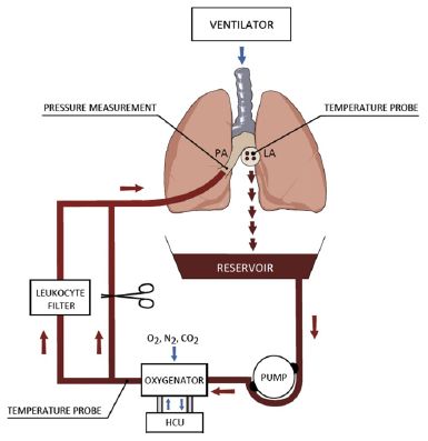
Figure 1.
                                               A schematic drawing of the typical EVLP unit.
                                               Adapted from Wallinder et al1
                                               PA (pulmonary artery), LA (left atrium), HCU
                                               (heater/cooler unit).

system. These devices are the Vivoline LS1 (Vivoline), the XPS (XVIVO Perfusion AB),
Lung assist (Organ assist) and Organ Care system Lung (OCS). They all apply a certain
protocol of EVLP, according to Lund, Toronto or OCS.

Three predominant protocols for the execution of EVLP have been developed (Table 1,
adapted from Andreasson et al65 and Maksidi et al66). The original, developed in Lund by
Stig Steen et al. and the protocol developed by the Toronto group, differ in the use of red
blood cells in the perfusate fluid, the flow at which evaluation is performed, the type of
pump used, and whether pulmonary venous blood drains passively from the lungs in an
open circuit or through a closed circuit with both pulmonary artery (PA) and left atrial
(LA) cannulas, and the application of a LA pressure.67,68 The Toronto protocol advocates
a low flow strategy and acellular perfusate, whereas the Lund protocol employs red blood
cells added to the perfusate and evaluates at physiological perfusate flows. All protocols
include corticosteroids to suppress immune response and broad-spectrum antibiotics in
the perfusate to treat any concomitant infection.69,70

In both the Lund and the Toronto protocols, lungs are initially harvested, cold-flushed
and kept in static cold storage, before being subjected to EVLP. After completion of
EVLP, awaiting transplantation, lungs are usually cooled and put in cold storage, however
this practice has been questioned.71 OCS differs in that aspect, providing EVLP
opportunities immediately after harvesting and pulmonary flush, during transportation,
thereby intending to minimise lung ischemia time.72 Protocols have over time been
adapted and developed locally and differ from centre to centre in some aspects.

The decision to proceed with transplantation after EVLP is based on the performance of
the organs in several different aspects: an adequate lung oxygenation capacity with an
adequate PaO2/FiO2 ratio (>40 kPa/>300 mmHg) during the evaluation phase; stable

6
hemodynamic and respiratory variables (pulmonary vascular resistance (PVR), peak
airway pressures, and lung compliance), the absence of any macroscopic signs of
pneumonic infiltrates, lung infarctions or other gross pathology, and a normal collapse
test indicating normal elastic and compliant properties of the lungs.
Table 1 - Three different protocols for EVLP
Lund Toronto OCS
Perfusion
Target flow 100% of CO, 70 ml/kg/min 40% of CO 2-2.5 l/min
PA pressure (mmHg) ≤20 ≤15 ≤20
LA pressure open LA 3-5 open LA
Type of pump Roller Centrifugal Pulsatile
Perfusate Steen Solution with RBC Steen Solution OCS lung solution
with RBC
Ventilation
Tidal volume (ml/kg) 6-8 7 6
Frequency 10-15 7 10
PEEP (cm H2O) 5 5 5
Fraction inspired oxygen 0.5 0.21 0.21
Temperature °C
Start of ventilation 32 32 32
Start of perfusion 15 25 32
Start of evaluation 37 37 37
PA = pulmonary artery; LA = left atrium; CO = cardiac output; RBC = red blood cells; PEEP = positive end-expiratory pressure
Clinical results - the Lund protocol
The Lund protocol was primarily designed as an assessment system, evaluating lungs
under physiologic conditions after rewarming, given a sufficient time for reconditioning.
The lungs are recruited and ventilated, bronchoscopy performed, the inflammatory
burden decreased by a leukocyte filter added to the circuit, time given for antibiotics and
glucocorticoids to have their effect, and time given for the hyperoncotic perfusate to
reduce oedema. Time on EVLP is not spent longer than needed if the organs are deemed
acceptable for transplantation, however time can be extended if there still is potential for
further improvement.1,49,73 The initial clinical series published by Ingemansson et al. has
been described above.51 The same group followed up with a review of their clinical
experience in 2011.45
Wallinder et al. published early results from a clinical EVLP program in Gothenburg,
based on the Lund protocol, in 2012.1 Soon after that, an EVLP program based on the
same protocol was initiated in Copenhagen.74 Wallinder et al. have in several publications
presented the outcome compared to a contemporary control group of conventional lung
transplantations.60,75 Long-term outcome in patients receiving lungs after EVLP, have in
these studies been shown to be comparable to conventional lungs.
Introduction 7Valenza et al. in Milano, published in 2012 a small study consisting of two patients on ECMO receiving marginal lungs after EVLP reconditioning, compared to a small contemporary control of conventional transplantations, with similar results between groups despite the critical state of the recipient population.76 They returned in 2014 with another study reporting transplantation after EVLP in seven cases compared to 28 controls with no significant differences in outcome and survival.77 Fildes et al. in Manchester compared early outcome up to one year after transplantation in a group of 9 patients transplanted with lungs after EVLP compared to a temporary control group without finding any significant differences in early clinical outcome between the two groups.78 Fisher et al. published results from the Develop UK study in 2016.79 In a non-randomized study of transplantation after EVLP versus standard-criteria lungs, only 18 out of 53 donor lungs were subsequently transplanted, and results showed a non-significant lower one-year survival in the EVLP, as well as, higher rate of early graft injury and postoperative ECMO support, at increased cost. Clinical results - the Toronto protocol The Toronto protocol, developed by Keshavjee and colleagues, has, since its introduction, gained widespread use all over the world.54,56,57,80-82 In addition to the evaluative possibilities of EVLP, focus is also on prolonging the perfusion with the possibility of treating and better reconditioning the organs.83 This approach is reflected in their early publication on repair of human donor lungs by IL-10 gene therapy.84 Just recently, the Toronto group, almost a decade later, published a study on the same subject, in a large animal model, demonstrating that ex vivo treatment with AdhIL-10 is safe and improves post-transplant lung function after EVLP.85 With a view to improving the Lund protocol, they apply acellular perfusate with the intention to avoid damaging haemolysis, a lower target flow to reduce any oedema formation and a closed circuit with the maintenance of a positive LA pressure through a specifically adapted LA cannula.86 Being the first prospective clinical trial in EVLP, the HELP trial, published by Cypel et al. in 2011 was ground-breaking.56 Twenty out of 23 sets of lungs considered high-risk were transplanted and outcome compared to a group of 166 patients receiving conventional lungs. No differences were seen with regards to primary graft dysfunction (PGD), days on ventilator, time in ICU or hospital- and 30-day mortality. In 2012 the same group reported their experience of 50 consecutive transplantations after 58 EVLP evaluations (86% conversion rate), with lungs from both DCD and DBD donors, compared to a contemporary control group.57 Short-term outcome were similar in both groups, and one-year survival was 87% in the EVLP group compared to 86% in the control group. 8
Aigner et al. in Vienna published a study in 2012 on 9 double-lung transplantations after
EVLP assessment, compared to a control group of 119 patients, reporting similar short-
term results between groups.87 The same year, Zych et al. in Harefield, after having
published a case report on a successful transplantation after EVLP in 2011,88 reported
transplantation of 6 pairs of donor lungs out of 13 EVLP evaluated, with a 100% 3-month
survival rate.89

A retrospective study of independently collected data from Toronto, Paris and Vienna,
encompassing 125 EVLP evaluations was presented at the ISHLT meeting in 2013.90
Both DCD and DBD donors were included in the cohort. Lungs were transplanted in
103 cases, mounting up to a conversion rate of 82.5%. Good short- and intermediate-
term outcome was reported, with a one-year survival rate of 88%.

The Paris group published their cohort in 2014, consisting of 31 double lung
transplantations after 32 EVLP evaluations, compared to a contemporary control group.
They experienced significantly longer times in ICU and in hospital stay in the EVLP
group, but one-year survival was excellent in both groups, 93% and 91% respectively.91

Turin reported no difference in incidence or severity of PGD in eight cases of
transplantation after EVLP compared to a control group of 28 patients.92

The result of the NOVEL trial,93 a prospective, non-randomised trial in six centres in the
USA was presented in 2014.94 76 EVLP:s were performed and 42 were transplanted (55%
utilization rate), and compared to a contemporary control group. Early outcome after
lung transplantation and one-year survival were not significantly different between
patients that received EVLP compared to standard criteria lungs.

Maryland reported short-term outcome data in 11 transplanted grafts out of 17 evaluated
with EVLP (65% conversion rate). There were no severe PGD at 72 hours in their
material.

In 2017, in a retrospective study of data from the Toronto Lung Transplant Program
database, Yeung et al. compared outcome in patients receiving lungs exposed to EVLP
for more than 12 hours (mean 14.6 hours) compared to a control group with shorter
EVLP-times than 12 hours (mean 6.7 hours).95 Longer EVLP did not significantly affect
short-term outcome. This is in line with the Toronto approach to EVLP, viewing EVLP
not just as a means of evaluation, but also as a treatment opportunity, and a method of
increasing flexibility around the transplantation process.

In 2017 Slama et al. published a study comparing two groups of donor lungs, that both
met standard inclusion criteria for transplantation.96 One of the groups underwent EVLP
prior to transplantation, and the other group was transplanted in a standard fashion
Functional results and perioperative outcome in the EVLP group were comparable to
those achieved with standard donor lung preservation techniques. It was concluded that
EVLP is an option to safely extend total preservation time.

                                                                       Introduction     9
Under way is the large multicentre “Normothermic EVLP As An Assessment Of Extended/Marginal Donor Lungs study”.97 Although short- and intermediate-term outcome has been widely published, long-term data is scarcer. In 2015 the Toronto group published a retrospective study of all transplants performed between 2008 and 2012 and reported up to five-year survival-rates that were similar between groups, as were freedom from chronic lung allograft dysfunction (CLAD), rejection episodes, 6-minute walk distance and FEV1.98 Clinical results - Hannover/Madrid – OCS In 2012, Warnecke et al. presented a new application of EVLP, evaluating the normothermic preservation and transportation of standard criteria lungs on a portable EVLP system, the Organ Care system™ Lung (Transmedics, Andover, USA).72 Twelve pairs of standard lungs were preserved under normothermic conditions in this study, as opposed to traditional cold preservation. Short-term outcome was non-inferior compared to control. The OCS protocol, in line with the Lund protocol, applies a cellular perfusate and open LA, but in agreement with the Toronto protocol, limit flows. As opposed to the other two protocols, Steen Solution has been replaced by a solution made by the system manufacturer. The principal initial focus of OCS was the transportation of standard criteria lungs using normothermic EVLP, setting it apart from the Lund and Toronto protocols that were primarily designed as protocols for evaluating and reconditioning marginal donor lungs, in-house. The INSPIRE-trial was a prospective randomized multicentre study comparing outcomes of standard criteria lungs preserved and transported by either normothermic EVLP in the OCS system or standard cold preservation.99,100 The use of the OCS Lung in the reconditioning of marginal lungs was studied in the EXPAND trial, results of which are to be presented at the 2018 ISHLT meeting. In 2016, Zeriouh et al. reported a study comparing short- and medium-term outcome in a group of 14 organs preserved with the OCS Lung, compared to a control group of 308 patients transplanted after cold preservation. Patients in the OCS group had a significantly better postoperative FEV1 at 3 and 6 months, and similar outcome in terms of cumulative survival and freedom from bronchiolitis obliterans syndrome (BOS).101 Schmack et al. published a recent review in 2016 on the OCS Lung concept.102 The focus of this thesis is, however, on the Lund and Toronto protocols and their application. EVLP in special cases Brown et al. reported a case of EVLP on donor lungs with pulmonary embolism, in which lung oxygenation capacity increased and PVR decreased during EVLP, suggesting that they could have been acceptable for lung transplantation.103 A second case report on the same subject, using urokinase during EVLP, and subsequently successfully transplanting a pair of lungs was published in 2014.104 Daine et al. published a case on successful 10
transplantations after EVLP in a series of five DBD donors after asphyxia by hanging.105
Patil et al. used EVLP to evaluate lungs that inadvertently during the harvesting procedure
were flushed with very high PA pressure.106 Costa et al. evaluated EVLP of lungs from
12 donors with a history of cardiac surgery with successful results and outcome.107 De
Wolf et al. used EVLP to gain time while waiting for a negative result in cross matching
in hyperimmunized patients, then moving forward with transplantation in three patients,
with all patients alive after three years.108

Establishing the baseline in human EVLP and predicting outcome

A series of descriptive publications with the primary aim of establishing baseline
parameters during EVLP have been presented. There has over time also been a growing
interest in investigating factors and markers during EVLP, predicting successful outcome
after transplantation.

In 2011, Sadaria et al. established a cytokine expression profile in human lungs undergoing
normothermic EVLP.109 The same year, Koike et al. reported on lactate metabolism,
during acellular normothermic EVLP, concluding that a gradual increase due to reduced
clearance was to be expected.110 In 2013, George et al. presented physiologic and
biochemical profiles of clinically rejected lungs on EVLP.111

In 2015 Machuca et al. in Toronto, using perfusate samples from 50 EVLP procedures in
human lungs collected after 1 and 4 hours of EVLP, investigated the expression of
cytokines, chemokines and growth factors. They showed that perfusate biomarkers could
potentially be used for more precise donor lung selection improving outcome after
transplantation.112 In a contemporary study they evaluated the endothelin-1 pathway as
potential predictors for lung function.113

There is a great interest in finding biochemical markers or combinations of markers
during EVLP that could predict the transplantability of the organs. None has so far
reached clinical use. Research is active in this field, and a range of preliminary smaller
studies have been published during 2017.114-118

EVLP as an experimental platform in animal studies

Although having been extensively applied in clinical practice for the last decade, animal
studies was the first step, binding theory and practice together, needed for the
development of the protocols later to be tested in human research. EVLP is still a novel
technique in constant development, and a steady stream of experimental animal studies
into different aspects of EVLP have emerged in parallel with clinical progress over the
last decade. In this section a brief description of the findings of the majority of these
experimental studies is given.

Because of their similarity to human lungs, albeit not identical, porcine lungs have been
the most extensively used.119,120 There are, however, also experimental studies performed
on lungs from other animals, such as rat, mice and dog.121-125

                                                                      Introduction      11
Emaminia et al. investigated in a small study in 2011 the use of an adenosine A2A agonist during EVLP, suggesting a positive effect on oedema and ischemia-reperfusion injury, confirming these findings in a study in mice in 2015.123,126 In 2016, on the same theme, they published a second study in mice on the attenuation of ischemia-reperfusion injury by an adenosine A2B antagonist.127 Several recent studies have reported positive results after adding agents to the perfusate, for example by inhibiting NF-κB128 or by adding trimerazidine to attenuate ischaemia/reperfusion injury during EVLP.129 None of these experimental approaches has yet been implemented into clinical practice. Meers et al. published in 2011 the work on an aspiration model in swine and demonstrated the feasibility of assessing the organs by EVLP.130,131 They then used this model to compare steroids to macrolides to improve gas-exchange in caustic-injured lungs.132 In 2014 Khalife-Hocquemiller et al., in an aspiration-model with gastric acid, found that surfactant attenuated lung injury during EVLP.133 Last year in a similar study in pigs, Nakajima et al. reported better outcome after lung lavage and surfactant replacement during EVLP after gastric-acid induced lung injury.134 Valenza et al. investigated glucose consumption during EVLP, and found that increased glucose consumption correlated with worse lung function.135 Following up on these results, in 2012, they showed that salbutamol infusion during EVLP was associated with lower pulmonary pressures and better lung mechanics.136 These findings were confirmed in a canine study by Kondo et al. in 2015, showing that inhalation of salbutamol attenuates lung injury.122 A third study in 2017, in a canine model, by Hijiya et al. supported earlier findings.137 The Toronto group performed prolonged EVLP during 12 hours in porcine lungs subjected to 24 hours of cold ischemia, whereby they organs were transplanted and evaluated in vivo.138 The organs developed oedema during EVLP and decreased compliance, and subsequently performed poor in vivo. The significant finding in this study was that ex vivo PO2 is a poor indicator of lung performance due to the linearization of the relationship between oxygen content and PO2 in acellular perfusate. The same group published an additional study, the same year, comparing ex vivo with in vivo intratracheal adenoviral vector gene delivery.139 The poor reliability of PO2 alone in assessing lung function during EVLP is well known, especially using acellular perfusate, as shown in the above-mentioned study. Conditions are different using cellular perfusate, however, it has been shown that it is important to relate the PaO2/FiO2 ratio to the inspired fraction of oxygen.140 PaO2 is related to shunt fraction and depending on the extent of shunt the relationship between PaO2 and different FiO2 varies.141 Pablo et al. in Maryland have adapted their EVLP protocol to put stronger emphasis on compliance dynamics in decision-making during EVLP.142 Okamoto et al. studied correlations between the PaO2/FiO2 ratio and airway and vascular parameters during cellular EVLP in both porcine and human lungs and found that airway parameters were complementary quantitative indicators of lung function in cellular EVLP.143 12
Thrombolytic therapy during EVLP in a rat model was studied in 2013 by Motoyama et
al., and again in 2014.144,145 It has, as described above, been applied in clinical cases as
well.

Maignan et al. have studied measurement of exhaled carbon monoxide (CO) during
EVLP, as a marker for ischemia reperfusion injury, showing a positive correlation
between exhaled concentration of CO and the degree of ischaemia/reperfusion
injury.146,147

Pierre et al. evaluated in a porcine model the effects of extending EVLP-times, reporting
increasing weight with increased time on EVLP. Results from this study could not in an
experimental setting show benefit from additional time on EVLP, contradicting later
clinical reports of successful outcome after extended EVLP in human lungs.148

Noda et al. have published several experimental studies in rat, beginning with the
establishment of a rat EVLP model in 2014.124 The same year they demonstrated that
lung grafts on EVLP exhibited prominent pro-inflammatory changes and compromised
metabolic profiles. It was possible to attenuate this by inhaling the lungs with hydrogen.149
In a study in 2015, they compared a novel technique of dual EVLP, also including the
bronchial artery circulation to the EVLP circuit, achieving superior outcome in this
group.150 Recently they published a study on optimal O2 levels in the perfusate during
EVLP. Deoxygenated perfusate exhibited significantly more inflammation with
compromised cellular metabolic activity and compromised post-transplant outcomes.151

In 2015, Harada et al. reported on a small study assessing the addition of a neutrophil
elastase inhibitor to the perfusate, indicating that the inflammatory response may be
attenuated and lung reperfusion injury be decreased.152 Lin et al. reported in 2017 positive
results of adding alpha-1-antitrypsin to the perfusate.153

In a study in rat, thermography was used to detect regional malperfusion during EVLP,
in lungs with induced thrombi. Thermographical evaluation may, in contrast to prevailing
modalities, detect regional damage in donor lungs.154 In 2017 Sage et al. reported on using
real-time CT to assess organs during EVLP.155

Martens et al. in a study in 2016 could not, in a porcine model, improve graft function
using inhalation of argon or xenon gases during EVLP.156 In a succeeding study by the
same group, exposing lungs to maximal xenon exposure, they again did not find support
for this approach.157 Several studies have found support for protective effects of inhaling
different agents during EVLP, such as sevoflurane.158

Renewed interest in cytokine filtration to reduce accumulation during prolonged EVLP
led Iskender et al. to publish a porcine study showing that cytokine removal decreased the
development of pulmonary oedema and electrolyte imbalance. These findings contrast
with the negative findings by Kakishita et al. back in 2010.159

                                                                        Introduction      13
Lately there has also been an interest into ventilation strategies applied during EVLP. In 2017, Mehaffey et al. demonstrated superior results with airway pressure release ventilation during EVLP.160 The same year, Aboelnazar et al. presented a negative pressure ventilation model, where they could demonstrate decreased inflammation and less oedema formation during EVLP.161 In 2017 Himmat et al. showed that a decrease in hypoxic pulmonary vasoconstriction during EVLP correlated with increased inflammation during extended EVLP.162 Although most experimental studies have been performed according to either the Lund or Toronto protocol, some studies adopting OCS have emerged. Among them a study in prolonged EVLP using OCS Lung and comparing cellular and acellular perfusates.163 EVLP in the context of DCD There is growing support of the use of EVLP after controlled DCD donation.57,164-166 In 2015 Machuca et al. published a study evaluating the impact of EVLP on outcome after transplantation of donation after DCD lungs.167 Between 2007 and 2013, out of a total of 673 lung transplantations performed, 55 were DCD transplantations (excluding bridged cases from analysis in this study). Twenty-eight (51%) of the DCD cases underwent EVLP. Outcome after transplantation with organs from DBD compared to DCD donors was similar, up to five years after transplantation. Organs from DBD donors that were transplanted after EVLP presented shorter hospital stay and a trend towards shorter time on ventilator, but without any difference in survival. EVLP has been extensively used as an experimental platform evaluating different procedures and approaches to performing transplantation of lungs after DCD.168-174 EVLP after DCD is however outside the scope of this thesis and will not be covered further. 14
AIMS

 General aims
 The general aims of this thesis were to increase the understanding of ex vivo lung
 perfusion (EVLP) the way it is contemporarily performed according to two different
 strategies, to experimentally investigate refinements and additions to the procedure and
 to compare two established protocols.

 Additional aims were to review the clinical outcome of patients that had been transplanted
 with lungs that had undergone EVLP in two Scandinavian centres, and to investigate
 whether it could be established which parameters during EVLP matter the most for
 clinical outcome.

 Study aims

  I.   To evaluate the effect of haemofiltration during EVLP on lung function, perfusate
       oncotic pressure, and lung weight as a measure of tissue oedema content, in a
       porcine model.

 II.   To compare two clinically used strategies for EVLP with respect to lung function,
       metabolism, inflammatory response, oxidative stress, and cell viability, in a porcine
       model.

III.   To review the combined clinical short- and medium-term outcome of patients
       undergoing lung transplantation after EVLP in Gothenburg and Copenhagen and
       compare it to a contemporary control group of patients transplanted with
       conventional (non-EVLP) lungs.

IV.    To assess the correlations between several physiological variables during EVLP,
       with short-term outcome variables in 47 double lung recipients of EVLP-evaluated
       lungs, to identify which factors are the most important in predicting outcome after
       transplantation.

                                                                              Aims       15
16
METHODS
The animal experiments were performed at the Laboratory for Experimental
Biomedicine, at Gothenburg University. Paper II is in collaboration with representatives
of the Laboratory for Microbiology, Parasitology and Hygiene, Antwerp University,
Antwerp, Belgium and the Department of Thoracic Surgery, Medical University of
Vienna, Austria. Paper III and VI are in collaboration with Rigshospitalet in Copenhagen
and the University of Copenhagen.
Ethical considerations
Papers I and II – experimental animal studies
The experimental animal studies were performed following approval of The Animal
Ethical Committee of the University of Gothenburg. The animals received care in
compliance with the European Convention for the Protection of Vertebrate Animals
Used for Experimental and Other Scientific Purposes (1986), the Principles of Laboratory
Animal Care formulated by the National Society for Medical Research, and the Guide for
the Care and Use of Laboratory Animals from the National Institutes of Health, USA. At
the end of the experiments animals were euthanized by exsanguination.
Papers III and IV – clinical studies
The Human Ethics Committees of the University of Gothenburg and the University of
Copenhagen approved the clinical studies. All patients were informed, both orally and in
writing, and consented in writing, when listed for transplantation, to the possibility of
receiving organs that had undergone EVLP. Participants were allowed to withdraw from
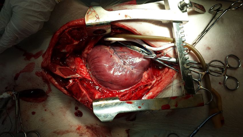
the study at any time. The organs were matched according to standard criteria.
Animal studies (papers I and II)
Animals
Swedish domestic pigs were supplied by a local breeder. All animals were delivered to the
laboratory one week in advance of performing the study. A veterinarian supervised care
and handling of the animals, before and during experiments.
Twenty-two animals were included in Paper I, of which sixteen were subjected to
induction of pulmonary oedema in vivo, and six controls were not. Two animals in the
oedema induction group were excluded due to them developing severe right heart failure
Methods 17and the inability to maintain study protocol targets. The remaining fourteen animals were randomized, in two equally sized groups, to EVLP either with or without haemofiltration. Twenty animals were included in Paper II, randomized to two equally sized study groups. Measurements during the experiments were when appropriate indexed to body surface area.175 Anaesthesia and preparation The animals were fasted overnight, with free access to water. They received premedication while in stables, with an intramuscular injection of a combination of 0.06 mg/kg body weight (BW) dexmedetomidin (Dexdomitor, Orion Pharma AB, Sollentuna, Sweden) and 5 mg/kg BW, respectively, of tiletamine hydrochloride and zolazepam hydrochloride (Zoletil, Virbac, Carros, France). The animals were then transferred to a preparation room and placed in the prone position. They were given an intramuscular injection of buprenorphine 0.03 mg/kg BW (Vetergesic vet, Orion Pharma Animal Health, Sollentuna, Sweden) and an intravenous catheter was placed in the ear. Tracheal intubation was performed under spontaneous breathing. The pigs were subsequently transferred to the operating room and placed in the supine position on the operating table. Volume-controlled ventilation was initiated with a tidal volume of 10 ml/kg, a positive end-expiratory pressure (PEEP) of 0 mmHg (Paper I) or 5 mmHg (Paper II), and a FiO2 of 0.5, adjusting the respiratory frequency to maintain end-tidal CO2 (et-CO2) in the normal range of 5-5.5 kPa, using a Servo Ventilator (Servo Ventilator 900C, Siemens- Elema AB, Solna, Sweden). Anaesthesia was maintained throughout the experiment using isoflurane (Isoba Vet, Intervet AB, Sollentuna, Sweden) at a minimal alveolar concentration (MAC) of 1.3. Lung harvesting A cannula was inserted in the pulmonary artery and secured in place with a purse string suture, and the left atrial appendage was incised widely to allow for free drainage of the organ preservation fluid. The lungs were then perfused antegradely at a low perfusion pressure (
You can also read