Mitochondrial Dysfunctions: A Red Thread across Neurodegenerative Diseases - MDPI
←
→
Page content transcription
If your browser does not render page correctly, please read the page content below

International Journal of
            Molecular Sciences

Review
Mitochondrial Dysfunctions: A Red Thread across
Neurodegenerative Diseases
Serena Stanga 1,2,3, * , Anna Caretto 1,2 , Marina Boido 1,2,3          and Alessandro Vercelli 1,2,3
 1    Department of Neuroscience Rita Levi Montalcini, University of Turin, 10126 Turin, Italy;
      anna.caretto@unito.it (A.C.); marina.boido@unito.it (M.B.); alessandro.vercelli@unito.it (A.V.)
 2    Neuroscience Institute Cavalieri Ottolenghi, University of Turin, 10043 Orbassano (TO), Italy
 3    National Institute of Neuroscience (INN), 10125 Turin, Italy
 *    Correspondence: serena.stanga@unito.it

 Received: 4 May 2020; Accepted: 22 May 2020; Published: 25 May 2020

 Abstract: Mitochondria play a central role in a plethora of processes related to the maintenance of
 cellular homeostasis and genomic integrity. They contribute to preserving the optimal functioning of
 cells and protecting them from potential DNA damage which could result in mutations and disease.
 However, perturbations of the system due to senescence or environmental factors induce alterations of
 the physiological balance and lead to the impairment of mitochondrial functions. After the description
 of the crucial roles of mitochondria for cell survival and activity, the core of this review focuses
 on the “mitochondrial switch” which occurs at the onset of neuronal degeneration. We dissect the
 pathways related to mitochondrial dysfunctions which are shared among the most frequent or disabling
 neurodegenerative diseases such as Alzheimer’s, Parkinson’s, and Huntington’s, Amyotrophic Lateral
 Sclerosis, and Spinal Muscular Atrophy. Can mitochondrial dysfunctions (affecting their morphology
 and activities) represent the early event eliciting the shift towards pathological neurobiological
 processes? Can mitochondria represent a common target against neurodegeneration? We also review
 here the drugs that target mitochondria in neurodegenerative diseases.

 Keywords: mitochondria; cellular homeostasis; neurodegenerative diseases; motor neuron
 diseases; neurodegeneration; oxidative stress; mitochondria biogenesis and dynamics; mitochondria
 targeting drugs

1. Introduction: The Peculiarity of Mitochondria
     Classically, mitochondria were considered the “powerhouse” of the cell, the energetic core
generating ATP for cell activities [1]. Intensive research on their morphology and functions showed
that mitochondria play several different roles.
     Interestingly, mitochondria possess their own genome, the mitochondrial DNA (mtDNA), packed
in nucleoids in the mitochondrial matrix in close association with the mitochondrial inner membrane [2].
Mutations at the mtDNA level are causative for many human diseases which are generally defined
as mitochondrial disorders [3]. Moreover, mitochondria are vital and dynamic organelles able to
modify their shape and size in order to respond to the cellular needs and, therefore, to maintain cellular
homeostasis [4]. Mitochondria are the site of the oxidation of metabolites, for example through Krebs’
cycle, and of the β-oxidation of fatty acids [5]. Mitochondria are also the main generators of the reactive
oxygen species (ROS) [6] and they participate to cell proliferation by maintaining a proper redox state
and by recycling oxidized electron carriers. Importantly, mitochondria buffer calcium ions regulate,
in turn, calcium homeostasis [7,8]. This is a crucial mitochondrial function impacting many cellular
pathways such as the neurotransmitters’ release from neurons and glial cells [9].

Int. J. Mol. Sci. 2020, 21, 3719; doi:10.3390/ijms21103719                              www.mdpi.com/journal/ijms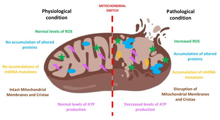
Int. J. Mol. Sci. 2020, 21, 3719                                                                     2 of 35

      In order to maintain the homeostasis of the cell, mitochondria play a crucial role in the choice of
cell fate since they are also able to control cellular programmed cell death [10]. Indeed, mitochondria
can induce apoptosis via caspase-dependent or independent mechanisms, the first by the activation
of pro-apoptotic members of the B-cell lymphoma 2 (Bcl-2) family, the latter by the release of toxic
mitochondrial proteins occurring after mitochondrial loss of function [11].
      Mitochondria are extremely sensitive to every subtle change perturbing the homeostasis of the
cell and, in turn, can modify their shape and number. Indeed, the processes of fusion and fission are
fundamental, respectively, to repair a damaged mitochondrion or to increment their number, as in
case of an increased demand of energy or to facilitate their removal when damaged in order to protect
cellular integrity [12].
      Additionally, the number and the size of cristae, which are dynamic bioenergetic compartments
of the inner mitochondrial membrane where the respiratory chain occurs, adapt to the needs.
Their plasticity guarantees a constant turnover to assure the balance between regeneration, biogenesis,
and elimination of damaged mitochondria [13]. On the other hand, a regressive event occurring to
mitochondria consists in “mitophagy”, i.e., the process of mitochondria degradation by autophagy [14],
aiming to maintain the correct turnover of mitochondria [15].
      Finally, mitochondria are also able to protect cell integrity by preventing the damage induced
by viral infection [16]; indeed, they can also stimulate the innate immune response against these
insults [17].

2. The “Mitochondrial Switch” and Its Impact on Neurodegeneration
      Mitochondria are extremely sensitive to the insults that occur and accumulate in the cell,
directly impacting on their function, consequently promoting disease development and progression.
Indeed, besides human hereditary diseases caused by mutations of the mtDNA or in nuclear genes
responsible for mitochondrial deregulation [18], mitochondrial dysfunctions are among the first events
which occur in a vast number of pathological conditions ranking from diabetes [19], inflammatory
diseases (such as multiple sclerosis [20]), to cancer [21] and neurodegenerative diseases [22]. We review
here the “mitochondrial switch” which occurs during neurodegeneration. Neurodegenerative diseases
consist of a group of heterogeneous disorders, but, nevertheless, they are all characterized by the
progressive loss of specific neuronal populations and circuits in the central nervous system (CNS)
triggered by mitochondria dysfunctions [23,24]. Another feature of the majority of neurodegenerative
diseases is the progressive and highly disabling motor decline. Indeed, besides neuronal cells, also
muscle cells are particularly enriched in mitochondria, since they need a high level of energy to
function, and are heavily impacted by mitochondrial dysfunctions. There is growing evidence in
the field exploring mitochondrial defects occurring also in the peripheral nervous system (PNS) and
peripheral cells. Due to the low potential of neural regeneration, mitochondrial damage results in
detrimental effects for neuron survival [25], while in peripheral cells the regeneration capacity is higher
and clinical symptoms are evident only later in the disease.
      Globally, in neurodegenerative diseases there is a “switch” in mitochondrial function which
contributes importantly to the transition from a normal physiological to a degenerative condition.
The accumulation of different stresses and the parallel impairment of a number of cell protective
processes elicits neurodegeneration (Figure 1).
      During neurodegeneration oxidative stress increases and intracellular ROS are formed, inducing
to mtDNA mutations [1] and also to the disruption of mitochondrial membranes and cristae with a
decrease in ATP production. Among the mechanisms eliciting oxidative stress, it is worth mentioning
the process of deuteronation. Deuterium is a hydrogen isotope, present in nature in a well-defined ratio
with hydrogen (1:6600) [26]: in body fluids, the probability of ATP synthase deuteronation process is
1/15,000 and the increase of this ratio can negatively interfere with ATP synthesis. Moreover, it has
also been demonstrated that deuteronation can enhance formation of free radicals, slowing the
electron trafficking in the mitochondrial electron transport chain (ETC) [27]. This mechanism has been
Int. J. Mol. Sci. 2020, 21, 3719 3 of 35
evaluated in several conditions or pathologies (i.e., cancer, diabetes, aging, depression) and it can
be reverted replacing heavy water with deuterium depleted water (DDW). Moreover, reduction of
deuterium content by DDW ingestion can rescue long-term memory in Wistar rats restoring altered
deuterium/hydrogen ratio and enhancing synaptic activity [28]. Indeed, DDW is able to promote
antioxidant enzymes’ expression (SOD) and restore glutathione (GSH) levels mitigating, in turn, ROS
Int. J. Mol. and
formation Sci. 2020, 21, x FOR PEER
preventing fattyREVIEW
acid oxidation [29]. 4 of 35
Figure 1. The mitochondrial switch. The sketch shows the appearance and the features of a
Figure 1. The mitochondrial switch. The sketch shows the appearance and the features of a
mitochondrion
mitochondrionbefore
beforeand
andafter
afterthe
theaccumulation
accumulation ofof damage responsiblefor
damage responsible forits
itsimpairment.
impairment.AtAt
thethe
onset of the neurodegenerative process the increase of reactive oxygen species (ROS), mitochondrial
onset of the neurodegenerative process the increase of reactive oxygen species (ROS), mitochondrial
DNADNA (mtDNA)
(mtDNA)mutations
mutationsandandaltered
altered proteins
proteins determines the swelling
determines the swellingofofmitochondria
mitochondria and
and thethe
disruption
disruptionofof
their membranes
their membranesand andcristae.
cristae. Such
Such an altered morphology
an altered morphologyheavily
heavilyimpacts
impactsonon function
function
determining,
determining,among
amongothers,
others,decreases
decreases ATP
ATP production, increasedROS,
production, increased ROS,leading
leading toto neuronal
neuronal death.
death.
Created
Createdwith BioRender
with BioRendersoftware.
software.
2.1.Moreover, the accumulation
Mitochondrial Dysfunctions inofAD altered proteins occurring in many neurodegenerative diseases can
determine a decline in mitochondrial biogenesis, impacting on mitochondrial turnover and inducing
AD is the most common and devastating neurodegenerative disease expected to affect more
structural and functional changes in cells. Different studies showed that various proteins involved
than 81 million people worldwide by 2040 [41,42]. The first evidence of pathogenic mechanisms in
in the above-mentioned processes and in mitophagy are affected in neurodegenerative diseases
AD has been the progressive accumulation of beta-amyloid peptide (Aβ) extracellularly in the brain
determining a dysregulation of these processes [30]. Indeed, mitophagy is required to remove damaged
and of neurofibrillary tangles of hyperphosphorylated tau inside neurons [43], determining the
mitochondria
progressive and lossthis
of process
cortical is
andfinely tuned to maintain
hippocampal neurons an andequilibrate
inducingturnover of mitochondria
brain atrophy, consequent and,
from a bigger picture, proper cellular development and differentiation.
cognitive and memory loss. More recently, advanced neuroimaging techniques showed evident Described for the first time
in 1998 [31], alterations
metabolic mitophagyinhas thegained
brain ofconsiderable
AD patients interest
at a veryinearly
the field
stageofofneurodegeneration in the
the disease [44]. Indeed, thelast
years; however,process
degenerative because of its fine
probably tuning,
starts 20–30ityears
is a difficult
before the target for onset
clinical designing
and onenewof therapies against
the major goals
neurodegeneration.
of AD research is theSome early authors
detectionhypothesized
before symptom a relation betweenthe
onset to prevent mitophagy dysregulation
disease outbreak. Oxidative and
neurodegenerative
stress-induced damage diseases,occurs
based on eventhebefore
impairment
amyloidin autophagosome–lysosome
and tau deposition, andfusion and defects
mitochondrial
in lysosomal
dysfunctions acidification, oftenwhich
are early events observed in neurodegeneration
precede neurodegeneration [45]. [32]. In AD there is also evidence of
neuroinflammation; indeed,can
Altogether, these events microglia andprocess
trigger the reactiveofastrocytes are closely
mitochondrial membraneassociated with amyloid
permeabilization and,
plaques in patients’ brains [46].
when severely injured mitochondria are not appropriately removed, they release their contents into the
cytosol Mitochondria appear environment:
and the extracellular morphologically thisand functionally
represents altered,
a starting pointimpacting on manyand
of both apoptosis processes
necrosis.
such as excessive ROS formation, and resulting in the decrease of brain
This process is called generation of damage-associated molecular patterns (DAMPs) and mitochondria energy because of the
reduction of ATP [47], alteration of calcium homeostasis, and of apoptosis
represent the major site of DAMP formation. By triggering cell death, mitochondria-derived ROS, induction [48]: indeed,
altered ATP,
calcium, levelsandof apoptotic
the other markers
signalingsuchmolecules
as Bcl-2, Bax,
alsoandleadcaspases have been observedThe
to neuroinflammation. in models
process of of
AD [49]. Moreover,
neuroinflammation alterations
refers in mitochondrial
to the proliferation dynamics
and activation appear and/or
of microglia early in AD development,
astrocytes (microgliosis,
contributing to impair neuronal autophagy because of defective mitophagy [50]. There is a vast
literature on the detrimental impact of the accumulation of damage and misfolded proteins inducing
dysfunctions at the cellular and mitochondrial level not only in the NS but also in peripheral tissues
such as patients fibroblasts [51] which show mitochondrial calcium dysregulation similar to those
occurring in the brain [52]. Moreover, AD-related proteins, such as the Amyloid Precursor Protein
(APP) and Presenilins (PSs), have been recently described as involved in peripheral processesInt. J. Mol. Sci. 2020, 21, 3719 4 of 35
astrogliosis) and, to note, neurodegenerative diseases all show evidence of neuroinflammation; the
specific disease-related peculiarities are described later on in the sections dedicated to each pathology.
Interestingly, neuroinflammation is closely related to mitochondria [33]: indeed, mitochondria can
regulate inflammatory signaling and can modulate the innate immunity by generating ROS and, as a
consequence, triggering neurodegeneration [34]. Closely related to ROS and DAMP formation, another
largely studied mechanism by which mitochondria drive neuroinflammation is their ability to activate
the NOD-, LRR-, and pyrin domain-containing protein 3 (NLRP3) inflammasome [35], in turn leading
to the release of proinflammatory cytokines [36]. Interestingly, NLRP3 localizes in the cytosol and
once activated it translocates into mitochondria and mitochondria-associated membranes (MAMs) [37].
Another mechanism by which mitochondria trigger and sustain neuroinflammation is closely related to
their dynamism: indeed, the inhibition/stimulation of fission induces/reduces NLRP3 inflammasome
assembly and activation, respectively [38].
In particular, mitochondrial dysfunctions (both affecting neuronal survival and triggering
neuroinflammation) have been reported in Alzheimer’s (AD), Parkinson’s (PD), and Huntington’s (HD)
diseases, Amyotrophic Lateral Sclerosis (ALS), and Spinal Muscular Atrophy (SMA), thus representing
a potential biomarker of the diseases, as one major unifying basic mechanism involved in aging and
neurodegeneration [39,40]. In the subparagraphs below we describe the mitochondrial dysfunctions
which are peculiar or shared among the above-mentioned diseases.
2.1. Mitochondrial Dysfunctions in AD
AD is the most common and devastating neurodegenerative disease expected to affect more than
81 million people worldwide by 2040 [41,42]. The first evidence of pathogenic mechanisms in AD has been
the progressive accumulation of beta-amyloid peptide (Aβ) extracellularly in the brain and of neurofibrillary
tangles of hyperphosphorylated tau inside neurons [43], determining the progressive loss of cortical and
hippocampal neurons and inducing brain atrophy, consequent cognitive and memory loss. More recently,
advanced neuroimaging techniques showed evident metabolic alterations in the brain of AD patients at a
very early stage of the disease [44]. Indeed, the degenerative process probably starts 20–30 years before
the clinical onset and one of the major goals of AD research is the early detection before symptom onset
to prevent the disease outbreak. Oxidative stress-induced damage occurs even before amyloid and tau
deposition, and mitochondrial dysfunctions are early events which precede neurodegeneration [45]. In AD
there is also evidence of neuroinflammation; indeed, microglia and reactive astrocytes are closely associated
with amyloid plaques in patients’ brains [46].
Mitochondria appear morphologically and functionally altered, impacting on many processes
such as excessive ROS formation, and resulting in the decrease of brain energy because of the reduction
of ATP [47], alteration of calcium homeostasis, and of apoptosis induction [48]: indeed, altered levels
of apoptotic markers such as Bcl-2, Bax, and caspases have been observed in models of AD [49].
Moreover, alterations in mitochondrial dynamics appear early in AD development, contributing to
impair neuronal autophagy because of defective mitophagy [50]. There is a vast literature on the
detrimental impact of the accumulation of damage and misfolded proteins inducing dysfunctions
at the cellular and mitochondrial level not only in the NS but also in peripheral tissues such as
patients fibroblasts [51] which show mitochondrial calcium dysregulation similar to those occurring
in the brain [52]. Moreover, AD-related proteins, such as the Amyloid Precursor Protein (APP) and
Presenilins (PSs), have been recently described as involved in peripheral processes associated to
muscle trophism, neuromuscular junction (NMJ) formation and mitochondrial morphology [53–55].
Mitochondrial dysfunction in peripheral cells could represent a new target for early and more accessible
diagnosis and, eventually, therapy for AD.
2.2. Mitochondrial Dysfunctions in PD
PD, the second most common neurodegenerative disease in aging, is characterized by the
reduction of dopamine (DA) levels in the striatum due to the degeneration of dopaminergicInt. J. Mol. Sci. 2020, 21, 3719 5 of 35
neurons in the SNpc. Patients show a progressive muscle rigidity and tremors due to decrease
in dopaminergic modulation on striatal neurons altering motor systems [56,57]. Inclusions of Lewy
bodies and presence of immunoreactive α-synuclein and ubiquitin are also hallmarks of the disease [58].
Moreover, neuroinflammation plays a role in PD; reactive microglia in the substantia nigra together
with positron emission tomography (PET) imaging studies of PD brains provided early evidence for
the role of neuroinflammation in PD [59,60].
The first evidence of mitochondrial dysfunction in PD came in 1983, when
Langston described chronic parkinsonism effects in young humans after the use of
1-methyl-4phenyl-1,2,3,6-tetrahydropyridine (MPTP), which is a prodrug to the neurotoxin
1-methyl-4-phenylpyridinium (MPP+); the latter interferes with the activity of the complex I of
the electron transport chain [61] whose functioning is fundamental for dopaminergic neuron survival.
Decades of research in the field confirmed that mitochondrial dysfunctions are a common feature in
PD and are present in animal models, in induced pluripotent stem cell (iPSCs)-derived neurons and in
patients’ brains [62–65]. Mitochondrial dysfunctions are related to both sporadic and familial PD and are
associated to disturbances of mitochondrial function, morphology, and dynamics (for a detailed review
see Bose and Beal [66]). High levels of ROS, due to the high metabolic demand, determine accumulation
of toxic oxidative species and structural alterations of complex I, affecting mitochondria functionality
in patient brains and mouse models [67,68]. Moreover, α-synuclein oligomers accumulation induces
the permeabilization of the mitochondrial membrane and direct toxicity by increasing ROS production
and consequently leading to neuronal death [69]. Disturbances in calcium homeostasis and calcium
overload could force the opening of the mitochondrial permeabilization transition pore, resulting in
ROS formation, Cyt C release, and apoptosis activation [70].
Mitochondrial dysfunctions in PD are also a result of changes in mitochondria biogenesis
caused by the dysregulation of transcription factors such as the peroxisome proliferator-activated
receptor gamma coactivator 1-alpha (PGC1α), as demonstrated both in mice and humans [71,72].
Furthermore, mitochondrial fragmentation, which occurs rapidly after the loss of membrane potential,
has been observed in PD: indeed, many proteins involved in fusion and fission (such as the
dynamin-related protein 1 (DRP1)) are altered in a model of familial PD [73]. Notably, genes
such as PINK1 and Parkin, which are related to familial-PD forms, are involved in the control of
mitochondrial dynamics [74] and several other genetic mutations, including PINK1, Parkin, DJ-1,
LRRK2, and α-Syn, have been linked to familial PD and the corresponding gene products are also
involved in mitophagy. Additionally, also in PD, mitochondrial dysfunctions are not limited to CNS,
but also involve peripheral tissues as it has been described in skeletal muscles and platelets [62,75].
2.3. Mitochondrial Dysfunctions in HD
HD is an autosomal dominant neurodegenerative disorder, caused by the expansion of CAG repeats
in exon 1 of the huntingtin gene (HTT), leading to the specific loss of GABAergic striatal medium spiny
neurons. Patients show impairments in behavior, muscle coordination, and a progressive mental decline
leading to death [76]. The huntingtin protein (HTT) is involved in many processes such as axonal transport,
signal transduction, and autophagy; its misfolding due to HTT mutation determines the disruption of
the above-mentioned biological processes and of many others [77]. Mitochondrial dysfunctions play a
central role in HD progression. Indeed, abnormal oxidative stress and ROS production, calcium imbalance,
and decreased enzymatic activity of the respiratory chain complexes resulting in the alteration of lactate
production have been observed both in mouse models of HD and in patients’ brains [78–80]. Several studies
have revealed reduced mitophagy in the brain of HD patients [81]. Moreover, accumulation of mtDNA
defects occurs early in HD to the point of being suggested as a potential biomarker of the disease [82,83].
Altogether, these mitochondria-derived defects recall pro-inflammatory activated innate immune cells;
indeed, in HD, a remarkable neuroinflammation has been observed in brains from HD patients and PET
imaging showed that microglia activation correlates with the pathology progression (for an extensive
review on the subject see [84]).Int. J. Mol. Sci. 2020, 21, 3719 6 of 35
As for the majority of neurodegenerative disorders, the approach in HD research field has always
been to focus on the neurological symptoms and phenotypes. However, the HTT gene is ubiquitously
expressed [85] and in HD there are important dysfunctions occurring outside the brain. In this regard,
impaired uptake and levels of glucose and cardiac dysfunctions have been shown in HD mouse
models [86,87]. Moreover, HD patients show a high predisposition to diabetes and muscle wasting,
actually before neurodegeneration [88,89]. Interestingly, these dysfunctions are closely connected
to mitochondrial activity and to tissues with high mitochondrial density. Moreover, mitochondrial
alterations are accompanied by oxidative stress in skin fibroblasts of patients [90]; additionally,
the enzymatic activity of the Aco2, which catalyzes the conversion of citrate to isocitrate in the TCA
cycle, is impaired in PBMCs of HD patients and PreHD carriers [91]. Authors suggest it as a potential
biomarker to assess the disease status of both patients and carriers, being an easy and affordable
non-invasive blood test in the clinical routine.
2.4. Mitochondrial Dysfunctions in ALS
ALS is a neurodegenerative disorder due to the loss of upper and lower motor neurons (MNs)
which entails muscle atrophy, progressive weakness, and respiratory failure leading to death [92].
Most cases are sporadic, whereas 10% are familial, with mutations mainly occurring on superoxide
dismutase 1 (SOD1), FUS, TDP43 genes causing the accumulation of the corresponding mutant
proteins [93–95]. However, the etiology of the disease is not yet understood: it includes excitotoxicity,
protein misfolding and aggregation, dysregulation of RNA metabolism, and neuroinflammation [96].
Inflammatory changes have been largely described in ALS patients: astrogliosis is evident within the
spinal cord (both ventral and dorsal horns) and brain (cortical gray matter and subcortical white matter),
whereas microgliosis in the spinal cord ventral horns, corticospinal tract, and motor cortex [97,98].
For sure, mitochondrial-dependent dysfunctions such as ATP deprivation, oxidative stress, impaired
cell signaling together with altered mitochondrial morphology/dynamics are also correlated to the
pathogenesis of ALS and represent very early phenomena [99]. More in details, (i) a high amount of ROS,
free radicals, and other toxic species such as peroxynitrite (ONOO− ) [100] and (ii) an alteration of the levels
of expression of proteins involved in mitochondrial fusion and fission (i.e., OPA1, Mitofusin for fusion,
and Fis1 and DRP1 for fission) [100], were observed and (iii) apoptotic signaling is impacted in different
ALS models. The latter triggers cell death, pivotal to keep the organism healthy by eliminating cells
which cannot be rescued from damage, and mitochondria are fundamental for this. Indeed, as previously
introduced, mitochondria induce apoptosis either via caspase-dependent or independent mechanisms.
In ALS, the apoptotic cascade is activated [101] and the mutant SOD1 is playing a major role in this cascade
by interacting with Bcl-2 [102]. Indeed, therapeutic strategies targeting mitochondrial dysfunction to
prevent ALS progression represent a possible treatment option [103].
As for the other neurodegenerative diseases, also in ALS the PNS is dramatically affected and
mitochondrial dysfunctions occur in peripheral tissues as well. MN degeneration is preceded by NMJ
denervation, determining a dying-back retrograde neuropathy. Such defects may cause MN death
by predisposing them to calcium-mediated excitotoxicity, increasing ROS generation and initiating
the apoptotic pathway [104]. Moreover, features of mitophagy in the presynaptic terminals of ALS’
NMJs have been observed [105], suggesting impaired mitophagy as one of the mechanisms causing
degeneration (for a review see Elfawy and Das [106]).
2.5. Mitochondrial Dysfunctions in SMA
SMA affects MNs in children and young adults with a mutation/deletion of the Survival Motor
Neuron 1 (SMN1) gene [107]. SMN has a role in the assembly of small ribonucleoprotein particles, that
function in pre-mRNA splicing and gene transcription and in mRNA transport in MN axons [108].
The outcome of its genetic alteration is a decrease in the levels of the functional protein which results
in motor impairment, muscle atrophy, and premature death [109]. The disease severity in humans
depends on SMN2 which is a highly homologous gene of SMN1. However, SMN2 produces only 10%Int. J. Mol. Sci. 2020, 21, 3719 7 of 35
of fully functional SMN; therefore, the degree of compensation relies on patient’s SMN2 copy numbers.
Although the genetic cause of SMA has been identified, many aspects of its pathogenesis remain unclear.
Interestingly, SMN deficiency has been associated to oxidative stress, mitochondrial dysfunction,
and impairment of bioenergetic pathways in different models. SMN silencing in NSC-34 cell lines
induces an increase in cytochrome c oxidase activity and mitochondrial membrane potential, generating
free radicals [110]; in human SMA iPSCs, axonal mitochondrial transport and mitochondrial number
and area are altered [111]. Studies in SMA murine models also show alterations in mitochondrial
respiration, mitochondrial membrane potential and mobility, and confirm an increased oxidative
stress level and fragmentation [112]. Additionally, proteomic studies show that bioenergetic pathways
associated to glycolytic enzymes, such as GAPDH and PGK1, are altered in SMA models [113,114].
Moreover, besides cell-autonomous motoneuronal toxicity, also glial-mediated inflammation seems to
negatively impact on neuronal survival and to promote progression and propagation of the degenerative
process by the activation of apoptotic cascades [115].
Even if SMA has been classically categorized as a MN disease, SMA pathogenesis is more
complex and many mechanisms and districts are involved. Indeed, since SMN protein is ubiquitously
expressed, its lack affects not only MNs, but also NMJs, which show dramatic alterations, including
immaturity, denervation, neurofilament accumulation, and impaired synaptic functions [116].
Additionally, skeletal [117] and heart muscle cells [118] are also dramatically affected: they require
high levels of energy and are, therefore, particularly enriched in mitochondria. Alterations in the
process of mitophagy have been described in the muscles of SMA patients [119]. Some researchers
suggest that these peripheral abnormalities may be the origin of the detrimental effects observed on
MN survival through retrograde signals coming from the muscles/NMJs [120,121]. On the contrary,
others support the idea that peripheral dysfunctions derive from MN degeneration which, in turn,
results in impairment of the nerve-muscle interplay [122]. This ‘chicken or the egg’ question about
where the degeneration starts could help with understanding the important mechanisms of the disease
and the development of disease-modifying interventions which could have greater therapeutic impact
by preferentially targeting the CNS, the PNS, or peripheral tissues. Above all, one possibility is that
mitochondrial impairment, which is an early event in SMA, occurs both at central and peripheral level,
probably representing a relevant target independently from the tissue.
3. Are Mitochondria the Red Thread in Neurodegenerative Diseases?
Considering the enormous economic and social impacts, finding a cure for neurodegenerative
disorders remains a priority in science. Researchers are focused on identifying the common pathogenic
processes shared among these diseases, in order to design new treatments and/or drug combinations
and repurposing. Indeed, scientists and pharmaceutical companies aim at the development of common
therapies for multiple neurodegenerative diseases. To this aim, understanding the co-occurrence and
overall interactions among these diseases is the first step for drug development.
The majority of neurodegenerative disorders have significant genetic components, with genetic
heritability such as for AD, PD, HD, ALS, and SMA. To date, the most common and methodologically
defined approaches used are based on microarrays, next-generation RNA sequencing, statistical and
bioinformatics methods to detect shared genes, single nucleotide polymorphisms (SNPs), and profile
networks of pathways underlying neurodegenerative mechanisms [123–126]. Genome wide association
studies, which represent the largest publicly available resource in the genomic domain, reveal the
similarities among diseases and can help in the process of drug screening and discovery (for a
comprehensive review see Arneson et al. [127]). In addition to genetics, considering that the majority
of the forms of neurodegeneration are sporadic, factors such as the ageing of the global population,
the lifestyle, and the exposure to chemicals are taken into account too and are nowadays known risk
factors for neurodegeneration [128,129].
Neurodegeneration can be defined as a process characterized by the loss of neuronal populations,
progressive cognitive decline, and/or motor symptoms. As largely described above, there isreveal the similarities among diseases and can help in the process of drug screening and discovery
(for a comprehensive review see Arneson et al. [127]). In addition to genetics, considering that the
majority of the forms of neurodegeneration are sporadic, factors such as the ageing of the global
population, the lifestyle, and the exposure to chemicals are taken into account too and are nowadays
Int. J. Mol. Sci. 2020, 21, 3719 8 of 35
known risk factors for neurodegeneration [128,129].
Neurodegeneration can be defined as a process characterized by the loss of neuronal
overwhelming progressive
populations, evidence ofcognitive
impaired decline, and/or motor
mitochondrial symptoms.
functions as a As largely factor
causative described above,the
driving
there is overwhelming evidence of impaired mitochondrial functions as a
development of neurodegenerative diseases together with the unavoidable elevation of oxidative causative factor driving the
development of neurodegenerative diseases together with the unavoidable elevation of oxidative
stress, the increase of free radicals in the brain, impaired DNA repair capability, and decreased tissue
stress, the increase of free radicals in the brain, impaired DNA repair capability, and decreased tissue
regeneration. Indeed, besides the intrinsic differences among diseases, mitochondria are a shared
regeneration. Indeed, besides the intrinsic differences among diseases, mitochondria are a shared key
key crossing point in the biological processes driving neurodegeneration and determining the clinical
crossing point in the biological processes driving neurodegeneration and determining the clinical
outcomes. Mitochondrial dysfunctions could be considered a possible red thread in neurodegenerative
outcomes. Mitochondrial dysfunctions could be considered a possible red thread in
diseases. This is the consequence of the fact that mitochondria, besides being the powerhouse of
neurodegenerative diseases. This is the consequence of the fact that mitochondria, besides being the
eukaryotic
powerhouse cells,
ofare fundamental
eukaryotic organelles
cells, are fundamentalinvolved in critical
organelles pathways
involved connected
in critical pathways to connected
cell growth
andtodifferentiation, cellular signaling, apoptosis, and cell cycle control. Together
cell growth and differentiation, cellular signaling, apoptosis, and cell cycle control. Together with the fact
withthat
mitochondrial dysfunctions appear at the early disease onset and contribute to
the fact that mitochondrial dysfunctions appear at the early disease onset and contribute to disease disease progression,
they are a hallmark
progression, they ofareneurodegeneration.
a hallmark of neurodegeneration.
Interestingly,
Interestingly,the the
mitochondrial
mitochondrialswitch driving
switch neurodegeneration
driving neurodegeneration occurs indistinctly
occurs in every
indistinctly tissue,
in every
eventissue, even in the periphery although neurodegeneration has always been approached and studiedthe
in the periphery although neurodegeneration has always been approached and studied focusing on
CNS. To some
focusing onextent,
the CNS.oneTo
could
some investigate
extent, oneneurodegenerative
could investigatedisorders as affectingdisorders
neurodegenerative mitochondria primarily.
as affecting
mitochondria
In Figure primarily. Indysfunctions
2 the mitochondrial Figure 2 the mitochondrial
and effects on dysfunctions
the CNS, PNS,and andeffects on theare
periphery CNS, PNS, andfor
represented
AD,periphery
PD, HD, ALS,are represented for AD, PD,
and SMA. Therefore, HD, ALS,
targeting and SMA.can
mitochondria Therefore,
represent targeting mitochondria
new therapeutic can
approaches
represent new therapeutic approaches for different neurodegenerative diseases.
for different neurodegenerative diseases. Below, molecules tested for neurodegeneration targeting the Below, molecules
maintested for neurodegeneration
pathways targeting the main
related to mitochondrial-dependent pathways related
dysfunctions, such as to mitochondrial-dependent
oxidative stress, mitochondrial
dysfunctions, such as oxidative stress, mitochondrial
biogenesis, mitochondrial membrane permeability and dynamics, are listed. biogenesis, mitochondrial membrane
permeability and dynamics, are listed.
Figure
Figure The
2. 2. process
The processofofneurodegeneration
neurodegenerationin inthe
the central nervous
nervoussystem
system(CNS),
(CNS),ininthe
theperiphery
peripheryandand
across diseases.
across diseases.Neurodegeneration
Neurodegeneration is is
a progressive
a progressiveprocess
processtaking
takingplace
placenot
notonly
onlyin inthe
theCNS
CNSbut
but also
also in
theinperiphery.
the periphery.
AfterAfter the accumulation
the accumulation of damage
of damage atcellular
at the the cellular
and,and, in particular,
in particular, mitochondrial
mitochondrial level,
level, the switch from physiology to pathology is fast and rarely reversible and it is
the switch from physiology to pathology is fast and rarely reversible and it is occurring in many celloccurring in many
cell Indeed,
types. types. Indeed, it occurs
it occurs in thebut
in the CNS CNS alsobut
in also in the periphery,
the periphery, in particular
in particular the neuromuscular
the neuromuscular junction
(NMJ), the skeletal muscles, blood cells, and fibroblasts. Moreover, the major pathways involved are
linked to ROS and free radical formation, alterations in ATP formation, calcium homeostasis, apoptosis,
mitochondrial turnover and dynamics, and neuroinflammation in terms of microglia and astrocytes
activation. These pathways are affected in all the neurodegenerative diseases discussed in this review.
Increase of ROS is represented by the black arrow pointing up and reduction in energy production by
the black arrow pointing down. Created with BioRender software.Int. J. Mol. Sci. 2020, 21, 3719 9 of 35
4. Therapies Targeting Mitochondria
Despite different time of onset, symptoms, and etiology, the above-mentioned neurodegenerative
pathologies share the progressive neuronal degeneration [130,131], resulting in the loss of neuronal
populations and impairment of neurotransmission (a highly energy-demanding process) [132].
However, even if these neurodegenerative diseases have been studied for decades, up to now,
most of them lack of disease-modifying therapies: the available treatments (summarized together with
their main effects and limitations in Table 1) are generally symptomatic and they are unable to fully
prevent the disease progression [133–138].
Nevertheless, in the last years the landscape of the potential drugs has expanded, in the attempt
not only to stop the disease progression, but also to prevent the onset of symptoms [139]. To this aim,
the identification of an early and possibly common target could represent a turning point in the treatment
of these pathologies: in this scenario, mitochondria could be considered promising targets, because (i) their
impairment can be detected since the earliest stages, influencing the onset and progression of the diseases,
(ii) their dysfunctions are common to all the pathologies described here [140,141]. In the next paragraphs
we will focus on the most common therapies that target these organelles in neurodegenerative diseases
and that have been object of preclinical studies and/or whose role has provided promising outcomes in
clinical trials, classifying them according to the mechanisms of action.
Table 1. Available treatments for Alzheimer’s disease (AD), Parkinson’s disease (PD), Huntington’s
disease (HD), Amyotrophic Lateral Sclerosis (ALS), and Spinal Muscular Atrophy (SMA).
The table summarizes the most common therapies for these neurodegenerative pathologies.
Unfortunately, the majority of these treatments are only symptomatic or palliative and cannot stop the
disease progression (especially for AD, PD, HD, and ALS) or are less effective in milder and late treated
patients (as in the case of SMA).
Mechanism of
Pathology Drug Main Effects Main Limitations References
Action
Low CNS selectivity;
Donepezil,
Cholinesterase Acetylcholine increase high doses cause
Galantamine,
AD inhibitors at synaptic level gastrointestinal [135,142–144]
and Rivastigmine
toxicity
Reduction of neuronal Low beneficial effects
Noncompetitive
dysfunctions due to in clinical trials
Memantine NMDA
glutamate (maybe due to late
antagonist
downregulation administration)
Ineffective in
DA precursor + mitigating some
Levodopa +
DOPA motor and
Carbidopa/Levodopa SNC DA level increase
decarboxylase non-motor
+ Benserazide
inhibitor symptoms;
dyskinesia
DA precursor +
Levodopa + DOPA
Motor fluctuations;
Carbidopa + decarboxylase SNC DA level increase
dyskinesia
Entacapone inhibitor +
COMT inhibitor
PD [138,145,146]
Less effective than
Pramipexole and Activation of DA Levodopa;
DA agonists
Apomorphine receptors dyskinesia;
expensive
Selegiline, Rasagiline, MAO-B Prevention of DA Mild efficacy in
and Safinamide inhibitors metabolism monotherapy
Bradykinesia, tremor Several side effects
and rigidity mitigation, such as hallucination,
Amantadine Antiviral drug
and Levodopa-induced confusion, blurred
dyskinesia reduction vision, and edema
Mild effects on motor
Trihexyphenidyl Anticholinergic Tremor reduction
symptomsInt. J. Mol. Sci. 2020, 21, 3719 10 of 35
Table 1. Cont.
Mechanism of
Pathology Drug Main Effects Main Limitations References
Action
Vesicular
Tetrabenazine Treatment of
monoamine
(XENAZINETM ) and pathology- associated Only symptomatic
HD transporter [134]
Deutetrabenazine chorea (synaptic DA treatment
(VMAT) type
(AUSTEDOTM ) reduction)
2 inhibitor
Glutamatergic Effectiveness limited
Riluzole transmission Antiexcitotoxic effects to the first six
ALS [137,147]
blocker months of therapy
Prescription only for
Edaravone Free radical scavenger
Antioxidant limited cohort of
(RADICAVATM ) (neuroprotection)
patients
Limited efficacy in
ASO acting on
milder or late-treated
Nusinersen SMN2 Increase of full-length
patients; expensive;
SMA (SPINRAZATM ) pre-mRNA SMN production [133,136,148]
repetitive intrathecal
splicing
injections
Smn1 delivering
by the
Onasemnogene adeno-associated Limited efficacy in
Increase of full-length
Abeparvovec virus AAV9 (only milder or late-treated
SMN production
(ZOLGENSMATM ) FDA approved, patients; expensive
not yet in
therapy)
AAV = adeno-associated virus; ASO = antisense oligonucleotide; COMT = catechol-O-methyltransferase; DA = dopamine;
FDA = Food and Drug Administration; MAO = monoamine oxidase; NMDA = N-methyl-D-aspartate; VMAT = vesicular
monoamine transporter.
4.1. Antioxidant
As previously mentioned, ROS are produced by mitochondria both in physiological and pathological
conditions [149] and can be responsible for onset and progression of many neurodegenerative
diseases [150,151]. Furthermore, as discussed above, one of the major events related to neurodegenerative
diseases, driven by the redox status, is microglial activation-derived neuroinflammation [152]: the induction
of the expression of proinflammatory genes leading to the release of cytokines and chemokines can represent
a consequence of the uncontrolled ROS production by mitochondria. This chronic inflammatory state is
characteristic of many neurodegenerative diseases [153].
Many synthetic or natural molecules (summarized in Table 2) have been investigated, since they
can reduce the ROS-induced effects, among which neuroinflammation, in a similar way due to their
molecular structure, through direct or indirect mechanisms.
4.1.1. Synthetic Antioxidants
As reviewed by Cenini and Voos [139], some compounds such as MitoQ play a key role in
the ETC preventing ROS formation due to the electron leakage to oxygen. It has a lipophilic tail
that lets it cross the inner mitochondrial membrane (IMM) and a benzoquinone ring that can be
reduced to ubiquinol and oxidized back to ubiquinone, performing the antioxidant activity [154,155].
It induced neuroprotective effects in preclinical studies in vitro on AD cortical neurons and determined
an increased lifespan and a milder cognitive decline, due to cholinergic neurons protection and
synaptic preservation in AD mice [139,155]; furthermore, MitoQ prevented PD mice dopaminergic cells
loss [156,157], increased lifespan and hindlimb strength of SOD1G93A mice [158], and finally mitigated
muscle wasting in R6/2 HD mice [159].
As reviewed by Weissig [160], SkQ1 belongs to the quinone derivative group and it is still analyzed
only in preclinical studies in OXYS rats providing learning and memory enhancing and behavioral
improvement [139,161]. SkQ1 and MitoQ share the TPP+ , a lipophilic cation that facilitates molecules
targeting to mitochondria; the same residue is also present in Mito-Apo, another compound thatInt. J. Mol. Sci. 2020, 21, 3719 11 of 35
displayed its neuroprotective effects in AD and PD both in vitro and in vivo providing, respectively,
a reduction of neuronal degeneration [162] and an attenuation of motor deficit in PD mouse models
when bound to apocynin, a NADPH oxidase inhibitor that counteracts ROS formation [158,163,164].
Beyond quinone structures, there are many other aromatic molecules that can provide themselves
not only redox activity but also lipophilic properties to cross the membrane. For instance,
the phenothiazine ring allows Methylene Blue both to reach the mitochondrial matrix and to perform
redox reactions reducing ROS formation [165,166]; indeed, this activity is ensured in vivo, increasing
attentional functions and preserving dopaminergic neurons in a PD rat model [157,167]. In Szeto-Schiller
tetrapeptides the tyrosine or dimethyl tyrosine residues can scavenge ROS but, differently from the
previous drugs, they act in the IMM without joining the mitochondrial matrix, as described by Rocha
et al. [168]; both in in vitro and in vivo studies they have shown multiple functions deriving from
the antioxidant activity, making it a promising compound for AD therapy [139], but they have also
provided interesting results in further studies on ALS cell models and in PD and ALS mice, ensuring
increased survival and motor performance enhancement due to neuroprotective effects [169].
Not only an aromatic structure can perform antioxidant activity scavenging ROS: thanks to its
dithiolane ring, α-Lipoic acid [170] can perform redox reactions promoting a reduction of cytotoxic
events in vitro and an amelioration of cognitive functions in AD animal models; the first promising
results on AD patients have been already obtained in association with other antioxidant compounds
but, unfortunately, there are no clinical trials to evaluate efficacy of isolated α-Lipoic acid [171].
Unlike the aforementioned compounds, inosine plays an indirect antioxidant activity: its derivative,
urate, exerts neuroprotective effects mediated by the nuclear factor erythroid 2-related factor 2 (Nrf2),
one of the main controllers of the response to oxidative stress; moreover, it seems to be able to increase
GSH levels and its release. This was highlighted in different experimental studies in vitro but positive
outcomes have also emerged in PD mouse models, encouraging scientists to consider preclinical
studies also on ALS models [172,173]. Nowadays, there are some clinical trials that have confirmed
safety and efficacy of inosine in increasing urate levels in both pathologies (NCT00833690; [174];
NCT02288091; [175]).
Finally, as reviewed by Reiter et al. [176], melatonin is able to counteract oxidative stress not
only directly interacting with free radicals [177], but also by stimulating antioxidant enzymes and
upregulating GSH synthesis, through an indirect pathway. For these reasons, its use in the treatment
of neurodegenerative diseases has been proposed: in vitro it exerted an antiapoptotic activity and
in vivo it improved behavioral and cognitive functions and extended the lifespan of AD and ALS mice
and rats [151,178–181]; furthermore, it enhanced locomotor performances in PD mouse models [182].
Otherwise, meta-analysis and clinical trials on AD and PD patients revealed only an improvement in
sleep quality without any ameliorations in terms of cognitive or motor functions and clinical trials
testing melatonin in a specific range dose to establish its neuroprotective activity are needed [181–183].
Similarly, N-Acetylcysteine (NAC) can exert both a direct and an indirect antioxidant effect, reacting
with ROS or restoring GSH levels [184–186]. Its efficacy has been already evaluated in vitro and
in vivo, where it, respectively, downregulated apoptotic markers in AD and PD cells and enhanced
mitochondrial activity (for example increasing brain connections or reducing lipid peroxidation [186])
and preventing the degeneration of MNs derived from SMA iPSCs [118]; furthermore, a mitigation
of cognitive and motor impairment has been observed on HD mice [187]. Finally, different clinical
trials have examined its efficacy on AD patients showing an amelioration in cognitive and behavioral
functions (NCT01320527; [188]) and an increase in GSH brain levels in PD patients, although this
outcome has not been obtained in all clinical trials, maybe because of NAC low oral bioavailability [186].
4.1.2. Natural Antioxidants
Beside the synthetic antioxidants, several natural compounds (taken as dietary supplements) can
play a direct antioxidant role. Among them, Vitamin C can scavenge free radicals when oxidized to
dehydroascorbate and recycled back to ascorbic acid through a radical mechanism [189]; its effectivenessInt. J. Mol. Sci. 2020, 21, 3719 12 of 35
in reducing apoptotic process and in regulating ROS balance and oxygen consumption has been already
proven, respectively, on Aβ1-42 peptide-treated human cortical neurons and in APP/PSEN1 and
5XFAD Tg mice [190–192]. Even Vitamin E, that includes its tocopherol and tocotrienol derivatives,
can regenerate tocopherol structure after lipid peroxidation inhibition, in a mechanism that can involve
Vitamin C or GSH, as described by Stahl and Sies [189]. Preclinical studies on aged mice and on
APP/PS1 mice showed promising results; indeed, ROS scavenging can recover mitochondrial damage,
but also enhance mitochondrial biogenesis and bioenergetics: this in turn has effects on cognitive and
behavioral impairment improvement [193–195]. Despite these encouraging outcomes in preclinical
stages, further clinical trials are needed to investigate the real effects of Vitamin E supplementation in
AD in order to consider all the factors that can influence patients’ responsiveness [196]. Even in ALS,
Vitamin E intake is debated because the positive outcomes obtained in preclinical trials on ALS onset
and progression cannot always be translated to patients [151,197].
Carotenoids (i.e., Astaxanthin) can exert a double antioxidant activity, since they can both quench
ROS through a radical mechanism (for an extended review see Fiedor and Burda [198]), or improve the
activity of several antioxidant enzymes like SOD, CAT, and GPX [199]; for instance, their efficacy has
been already proven on an AD cell model showing a prevention of synaptotoxic events due to ROS
production [200].
Table 2. Antioxidant compounds for neurodegenerative disease treatment. The table lists synthetic
and natural antioxidant compounds tested for the treatment of neurodegenerative diseases both in
preclinical and clinical studies. The most recent and promising studies are collected here. For clinical
trial ID we referred to https://clinicaltrials.gov.
Therapeutic Drug / Preclinical Clinical Trials /
Pathology Preclinical Results Clinical Results
Function Molecule Studies / PMCID Trial ID
In vitro studies:
mitigation of cytotoxic
α-Lipoic Preclinical in vitro effects (reduction of Clinical trials Safety and neuroprotection are
AD and in vivo information is confirmed in combination with other
acid ROS production and
PMC6914903 [171] lipid peroxidation). collected here antioxidants conventional treatments but
In vivo studies: PMC6914903 further studies on interactions between
memory and learning [171] them are needed. Isolated α-lipoic acid
improvement activity has to be tested
Clinical trial
Phase
1 Completed, Safety, tolerability, and efficacy in
Inosine ALS Under evaluation
PMC6292193 increasing urate serum levels
[175]
NCT02288091
In vitro:
neuroprotection
Synthetic (Nrf2 transcription
antioxidant Preclinical in vitro Clinical trial Safety, tolerability, and effectiveness in
Inosine/Urate PD and in vivo and nuclear Phase increasing urate serum levels
PMC5233635 [173] translocation; GSH 2 Completed,
increasing) PMC3940333
In vivo: behavioral [174]
improvement; NCT00833690
reduction of
dopaminergic
neurons loss
In vitro: protection
Preclinical in vitro from apoptosis and Meta-analysis of Improvement in sleep quality but no
Melatonin AD and in vivo controlled trials ameliorations in cognitive functions
neuroinflammation.
PMC6826722 [181] In vivo: improvement information is when melatonin is administered not in
in cognitive functions collected in the combinations with other AD treatments
and behavioral following work
activities (reduction of PMC6826722
neuronal death and [181]
beneficial effects on
synapses), protection
against
neuroinflammationInt. J. Mol. Sci. 2020, 21, 3719 13 of 35
Table 2. Cont.
Therapeutic Drug / Preclinical Clinical Trials /
Pathology Preclinical Results Clinical Results
Function Molecule Studies / PMCID Trial ID
Preclinical in vitro In vitro: apoptosis Clinical safety Safety, improvement of sleep quality,
Melatonin ALS and in vivo inhibition trial information and reduction of oxidative stress
PMC7016185 [151] In vivo: survival is collected in biomarkers. Further studies to confirm
extension and delay the following its efficacy alone or combined to other
in disease progression work PMID: drugs different from Riluzole are needed
(oxidative damage 22739839 [183]
reduction and
protection against
neuroinflammation)
Reduction of
Clinical trials
locomotor deficit
information is
(downregulation of
Preclinical in vivo collected in the Improvement in sleep quality but no
Melatonin PD lipid peroxidation and
PMC6646522 [182] following work benefits on motor activity
dopaminergic cells
PMC6646522
loss) and of
[182]
neuroinflammation
Attentional functions
Preclinical in vivo
Methylene and motor
PD PMID: 30219247
Blue improvement and
[157]
neuroprotection
Mito-Apo on
dopaminergic
neuronal cell line,
mouse primary
cortical neurons,
Preclinical in vitro
Mito-Apo AD and a human
PMC5392427 [162]
mesencephalic cell
line: reduction of
neuronal
degeneration and of
neuroinflammation
In vitro:
Preclinical in vitro neuroprotection
Mito-Apo PD against oxidative
and in vivo
PMC4995106 [163] stress
PMC5651937 [164] In vivo: motor deficit
and
neuroinflammation
attenuation
(neuroprotection)
In vitro:
neuroprotection
Preclinical in vitro against oxidative
MitoQ AD and in vivo stress and neurites
PMC6716473 [139] outgrowth
In vivo: mitigation of
cognitive decline and
elongation of lifespan
MitoQ increases
Preclinical in vivo hindlimb strength and
MitoQ ALS PMID: 24582549 promotes lifespan
[158] elongation of
SOD1G93A mice
MitoQ on R6/2 HD
mouse model:
Preclinical in vivo
MitoQ HD reduction of
PMC6970224 [159]
ROS-induced
autophagy
MitoQ prevents
dopaminergic
Preclinical in vivo
neurons loss in a
MitoQ PD PMID: 29842922
6-OHDA PD mouse
[156]
model promoting
mitochondrial fusion
In vitro: apoptosis
Preclinical in vitro inhibition and Clinical Trial
N-Acetylcysteine AD protection against Cognitive and behavioral improvement
and in vivo Phase
PMC6320789 [186] neuroinflammation 2 Completed
In vivo: increase of PMID: 25589719
brain connections, [184]
GSH levels, TH and NCT01320527
Complex 1 activity
and protection against
neuroinflammationInt. J. Mol. Sci. 2020, 21, 3719 14 of 35
Table 2. Cont.
Therapeutic Drug / Preclinical Clinical Trials /
Pathology Preclinical Results Clinical Results
Function Molecule Studies / PMCID Trial ID
Preclinical in vivo Cognitive and motor
N-Acetylcysteine HD
PMC3967529 [187] deficits improvement
Preclinical in vitro In vitro: apoptosis Clinical trials
N-Acetylcysteine PD inhibition Increase of GSH brain levels
and in vivo, information is
PMC6320789 [186] In vivo: increase of collected in the
GSH levels and following work
reduction of lipid PMC6320789
peroxidation [186]
NAC on iPSCs:
mitigation of motor
neuron degeneration
(increasing in
Preclinical in vitro mitochondrial
N-Acetylcysteine SMA
PMC4728333 [119] number and axonal
transport, reduction
of axonal swelling,
and apoptosis
inhibition)
Cognitive and
behavioral
improvement
(reduction of ROS
formation,
Preclinical in vivo
SkQ1 AD improvement of
PMC6716473 [139]
mitochondrial
biogenesis and
bioenergetics and
mitochondrial
structure protection)
In vitro:
mitochondrial
biogenesis,
Preclinical in vitro bioenergetics and
Szeto-Schiller
AD and in vivo dynamics
tetrapeptides
PMC6716473 [139] improvement,
and apoptosis
inhibition
In vivo: anterograde
axonal transport and
synaptic activity
enhancement
Preclinical in vitro In vitro: mutant cells
Szeto-Schiller
ALS and in vivo apoptosis inhibition
tetrapeptides
PMC4267688 [169] In vivo: increase of
survival and
behavioral
improvement in
SOD1G93A mice
(neuroprotection)
Lifespan extension
and motor
Szeto-Schiller Preclinical in vivo
PD performances
tetrapeptides PMC4267688 [169]
improvement
(neuroprotection)You can also read