Host matrix metalloproteinases in cerebral malaria: new kids on the block against blood-brain barrier integrity?
←
→
Page content transcription
If your browser does not render page correctly, please read the page content below
Polimeni and Prato Fluids and Barriers of the CNS 2014, 11:1
                                                                                                                                           FLUIDS AND BARRIERS
http://www.fluidsbarrierscns.com/content/11/1/1
                                                                                                                                           OF THE CNS

 REVIEW                                                                                                                                         Open Access

Host matrix metalloproteinases in cerebral
malaria: new kids on the block against
blood–brain barrier integrity?
Manuela Polimeni1 and Mauro Prato2,3*

  Abstract
  Cerebral malaria (CM) is a life-threatening complication of falciparum malaria, associated with high mortality rates,
  as well as neurological impairment in surviving patients. Despite disease severity, the etiology of CM remains
  elusive. Interestingly, although the Plasmodium parasite is sequestered in cerebral microvessels, it does not enter
  the brain parenchyma: so how does Plasmodium induce neuronal dysfunction? Several independent research
  groups have suggested a mechanism in which increased blood–brain barrier (BBB) permeability might allow toxic
  molecules from the parasite or the host to enter the brain. However, the reported severity of BBB damage in CM is
  variable depending on the model system, ranging from mild impairment to full BBB breakdown. Moreover, the
  factors responsible for increased BBB permeability are still unknown. Here we review the prevailing theories on CM
  pathophysiology and discuss new evidence from animal and human CM models implicating BBB damage. Finally,
  we will review the newly-described role of matrix metalloproteinases (MMPs) and BBB integrity. MMPs comprise a
  family of proteolytic enzymes involved in modulating inflammatory response, disrupting tight junctions, and
  degrading sub-endothelial basal lamina. As such, MMPs represent potential innovative drug targets for CM.
  Keywords: Cerebral malaria (CM), Plasmodium, Blood–brain barrier (BBB), Matrix metalloproteinases (MMPs),
  Inflammation

Introduction                                                                          Southern America where mortality mainly affects adults.
Human malaria is a widespread infectious disease caused                               Additionally, occasional cases are observed in non-immune
by Plasmodium protozoan parasites and is associated with                              adult travelers from developed countries returning from
high morbidity and mortality rates, resulting in 627,000                              these areas. Despite the intense efforts made by the re-
deaths among 207 million cases estimated in 2012 [1].                                 search community and the Global Eradication program [2],
Human malaria is caused by five different Plasmodium                                  no effective vaccines or adjuvant therapies are available for
species: P. falciparum, P. malariae, P. ovale, P. vivax and                           complicated malaria. It is projected that in the next few
P. knowlesi. P. falciparum and P. vivax are the most com-                             years the dramatic issue of drug-resistant malaria could be-
mon, correlating with the most severe forms of malaria                                come a serious threat [3-5].
and the highest death rate, whereas other Plasmodium                                     P. falciparum is unique in that it causes mature in-
species generally cause milder forms of malaria which are                             fected red blood cells (iRBCs) to sequester and adhere to
rarely fatal [1]. The majority of deaths occur among chil-                            microvascular beds in numerous organs. A paradigmatic
dren under the age of five years living in sub-Saharan                                complication of falciparum malaria is cerebral malaria
Africa, and in Southern/South-Eastern Asia and Central/                               (CM), which develops after iRBCs sequester in the mi-
                                                                                      crovasculature of the central nervous system (CNS). Un-
                                                                                      like the other human malarial parasites which rarely
* Correspondence: mauro.prato@unito.it                                                cause neurological dysfunction, P. falciparum-induced
2
 Dipartimento di Neuroscienze, Università di Torino, C.so Raffaello 30, 10125
Torino, Italy
                                                                                      CM often leads to death or severe neurological sequelae
3
 Dipartimento di Scienze della Sanità Pubblica e Pediatriche, Università di           [6]. Curiously, P. falciparum appears to remain in the vas-
Torino, Torino, Italy                                                                 cular space without ever entering the brain parenchyma,
Full list of author information is available at the end of the article

                                         © 2014 Polimeni and Prato; licensee BioMed Central Ltd. This is an Open Access article distributed under the terms of the
                                         Creative Commons Attribution License (http://creativecommons.org/licenses/by/2.0), which permits unrestricted use,
                                         distribution, and reproduction in any medium, provided the original work is properly cited. The Creative Commons Public
                                         Domain Dedication waiver (http://creativecommons.org/publicdomain/zero/1.0/) applies to the data made available in this
                                         article, unless otherwise stated.
Polimeni and Prato Fluids and Barriers of the CNS 2014, 11:1                                                                        Page 2 of 24
http://www.fluidsbarrierscns.com/content/11/1/1

in contrast to other encephalitis-causing pathogens, such                 hypoglycemia or other CNS infections [10]. It is difficult
as Trypanosoma spp. or Toxoplasma gondii [7], thus rais-                  to confirm diagnoses of CM in endemic areas because of
ing question of how intravascular Plasmodium parasites                    overlapping infections such as bacterial meningitis in
are capable of inducing such a devastating neural dysfunc-                patients showing incidental malarial parasitaemia [11].
tion in CM.                                                               Children from areas endemic for malaria or non-immune
  Recent evidence suggests that a compromised integrity                   adults traveling from developed countries are at higher
of the blood–brain barrier (BBB) results in a subsequent                  risk for developing CM. On the contrary, CM is rarely en-
increase in BBB permeability which enables toxic soluble                  countered in > 10-year-old patients who have been ex-
factors released either by host or parasite to cross this                 posed to P. falciparum since birth. Mortality ranges from
barrier and exert neurological effects. This review fo-                   15–30%, and 11% of children display neurological deficits
cuses on CM pathophysiology and novel insights from                       upon discharge [12].
animal and human models into the role of BBB func-                           The pathophysiological mechanisms underlying CM
tional impairment in CM. Finally, we discuss the emer-                    are not fully understood so far. As seen in Figure 1 and
ging role of host matrix metalloproteinases (MMPs), a                     discussed in the next paragraphs, there are currently
family of proteolytic enzymes related to inflammation                     three distinct theories on the etiology of CM typical fea-
and BBB damage in CM, opening the possibility for dis-                    tures: i) the mechanical hypothesis; ii) the permeability
covery of new effective adjuvant therapies for CM.                        hypothesis; and iii) the humoral hypothesis [4,9,13-16].
                                                                          It is possible that these theories are all pieces of that
Pathophysiology of cerebral malaria                                       puzzle that need to be combined as they likely constitute
CM appears as a diffuse encephalopathy commonly pre-                      more complementary than alternative models [6,17].
senting with headache, agitation, frank psychosis, sei-
zures and impaired consciousness, and occasionally with                   Mechanical hypothesis
brainstem signs or focal neurological signs such as hemi-                 The mechanical hypothesis proposes CM is caused by a
plegia and cranial nerve palsies [8,9]. According to the                  mechanical obstruction of the cerebral microvasculature,
World Health Organization (WHO) clinical criteria, CM                     with coma resulting from impaired brain perfusion [9,14,18].
is defined as a potentially reversible, diffuse encephalop-               Such a hypothesis was made after one of the first
athy causing a Glasgow coma score of 11/15 or less,                       pathological studies on human CM showed that brain
often associated with fitting, in the absence of other fac-               capillaries were packed with iRBCs [18]. In the mech-
tors that could cause unconsciousness such as coexistent                  anical hypothesis, specific interactions between iRBCs

 Figure 1 Most commonly accepted hypotheses for pathophysiological mechanisms underlying clinical progress towards cerebral
 malaria (CM). The diagram summarizes the three distinct hypotheses on CM etiology and their typical features: i) the mechanical hypothesis is
 associated with iRBC cytoadherence and their reduced deformability, causing following anemia, rosette formation and microvascular obstruction;
 ii) the permeability hypothesis is based on BBB impairment and subsequent increase in vascular permeability, allowing toxic compounds to reach
 the brain parenchyma and causing neurological dysfunction; iii) the humoral hypothesis focuses on the enhanced production by the host of
 pro-inflammatory molecules, including cytokines and chemokines, and other soluble factors such as ROS, which are putatively responsible for
 inflammation, fever and coma during CM.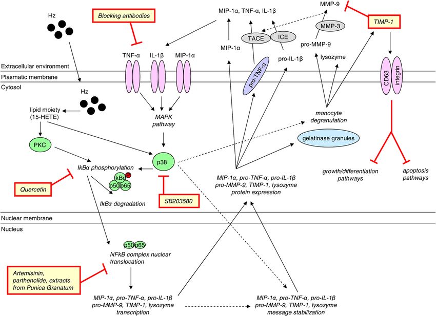
Polimeni and Prato Fluids and Barriers of the CNS 2014, 11:1                                                       Page 3 of 24
http://www.fluidsbarrierscns.com/content/11/1/1

and vascular endothelium are thought to mediate seques-             Enwonwu and colleagues implicated histamine as one of
tration of iRBCs within the brain resulting in removal            these toxic molecules that enters the brain parenchyma
from peripheral circulation [19-21]. The molecules in-            after BBB impairment and contributes to the neurological
volved in these interactions are parasite proteins expressed      manifestions of CM [33-37]. The authors observed altered
on iRBC surface, such as P. falciparum erythrocyte mem-           neural histidine uptake in children with severe falciparum
brane protein-1 (PfEMP-1), and specific host receptors            malaria providing an explanation for the enhanced cere-
in the microvascular endothelium, including intracel-             bral production of histamine [33]. They also found in-
lular adhesion molecule-1 (ICAM-1), vascular cellular ad-         creased plasma levels of histamine in severe malaria
hesion molecule-1 (VCAM-1), thrombospondin, CD36, and             patients, further supporting their hypothesis [34]. More-
E-elastin [22-25].                                                over, the involvement of histamine in CM has also re-
   Cytoadherence and decreased pliability are the main            cently been confirmed in a murine model [35-37]. In this
mechanisms underlying vascular obstruction [9,17-21].             study, histidine decarboxylase-deficient mice were unable
It is speculated that cytoadherence evolved as a mechan-          to synthesize free histamine and did not develop CM after
ism for the parasite to evade triggering a host immune            infection with P. berghei ANKA. These mice displayed
response and being cleared from the spleen. Cytoadherence         preserved BBB integrity, were void of iRBC aggregation in
is also beneficial for the parasite as to provide an optimal      the brain vessels, and did not sequester CD4+ and CD8+
environment of low oxygen tension for parasite growth.            T cells [36]. Further investigation of histamine receptors
Decreased deformability along with increased membrane             revealed histamine-1-receptor (H1R) and histamine-2-
stiffness and rigidity of iRBCs are due to changes in the         receptor (H2R) are associated with severe malaria devel-
cytoskeleton triggered by growing intracellular parasites.        opment [37], whereas histamine-3-receptor (H3R) has a
Cell deformability has been indicated as a predictor of           neuroprotective role [36].
anemia development [26], whereas cell rigidity correlates
with a higher fatality rate [27]. Another phenomenon              Humoral hypothesis
occurring along with iRBC sequestration is rosetting, char-       The humoral hypothesis is a natural extension of the per-
acterized by iRBCs forming a flower-like cluster around a         meability hypothesis. This hypothesis suggests that host
non-iRBC, making a tight rigid structure [28]. Rosetting is       factors such as leukocyte-derived cytokines and chemo-
more frequent in patients with CM than in those with un-          kines can enter the brain parenchyma after increased BBB
complicated malaria. However, rosette formation has also          permeability, thus causing CM symptoms such as fever
been reported for other Plasmodium strains (P. vivax and          and coma [9,13,14,16,38-40].
P. ovale) which do not cause CM [29]. Since rosetting oc-            Effector cells including T cells, NK cells, and monocytes,
curs in all manifestations of the disease, it is not associated   along with inflammatory responses mediated by cytokines
with severity or clinical outcome of CM [30]. One question        such as tumor necrosis factor-α (TNF-α), limphotoxin-α
the mechanical hypothesis by itself does not explain is why       (LT-α), and interferon-γ (IFN-γ), are proposed to contrib-
most patients recovering from CM do not show any evi-             ute to the development of murine CM [41-48]. However,
dence of ischemic brain damage [12].                              the extent of their involvement and molecular mecha-
                                                                  nisms in human CM is still topic of debate [48,49].
Permeability hypothesis                                              CD8+ T cells have been reported to initiate BBB tight
The permeability hypothesis proposes that BBB damage              junction disruption and promote CNS vascular permeabil-
is the underlying mechanism of CM, such that a leaky              ity under neuroinflammatory conditions [50-52]. Consist-
BBB allows toxic compounds to enter the brain and cause           ently, CD8+ T cell sequestration in cerebral microvessels
neurological dysfunction [9,13-15,31]. Several animal CM          and subsequent brain infiltration have been demonstrated
models have confirmed that the BBB is disrupted and that          in murine CM [43,44], where Plasmodium antigens can
cerebral edema is present in CM, although this is less evi-       be cross-presented during infection by dendritic cells
dent in humans [15]. Nevertheless, iRBCs remain attached          (DCs) [53,54] and brain endothelial cells in association
to endothelium, without entering the brain parenchyma             with MHC class I molecules [55]. Recent human studies
[7,9]. Interestingly, Adams and colleagues have suggested         support the idea that malaria antigens can be transferred
that iRBC cytoadherence might activate secondary signaling        to endothelial cells [56]. However, it is currently unknown
events similar to those occurring in leukocytes [32]. These       whether Plasmodium-specific CD8+ T cells are in-
secondary signaling events are thought to cause functional        volved in the pathogenesis of human CM [57]. Furthermore,
alterations in the BBB, which could allow toxic compounds         lymphocyte infiltration into brain parenchyma remains to
to pass into the CNS. These events might be reversible,           be investigated [49].
therefore explaining why neurological manifestations are             TNF-α relevance in CM is also unclear. TNF-α involve-
just transient in most cases and why a large number of re-        ment in murine CM was first proposed in 1987 [58]. Since
covering patients lack neurological sequelae [32].                then there have been numerous studies investigating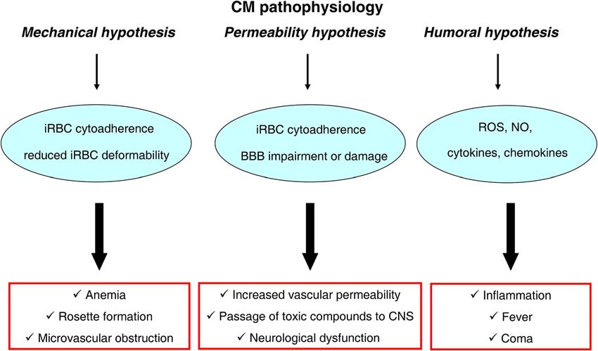
Polimeni and Prato Fluids and Barriers of the CNS 2014, 11:1                                                    Page 4 of 24
http://www.fluidsbarrierscns.com/content/11/1/1

TNF-α levels in CM mice albeit the results are inconsist-      junction damage and facilitating blood flow [81]. Lastly,
ent. For example, some works confirmed the association         treatment with exogenous L-arginine, the substrate for
of high TNF-α levels with murine CM [59-61], whereas           NOS, recently proved to be safe in a pilot study on CM
others argued against such correlation, finding LT-α and       patients, although effective doses still need to be opti-
IFN-γ as more suitable markers [44,47,62,63]. To recon-        mized [82].
cile such discrepancies, it has been proposed that low con-       Furthermore, during malaria infection both host and
centrations of TNF-α could enhance parasite killing,           parasite undergo strong oxidative stress, which leads to in-
whereas higher concentrations might be associated with         creased production of reactive oxygen species (ROS) and
increased incidence of murine CM [46]. However, data on        subsequent protein and lipid peroxidation [83,84]. The co-
TNF-α also appear inconclusive in human CM studies.            existence of both parasite and erythrocyte is a matter of a
Indeed, clinical studies tend to exclude any association       delicate balance: low ROS concentrations seem to inhibit
between CM and increased plasma, serum or CSF levels           parasite growth, whereas larger amounts may damage vas-
of TNF-α [64-67], although a few works have proposed a         cular endothelial cells and increase vascular permeability
correlation in two different Asian populations [68,69].        [85]. Oxidative stress paradoxically has both a pathogenic
Instead, in some of these studies, high CXCL10/IP-10           and protective role in CM [86]. An anti-oxidant diet was
plasma levels and low angiogenic factors such as vas-          shown to reduce BBB damage and counteract CM devel-
cular endothelial growth factor (VEGF) and angiopoietin-       opment in CM-sensitive mice [87], and anti-oxidant adju-
1 (Ang-1) in children with CM, predicted subsequent            vant therapy, provided at the initial stages of murine CM,
mortality [65,66,68]. Moreover, a protective role for IL-12    prevented the development of persistent cognitive damage
has been proposed in human CM [70,71].                         [88]. In contrast, NADPH-deficient mice were shown
   Among soluble factors involved in CM, a critical role       to develop CM despite the lack of ROS production,
for nitric oxide (NO) has also been suggested. It was hy-      suggesting that ROS did not contribute to CM pathogen-
pothesized that NO levels correlate with disease severity,     esis [89,90]. To reconcile such an apparent inconsistency,
since the sequestration of iRBCs might contribute to CM        Linares and colleagues have recently shown that glutathi-
pathogenesis by causing hypoxia, which is related to en-       one peroxidase and heme oxygenase-1 up-regulation co-
hanced production of cytokine-induced NO, compensa-            operate to suppress superoxide dismutase, catalase, heat
tory vasodilatation, and subsequent brain volume increase      shock protein-70 and thioredoxin-1 down-regulation ef-
[39]. However, activation of inducible NO synthase (NOS)       fects in murine CM, counteracting oxidative damage and
might also serve protective functions, since NOS inhibits      maintaining redox equilibrium [91]. In human CM, ROS
the side effects of brain indoleamine 2,3-dioxygenase          have been associated with a pathogenic role thus far. In
(IDO) and quinolinic acid accumulation [72], although          vitro, ROS inhibition was shown to protect brain endothe-
IDO systemic distribution is independent of malaria dis-       lial cells against P. falciparum-induced apoptosis and to
ease severity [73]. In a study performed on Tanzanian          decrease iRBC cytoadherence through ICAM-1 down-
children infected with malaria, the plasma levels of NOS-      regulation and iNOS induction [92,93]. Consistently, in a
suppressing IL-10 increased with disease severity, suggest-    recent clinical study performed on fifty Indian children
ing that a reduced NO production may contribute to CM          with severe malaria, oxidative stress was associated with
[74]. Moreover, a genetic single nucleotide polymorphism       disease severity [94].
found in the NOS2 promoter region causes elevated NO
production and was significantly associated with protec-       Blood–brain barrier impairment in cerebral malaria
tion against CM in Tanzanian and Kenyan children [75].         The BBB is one of three main barrier defences protecting
In line with these observations, Anstey and colleagues         the CNS. It is constituted of cerebral vascular endothelial
demonstrated that decreased NO production was associ-          cells, which do not form a rigid structure, but rather
ated with endothelial dysfunction in human CM [76,77].         a dynamic interface with a range of physical, biochemical
Similarly, van der Heyde and his group demonstrated            and immune properties and functions, built from effective
that low NO bioavailability was associated with mur-           inter-cellular junctions and cell-matrix adhesion mole-
ine CM [78,79]. Interestingly, prophylaxis with inhaled        cules, enzymes, and trans-endothelial transport systems
NO in CM-sensitive mice significantly reduced systemic         [95]. In particular, BBB integrity is dictated by tight junc-
inflammation and endothelial activation by lowering            tions between adjacent endothelial cells, forming a network
TNF-α, IFN-γ, monocyte chemotactic protein-1 (MCP-1),          of strands composed by several proteins, including junc-
sICAM-1 and von Willebrand factor, and by increasing           tional adhesion molecules, claudins (mainly -1 and -5) and
Ang-1 levels in peripheral blood [80]. The protective          occludin, which interact with cellular actin through cyto-
effect of exogenous NO on mouse CM appears asso-               plasmic proteins such as zonula occludens-1 (ZO-1) [96].
ciated with decreased brain vascular expression of in-         Figure 2 depicts the structure of neural inter-endothelial
flammatory markers, resulting in attenuated endothelial        tight junctions, along with cell-matrix adhesion complexes
Polimeni and Prato Fluids and Barriers of the CNS 2014, 11:1                                                                                 Page 5 of 24
http://www.fluidsbarrierscns.com/content/11/1/1

 Figure 2 Blood–brain barrier structure: cerebral microvascular inter-endothelial junctions (adherens and tight junctions) and
 cell-matrix adhesion molecules. Diagram showing the structures of CNS inter-endothelial junctions, including adherens junctions and tight
 junctions, and of cell-matrix adhesion complexes, including talin, filamin, tensin or α-actinin filaments associated with integrins in the extracellular
 matrix. The core of adherens junctions results after the interactions among transmembrane glycoproteins, such as VE-cadherin, whose cytoplasmic
 face is linked to the catenin family members, including p120-catenin, β-catenin, and α-catenin. Tight junctions are composed of a branching
 network of sealing strands, each of which is formed by extracellular domains of transmembrane proteins, claudins and occludin, joining one
 another directly. These transmembrane proteins associate with different peripheral membrane proteins such as ZO-1 located on the intracellular
 side of plasma membrane, anchoring the strands to the actin component of the cytoskeleton.

including talin, filamin, tensin or α-actinin filaments                        BBB status during CM are high variable among different
associated with integrins. We will next discuss how                            model systems [98].
the disruption of these molecules by host proteolytic en-
zymes such as MMPs could play a relevant role in CM                            Phenotype of brain and non-brain endothelial cells
pathophysiology.                                                               co-cultured with Plasmodium iRBCs in vitro
  BBB functional integrity and permeability are generally                      As discussed below and summarized in Table 1, evidence
assessed by evaluating the passage of molecules from the                       showing differential phenotypes between neural and non-
blood into the cerebral-spinal fluid (CSF). BBB perme-                         neural endothelial cells after co-culture with Plasmodium
ability is determined by size and charge of the molecules,                     iRBCs comes from several in vitro studies [56,93,99-108].
and the presence of specific BBB receptors to aid in the                         First, the effects of P. falciparum infection were inves-
transport of certain molecules. The importance of BBB                          tigated in a BBB model of cultured primary porcine brain
physiology and pathology has led to the development of                         capillary endothelial cells (PBCECs) [99]. In this study,
several BBB models to better investigate the physio-                           membrane-associated malaria antigens obtained from
logical, anatomical and functional characteristics [97].                       lysed P. falciparum schizont-iRBCs increased endothelial
However, once again the current experimental data on                           E-selectin and ICAM-1 expression, reduced the trans-
Table 1 Phenotype of endothelial cells after co-culture with infected red blood cells in vitro

                                                                                                                                                                                                             http://www.fluidsbarrierscns.com/content/11/1/1
                                                                                                                                                                                                             Polimeni and Prato Fluids and Barriers of the CNS 2014, 11:1
                    Endothelial cell type                           Plasmodium strain                Evaluated parameters                               Endothelial phenotype                        Ref.
       Porcine brain capillary endothelial cells (PBCEC)                P. falciparum            - ICAM-1, E-selectin expression;                  - increased ICAM-1 and E-selectin                 [99]
                                                                                                              - TEER;                                  - decreased BBB function;
                                                                                                   - tight junction expression                         - tight junction disruption
Human umbilical vascular endothelial cells (HUVEC) co-cultured   P. falciparum from patients          mRNA expression of:               - increased adhesion molecule mRNA (not CM-specific);        [100]
     with iRBC-fed peripheral blood mononuclear cells            with uncomplicated malaria,
                                                                                                      - adhesion molecules                    - reduced tight junction mRNA (CM-specific)
                                                                     severe malaria, or CM
                                                                                                  (ICAM-1, VCAM-1, E-selectin);
                                                                                                        - tight junctions
                                                                                                    (occludin, vinculin, ZO-1)
  TNF-α- or LT-α-activated human brain endothelial cell line            P. falciparum           - permeability to 70-kDa dextran;                     - increased BBB permeability;                  [101]
         (HBEC-5i) (with/without platelet co-culture)
                                                                                                              - TEER;                                  - decreased BBB function;
                                                                                               - endothelial microparticle release;                 - increased microparticle release;
                                                                                                     - endothelial apoptosis            - increased endothelial apoptosis (all effects potentiated
                                                                                                                                                              by platelets)
Human brain microvascular endothelial cells (HBMEC); HUVEC              P. falciparum                  - ICAM-1 expression                 increased ICAM-1 expression in HBMEC but not in           [93]
                                                                                                                                                               HUVEC
                           HBMEC                                        P. falciparum           - electrical cell substrate sensing;                    - reduced BBB function;                      [102]
                                                                                                              - TEER                                  - increased BBB permeability
   Human dermal microvascular endothelial cells (HDMEC);                P. falciparum           - immunofluorescence staining of               - loss in total protein content of claudin-5;         [103]
    human lung microvascular endothelial cells (HLMEC)                                             ZO-1, claudin-5, VE-cadherin;
            (with parasite sonicates or iRBCs)
                                                                                                - observation of inter-endothelial      - redistribution of ZO-1 from cytoskeleton to membrane
                                                                                                      gaps in monolayers;                              and cytosolic/nuclear fractions;
                                                                                                 - evaluation of pro-inflammatory       - minimal inflammation and death (all effects only with
                                                                                               response, direct cellular cytotoxicity                        sonicates)
                                                                                                           or cell death.
                           HBMEC                                        P. falciparum             - expression of transcriptome         - increased expression of ICAM-1 and pro-inflammatory        [104]
                                                                                                      (including ICAM-1 and                                    molecules
                                                                                                  pro-inflammatory molecules)
   HBEC-5i; immortalized human cerebral microvascular cell              P. falciparum          - immunofluorescent microscopy to          - malaria antigen presentation by endothelial cells;       [56]
                       line hCMEC/D3                                                                 evaluate malaria antigen
                                                                                                 presentation by endothelial cells;                     - tight junction opening;

                                                                                                              - TEER                                  - increased BBB permeability
                         hCMEC/D3                                       P. falciparum            - fluorescent permeability assay;                    - increased BBB permeability;                  [105]
                                                                                                 - expression of cell adhesion                       - increased ICAM-1 expression;
                                                                                                 molecules and tight junctions
                                                                                                                                                            - cytoadherence;

                                                                                                                                                                                                                                               Page 6 of 24
                                                                                                                                                       - altered ZO-1 distribution
Table 1 Phenotype of endothelial cells after co-culture with infected red blood cells in vitro (Continued)
http://www.fluidsbarrierscns.com/content/11/1/1
Polimeni and Prato Fluids and Barriers of the CNS 2014, 11:1
TNF-α-activated subcutaneous fat tissue-derived EC from P. falciparum - adhesion molecule expression - higher ICAM-1, VCAM-1, CD61; [106]
patients with uncomplicated malaria or CM (ICAM-1, VCAM-1, CD61, CD62-E)
- enhanced microparticle release;
- microparticle production;
- induced MCP-1 and IL-6 release;
- MCP-1, RANTES, IL-6 release ; - higher caspase-3 activation (all effects CM-specific)
- caspase-3 activation
HBEC-5i P. falciparum (various strains) parasite strain selection assay based CM-associated cytoadherence [107]
on cytoadherence
Murine brain vascular endothelial cells (MBVEC) murine P. berghei ANKA (CM model); - study of cytoadherence higher VCAM-1-mediated cytoadherence in CM model [108]
lung vascular endothelial cells (MLVEC) P. berghei K173 mechanisms; compared to non-CM model
(non-CM model)
Page 7 of 24Polimeni and Prato Fluids and Barriers of the CNS 2014, 11:1 Page 8 of 24 http://www.fluidsbarrierscns.com/content/11/1/1 endothelial electrical resistance (TEER), and promoted the CM) after co-culturing with iRBC-fed mononuclear cells, disruption of tight junctions, indicative of increased BBB however such increase did not appear specific for CM. On permeability. the contrary, reduced mRNA levels of tight junction pro- Consistently in various types of human brain endothe- teins (occludin, vinculin, and ZO-1) were strictly associated lium, including HMBEC primary cultures and HBEC-5i with CM [100]. or hCMEC/D3 cell lines, iRBCs were also shown to Genetic differences between Plasmodium strains might increase ICAM-1 expression [93,99,104,105], to reduce also play a role in CM development. Indeed, it has been TEER [56,101,102], to alter tight junction expression and shown that different strains of P. falciparum display variable distribution [56,105], and to enhance BBB permeabil- degrees of cytoadherence to HBEC-5i [107]. Additionally, ity to 70-kDa dextran [101]. Interestingly, platelets were P. berghei ANKA, a murine CM-associated Plasmodium suggested to play a key role in iRBC-dependent in- strain, induces a higher VCAM-1-mediated cytoadherence crease in BBB permeability, releasing microparticles and compared to P. berghei K173 (non-CM strain) in either causing cell apoptosis in TNF-α- and LT-α-activated brain or lung mouse vascular endothelial cells [108]. HBEC-5i [101]. In hCMEC/D3 cells, iRBC-increased cell adhesion and paracellular permeability correlated with Blood–brain barrier and in vivo animal models of cerebral ZO-1 disorganization, but the latter effect appeared medi- malaria ated by parasite-induced metabolic acidosis, independent Several in vivo animal models have reported alterations from cytoadherence [105]. Moreover, differential global in BBB after exposure to Plasmodium parasites or mal- gene expression in HBMEC after interacting with iRBCs aria products such as hemozoin (Hz, malarial pigment) revealed significantly up-regulated transcripts related to [47,109-124]. As summarized in Table 2 and described immune and inflammatory responses, apoptosis, cell- below, these studies provide insightful findings regarding cell signaling, signal transduction and nuclear factor- BBB breakdown in animal CM models. kB (NF-kB)-activation cascade [104]. After co-culturing The first animal studies on BBB permeability in mal- with iRBCs, the mRNA expression of neural endothelial pro- aria date back to 1968, when Migasena and Maegraith inflammatory chemokines (IL-6, CXCL-8/IL-8, CXCL-1/ demonstrated the movement of albumin across the BBB GRO-α, CXCL-2/MIP-2α, and CCL-20/MIP-3α) increased in Macaca mulatta monkeys infected with P. knowlesi more than 100-fold, highlighting the strong inflamma- [109-111]. However, P. knowlesi does not induce CM. As tory component and the active role of the endothelium in such, the rhesus monkey infected with primate malaria CM pathogenesis [104]. Furthermore, in TNF-α-activated parasites, P. coatneyi and P. fragile, is considered to be a subcutaneous fat tissue-derived endothelial cells, a model more valid primate model to study in the context of se- comparable to cerebral endothelium, P. falciparum vere malaria with cerebral involvement [112-114]. iRBCs induced several CM-specific effects, including Of the four species of rodent malaria parasites (P. berghei, up-regulation of ICAM-1, VCAM-1, and CD61, en- P. yoelii, P. chabaudi, P. vinckei), only a few P. berghei strains hancement of microparticle, MCP-1 and IL-6 release, can induce experimental CM in mice, with the ANKA strain and higher caspase-3 activation [106]. Increased levels of being the most widely studied. Symptoms of experimental inflammatory cytokines may have direct systemic effects CM in P. berghei ANKA-infected susceptible mice include and adversely affect the clinical outcome by increasing the paralysis, ataxia, head deviation, convulsion and coma cytoadherence of infected RBCs to venular endothelium [98]. In P. berghei K173-infected mice an excessive move- through up-regulation of adhesion molecules, such as ment of water, albumin and other proteins into the brain, ICAM-1 [93]. as well as severe brain edema, microthrombosis, sludging To assess the specificity of these effects for human of mononuclear cells, arteriolar spasms, scattered distur- cerebral endothelium, additional comparative studies bances of the microcirculation, and occasional prolifera- were also performed using non-neural endothelial cells. tion of gliocytes were observed, suggesting a progressive Interestingly, P. falciparum iRBCs did not affect the ex- deterioration of BBB integrity culminating in endothelial pression and distribution of tight junctions (as measured by lesions and haemorrhages [31,115-118]. Of note, mouse claudin-5 and ZO-1) and did not induce pro-inflammatory CM models present neurological signs (ataxia, hemiplegia response or cell death in human dermal or lung micro- and coma) similar to the clinical features reported in hu- vascular endothelium, although parasite sonicates did [103]. man CM [119]. Additionally, the up-regulating effects of iRBCs on ICAM-1 In a recent work, Penet and colleagues presented the expression observed in HBMEC were not reproduced in first in vivo magnetic resonance study of mouse CM, human umbilical vascular endothelial cells (HUVEC) from demonstrating BBB breakdown in CM. Multimodal mag- healthy donors [93]. An increase in ICAM-1, VCAM-1, netic resonance neuroimaging techniques (imaging, diffu- and E-selectin mRNA was found in HUVEC from patients sion, perfusion, angiography, spectroscopy) of P. berghei with different degrees of malaria (uncomplicated, severe, or ANKA-infected mice revealed vascular damage, including
Polimeni and Prato Fluids and Barriers of the CNS 2014, 11:1                                                                               Page 9 of 24
http://www.fluidsbarrierscns.com/content/11/1/1

Table 2 Evidence of blood–brain barrier (BBB) impairment in animal models with cerebral malaria (CM)
     Animal source             Plasmodium            Method to evaluate BBB integrity                    Degree of impairment                 Reference
                                  strain
      Rhesus monkey             P. knowlesi         Examination of movement of proteins                Increase of BBB permeability           [109-111]
     (Macaca mulatta)                                 across the BBB by radiometric and
                                                             fluorimetric methods
      Rhesus monkey              P. fragile      Electron microscopy, immunohistochemical        Parasitized red blood cells sequestration      [112]
     (Macaca mulatta)                             analysis (CD36, thrombospondin, ICAM-1),         and adherence to endothelial cells in
                                                             formation of rosettes                the cerebral microvessels, neurological
                                                                                                       symptoms similar to humans
      Rhesus monkey             P. coatneyi                    Clinical observation               Anemia, coagulopathy, and renal and           [113]
     (Macaca mulatta)                                                                                   metabolic dysfunction
      Rhesus monkey             P. coatneyi      Tissue samples from the brain (cortex and         Expression of pro-inflammatory and T         [114]
     (Macaca mulatta)                            white matter of the cerebrum, cerebellum,        helper-1 cytokines, adhesion molecules,
                                                 and midbrain) collected for quantitation of     and iNOS appears to predominate in the
                                                  mRNA expression of cytokines, adhesion          cerebellum of infected rhesus monkeys
                                                           molecules, and iNOS
   A/J and CBA/H mice        P. berghei (ANKA)     Detection of the movement of the dye                     Breakdown of BBB                    [115]
                                                  Evans blue, radioisotope labelled albumin
                                                               and erythrocytes
          mouse              P. berghei (K173)   Histochemical and histological evaluation of Progressive deterioration of BBB integrity      [116-118]
                                                     cerebral lesions and their distribution
     CBA/T6, Balb/c             P. berghei             Evaluation of neurological signs               Increased permeability of BBB             [119]
    and DBA/2 J mice         (ANKA and K173)            (ataxia, hemiplegia and coma)
          Mouse              P. berghei (ANKA)   Multimodal magnetic resonance techniques                    BBB breakdown                      [120]
                                                 (imaging, diffusion, perfusion, angiography,
                                                                spectroscopy).
CM- resistant BALB/c mice    P. berghei (ANKA)    Evaluation of pro-inflammatory cytokines                   BBB breakdown                      [121]
                                                                  produced
C57BL/6 and BALB/c mice      P. berghei (NK65)   Histopathological analysis of cerebral tissue        Increased permeability of BBB             [122]
        TNF-α-and            P. berghei (ANKA)    Histochemical and histological evaluation       Neurological signs of CM, associated          [47]
    LT-α-deficient mice                                                                          with perivascular brain haemorrhage in
                                                                                                 TNF-α -/- mice; completely resistant to
                                                                                                           CM in LT-α -/- mice
          Mouse              P. berghei (ANKA)   Examination of the outcome of TGF-β and           Critical balance between TGF-β and           [123]
                                                    TNF-α production in the context of             TNF-α might have a key role in BBB
                                                           splenocyte apoptosis                                  breakdown
 Different murine models:   P. berghei (ANKA)    Examination of histopathological alterations,        CM related to the opening of              [124]
CBA/CaJ and Swiss Webster     P. yoelii (17XL)      BBB dysfunction, or neurological signs       paracellular-junctional and transcellular-
mice (CM sensitive), Balb/c P. berghei (NK65)                                                    vesicular fluid transport pathways at the
and A/J mice (CM resistant) and P. yoelii (YM)                                                           neuroimmunological BBB

BBB disruption and haemorrhages, major edema forma-                           hand, P. berghei NK65-infected mice showed enhanced pro-
tion, reduced brain perfusion and ischemic metabolic pro-                     duction of LT-α and several chemokines (CXCL-9/MIG,
file, with reduced high-energy phosphates and enhanced                        CCL-2/MCP-1, CCL-3/MIP-1α and CCL-5/RANTES), but
brain lactate. These data strongly point to the coexistence                   no neurological symptoms [47,122]. A complementary study
of inflammatory response and ischemic lesions [120].                          performed on the same model proposed a concurrent role
   Other recent works illustrated a complex strain-dependent                  for Transforming Growth Factor-β (TGF-β) and TNF-α in
relationship between leukocyte recruitment, BBB perme-                        promoting splenocyte apoptosis [123].
ability and chemokine production. Major pathological con-                       It should be noted that the cerebral microvascular tree
sequences of malaria arise from inappropriate or excessive                    contains two functionally distinct BBB: i) the physio-
immune response mounted by the host in an attempt to                          logical BBB, formed by capillaries 4–8 mm in diameter,
eliminate the parasite. In P. berghei ANKA-infected                           consisting of a single layer of endothelia, gliovascular mem-
mice, inflammation of the cerebral microvasculature and                       brane, and astrocyte endfeet; and ii) the neuroimmunologi-
leukocyte recruitment were clearly evident and found to                       cal BBB, formed by postcapillary venules 10–60 mm in
be driven by production of pro-inflammatory cytokines                         diameter and encompassing two layers - the endothelium
(IL-12, IFN-γ) and CM development [121]. On the other                         with its basement membrane and the glia limitans with
Polimeni and Prato Fluids and Barriers of the CNS 2014, 11:1 Page 10 of 24
http://www.fluidsbarrierscns.com/content/11/1/1
associated astrocyte endfeet - separated by the perivascular Regarding African populations, a study on Zairean chil-
space [125]. The physiological BBB serves as a tight diffu- dren showed no difference in CSF albumin compared to
sion barrier for small solutes while the neuroimmunological controls [128]. However, in Malawian children with CM,
BBB permits transport of macromolecules and diapedesis the activation of endothelial cells and macrophages, along
of immune cells [125]. In a very recent study comparing with the disruption of endothelial intercellular junctions
different mouse models of experimental CM (P. berghei in vessels containing sequestered iRBCs, and subtle but
ANKA infection), human CM-like histopathology (P. yoelii measurable changes in albumin CSF versus albumin serum
17XL) and non-CM (P. berghei NK65 and P. yoelii YM), levels were observed. Nevertheless, negligible leakage of
Nacer and colleagues observed that the physiological plasma proteins was still apparent [129]. In Kenyan chil-
BBB in the experimental CM model remained intact, dren with CM, protein and amino acid levels in paired
whereas regulated fluid transport across the neuroimmu- plasma and CSF samples were measured, showing that
nological BBB led to brain swelling, intracranial hyperten- BBB was mildly impaired in some children with severe
sion, coma, and ultimately death due to dysfunction of falciparum malaria [130]. However, this impairment was
respiratory centers in pons and the medulla oblongata as a not confined to CM, as it was also reported in children with
result of brain stem compression [124]. Thus, they pro- prostration-associated malaria and, to a lesser extent, in
posed that CM may occur in two steps: 1) induction of children with malaria and seizures. Evidence of intrathecal
coma based on regulated, preventable and reversible immunoglobulin synthesis in children with malaria was also
opening of the neuroimmunological BBB; and 2) endothe- observed [130]. Finally, data obtained in a recent work per-
lial death-associated haemorrhaging, which is difficult to formed on Malawian children are consistent with the pro-
reverse by treatment and eventually fatal [124]. A similar posed link between iRBCs sequestration and intravascular/
mechanism for neuroimmunological BBB opening in hu- perivascular pathology in fatal pediatric CM, resulting in
man CM would explain the reversibility of coma with myelin damage, axonal injury, and BBB breakdown; how-
treatment, the scarce traces of tissue necrosis in surviving ever, no Hz-laden monocyte extravasation was found [131].
patients, and the different neurological outcomes of pa- Pathological studies on post mortem samples of CM
tients despite similar clinical presentation [6,8,9,13,102]. patients showed cerebral edema and raised intracranial
pressure in 50% of West African children [132] but not
Blood–brain barrier and human studies on cerebral malaria in South Asian adults [133,134] or Malawian children
BBB functional impairment during human CM has been [129]. Nevertheless, an important correlation between
investigated in several clinical and post mortem studies sequestration of iRBCs in the brain microvessels and the
[126-143]. Table 3 summarizes the most relevant results. malaria-related encephalopathy was shown in Asian pa-
Here, the investigations on human CM patients were tients [133]. The adhesion of iRBCs to brain microvessels
performed using albumin CSF/serum ratio as an indica- is mediated by specific receptors on the host endothelium,
tor of BBB integrity [126-128], by post mortem immuno- including ICAM-1, CD36 and CD31 [22-25]. Immunohis-
histochemical analysis [129-135], or through brain imaging tochemistry showed altered distribution of the cell junc-
techniques [136-144]. Interestingly, the BBB seems to be tion proteins occludin, vinculin and ZO-1 in Vietnamese
more impaired in children than in adults. Moreover, it ap- adults and Malawian children with CM [129,135]. Seques-
pears that African and Asian patients display a different tration of iRBCs in cerebral microvessels was significantly
degree of BBB damage, with BBB breakdown being more higher in the brains of patients with CM compared with
likely to occur in African than Asian populations. non-CM patients in all parts of the brain (cerebrum, cere-
One of the first studies on Asian patients was conducted bellum, and medulla oblongata), and was quantitatively as-
in Thailand [126]. In this work, albumin CSF/serum ratios sociated with pre mortem coma [129].
were higher in CM patients than in controls, but it did In recent years, several imaging studies have been also
not correlate with coma and mortality. Thus, the authors conducted on the brains of CM patients during disease pro-
concluded that their data did not support the idea that gress or after recovery [136-144]. Using magnetic resonance
cerebral edema might be the cause of coma. More than a or computed tomography, several common features impli-
decade later, albumin and Immunoglobulins G plasma/ cating BBB damage have been observed, including cerebral
CSF ratios were found to be only mildly impaired in edema, increased brain volume, ischemia and large vessel
Vietnamese patients, suggesting only minimal degree of infarcts, hemorrhagic cortical lesions, focal and multifocal
BBB breakdown in few CM cases [127]. Therein, human atrophy, and limited CSF circulation [136-139,141-144].
CM appeared to cause only subtle functional changes in Interestingly, magnetic resonance in a recently published
BBB integrity, with minimal intra-parenchymal inflamma- case-report of a 37-year-old French patient with malaria
tory response compared with other neurologic infections, travelling back from Equatorial Guinea, showed that he
such as cryptococcal, tubercular, and acute bacterial men- developed posterior reversible encephalopathy syndrome,
ingitis [127]. which is characterized by diffuse symmetric signal-intensityPolimeni and Prato Fluids and Barriers of the CNS 2014, 11:1 Page 11 of 24
http://www.fluidsbarrierscns.com/content/11/1/1
Table 3 Evidence of blood–brain barrier (BBB) impairment in human cerebral malaria (CM) patients
Group type Plasmodium Number of Method to evaluate BBB integrity Degree of impairment Reference
strain patients per
cohort
Thai patients P. falciparum 157 Albumin CSF/serum ratio BBB intact [126]
Vietnamese patients P. falciparum 20 Albumin and Immunoglobulins G Minimal BBB breakdown in a few [127]
plasma/CSF ratios cases of CM
Zairean children P. falciparum 21 Albumin CSF/serum ratio BBB not impaired [128]
Malawian children P. falciparum 72 Immunohistochemistry on autopsy Disruption of endothelial intercellular [129]
brain tissues junctions and impaired BBB function
Kenyan children P. falciparum 100 Protein and immunoglobulin CSF/ Mild BBB impairment in some cases [130]
serum ratio
Malawian children P. falciparum 50 Immunohistochemistry on autopsy BBB breakdown [131]
brain tissues
Nigerian children P. falciparum 61 Examination of the possible risk Cerebral edema and raised [132]
factors for poor prognosis and intracranial pressure in 50%
studies on post mortem samples
Thai and Vietnamese P. falciparum 65 Studies on post mortem samples Cerebral sequestration of [133]
children P. falciparum-infected erythrocytes
Vietnamese patients P. falciparum 20 Studies on post mortem samples Heterogeneous cerebral edema [134]
and plasma protein leakage
Vietnamese adults P. falciparum 14 Immunohistochemistry Alteration of cell junction proteins [135]
and Malawian occludin, vinculin and ZO-1
children
Kenyan children P. falciparum 14 Computed tomography Cerebral edema and ischemia [136]
French adults back P. falciparum 3 Magnetic resonance Hemorrhagic cortical lesions [137]
from Cameroon,
Niger, and Thailand
Malian children P. falciparum 8 Computed tomography Diffuse atrophy with asymmetrical ventricle [138]
dilation, suggesting limited CSF circulation
French adult back P. falciparum 1 Magnetic resonance BBB breakdown [140]
from Equatorial
Guinea
Malawian children P. falciparum 14 Computed tomography Fatal CM: cerebral edema, large vessel [141]
infarcts; Non fatal CM with neurological
sequelae: focal/multifocal atrophy
Indian adults P. falciparum 4 Magnetic resonance Bithalamic infarctions with or [142]
without haemorrages
Malawian children P. falciparum 120 Magnetic resonance increased brain volume; abnormalities [143]
in cortical, deep gray, and white
matter structures
Malawian children P. falciparum 38 Magnetic resonance periventricular and subcorical T2 signal
changes, atrophy, and focal cortical defects
abnormalities of white matter in the posterior circula- several studies performed on mouse CM models suggest a
tion territory [140]. Since data from previous litera- strong BBB breakdown [115-124], data on increased BBB
ture suggest one of the mechanisms of posterior reversible permeability in human CM are somehow less evident,
encephalopathy involves capillary leakage and acute generally suggesting the occurrence of only mild BBB im-
disruption of the BBB, the authors concluded that this pairment, characterized by a relevant degree of tight junc-
case-report supports the theory of BBB disruption as a key tion disruption, but lacking molecule exchange between
factor for CM development [140]. serum and CSF [126-135].
In this context, it should be noted that the relevance
Blood–brain barrier impairment in cerebral malaria: some of murine CM models for studying CM pathophysiology
reflections upon the available studies has been a topic of big debate in the recent years [49].
Clearly there is much discrepancy on the extent of BBB Being clearly an inflammatory syndrome with local vas-
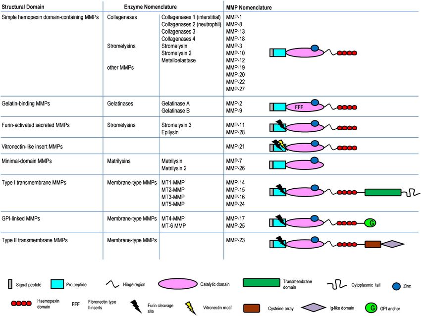
damage between animal and human models of CM. While cular endothelial activation, murine CM displays obviousPolimeni and Prato Fluids and Barriers of the CNS 2014, 11:1 Page 12 of 24 http://www.fluidsbarrierscns.com/content/11/1/1 differences and some similarities to the clinical and BBB permeability in pediatric CM is still unknown. Future pathological features of human CM, such as signs of research aimed at shedding light on this topic will cer- vascular inflammation/damage [145]. A recurring issue tainly be useful. concerns the degree of iRBC sequestration in the brain and other organs of P. berghei ANKA–infected mice. Al- Involvement of matrix metalloproteinases in cerebral though recent data find increased iRBC accumulation malaria during murine CM in multiple organs including the In the last decade, experimental evidence implicated a spe- brain [146], P. berghei infection is generally acknowledged to cific family of host proteolytic enzymes known as MMPs promote marked accumulation of leukocytes (particularly in malaria pathogenesis [2,151-154]. MMPs are either se- monocyte, macrophages and T cells), which is in stark con- creted or membrane-bound zinc-dependent proteases, and trast to human CM [147]. Thus, despite several processes their role is also related to the inflammatory response and shared either by murine or human CM, the changes in the the BBB function [155-163]. Members of the MMP family endothelial cell microenvironment induced by cytoadher- are produced by a broad spectrum of specialized cells, in- ence and inflammation are not the same [104]. Addition- cluding fibroblasts, endothelial cells, lymphocytes, mono- ally, mouse studies suggesting associations between high cytes, macrophages, smooth muscle cells, glial cells, and levels of cytokines and CM [58-61] have been recently neurons [155,158]. challenged by works showing that high levels of pro- As detailed in Figure 3, the mammalian MMP family inflammatory cytokines such as TNF-α are poor indicators encompasses 25 members, categorized by different num- of human CM in African children [64-67]. Thus, future bers or named depending on their matrix substrates. experimental studies on alternative animal models (non- MMPs are evolutionarily conserved and tightly regu- human primates and other mouse models) are encouraged lated. Conserved protein domains include an N-terminal and urgently necessary to better understand the patho- signal peptide required for secretion, a cleavable pro- logical processes underlying human infection [49]. domain maintaining enzymatic latency, a catalytic domain, Another interesting point emerging from clinical data a Zn-binding domain, and (besides the minimal MMPs) a is that the BBB appears more impaired in children than C-terminal hemopexin domain thought to be important in adults [126-143]. Since CM often strikes children at a for protein-protein interactions [155,157,160,162,163]. critical time in brain development, Hawkes and col- The active domain and the Zn-binding domain are essen- leagues have nicely hypothesized that developmental tial for catalytic activity: upon pro-domain cleavage a changes in the cerebral vasculature may account for Zn2+-ion becomes available to coordinate with a hydrolytic some of the differences in disease presentation and out- water molecule to enable nucleophilic attack of a substrate, come between children and adults, including mortality, and the enzyme is functionally active [164]. Additional seizures and neurocognitive sequelae, rates of associated MMP motifs include a gelatin-binding fibronectin-like do- anemia and renal dysfunction, retinal vessel changes, main, a serine-, threonine- and proline-rich collagen type frequency of ring haemorrhages, and inflammatory cell V-like domain, a C-terminal transmembrane (TM) domain accumulation in brain microvessels [148]. To avoid any or GPI anchor, and in some cases a cytoplasmic domain misunderstanding, it has been pointed out that the wide- [155,157,160,162,163]. MMP-2 and MMP-9 (also named spread belief amongst neurotoxicologists that BBB is im- gelatinase A and B) are further characterized by the pres- mature or even absent in the newborn is contrasted by a ence of three head-to-tail cysteine-rich repeats within substantial body of evidence supporting the concept of the catalytic domain reminiscent of the collagen-binding well-developed barrier mechanisms in the developing type II repeats of fibronectin and this domain is necessary brain [149,150]. For instance, inter-cellular tight junc- for the binding and cleaving activities of these MMPs tions between cerebral endothelial cells and between [161,165,166]. Six membrane-anchored MMPs (MT1- choroid plexus epithelial cells are functionally effective to MT6-MMPs) have a basic RX(K/R)R motif at the as soon as they differentiate [150]. Nevertheless, the brain C-terminal end of their pro-domains. This motif is recog- develops within an environment that is different from that nized and cleaved intracellularly by furin-like proteases. of the rest of the body, and the developing brain possesses Four MT-MMPs (MT1- to MT4-MMPs) are anchored to a number of unique features not generally present in the the cell membrane through a type I TM domain while the adult [149]. Interestingly, certain genes coding for influx/ other two MT-MMPs (MT5- and MT6-MMPs) are teth- efflux proteins are expressed at much higher levels early ered to the membrane via a GPI moiety. An additional in development than in the adult, and there is physio- MT-MMP (MMP-23) has an N-terminal type II TM do- logical evidence that these transport systems are function- main [162]. ally more active in the developing brain [150]. How such MMPs are regulated at multiple levels including transcrip- differences between the pediatric and adult BBB can tion, translation, compartmentalization, secretion, activation, affect CM pathogenesis and correlate with enhanced and inhibition by protein inhibitors. Most MMPs are found
Polimeni and Prato Fluids and Barriers of the CNS 2014, 11:1                                                                            Page 13 of 24
http://www.fluidsbarrierscns.com/content/11/1/1

 Figure 3 Nomenclature and structure of mammalian matrix metalloproteinases(MMPs). The mammalian MMP family encompasses 25
 members, categorized by different numbers (standard MMP nomenclature) or named depending on their matrix substrates (enzyme nomenclature).
 Each MMP displays some conserved structural domains, including: i) an N-terminal signal peptide required for secretion; ii) a cleavable pro-domain
 maintaining enzymatic latency; iii) a catalytic and Zn-binding domain; and iv) a C-terminal hemopexin domain. Optional MMP motifs include a
 fibronectin-type domain, a vitronectin motif, a furine cleavage site, three head-to-tail cysteine-rich repeats, and (for MT-MMPs) a
 C-terminal transmembrane domain or GPI anchor occasionally associated with a cytoplasmic domain.

at low levels and not constitutively transcribed, but are                    pro-MMP-9 is activated through a proteolytic cascade
expressed after external induction by pro-inflammatory                       sequentially involving plasminogen, MMP-3 and MMP-1
molecules, growth factors, NO, cell-cell interactions, cell-                 [165]. The activation of proMMP-2 requires previous forma-
matrix interactions, UV radiations [155,157,159-162]. Sev-                   tion of a pro-MMP-2/tissue inhibitor of metalloproteinase-2
eral signalling pathways and transcription factors are                       (TIMP-2)/MT1-MMP (MMP-14) multimeric complex
known to regulate MMP expression, including mitogen-                         [166]. In addition to pro-MMP-2 activation, the bind-
activated protein kinases (MAPKs), NF-kB, and activator                      ing of TIMP-2 to MT-1-MMP and MT-3-MMP slows
protein-1 (AP-1) [155,165,167]. After synthesis, MMPs are                    down the autocatalytic turnover of these MT-MMPs, para-
stored in inflammatory cell granules, which restrict their ac-               doxically enhancing surface proteolysis further by stabiliz-
tion [161]. Furthermore, MMPs are produced as inactive                       ing the pool of active enzyme at the cell surface [160,168].
zymogens, referred to as pro-MMPs. Activation is achieved                    Once MMPs have been released into the extracellular
by various proteases (other activated MMPs and several                       space or anchored to the membrane and activated, they
serine proteases) or ROS that disrupt the interaction be-                    are kept in check by their endogenous tissue inhibitors
tween the active site zinc atom in the catalytic domain                      (four different forms, from TIMP-1 to TIMP-4). TIMPs
and the conserved cysteine within the pro-domain. Pro-                       inhibit MMP activity with relatively low selectivity in a 1:1
MMPs can be cleaved and activated through different mech-                    stoichiometric ratio. Interestingly, the ratio of MMP:TIMP
anisms and in a context-specific manner. For example,                        can also influence activation mechanisms [157,167].
Polimeni and Prato Fluids and Barriers of the CNS 2014, 11:1                                                     Page 14 of 24
http://www.fluidsbarrierscns.com/content/11/1/1

  MMPs were originally discovered in tadpoles as the agents      enhanced, suggesting a protective role for this MMP in CM
responsible for tail resorption during frog metamorphosis.       [172]. In another study, CM mice showed increased neural
Thus, they were first characterized as proteases involved in     MMP-7 protein levels [151]. Interestingly, urokinase-type
degrading structural proteins comprising the extracellular       plasminogen activator (uPA) -/- or urokinase-type plas-
matrix (ECM) and sub-endothelial basement membranes              minogen activator receptor (uPAR) -/- knock-out mice
[155]. However, MMPs are now known to have more so-              with CM displayed enhanced survival and attenuated
phisticated processes than mere ECM turnover. MMPs               thrombocytopenia [174].
can also cleave a growing spectrum of other substrates, in-         A parasite molecule, malarial pigment Hz - a lipid-
cluding cytokines, chemokines, growth factors, hormones,         bound ferriprotoporphyrin IX produced by P. falciparum
chemotactic and adhesion molecules, membrane recep-              after hemoglobin catabolism [175] - is proposed to play
tors, intercellular junctions, as well as other proteases, in-   a role in experimental CM. Indeed, using a sensitive
cluding some hemostasis-related molecules and MMPs               fluorometric method to determine Hz content in blood
themselves, protease inhibitors, clotting factors, and anti-     and tissue samples from mice infected with P. berghei
microbial peptides [169-171]. MMP-dependent cleavage             NK65 (non-CM model) or ANKA (CM model), Sullivan
can serve to activate, inhibit, process, release, shed, or re-   and colleagues observed increased Hz levels in tissue
veal cryptic codes in the substrates they act on. Therefore,     correlating with the duration of infection, with neural
the once formidable proteolytic potential of MMPs is now         Hz levels being higher in CM than non-CM mice, rais-
realized to serve essential roles in promoting or inhibiting     ing the possibility that Hz presence may be associated
cell survival, proliferation, migration, invasion, hemostasis    with cerebral pathology [176].
and inflammation in either physiological or pathological            Interestingly, in vitro, Hz appears to play a major role
processes [159,167,169,170].                                     in MMP dysfunction. Phagocytosis of Hz by RAW 264.7
  In physiology, MMPs are involved in diverse biological         rat macrophage cell line was shown to impair expression
mechanisms ranging from wound repair to pregnancy                of several inflammatory molecules [177] and, after an
[155,157,159,167]. In pathology, MMP dysfunction has             early inhibitory peak, to increase the long-term mRNA
been implicated in cancer, cardio-vascular diseases, em-         expression of MMP-9 [178]. This effect was related to the
physema, acute renal failure, ophthalmic pathologies, neu-       lipid moiety of Hz, since lipid-free synthetic Hz (β-hematin)
roinflammation, neurodegenerative disorders, autoimmune          did not modulate MMP-9 expression. The Hz-dependent
diseases [156-160,167,169,170] and, very recently, malaria       enhancement of MMP-9 transcription and protein re-
[2,151-154].                                                     lease was mimicked by 4-hydroxy-2-nonenal (4-HNE)
                                                                 [178], a molecule generated by Hz from polyunsaturated
Matrix metalloproteinases and animal models                      fatty acids [179].
In vivo mouse models of CM have recently implicated
MMP dysfunction in disease pathology, although it should         Matrix metalloproteinases and human studies
be kept in mind that experimental CM presents important          In vitro studies using human monocytes and endothelial
differences compared to human CM, such as leukocyte              cells [152,180-195] provide convincing and homoge-
sequestration in cerebral microvessels and subsequent mi-        neous evidence for Hz-dependent mechanisms underlying
gration into brain parenchyma [49]. An excellent study           aberrant MMP-9 function (see Figure 4). In a series of
performed by Van den Steen and his group comprehen-              works performed with human adherent or immunopurified
sively investigated mRNA expression levels of MMPs and           monocytes from peripheral blood, the phagocytosis of free
protein release or pro-enzyme activation in five differ-         Hz or Hz-containing trophozoites enhanced MMP-9
ent organs (brain, lung, spleen, liver, and kidney) from         mRNA levels, protein expression, and activity [180-186].
CM-sensitive C57B1/6 mice infected with P. berghei ANKA          This observation was also investigated using THP-1 mono-
(CM model) or P. berghei NK65 (non-CM model) and                 cyte cell line [187]. Hz-fed monocytes display increased
CM-resistant Balb/C mice infected with P. berghei ANKA           total gelatinolytic activity [186] and invasiveness [180]
(CM-resistant model) [172]. Importantly, they observed en-       caused by MMP-9 - but not MMP-2 [188] - enhancement.
hanced expression and activation of monocytic (CD11b+)              Increased MMP-9 function in human monocytes ap-
MMP-9 in brains of CM mice [172] specific to CM, as sug-         pears to be mediated by Hz-dependent over-production
gested by comparison with non-CM models, such as lung            of several pro-inflammatory molecules, including TNF-α
pathology [173]. Additionally, tissue-specific increases in      [180], IL-1β [181], and CCL-3/MIP-1α [184]. Further in-
mRNA expression were found for several MMPs, in-                 vestigation revealed increases in MMP-9 [181], TNF-α
cluding MMP-3, -4, -8, and -13 in spleen, MMP-8, -12, -13,       [189,190] and IL-1β [181,190], but not CCL-3/MIP-1α
and -14 in liver, and MMP-8 and -13 in brain. All of these       [183,190], were dependent on the lipid moiety of Hz.
increases were more pronounced in the CM model. In a             These studies unveiled a major role for 15-HETE, a potent
CM-resistant model, MMP-3 expression was significantly           lipid peroxidation derivative generated by Hz autocatalysis.
You can also read