Selective inhibition of CDK7 reveals high-confidence targets and new models for TFIIH function in transcription - Genes Dev
←
→
Page content transcription
If your browser does not render page correctly, please read the page content below
Downloaded from genesdev.cshlp.org on November 11, 2020 - Published by Cold Spring Harbor Laboratory Press

Selective inhibition of CDK7 reveals
high-confidence targets and new models
for TFIIH function in transcription
Jenna K. Rimel,1,15 Zachary C. Poss,2,15 Benjamin Erickson,3,4 Zachary L. Maas,1,2,5
Christopher C. Ebmeier,2 Jared L. Johnson,6 Tim-Michael Decker,1 Tomer M. Yaron,6,7,8,9
Michael J. Bradley,10 Kristin B. Hamman,10 Shanhu Hu,10 Goran Malojcic,10 Jason J. Marineau,10
Peter W. White,11 Martine Brault,11 Limei Tao,11 Patrick DeRoy,11 Christian Clavette,11
Shraddha Nayak,12 Leah J. Damon,1,5 Ines H. Kaltheuner,13 Heeyoun Bunch,14 Lewis C. Cantley,6
Matthias Geyer,13 Janet Iwasa,12 Robin D. Dowell,2,5 David L. Bentley,3,4 William M. Old,2
and Dylan J. Taatjes1
1
 Department of Biochemistry, University of Colorado, Boulder, Colorado 80303, USA; 2Department of Molecular, Cellular, and
Developmental Biology, University of Colorado, Boulder, Colorado 80309, USA; 3Department of Biochemistry and Molecular
Genetics, University of Colorado School of Medicine, Aurora, Colorado 80045, USA; 4UC-Denver RNA Bioscience Initiative,
University of Colorado School of Medicine, Aurora, Colorado 80045, USA; 5BioFrontiers Institute, University of Colorado, Boulder,
Colorado 80309, USA; 6Meyer Cancer Center, Weill Cornell Medicine, New York, New York 10065, USA; 7Englander Institute for
Precision Medicine, 8Institute for Computational Biomedicine, 9Department of Physiology and Biophysics, Weill Cornell
Medicine, New York, New York 10065, USA; 10Syros Pharmaceuticals, Massachusetts 02140 USA; 11Paraza Pharma, Inc.,
Montreal, Quebec H4S 1Z9, Canada; 12Department of Biochemistry, University of Utah, Salt Lake City, Utah 84112, USA;
13
  Institute of Structural Biology, University of Bonn, Bonn 53127, Germany; 14School of Applied Biosciences, College of Agriculture
and Life Sciences, Kyungpook National University, Daegu 41566, Republic of Korea

CDK7 associates with the 10-subunit TFIIH complex and regulates transcription by phosphorylating the C-terminal
domain (CTD) of RNA polymerase II (RNAPII). Few additional CDK7 substrates are known. Here, using the covalent
inhibitor SY-351 and quantitative phosphoproteomics, we identified CDK7 kinase substrates in human cells.
Among hundreds of high-confidence targets, the vast majority are unique to CDK7 (i.e., distinct from other tran-
scription-associated kinases), with a subset that suggest novel cellular functions. Transcription-associated factors
were predominant CDK7 substrates, including SF3B1, U2AF2, and other splicing components. Accordingly, wide-
spread and diverse splicing defects, such as alternative exon inclusion and intron retention, were characterized in
CDK7-inhibited cells. Combined with biochemical assays, we establish that CDK7 directly activates other tran-
scription-associated kinases CDK9, CDK12, and CDK13, invoking a “master regulator” role in transcription. We
further demonstrate that TFIIH restricts CDK7 kinase function to the RNAPII CTD, whereas other substrates (e.g.,
SPT5 and SF3B1) are phosphorylated by the three-subunit CDK-activating kinase (CAK; CCNH, MAT1, and CDK7).
These results suggest new models for CDK7 function in transcription and implicate CAK dissociation from TFIIH as
essential for kinase activation. This straightforward regulatory strategy ensures CDK7 activation is spatially and
temporally linked to transcription, and may apply toward other transcription-associated kinases.
[Keywords: CDK12; CDK13; CDK7; CDK9; SF3B1; SILAC-MS; TFIIH; kinase inhibitor; splicing; transcription]
Supplemental material is available for this article.
Received June 15, 2020; revised version accepted September 18, 2020.

TFIIH is essential for RNA polymerase II (RNAPII) tran-                     tion because it “melts” the promoter to allow single-
scription and is a component of the preinitiation complex                   stranded DNA to enter the RNAPII active site (Tirode
(PIC), which assembles at transcription start sites of all                  et al. 1999). Another catalytic subunit in TFIIH, the
RNAPII-regulated genes. TFIIH contains the ATPase/                          CDK7 kinase (Kin28 in yeast), also appears to be broadly
translocase XPB that appears to be essential for transcrip-                 required for proper regulation of RNAPII transcription.
                                                                            During early stages of transcription initiation, CDK7
15
                                                                            phosphorylates the RNAPII CTD and this initiates a
  These authors contributed equally to this work.
Corresponding authors: taatjes@colorado.edu, william.old@colorado.edu
Article published online ahead of print. Article and publication date are   © 2020 Rimel et al. This article, published in Genes & Development, is
online at http://www.genesdev.org/cgi/doi/10.1101/gad.341545.120. Free-     available under a Creative Commons License (Attribution-NonCommer-
ly available online through the Genes & Development Open Access             cial 4.0 International), as described at http://creativecommons.org/licens-
option.                                                                     es/by-nc/4.0/.

                  GENES & DEVELOPMENT 34:1–22 Published by Cold Spring Harbor Laboratory Press; ISSN 0890-9369/20; www.genesdev.org                  1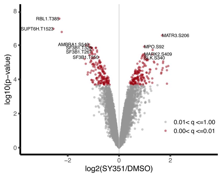
Downloaded from genesdev.cshlp.org on November 11, 2020 - Published by Cold Spring Harbor Laboratory Press

Rimel et al.

cascade of events that correlate with RNAPII promoter es-           A                                   C
cape and transcription elongation (Corden 2013; Eick and
Geyer 2013). CDK7 can also phosphorylate CDK9, which,
together with CCNT1, comprises the P-TEFb complex. P-
TEFb represents another transcription-associated kinase
that is capable of phosphorylating the RNAPII CTD, and              B
                                                                                                         D
CDK7 phosphorylation of CDK9 enhances its kinase ac-
tivity (Larochelle et al. 2012). Phosphorylation of the
RNAPII CTD appears to be essential for proper processing
of RNA transcripts (Kanin et al. 2007; Glover-Cutter et al.
2009; Hong et al. 2009), due in part to recruitment of fac-
tors involved in 5′ capping and splicing (Cho et al. 1997;
McCracken et al. 1997; David et al. 2011; Ebmeier et al.
                                                                    Figure 1. SY-351 is a potent and highly selective covalent inhibitor
2017).                                                              ofhuman CDK7. (A)SY-351 structure.(B) Kinome selectivity inA549
   The biological roles for CDK7 remain incompletely un-            cell lysate, with 0.2 µM and 1 µM SY-351. The top six hits shown are
derstood, in part because identification of its kinase sub-         kinases inhibited > 50% by 1 µM SY-351. Note that SY-351 was used
strates has been limited. CDK7 can also function apart              at 0.05 µM (50 nM) throughout this study. (C) CDK7 and CDK12 tar-
from TFIIH, as the three-subunit CDK activating kinase              get occupancy in HL-60 cells after 1-h treatment. The SY-351 EC50 is
(CAK: CDK7, CCNH, and MAT1). In the cytoplasm, the                  8.3 nMfor CDK7 and 36 nMfor CDK12. The SY-351 EC90 is 39 nM for
CAK phosphorylates and activates other CDKs (e.g.,                  CDK7 and 172 nM for CDK12. The dashed line indicates 50 nM SY-
CDK1 and CDK2) to regulate the cell cycle (Fisher                   351, the dose used throughout this study. (D) SY-351 inhibition of ac-
2005). It is not known whether the CAK may also function            tive kinases CDK7/CCNH/MAT1, CDK2/CCNE1, CDK9/CCNT1,
                                                                    CDK12/CCNK at KM ATP for each enzyme. The best fit IC50 values
in the nucleus, but current models assume that nuclear
                                                                    are 23 nM, 321 nM, 226 nM, and 367 nM, respectively.
CDK7 regulates transcription through its association
with TFIIH.
   Viruses are known to target TFIIH to hijack the RNAPII
transcription response during infection (Qadri et al. 1996;         centration of 1 µM SY-351, only six other kinases were in-
Cujec et al. 1997; Le May et al. 2004), and TFIIH muta-             hibited >50%, including CDK12 and CDK13 (Fig. 1B).
tions (i.e., those that are not lethal) are linked to congeni-         Similar to other compounds in this class (Kwiatkowski
tal and somatic diseases such as xeroderma pigmentosum,             et al. 2014; Hu et al. 2019), the acrylamide moiety of SY-
Cockayne syndrome, and various types of cancer                      351 covalently reacts with cysteine residue 312 of CDK7
(Schaeffer et al. 1993; Manuguerra et al. 2006). Numerous           and exhibits time-dependent inhibition of CDK7 with a
compounds have been developed that target the kinase ac-            KI of 62.5 nM and kinact of 11.3 h−1 (Supplemental Fig.
tivity of CDK7 (Kelso et al. 2014; Kwiatkowski et al. 2014;         S1A). Due to the ability of SY-351 to covalently interact
Olson et al. 2019), including several that advanced to clin-        with cysteine residues, we sought to identify other pro-
ical trials (Hu et al. 2019). Here, we used SY-351, a potent        teins that might react in cells. We used activity-based pro-
and selective covalent CDK7 inhibitor. By combining                 tein profiling (ABPP) with a panel of alkyne functionalized
SILAC-based phosphoproteomics with transcriptomics                  analogues of SY-351 (Supplemental Fig. S2) in HL60 cells,
and biochemical assays, we were able to identify high-              as described (Cravatt et al. 2008; Lanning et al. 2014). For-
confidence CDK7 substrates, a surprisingly widespread re-           ty-six proteins were identified as competitive hits, includ-
quirement for CDK7 activity in splicing and unexpected              ing expected kinase targets CDK7 and the related CDK12
aspects of CDK7 kinase regulation that involve its associ-          and CDK13 kinases, which have cysteine residues that
ation with TFIIH.                                                   align with CDK7: C1039 in CDK12 and C1017 in
                                                                    CDK13 (Supplemental Table S2).
                                                                       The ability of SY-351 to covalently react with CDK12
Results                                                             and CDK13 must be considered to ensure that cellular ef-
                                                                    fects in this study were due primarily to CDK7 inhibition.
SY-351: a potent, highly selective covalent CDK7
                                                                    A 1-h treatment of 50 nM SY-351 reached the EC90 of
inhibitor
                                                                    CDK7 target engagement (39 nM), while minimizing
Potent covalent inhibition of CDK7 by SY-351 (Fig. 1A)              CDK12 target engagement in HL60 cells (Fig. 1C). More-
has been recently described (Hu et al. 2019). Although in           over, as shown previously (Hu et al. 2019), SY-351 selec-
vivo properties, including high clearance, limited develop-         tively inhibited catalytically active CAK complex
ment of this compound, SY-351 is useful as a molecular              (CDK7 in complex with CCNH and MAT1) over other cy-
probe to understand CDK7 inhibition in cells. Selectivity           clin-dependent kinases CDK2:CCNE1, CDK9:CCNT1,
was evaluated in a panel of 252 kinases using KiNativ pro-          and CDK12:CCNK (Fig. 1D; Supplemental Fig. S1B).
filing (Supplemental Table S1). CDK7 was the top hit with           Based on these results, we concluded that inhibition of
>90% inhibition, and was the only kinase inhibited more             CDK7 predominates at 50 nM SY-351 and 1-h treatment.
than 50% with 0.2 µM SY-351. To minimize off-target ef-             This condition was used for stable isotope labeling of ami-
fects, we used SY-351 at a concentration of 50 nM (0.05             no acids in cell culture (SILAC)-based phosphoproteomics
µM) throughout this study (see below). At a higher con-             experiments in HL60 cells.

2       GENES & DEVELOPMENT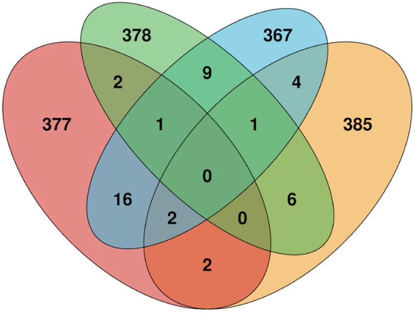
Downloaded from genesdev.cshlp.org on November 11, 2020 - Published by Cold Spring Harbor Laboratory Press
CDK7 substrates and TFIIH function
Identification of high-confidence CDK7 targets using ods), which improved our statistical power to detect phos-
SILAC phosphoproteomics phorylation sites that changed in abundance with SY-351
treatment (Supplemental Fig. S3B). We also confirmed
We performed a double-label SILAC phosphoproteomics that metabolic labeling did not transfer to proline through
experiment in which we metabolically labeled HL60 cells any salvage pathways (Supplemental Fig. S3C).
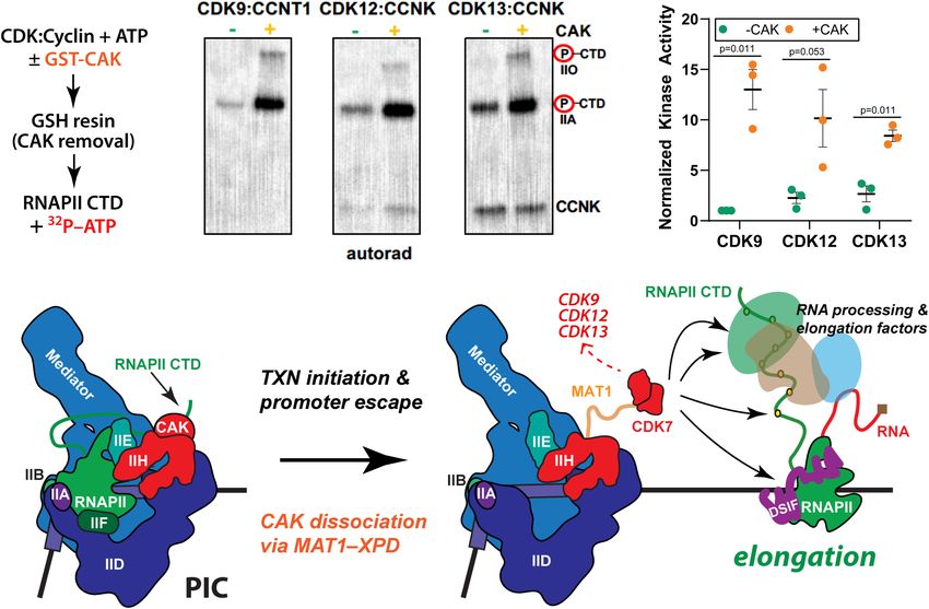
with “light”-labeled (Lys0 and Arg0) or “heavy”-labeled Overall, we detected 621 phosphosites that decreased
(Lys8 and Arg10) amino acids, which were treated with ve- and 732 sites that increased (FDR < 0.05) (Fig. 2B) upon
hicle (DMSO) or 50 nM SY-351, followed by phosphopep- SY-351 treatment, with quantification of nearly 40,000 to-
tide enrichment and mass spectrometry analysis. The tal phosphosites and 16,616 phosphosites quantified in all
experimental design included two biological replicates of four replicates. Representative MS data are shown in Sup-
SY-351-treated heavy cells compared with DMSO treated plemental Figure S4A. Although decreased or increased
light cells, a “label-flip” biological replicate of SY-351 light phosphosite abundance could simply reflect changes in
cells compared with DMSO treated heavy cells, and a protein levels, this was not observed within the time
“null” condition for which both heavy and light were frame of the experiment (Supplemental Fig. S4B). The
treated with DMSO (Fig. 2A). The label-flip and null condi- large number of increasing sites was not unexpected and
tions control for systematic errors in phosphopeptide likely reflects compensatory functions of kinases and
Heavy:Light SILAC ratios that are independent of the phosphatases (see the Discussion).
SY-351 treatment effect, which we observed after metabol- Comparison of CDK7 phosphosites with those from oth-
ic labeling of HL-60 cells using the SILAC protocol (Sup- er transcription-associated kinases CDK8/CDK19 (Poss
plemental Fig. S3A). We used a linear statistical model to et al. 2016), CDK9 (Sansó et al. 2016; Decker et al. 2019),
capture these systematic errors (see Materials and Meth- and CDK12/CDK13 (Krajewska et al. 2019) revealed few
A B
C
Figure 2. SILAC phosphoproteomic overview and identification of phosphorylation sites significantly changed by SY-351 treatment. (A)
Experimental design and workflow using SILAC double-labeled HL-60 cells (Light:Heavy) and phosphoproteomics. Labeled cell popula-
tions were treated with vehicle (DMSO) or 50 nM SY-351 as indicated, with two biological replicates of SY-351-treated heavy cells (vs.
DMSO light cells), a “label-flip” biological replicate of SY-351 light cells versus DMSO heavy cells, and a “null” condition for which
both heavy and light were treated with DMSO. Tryptic peptides were enriched using titanium dioxide beads and analyzed by high-reso-
lution mass spectrometry to identify phosphopeptides and quantify their light:heavy SILAC ratios. (B) Volcano plot showing the statistical
significance and magnitude of change with SY-351 treatment for phosphorylation site SILAC ratios. The adjusted P-value range for phos-
phorylation site ratios is indicated in color. (C ) Venn diagram showing phosphorylation sites shared or mutually exclusive for transcrip-
tion-associated kinases CDK7, CDK8, CDK9, and CDK12/CDK13. For each kinase, the top 400 identified phosphorylated proteins, based
upon the largest negative log fold change values and P < 0.05, were used for comparisons, and the number of overlapping proteins was cal-
culated for all combinations of samples. CDK7 targets identified here were compared with proteins identified as substrates for CDK8 in
HCT116 (Poss et al. 2016), CDK9 in Raji B lymphocytes (Decker et al. 2019), and CDK12/13 in IMR-32 and Kelly cells (Krajewska et al.
2019).
GENES & DEVELOPMENT 3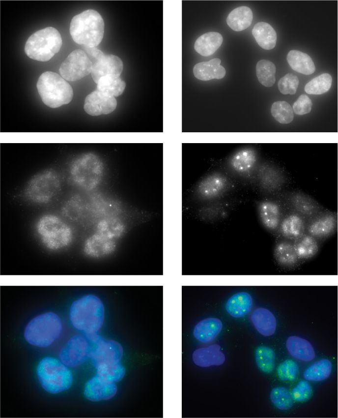
Downloaded from genesdev.cshlp.org on November 11, 2020 - Published by Cold Spring Harbor Laboratory Press
Rimel et al.
shared substrates (Fig. 2C; Supplemental Fig. S4C; Supple- in the PhosphoSitePlus database (Hornbeck et al. 2015),
mental Table S3), suggesting nonredundant roles for these we mapped significant sites (q-value < 0.05) onto a ki-
kinases. Although these MS-based analyses used different nase-substrate network generated in Cytoscape. A subnet-
human cell types, the cellular targets for CDK7, CDK8/19, work was selected from the larger parent network to
CDK9, or CDK12/13 are expected to be shared across emphasize only kinases and substrates within a few de-
cell types. A STRING analysis of the decreased sites (q val- grees of separation from CDK7, CDK9, and POLR2A (Sup-
ue < 0.05) displays high-confidence CDK7 targets within plemental Fig. S5). CDK9 was included here because
known interaction networks (Fig. 3). Using a more strin- others have shown that CDK7 can activate CDK9 through
gent cutoff (log2 [SY-351/DMSO] < –0.5 and q-value < phosphorylation of the CDK9 T-loop (Larochelle et al.
0.01), 120 unique phosphorylation sites were identified 2012). Whereas phosphorylation at the CDK9 T-loop site
that decreased with SY-351 treatment, corresponding to (Thr186) was detected in our SILAC-MS data, it did not
72 phosphoproteins (Table 1). The CDK7 substrates iden- significantly change in SY-351-treated HL60 cells (Supple-
tified in Figure 3 and Table 1 included many splicing and mental Table S4). As expected, the kinase-substrate sub-
RNA processing factors (Supplemental Fig. S4D); most no- network showed known CDK7 phosphorylation sites on
table was SF3B1, with decreased phosphorylation at 18 CDK1 (Thr161) and within the RNAPII CTD (Ser1878).
high-confidence sites in SY-351-treated cells. Notably, known T-loop sites in CDK12 (Thr893) and
Finally, to visualize significantly changing phosphory- CDK13 (Thr871) were also identified, suggesting that
lation sites with known kinase-substrate relationships CDK7 might directly activate these kinases (see below).
DYNC1LI1
EXOC1 TBC1D1 BICD2 SPTAN1
NSF
DYNC1LI2
RAB8A
KHDRBS1 COPA
NCF2
LEMD3
PKN1 KIF22
RIF1
HIST1H1D AKAP9 KIF18B
CDK11A
GAPVD1
AGFG1 CENPE
KIF13B
LMNB1 PCM1 CCP110 XRCC6
HIST1H1E
CLASP1
LDLRAP1
AHCTF1 MKL1
TFRC DIAPH1 MSH6
HIST1H1B
CDK1
HMGA1 CENPF
CDCA5
POLA2 KPNA2
RB1
NEK6
MTDH MYC
RNF123 ORC6 RANBP2
TRIP12 NUP133
DNMT1 MCM7
RBL1 ORC1 SLBP
NDRG1
HUWE1
WEE1 NUP188 NUP205 FYTTD1
HERC1 CHD8
UBR4 MCM3 KAT8
RBL2
SIN3A SFPQ
POLDIP3
UBE2O THOC2
CHEK1
NCOR2
UBA6 SMAD2
HMGN1 NONO
SRSF2
DHX38 SRRM1
MKI67IP
WIBG
MED1 HNRNPC
NR1D1 CASC3
RPRD2 EIF3A
U2AF2 SMG1
POLR2A
PABPN1 EIF4G2
SRA1 ELAVL1 RPL18
CDK12 EIF5B
CCNK HNRNPM
THRAP3
SRRM2 SMG9
CTDP1
CWC22
GTF2F1 HNRNPA3 SEC61B
SUPT16H
CDK13 IRAK1
CDC5L
HNRNPA2B1
LEO1
HNRNPD
PRPF40A
SF3B1 MYD88 WIPF1
PAK4 MYO9B
SUPT5H ARHGEF2
STRING analysis:
RBM25
ARHGEF7 WASF2
MYBBP1A
decreased phospho- MAP3K11
DDX21
NCK1
WAS
sites with SY-351 KSR1 SH3KBP1 LCP2
FDR < 0.05 ARAF
HLA-A
DBNL
PIK3AP1
MARK3 TYROBP
APBB1IP
LCP1
HLA-C
Figure 3. Protein–protein interaction network of phosphoproteins with significantly decreasing phosphorylation sites upon SY-351
treatment. Proteins with decreasing phosphorylation sites (log2FC < 0 and FDR < 0.05) were visualized using the STRING database web
application, and clustered using the MCL algorithm, with clusters indicated by node color.
4 GENES & DEVELOPMENT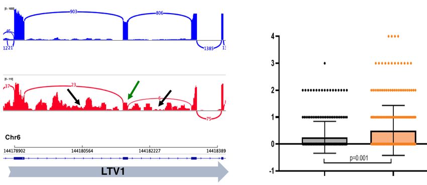
Downloaded from genesdev.cshlp.org on November 11, 2020 - Published by Cold Spring Harbor Laboratory Press

                                                                                                   CDK7 substrates and TFIIH function

Table 1. High-confidence CDK7 targets

                                                                                             No.
Uniprot_acc      Gene                   Protein_function             High_conf_sites      sig.down       Min.log2FC     Min.adj.pval

O75533        SF3B1         Splicing                               S322,S349,T207,           18             −1.16       1.60 × 10⁻³
                                                                     T211,T223,
                                                                     T244,
                                                                     T267,T273,
                                                                     T278,
                                                                     T296,T299,
                                                                     T303,
                                                                     T313,T326,
                                                                     T328,
                                                                     T350,T354,T434
P28749        RBL1          TF; repressor of E2F; tumor            S749,S762,T369,             4            −2.33       4.22 × 10⁻⁴
                              suppressor                             T385
Q96DF8        DGCR14        Splicing                               S292,T110,T3,               4            −1.12       2.85 × 10⁻³
                                                                     T437
Q99459        CDC5L         ell cycle reg’n; splicing              T377,T404,T438              3            −1.21       4.54 × 10⁻³
Q5VT52-3      RPRD2         Pol II CTD binding                     S588,T456,T491              3            −1.04       3.32 × 10⁻³
Q9HC98        NEK6          Kinase; cell cycle reg’n               S199,T201,T202              3            −0.81       7.76 × 10⁻³
P06400        RB1           TF; repressor of E2F; tumor            S608,T601,Y606              3            −0.64       6.41 × 10⁻³
                               suppressor
Q9BTA9        WAC           TXN, cell cycle reg’n                  T457,T471                   2            −2.23       8.45 × 10⁻⁴
P24928        POLR2A        Pol II large subunit                   S1878,S1920                 2            −1.34       2.36 × 10⁻³
Q14004-2      CDK13         Kinase; RNA processing                 S525,T1087                  2            −1.30       1.93 × 10⁻³
O94762-4      RECQL5        Helicase; TXN, DNA repair              S788,T812                   2            −1.15       1.93 × 10⁻³
O00267        SUPT5H        Pol II elongation                      S666,S671                   2            −1.08       2.63 × 10⁻³
P10398        ARAF          Kinase; cell cycle reg’n               S257,T253                   2            −1.08       7.21 × 10⁻³
Q9Y520-6      PRRC2C        Stress granule protein                 S2143,T2608                 2            −0.86       1.69 × 10⁻³
Q8IVT5-2      KSR1          Pseudokinase; activates BRAF           S331,S334                   2            −0.84       3.38 × 10⁻³
P16333        NCK1          RAS signaling                          S91,S96                     2            −0.78       5.94 × 10⁻³
Q08999        RBL2          Repressor of E2F; heterochromatin      S1068,S413                  2            −0.76       4.26 × 10⁻³
Q8WWM7-6      ATXN2L        Stress granule protein                 S390,S391                   2            −0.73       2.68 × 10⁻³
P49792        RANBP2        Nuclear pore, import/export            S21,T19                     2            −0.70       3.68 × 10⁻³
Q8N9Q2        SREK1IP1      Splicing                               S145,T146                   2            −0.70       3.32 × 10⁻³
Q7KZI7-16     MARK2         Kinase; microtubule dynamics           S390,T466                   2            −0.66       4.16 × 10⁻³
Q96Q15        SMG1          Kinase; mRNA surveillance              S3570,T3573                 2            −0.64       3.86 × 10⁻³
Q86VM9        ZC3H18        mRNA biogenesis                        S795,T796                   2            −0.62       6.48 × 10⁻³
Q92597        NDRG1         Lipid metabolism                       S330,T366                   2            −0.59       4.39 × 10⁻³
Q7KZ85        SUPT6H        Pol II elongation; RNA processing      T1523                       1            −2.56       8.23 × 10⁻⁴
Q9C0C7-4      AMBRA1        Autophagy                              S549                        1            −1.17       1.60 × 10⁻³
Q14103-3      HNRNPD        RNA processing                         S87                         1            −1.12       2.68 × 10⁻³
Q9UQ88-4      CDK11A        Kinase; cell cycle reg’n               T570                        1            −1.07       2.28 × 10⁻³
Q9UNA1-2      ARHGAP26      Endocytosis; membrane remodeling       S584                        1            −1.01       3.25 × 10⁻³
Q96N67-4      DOCK7         Cell motility                          S30                         1            −1.01       2.80 × 10⁻³
P49454        CENPF         Kinetochore function                   S276                        1            −1.00       7.46 × 10⁻³
Q13586        STIM1         Calcium signaling                      S575                        1            −0.95       9.60 × 10⁻³
P49756        RBM25         Splicing                               S677                        1            −0.94       2.90 × 10⁻³
Q92609        TBC1D5        Autophagy; vesicle trafficking         S44                         1            −0.92       5.10 × 10⁻³
P23246        SFPQ          Splicing                               T226                        1            −0.89       6.62 × 10⁻³
Q14847        LASP1         Proliferation                          T104                        1            −0.87       6.55 × 10⁻³
P29350-4      PTPN6         Phosphatase                            S570                        1            −0.85       7.17 × 10⁻³
P48634        PRRC2A        Splicing; m6A reader                   S1004                       1            −0.81       3.32 × 10⁻³
P10412        HIST1H1E      Histone H1                             T146                        1            −0.78       9.40 × 10⁻³
Q9H0W8-2      SMG9          mRNA surveillance                      S32                         1            −0.75       2.76 × 10⁻³
Q8NEC7-3      GSTCD         Glutathione S-transferase              S146                        1            −0.74       5.66 × 10⁻³
O14647        CHD2          Helicase; chromatin remodeling         S1789                       1            −0.74       7.76 × 10⁻³
Q7Z6Z7-2      HUWE1         E3 ubiquitin ligase                    T1722                       1            −0.73       3.12 × 10⁻³
O96013        PAK4          Kinase; cell cycle reg’n               S148                        1            −0.73       4.66 × 10⁻³
Q2KHR2-2      RFX7          TF; metabolism                         S225                        1            −0.73       8.88 × 10⁻³
Q9NYV4-2      CDK12         Kinase; RNA processing                 T893                        1            −0.72       2.68 × 10⁻³
Q9UK76        JPT1          Cell cycle reg’n                       S92                         1            −0.71       8.93 × 10⁻³
                                                                                                                         Continued

                                                                                                    GENES & DEVELOPMENT            5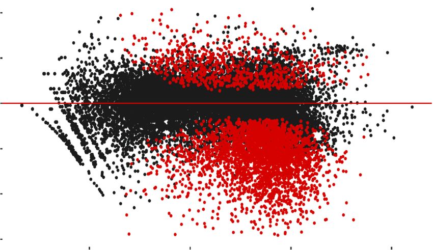
Downloaded from genesdev.cshlp.org on November 11, 2020 - Published by Cold Spring Harbor Laboratory Press

Rimel et al.

Table 1. Continued

                                                                                                 No.
Uniprot_acc       Gene                   Protein_function                High_conf_sites      sig.down     Min.log2FC   Min.adj.pval
Q15717         ELAVL1         mRNA biogenesis                          S202                       1           −0.69      3.32 × 10⁻³
Q99836-2       MYD88          Inflammatory response                    S199                       1           −0.69      7.62 × 10⁻³
P98171         ARHGAP4        Cell motility                            T898                       1           −0.68      5.73 × 10⁻³
Q7Z460-4       CLASP1         Microtubule dynamics; kinetochore        S663                       1           −0.68      8.47 × 10⁻³
Q9NS56         TOPORS         E3 ubiquitin ligase                      S194                       1           −0.67      6.41 × 10⁻³
Q6IQ22         RAB12          Autophagy                                S21                        1           −0.65      7.30 × 10⁻³
Q9Y3X0         CCDC9          Cell motility                            S376                       1           −0.64      6.61 × 10⁻³
Q96T58         SPEN           TXN reg’n; X-inactivation                S1278                      1           −0.63      3.95 × 10⁻³
Q5SRE5-2       NUP188         Nuclear pore complex                     S1606                      1           −0.61      4.39 × 10⁻³
Q92974-2       ARHGEF2        Cell motility; cell cycle reg’n          S163                       1           −0.61      9.27 × 10⁻³
Q9Y618-3       NCOR2          TXN corepressor                          S2008                      1           −0.60      6.68 × 10⁻³
Q8IUH3-3       RBM45          RNA binding                              S462                       1           −0.60      8.81 × 10⁻³
Q9Y6G9         DYNC1LI1       Mitosis; vesicle trafficking             S398                       1           −0.60      9.21 × 10⁻³
Q8IWZ3-6       ANKHD1         Vesicle formation                        S2539                      1           −0.59      9.52 × 10⁻³
Q9P107         GMIP           RhoA GTPase reg’n                        S243                       1           −0.58      6.72 × 10⁻³
Q9P270         SLAIN2         Microtubule dynamics                     S48                        1           −0.57      6.30 × 10⁻³
Q96C19         EFHD2          Cell motility                            S74                        1           −0.57      3.12 × 10⁻³
Q5SW96         LDLRAP1        Endocytosis                              S14                        1           −0.56      8.05 × 10⁻³
Q8N163         CCAR2          Pol II elongation; splicing              S671                       1           −0.56      4.67 × 10⁻³
Q9BRZ2         TRIM56         E3 ubiquitin ligase                      T442                       1           −0.55      6.61 × 10⁻³
O43586         PSTPIP1        Phosphatase reg’n; inflammation          S318                       1           −0.54      8.36 × 10⁻³
Q9NR30         DDX21          RNA helicase; TXN reg’n                  S71                        1           −0.54      3.68 × 10⁻³
Q9BY77         POLDIP3        mRNA export; translation                 S275                       1           −0.53      8.94 × 10⁻³
Q5HYJ3         FAM76B         Nuclear speckles                         S193                       1           −0.50      7.02 × 10⁻³
Genes in bold indicate proteins are nuclear; bold italic indicates nuclear or cytoplasmic.

Inhibition of CDK7 causes diverse and widespread                       that CDK12 inhibition results in premature, intronic pol-
splicing changes                                                       yadenylation (iPA) (Dubbury et al. 2018; Krajewska et al.
                                                                       2019; Fan et al. 2020), which causes reduced exon read
To probe potential CDK7-dependent effects on RNA pro-                  counts toward gene 3′ ends. A DEXSeq analysis (Anders
cessing, we completed spike-in normalized (Supplemen-                  et al. 2012) showed no evidence for iPA genome-wide in
tal Fig. S6A) RNA-seq analyses in HL60 cells treated                   cells treated with SY-351 (Supplemental Fig. S6E), which
with SY-351 (50 nM for 5 h) along with DMSO controls.                  further validated our experimental strategy and confirmed
Biological replicates (CTRL vs. SY-351) (Supplemental                  the selectivity of SY-351 for CDK7.
Fig. S6B) were sequenced to high depth (>120 million                      To investigate possible effects of SY-351 on alternative
mapped reads/replicate) to facilitate splicing analysis,               splicing, we used MAJIQ (Vaquero-Garcia et al. 2016),
and the 5-h treatment time was determined empirically                  which detects altered exon skipping and intron retention
by assessing total RNA and cell viability across a series              events as well as more complex splicing changes. We iden-
of time points (Supplemental Fig. S6C,D). At 5 h, total                tified 11,348 changes in exon inclusion upon SY-351 treat-
cellular RNA levels remained relatively unchanged versus               ment versus DMSO controls (ΔPSI [percent spliced in] ≥
t = 0 controls; moreover, a 5-h time point enabled poten-              0.2, P < 0.05) (Fig. 4C), and SY-351 treatment increased in-
tial splicing changes to manifest in steady-state mRNA.                clusion/reduced skipping of alternative exons more fre-
Significant changes in mRNA abundance (P < 0.05; fold                  quently than it decreased their inclusion/increased
change > 2) were identified by DEseq2 in SY-351-treated                skipping (6738 vs. 4610) (Fig. 4C). The 5′ and 3′ splice sites
samples, with an excess of down-regulated transcripts                  at alternative exons whose inclusion was affected by SY-
over up-regulated transcripts (Fig. 4A). Gene set enrich-              351 did not significantly differ in strength (Yeo et al.
ment analysis (GSEA) (Fig. 4B) showed decreases in prolif-             2004) from those that were unaffected (Supplemental
erative hallmarks with SY-351 treatment (e.g., E2F                     Fig. S7A).
targets, MYC targets, G2/M checkpoint), as expected.                      Notably, both reduced and increased exon skipping
   SY-351 is highly selective for CDK7, although some in-              were frequently associated with abnormal retention of in-
hibition of CDK12 and CDK13 was observed at higher                     trons flanking the alternative exon; this phenomenon was
concentrations in kinome-wide profiling (Fig. 1B). Where-              the major source of abnormal transcripts detected in SY-
as our experimental design minimized potential con-                    351-treated cells. Reduced exon inclusion with retention
founding effects from CDK12/13 inhibition (Fig. 1C,D),                 of flanking introns is illustrated in Figure 4D (see also Sup-
we nevertheless probed the RNA-seq data for evidence                   plemental Fig. S7B). Increased exon inclusion with (1) re-
that CDK12 inhibition was occurring. Others have shown                 tention of a downstream flanking intron (Supplemental

6       GENES & DEVELOPMENT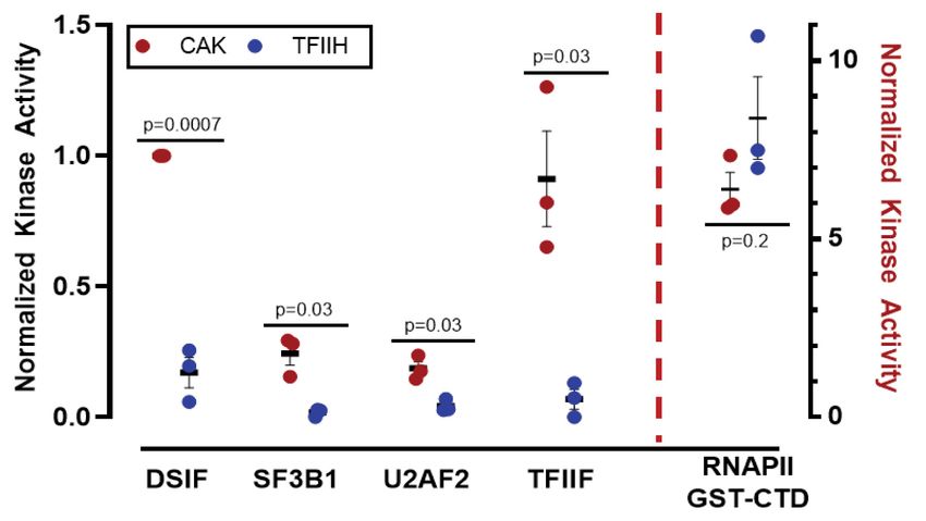
Downloaded from genesdev.cshlp.org on November 11, 2020 - Published by Cold Spring Harbor Laboratory Press

                                                                                                       CDK7 substrates and TFIIH function

  A                                         B                                                             E

 C                                                         D                                              F

Figure 4. CDK7 inhibition with SY-351 causes widespread defects in splicing. (A) Differentially expressed mRNAs following SY351
treatment (DESeq2). Replicate RNA-seq data sets were generated from rRNA-depleted total RNA from HL60 cells treated with DMSO
(CTRL) or SY-351 (50 nM in DMSO) for 5 h. (B) GSEA analysis. (C) SY-351 favors exon inclusion over exon skipping, splicing over retention
of introns, and alters 5′ and 3′ splice site selection equally, without preference for upstream or downstream sites. Alternative splicing
events significantly affected by SY-351 were identified in replicate RNA-seq data sets using MAJIQ (Vaquero-Garcia et al. 2016). (D)
IGV genome browser Sashimi plot of a segment of the LTV1 gene. Normalized read numbers for DMSO control and SY-351 treated sam-
ples are shown on the Y-axis. Splice junction read numbers for sense strand transcripts are shown in blue and red. Note reduced exon in-
clusion (green arrow) and retention of both flanking introns (black arrows) in SY-351. (E) Immunofluorescence microscopy of endogenous
SF3B1 (green) and Hoechst (blue) in DMSO and SY-351 conditions. (F) Quantitation of SF3B1 puncta per nucleus in DMSO and SY-351
conditions, P = 0.001 (n = 3 biological replicates).

Fig. S7C,D), (2) an upstream flanking intron (Supplemen-               SF3B1 sites were identified by SILAC phosphoproteomics,
tal Fig. S7E), or (3) both downstream and upstream introns             we asked whether defects in SF3B1 function would be ev-
(Supplemental Fig. S7F) was also observed. We detected                 ident in cells treated with SY-351. As shown in Table 1,
significant effects of SY-351 on use of annotated alterna-             all 18 high-confidence phosphorylation sites reside be-
tive 5′ and 3′ splice sites in approximately equal numbers             tween SF3B1 residues 207–434. Previous studies (Eil-
(753 5′ ss vs. 790 3′ ss), with similar numbers of either up-          bracht and Schmidt-Zachmann 2001) linked this region
stream of or downstream from alternative splice sites                  to SF3B1 nuclear localization (residues 196–216) and
(Fig. 4C). In addition, SY-351 significantly altered the               SF3B1 association with nuclear speckles (TP-rich domain;
splicing of over 1500 annotated retained introns with a                residues 208–440). Therefore, we tested whether SF3B1
strong bias toward increased (Supplemental Fig. S8A–D)                 nuclear localization or speckle association would be af-
versus decreased (Supplemental Fig. S8E) splicing of these             fected by SY-351 treatment. As shown in Supplemental
introns (1151 vs. 522 cases) (Fig. 4C).                                Figure S9A,B, nuclear versus cytoplasmic localization of
   In summary, SY-351 inhibited splicing of introns flank-             SF3B1 did not appear to be altered in SY-351-treated cells,
ing many alternative exons, but did not generally inhibit              at least under our assay conditions (4 h SY-351, 50 nM). In
splicing. In fact, SY-351 actually increased splicing of               contrast, immunofluorescence (IF) experiments showed
many retained introns (Supplemental Fig. S8A–D).                       changes in SF3B1 association with nuclear speckles in
                                                                       SY-351-treated cells versus untreated controls (Fig. 4E,
                                                                       F), although total nuclear IF signal remained largely un-
Splicing changes can be linked in part to SF3B1
                                                                       changed (Supplemental Fig. S9C). Moreover, the SY-351
SF3B1 is a component of the U2 small nuclear ribonucleo-               effects were distinct from THZ-531, an inhibitor of
protein (snRNP) and facilitates hybridization of the U2                CDK12/13 (Supplemental Fig. S9D,E). Although we can-
snRNA with the pre-mRNA branch point sequence with-                    not decouple the changes in SF3B1 speckle localization
in the spliceosome (Maji et al. 2019). Because 18 different            from transcriptional changes triggered by SY-351, the IF

                                                                                                         GENES & DEVELOPMENT           7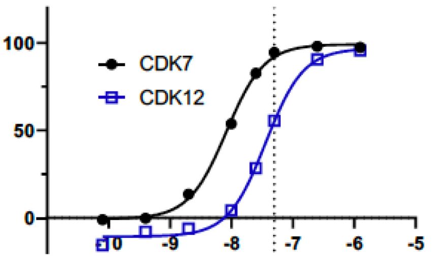
Downloaded from genesdev.cshlp.org on November 11, 2020 - Published by Cold Spring Harbor Laboratory Press

Rimel et al.

data further implicate the CDK7 kinase as a regulator of            be modifying these substrates in the context of the three-
SF3B1 function.                                                     subunit CAK module (CDK7, CCNH, and MNAT1) (Fig.
   We also compared splicing changes induced by SY-351              5C), which can exist as a stable complex apart from TFIIH
with those induced by the SF3B1 inhibitor Pladienolide              (Rimel and Taatjes 2018). We tested whether the same set
B (PladB) (Kotake et al. 2007). Using MAJIQ2 (Vaquero-              of substrates would be differentially modified by the three-
Garcia et al. 2016), we identified 1152 local splicing vari-        subunit CAK module versus TFIIH (Fig. 5B,D). The data re-
ations (LSVs) in common between SY-351 and PladB.                   vealed that CDK7 efficiently phosphorylated DSIF, TFIIF,
These events met the following criteria: (1) P-value <              U2AF2, and SF3B1 within the three-subunit kinase mod-
0.05, (2) absolute ΔPSI ≥ 0.2, (3) used in >10% of reads,           ule, in stark contrast to the 10-subunit TFIIH complex.
and (4) the coordinates of the 5′ and 3′ splice sites were          In addition, the comparison of TFIIH versus CAK showed
within 10 bases in both cell lines (HL60 for SY-351 and             that CDK7 was slightly more active toward the RNAPII
K562 for PladB). Among these 1152 events, the ΔPSI value            CTD within TFIIH versus the CAK (Fig. 5D). TFIIH also
was altered by both inhibitors in the same direction in 765         phosphorylated the RNAPII CTD within promoter-assem-
cases (P-value = 2.3307 × 10−79, hypergeometric test). Ex-          bled, transcriptionally active preinitiation complexes
amples of inhibitor effects on splicing of individual in-           (PICs), as expected (Supplemental Fig. S11A). NELF and
trons are shown in Supplemental Figure S9F. From this               MYC were not efficiently phosphorylated by CDK7 within
comparative analysis, we conclude that a subset of the              the CAK or TFIIH (Supplemental Fig. S11B,C), suggesting
splicing defects observed in SY-351-treated cells result            these are not directly targeted by CDK7.
from changes in SF3B1 function.                                        We conducted Western blotting experiments with anti-
                                                                    bodies specific to the Ser2, Ser5, or Ser7 phosphorylated
                                                                    CTD to assess whether selectivity for these sites might
CDK7 kinase activity is regulated by TFIIH                          differ between the CAK and TFIIH. Consistent with the
We performed in vitro kinase assays with the 10-subunit             bulk 32P-ATP kinase assays, the data revealed increased
human TFIIH complex (Fig. 5A) to test whether high-con-             modification of each site by TFIIH (vs. CAK); however,
fidence CDK7 substrates were, in fact, direct targets. Sub-         the Ser2, Ser5, and Ser7 phosphorylated CTD species mi-
strates tested were DSIF, NELF, SF3B1, U2AF2, TFIIF,                grated differently in each case (CAK vs. TFIIH), suggesting
MYC, and the RNAPII CTD (Supplemental Fig. S10),                    that a different number or different pattern of sites within
which were identified as CDK7 kinase targets from the               the 52-repeat sequence were being modified (Supplemen-
phosphoproteomics data (Fig. 3). Contrary to expectations,          tal Fig. S11D,E).
TFIIH was unable to efficiently phosphorylate these sub-               The data shown in Figure 5 established distinct sub-
strates in vitro, with the exception of the RNAPII CTD              strate specificities for CDK7 in the free kinase module
(Fig. 5B). Because CDK7 can activate other CDKs (Rimel              versus TFIIH. To test this further, we compared the
and Taatjes 2018), these data suggested that DSIF, NELF,            three-subunit CAK module with the 10-subunit TFIIH
SF3B1, U2AF2, TFIIF, and MYC may be indirect targets                complex in positional scanning peptide array experi-
of CDK7. It was also plausible, however, that CDK7 might            ments, which were completed with 32P-ATP (Begley

A                B                                                                      Figure 5. CDK7 kinase activity is negative-
                                                                                        ly regulated by TFIIH. (A) SYPRO-stained
                                                                                        gel of human TFIIH. (B) TFIIH activates
                                                                                        CDK7 kinase toward the CTD, but reduces
                                                                                        or prevents CDK7 phosphorylation of
                                                                                        DSIF, SF3B1, U2AF2, and TFIIF. Within
                                                                                        the CAK, CDK7 is activated toward these
                                                                                        substrates. (C ) SYPRO-stained gel of human
                                                                                        CAK complex (MAT1 is GST-tagged).
                                                                                        (D) Quantitation of CDK7 kinase results
                                                                                        across biological replicates (n = 3) with auto-
                                                                                        rad signal normalized to CAK phosphorylat-
                                                                                        ed DSIF; P-values as shown.
C                D

8       GENES & DEVELOPMENT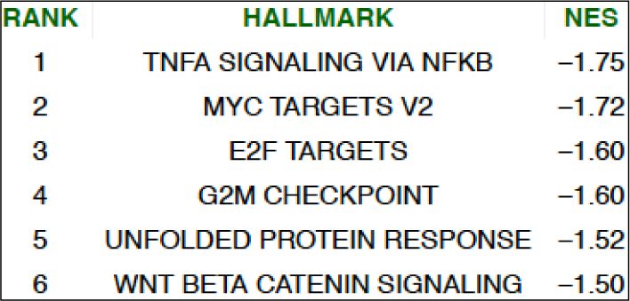
Downloaded from genesdev.cshlp.org on November 11, 2020 - Published by Cold Spring Harbor Laboratory Press

                                                                                                CDK7 substrates and TFIIH function

et al. 2015). Although peptide arrays are limited in their        toward a common substrate, the RNAPII CTD. Impor-
ability to match the structure of native, physiological sub-      tantly, the CAK was removed prior to these kinase assays
strates, the assay provided an independent means to com-          to ensure no contaminating CDK7 was present, which
pare CDK7 substrate preferences in the context of the             would otherwise confound the analysis (Supplemental
CAK versus TFIIH. Consistent with the in vitro kinase as-         Fig. S13A). In parallel, we tested P-TEFb (CDK9,
says with native substrates (Fig. 5), the peptide array data      CCNT1) complexes and verified CDK7-dependent activa-
showed differential phosphorylation site preferences (Sup-        tion of CDK9 (Fig. 6B,C), in agreement with previous stud-
plemental Fig. S12A), with TFIIH showing greater selec-           ies (Larochelle et al. 2012).
tivity compared with the three-subunit CAK.                          We next tested whether the 10-subunit TFIIH complex
   The results summarized in Figure 5 suggested that              would similarly enable CDK7-dependent kinase activa-
CDK7 phosphorylates substrates other than the RNAPII              tion, following the protocol outlined in Supplemental Fig-
CTD as part of the CAK, not TFIIH. Many high-confi-               ure S13B. As shown in Supplemental Figure S13C–E,
dence CDK7 substrates (Table 1; Supplemental Table                TFIIH was unable to activate the CDK9 or CDK12 kinas-
S3) represent nuclear factors associated with elongating          es, in contrast to the CAK. These results are consistent
RNAPII. Because TFIIH assembles at transcription start            with data summarized in Figure 5 and further implicate
sites as part of the RNAPII preinitiation complex (Rimel          CAK structural reorganization or dissociation from TFIIH
and Taatjes 2018), it is plausible that CDK7 might dissoci-       as essential for CDK7 kinase activation. To further probe
ate from TFIIH to access such substrates. To test this idea,      these results, we tested whether a distinct kinase, ERK,
we performed size exclusion chromatography on HCT116              could activate CDK9, CDK12, or CDK13 in a manner sim-
nuclear or cytoplasmic fractions. As shown in Supplemen-          ilar to the CAK. As shown in Supplemental Figure S13, F–
tal Figure S12B, CDK7 was detected at molecular weights           H, ERK was unable to activate CDK9, CDK12, or CDK13
consistent with the CAK complex in cytoplasmic frac-              in the same series of assays, demonstrating a selective role
tions, as expected. In contrast, CDK7 migrated only at mo-        for the CAK.
lecular weights consistent with the 10-subunit TFIIH                 Combined with the phosphoproteomics results that
complex (∼500 kDa) in nuclear extracts (Supplemental              identified T-loop sites in CDK12 and CDK13 as CDK7
Fig. S12B). These results suggest that the CAK remains as-        substrates, the results summarized in Figure 6 revealed
sociated with TFIIH in cell nuclei; however, we cannot            that CDK7 directly activates the CDK12 and CDK13 ki-
rule out that a small percentage of CDK7 (below detection         nases, as well as CDK9. Taken together, these results im-
limit) completely dissociates as the CAK complex and re-          plicate CDK7 as a master regulator of transcription-
mains in the nucleus.                                             associated kinases, analogous to its role as the master reg-
                                                                  ulator of cell cycle kinases (Larochelle et al. 2007).
CDK7 directly activates transcription-associated kinases
CDK9, CDK12, and CDK13
                                                                  Discussion
CDK7 has been shown to activate another transcription-
associated kinase, CDK9, via phosphorylation of its T-            The regulatory roles of CDK7 in RNAPII transcription have
loop residue T186 (Larochelle et al. 2012). The SILAC             remained elusive and enigmatic. Various methods to inhib-
phosphoproteomics data with the CDK7 inhibitor SY-                it CDK7 activity have been implemented over the years, in
351 implicated CCNK, CDK12, and CDK13 as direct tar-              both yeast and mammalian systems. Confounding issues
gets of CDK7 (Table 1); in fact, high-confidence sites in         have included cytotoxicity (Kwiatkowski et al. 2014), in-
CDK12 and CDK13 included T-loop residues (T893 and                complete CDK7 inhibition (e.g., with analog-sensitive al-
T871, respectively). This suggested that, as with CDK9            leles, which must compete with cellular ATP) (Kanin
(Larochelle et al. 2012), CDK7 might activate CDK12               et al. 2007; Hong et al. 2009), chemical probes that inhibit
and CDK13 as well. CDK12 and CDK13 are essential for              other transcription-associated kinases (Kwiatkowski et al.
normal cotranscriptional phosphorylation of the RNAPII            2014), and masking of transcriptional inhibitory effects
CTD (primarily in gene bodies, at Ser2) and therefore             through global mRNA stabilization (Rodríguez-Molina
help regulate splicing and RNAPII termination (Greenleaf          et al. 2016). Additionally, because of differing transcription
2019; Chou et al. 2020).                                          regulatory mechanisms, data from yeast (e.g., Kin28 in S.
   Because CDK12:CCNK and CDK13:CCNK are highly                   cerevisiae) may have limited relevance to human CDK7.
active in vitro and efficiently autophosphorylate, we             Despite these limitations, a basic role for CDK7 in tran-
could not reliably assess whether CDK12, CDK13, or                scription initiation, elongation, and pre-mRNA capping
CCNK were direct targets of CDK7 in kinase assays. How-           has emerged. Our results, which involved a combination
ever, we were able to test whether CDK7 was capable of            of biochemistry, chemical biology, transcriptomics, and
activating CDK12 and/or CDK13 using an experimental               the first large-scale identification of CDK7 kinase sub-
strategy outlined in Figure 6A. CDK12:CCNK or                     strates, build and expand upon these themes.
CDK13:CCNK were prephosphorylated by the CAK and
subsequently tested versus mock-treated complexes
                                                                  CDK7 as a master regulator of transcriptional kinases
(identical incubation but without added CAK). As shown
in Figure 6B,C, CDK7-dependent prephosphorylation of              Although CDK7 is a well-known activator of cell cycle
CDK12:CCNK or CDK13:CCNK activated these kinases                  CDKs (Larochelle et al. 2007; Schachter et al. 2013), our

                                                                                                  GENES & DEVELOPMENT           9
Downloaded from genesdev.cshlp.org on November 11, 2020 - Published by Cold Spring Harbor Laboratory Press

Rimel et al.

A              B                                      C                         Figure 6. CDK7 is a master regulator of transcription-
                                                                                associated kinases; model. (A) Experimental overview;
                                                                                CDK:cyclin complexes were incubated with the CAK,
                                                                                followed by CAK removal. CDK:cyclin complexes
                                                                                were then tested for activity against the RNAPII CTD.
                                                                                (B) Representative kinase data in which purified
                                                                                CDK9:CCNT1, CDK12:CCNK, or CDK13:CCNK
                                                                                were tested against a common substrate, the RNAPII
                                                                                CTD. Each kinase complex was prephosphorylated by
D                                                                               CDK7 (vs. controls) as shown. (C) Quantitation of ki-
                                                                                nase results across biological replicates (n = 3) with auto-
                                                                                rad signal normalized to CDK9 CAK pretreatment; P-
                                                                                values as shown. (D) Working model for TFIIH/CDK7
                                                                                function in RNAPII transcription. Within the PIC, the
                                                                                CAK is assembled with TFIIH and CDK7 is activated to-
                                                                                ward the RNAPII CTD. The CDK7 phosphorylated
                                                                                CTD can be released from Mediator (Søgaard and
                                                                                Svejstrup 2007) and helps recruit chromatin modifiers,
                                                                                capping enzymes, and other RNA-processing factors
                                                                                (Ebmeier et al. 2017). Following promoter escape, the
CAK may dissociate from TFIIH while maintaining interaction with XPD in the TFIIH core, via the flexible CAK subunit MAT1 (Greber
et al. 2019). This “CAK release” allows access to cotranscriptional substrates (e.g., splicing and elongation factors) and also activates
CDK7 toward these substrates. In this way, CDK7 activity is spatially and temporally regulated by TFIIH. Moreover, CDK7-dependent
activation of CDK9, CDK12, and CDK13 would promote coordinated regulation of promoter-proximal pausing, transcription elongation,
and mRNA biogenesis.

biochemical and phosphoproteomics data suggest CDK7                      therefore have increased effects on gene expression, which
is also a master regulator of transcription-associated ki-               could be both beneficial and counterproductive in a clini-
nases, with the potential to activate CDK9, CDK12, and                   cal setting. With respect to the data in this study, the
CDK13. Whereas the Mediator kinases CDK8 and                             large-scale splicing defects we observed could represent
CDK19 represent other transcription-associated kinases,                  the combined effect of blocking CDK7 activity and reduc-
these lack evidence of activation via T-loop phosphoryla-                ing kinase activity of CDK9, CDK12, and CDK13, each of
tion; in fact, the T-loops of CDK8 and CDK19 have T-to-D                 which is linked to splicing regulation (Chou et al. 2020).
substitutions, which mimic a phosphorylated state. The                   Furthermore, it is likely that a subset of high-confidence
Fisher laboratory (Larochelle et al. 2012) previously impli-             substrates identified in the phosphoprotemics data repre-
cated CDK7 in the activation of CDK9, the P-TEFb ki-                     sent indirect targets of CDK7 that result from CDK7-de-
nase, through phosphorylation of the CDK9 activation                     pendent reduction in CDK9, CDK12, and/or CDK13
loop. We confirmed this observation for CDK9 and also                    activity. However, it appears that our experimental strat-
identified high-confidence CDK7 sites in the activation                  egy minimized indirect effects (see below). The ability of
loops of CDK12 and CDK13, which suggested that                           CDK7 to activate CDK9, CDK12, and CDK13 will make
CDK7 activates these kinases. This hypothesis was veri-                  it challenging to completely decouple CDK7 kinase-spe-
fied with in vitro kinase assays using purified CDK12:                   cific functions in mammalian cells.
CCNK or CDK13:CCNK (Fig. 6B,C). Importantly, this
CDK7-dependent activation of transcription-associated
                                                                         CDK7 inhibition: distinct targets and cellular
kinases appeared to be blocked by TFIIH; only within
                                                                         compensation
the CAK was CDK7 capable of activating CDK9,
CDK12, or CDK13. These results were consistent with ki-                  Although phosphoproteomics experiments revealed a
nase assays with other substrates that showed negative                   large number of decreased phosphosites in SY-351-treated
regulation of CDK7 function by TFIIH (Fig. 5). A key ex-                 cells (50 nM, 1 h), a significant number of phosphosites
ception was the RNAPII CTD, which was efficiently                        also increased (Fig. 2B). This likely reflects a combination
phosphorylated by CDK7 as part of the CAK or TFIIH.                      of phosphatase activity changes and mobilization of other
CDK7 activation of the RNAPII Ser2 CTD kinases                           cellular kinases upon CDK7 inhibition. A common theme
CDK12 and CDK13 is consistent with reduced Ser2 phos-                    among CDK knockout studies has been functional com-
phorylation observed toward gene 3′ ends in CDK7-inhib-                  pensation by other kinases (Malumbres et al. 2004; Santa-
ited cells (Ebmeier et al. 2017).                                        maría et al. 2007). This phenomenon may also explain an
   The identification of CDK7 as a master regulator of oth-              intriguing finding reported for the covalent CDK7 inhibi-
er transcriptional kinases suggests a larger and more cen-               tor YKL-5-124, which was evaluated primarily in HAP-1
tral role for CDK7 in controlling transcription and RNA                  cells (Olson et al. 2019). The data revealed that CDK7 in-
processing. The ability to activate CDK9, CDK12, and                     hibition had no major effect on global levels of RNAPII
CDK13 also reveals a new mechanistic basis for CDK7                      CTD phosphorylation. Although these data contradict
amplification in certain cancers; CDK7 inhibitors may                    current models of CDK7 function, it is noteworthy that

10       GENES & DEVELOPMENT
Downloaded from genesdev.cshlp.org on November 11, 2020 - Published by Cold Spring Harbor Laboratory Press
CDK7 substrates and TFIIH function
analysis of RNAPII CTD phosphorylation occurred 6 h af- mechanisms by which CDK7 regulates splicing, likely
ter treatment, which would allow time for other kinases through direct phosphorylation of SF3B1 and other splic-
to compensate. Many kinases have been shown to be capa- ing factors, as well as the RNAPII CTD. The surprisingly
ble of CTD phosphorylation, including ERK, DYRK1A, large effect of this “transcription-associated” kinase on
PLK3, CDK9, CDK12, and CDK13. mRNA processing probably reflects strict mechanistic
Kinome profiling data indicated SY-351 is among the coupling between transcription and splicing (Bentley
most potent and selective CDK7 inhibitors (Supplemental 2014; Herzel et al. 2017).
Table S1); however, low-level inhibition persists for sever-
al other kinases, including CDK12. Compared with other
Mechanistic model for regulation of CDK7 function
transcription-associated kinases CDK8, CDK9, and
during transcription
CDK12/CDK13, CDK7 phosphorylates a distinct set of
proteins (Fig. 2C). Because CDK7 itself activates CDK9, A simple way to ensure that CDK7 phosphorylates its
CDK12, and CDK13, the distinct set of substrates identi- substrates at the appropriate time (e.g., transcription ini-
fied for CDK7 indicated that our experimental strategy (50 tiation vs. elongation) and place (e.g., genomic loci being
nM SY-351, 1 h) minimized secondary or off-target effects. actively transcribed) is to restrict its activity at gene pro-
Moreover, analysis of exon usage across long genes (Sup- moters. In our efforts to verify high-confidence CDK7
plemental Fig. S6E) lacked any hallmarks of CDK12 inhi- substrates, we noted stark differences between the
bition (Dubbury et al. 2018; Krajewska et al. 2019) in cells three-subunit CAK and the 10-subunit TFIIH complex
treated with SY-351. Nevertheless, we cannot rule out the (Fig. 5). As part of TFIIH, CDK7 was unable to efficiently
possibility that some identified phosphorylation sites re- phosphorylate any substrate tested, except for the RNA-
sult from reduced activity of CDK9, CDK12, or CDK13, PII CTD. In contrast, CDK7 efficiently phosphorylated
due to loss of CDK7-dependent activation. transcription elongation and splicing factors SPT5
The most well-represented classes of proteins whose (DSIF subunit), SF3B1, U2AF2, and the RNAPII CTD as
phosphorylation levels decreased upon CDK7 inhibition part of the CAK complex. The CAK, but not TFIIH,
included cell cycle and transcription factors and regula- was also capable of directly activating CDK9, CDK12,
tors of mRNA biogenesis. These targets are consistent and CDK13 in vitro, presumably through T-loop phos-
with known biological roles for CDK7 but reveal potential phorylation of each kinase. These results suggest that
mechanisms by which CDK7 controls these basic cellular TFIIH, which associates with the PIC at transcription
processes. We also identified factors important for cell start sites, represses CDK7 function toward substrates
motility, vesicle trafficking, stress granule formation, except the RNAPII CTD; this repression then appears
and endocytosis, suggesting novel cellular roles for to be relieved upon CDK7 dissociation from TFIIH (Fig.
CDK7 that warrant further investigation. 6D). In this way, CDK7 activity could be responsive to
distinct transcriptional stages, such that RNAPII CTD
phosphorylation will occur within the PIC, whereas sub-
CDK7 governs alternative splicing
strates relevant to elongation and RNA processing will
Unexpectedly, SY-351 caused profound and widespread be phosphorylated only after RNAPII initiation and pro-
changes in alternative mRNA splicing, and we hypothe- moter escape.
size that reduced phosphorylation of the universal splic- CDK7 dissociation could result from complete separa-
ing factor SF3B1 contributes to these defects (Kfir et al. tion from TFIIH as the CAK; however, size exclusion chro-
2015). In support, we observed that SF3B1 association matography showed no evidence of the free CAK in the
with nuclear speckles was altered in cells treated with nucleus (Supplemental Fig. S12B). Alternately, a struc-
SY-351 versus controls (Fig. 4E,F). Furthermore, a compar- tural reorganization could occur in which the CAK re-
ative analysis of splicing defects in SY-351 versus PladB- mains flexibly tethered to core TFIIH. The CAK
treated cells showed some commonalities (Supplemental associates with core TFIIH through its MAT1 subunit
Fig. S9F); however, the variety and scope of splicing de- (Luo et al. 2015), which interacts with both XPB and
fects was greater with SY-351, suggesting that additional XPD (Greber et al. 2017, 2019). Although it is not known
factors contribute to the splicing changes caused by how CAK dissociation from TFIIH is controlled, evidence
CDK7 inhibition. for CAK dissociation during transcription was recently re-
SF3B1 is implicated in alternative splicing, as revealed ported by the Egly group (Compe et al. 2019), and struc-
through the impact of oncogenic SF3B1 mutations on tural data from the Nogales laboratory (Greber et al.
the transcriptome in leukemias and other cancer types 2019) suggest that the MAT1–XPB interaction is disrupted
(Darman et al. 2015; Obeng et al. 2016; Seiler et al. upon TFIIH binding to promoter DNA. Loss of the MAT1-
2018). Although SF3B1 (and U2AF2) functions in 3′ splice XPB interaction could release the CAK from core TFIIH,
site specification, our results showed that SY-351 alters while retaining a long, flexible tether to XPD. This would
use of both alternative 3′ and 5′ splice sites equally (Fig. enable CDK7 to freely sample a large volume around the
4C). SY-351 preferentially favored inclusion versus skip- promoter (and a diverse array of kinase substrates) with-
ping of alternative exons, with frequent inhibition of out requiring its complete dissociation from TFIIH (Fig.
splicing of introns flanking alternative exons (Fig. 4C,D). 6D). Based on cryoEM structural data (Greber et al.
SY-351 also frequently enhanced splicing of retained in- 2019), we modeled how the CAK may dissociate from
trons (Fig. 4C). These diverse effects suggest complex TFIIH while retaining the MAT1–XPD interaction
GENES & DEVELOPMENT 11Downloaded from genesdev.cshlp.org on November 11, 2020 - Published by Cold Spring Harbor Laboratory Press
Rimel et al.
(Supplemental Movie S1). Further structural and function- SY-351 was screened at both 1.0 µM and 0.2 µM concentrations in
al studies will be required to test this model. A549 cell lysate to determine percentage inhibition against 252
Similar to CDK7, other transcription-associated kinas- kinases. Compound was incubated for 15 min at room tempera-
es assemble within different protein complexes. For exam- ture in cell lysate, followed by probe addition and a 10-min incu-
bation, also at room temperature.
ple, CDK9 is a component of several biochemically
distinct complexes (Luo et al. 2012), and the CDK8 mod-
ule reversibly associates with the Mediator complex Synthesis of chemical compounds
(Knuesel et al. 2009a). CDK12 and CDK13 also appear to
assemble into compositionally distinct complexes that For synthesis of chemical compounds, see the Supplemental
Material.
may be cell type-specific (Bartkowiak and Greenleaf
2015; Liang et al. 2015; Huttlin et al. 2017). Our results
with CDK7 suggest that other transcription-associated ki- Cell treatment for ABPP experiments
nases may be regulated by similar means; that is, substrate
preference may be dependent upon CDK-associated fac- Three separate experiments, giving 27 paired samples, were run
and analyzed. The first consisted of three paired samples, one
tors. Although further research is needed to rigorously
for each of the probes at 1 µM, with (light medium) or without
test this hypothesis, it is noteworthy that CDK9 activity (heavy medium) SY-351 preincubation. The second and third
increases within the Super Elongation Complex (Luo each consisted of duplicate samples for each of the three probes,
et al. 2012) and CDK8 modifies chromatin templates (his- at both 100 nM and 1 µM, with or without 10 µM SY-351 prein-
tone H3) only upon association with Mediator (Knuesel cubation. For each sample, a total of 10 million cells in 20 mL of
et al. 2009b). medium, in a T25 flask were used. Twenty microliters of DMSO
or 10 mM SY-351 inhibitor (1000× 0.1% DMSO) was added
directly to heavy or light cells, respectively. After 1 h of incuba-
CDK7 as a therapeutic target tion, 20 µL of the appropriate probe solution was added (1000×
the final concentration, 0.2% DMSO final) for another hour. Cells
Given its central role in cell cycle regulation and tran-
were washed twice with 10 mL of cold Dulbecco’s PBS (D-PBS),
scription, CDK7 has broad biomedical relevance. An im- lacking calcium and magnesium (Wisent) and then lysed in 200
portant step in understanding the biological roles for any µL freshly prepared ice cold D-PBS plus 1% IGEPAL CA-630
kinase is to define its substrates; this effort led us toward (Sigma; referred to below as NP40) plus Complete protease inhib-
unexpected insights about CDK7 function and its regula- itors without EDTA (Roche). After 30 min on ice with occasional
tion that may advance development of next-generation mixing using a P-1000 pipette tip, supernatants were recovered
molecular therapeutics. For instance, given the distinct following spinning at 1000×g for 5 min. Total protein concentra-
substrate preferences, it may be possible to develop tion was determined using BCA assay (Thermo) with BSA stan-
CDK7 inhibitors that are selective for TFIIH versus the dards, and samples were frozen at −80°C. The procedure
typically yielded 1 mg of protein at ∼5 µg/µL.
CAK.
Lysates were treated following the reaction protocol described
Recent data (Olson et al. 2019), including this study,
by Lanning et al. (2014). In brief, 0.5 mg (∼100 µL each) of corre-
suggest that other kinases can compensate for CDK7 inhi- sponding light and heavy lysates were mixed with room temper-
bition. Here, we identified CDK7 as a master regulator of ature D-PBS in a 1.5-mL microtube to give 500 µL of suspension at
transcription-associated kinases, which mirrors its role as 2 mg/mL total protein. Click reactions were initiated immediate-
an activator of cell cycle kinases (Larochelle et al. 2007; ly by adding biotin-azide (Thermo) to a final concentration of 200
Schachter et al. 2013). This new understanding suggests µM, followed by TCEP to 1 mM final (added and vortexed). TBTA
that CDK7 inhibitors may more effectively circumvent dissolved in 4:1 DMSO/t-butanol (30 µL per sample) and CuSO4
compensatory mechanisms by other transcription-associ- (10 µL per sample) were premixed and added to reach 100 µM
ated kinases, which may yield therapeutic advantages. TBTA/1 mM CuSO4. Reactions were allowed to proceed in a
Thermomixer for 1 h at 25°C in the dark with gentle vortexing af-
ter 30 min (some visible protein precipitation was typically ob-
Materials and methods served). Excess reagents were then removed by adding 2 mL of
methanol, 1.5 mL of water, and 0.5 mL of chloroform (HPLC
Activity assay: KI/kinact and selectivity grade) and mixing vigorously by vortexing in 15-mL tubes. The
biphasic solution was then centrifuged at 4000 rpm for 20 min
SY-351 biochemical potency and selectivity was determined as in at 4°C. The protein precipitant is found at the phase interface
Hu et al. (2019). Briefly, covalent potency was determined by as a solid disk. Both the bottom organic and upper aqueous layers
measuring the KI and kinact with CDK7/CCNH/MAT1 complex. were carefully removed as completely as possible. A second wash
Selectivity over a panel of CDK enzymes were determined at both was performed by adding 600 μL of methanol, 600 μL of water, and
2mM ATP and Km ATP concentration as determined for each 150 μL of chloroform, vortexing, then transferring to a 1.5-mL
CDK complex. The Km ATP concentrations were as follows for tube. Samples were spun at 20,000g for 10 min at 4°C to repellet
each enzyme: 50 µM with CDK7/CCNH/MAT1, 100 µM with and then air-dried for ∼15 min. Proteins were resuspended in 500
CDK2/CCNE1, 30 µM with CDK9/CCNT1, and 30 µM with μL of 6 M urea (Sigma) plus 25 mM NH4HCO3 (Sigma) freshly pre-
CDK12/CCNK. pared. Cysteines were reduced and alkylated by first DTT to 10
mM for 30 min at room temperature in the dark, followed by add-
ing iodoacetamide to a concentration of 25 mM, with a further 30-
Kinase selectivity
min incubation at room temperature in the dark. The mixture
Broad kinase selectivity was determined by KiNativ in situ profil- was transferred to a 15-mL tube containing 6 mL of PBS (pH
ing (ActivX Biosciences, Inc.), as described (Patricelli et al. 2011). 7.4; Wisent) plus 0.25% sodium deoxycholate (Sigma) freshly
12 GENES & DEVELOPMENTYou can also read