Triple-Negative Breast Cancer: A Review of Conventional and Advanced Therapeutic Strategies - MDPI
←
→
Page content transcription
If your browser does not render page correctly, please read the page content below
International Journal of
           Environmental Research
           and Public Health

Review
Triple-Negative Breast Cancer: A Review of
Conventional and Advanced Therapeutic Strategies
Mauricio A. Medina 1, * , Goldie Oza 2, * , Ashutosh Sharma 3 , L.G. Arriaga 2 ,
José Manuel Hernández Hernández 4 , Vincent M. Rotello 5 and Jose Tapia Ramirez 6, *
 1    Department of Nanoscience and Nanotechnology, CINVESTAV, Zacatenco, Avenida Instituto Politécnico
      Nacional 2508, Mexico City 07360, Mexico
 2    Centro de Investigación y Desarrollo Tecnológico en Electroquímica (CIDETEQ), Parque Tecnológico
      Querétaro s/n, Sanfandila. Pedro Escobedo, Querétaro 76703, Mexico; larriaga@cideteq.mx
 3    Tecnologico de Monterrey, School of Engineering and Sciences, Campus Queretaro, Av. Epigmenio González
      No. 500, Fracc. San Pablo, Queretaro 76130, Mexico; asharma@tec.mx
 4    Department of Cell Biology, CINVESTAV, Zacatenco, Avenida Instituto Politécnico Nacional 2508, Mexico
      City 07360, Mexico; manolo@cell.cinvstav.mx
 5    Department of Chemistry, University of Massachusetts, 710 North Pleasant Street, Amherst, MA 01003, USA;
      rotello@umass.edu
 6    Department of Genetics and Molecular Biology, CINVESTAV, Zacatenco, Avenida Instituto Politécnico
      Nacional 2508, Mexico City 07360, Mexico
 *    Correspondence: mauricio.medina@cinvestav.mx (M.A.M.); goza@cideteq.mx (G.O.);
      jtapia@cinvestav.mx (J.T.R.)

 Received: 27 January 2020; Accepted: 3 March 2020; Published: 20 March 2020

 Abstract: Triple-negative breast cancer (TNBC) cells are deficient in estrogen, progesterone and
 ERBB2 receptor expression, presenting a particularly challenging therapeutic target due to their highly
 invasive nature and relatively low response to therapeutics. There is an absence of specific treatment
 strategies for this tumor subgroup, and hence TNBC is managed with conventional therapeutics,
 often leading to systemic relapse. In terms of histology and transcription profile these cancers
 have similarities to BRCA-1-linked breast cancers, and it is hypothesized that BRCA1 pathway is
 non-functional in this type of breast cancer. In this review article, we discuss the different receptors
 expressed by TNBC as well as the diversity of different signaling pathways targeted by TNBC
 therapeutics, for example, Notch, Hedgehog, Wnt/b-Catenin as well as TGF-beta signaling pathways.
 Additionally, many epidermal growth factor receptor (EGFR), poly (ADP-ribose) polymerase (PARP)
 and mammalian target of rapamycin (mTOR) inhibitors effectively inhibit the TNBCs, but they
 face challenges of either resistance to drugs or relapse. The resistance of TNBC to conventional
 therapeutic agents has helped in the advancement of advanced TNBC therapeutic approaches
 including hyperthermia, photodynamic therapy, as well as nanomedicine-based targeted therapeutics
 of drugs, miRNA, siRNA, and aptamers, which will also be discussed. Artificial intelligence is another
 tool that is presented to enhance the diagnosis of TNBC.

 Keywords: nanomedicine; triple negative breast cancer; artificial intelligence; theranostics;
 immunotherapy

1. Introduction
     Breast cancer is a pathology that emerges from the breast tissue, especially milk duct (ductal
carcinoma representing 80% of the cases) as well as the lobules. The cancer emerging from the ductile
region is known as ductal carcinoma while those emerging from the mammary lobules are known as
lobular carcinomas [1].

Int. J. Environ. Res. Public Health 2020, 17, 2078; doi:10.3390/ijerph17062078     www.mdpi.com/journal/ijerph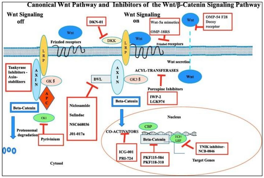
Int. J. Environ. Res. Public Health 2020, 17, 2078                                                  2 of 32

      As per the World Health Organization (WHO), breast cancer (BC) leads to mortality of women
worldwide (age group: 20–59 years) (World Health statistics 2013). According to the global cancer
project (GLOBOCAN 2012) breast cancer is considered as second most commonly occurred pathology
in the world [2]. Breast cancer is a commonly occurring disease in less-developed and industrialized
countries as well as the second notable cause of mortality in Europe and the United States after lung
cancer [2,3].
      In everyday medical practice, breast cancer diagnosis relies on three different types of analysis:
(A) clinical examination; (B) radiological/image examinations (that includes mammography, magnetic
resonance imaging (MRI) ultrasonography etc.) and (C) immunohistopathological examinations [4].
Employing all these tools, the clinical oncologist can stage the disease using TNM classification [5]
and reviewing the guidelines established in rigorous clinical trials, although, they can also use genetic
profiling tests such as MammaPrint [6] and Oncotype DX [7] to understand disease prognosis better.
      However, two decades ago, before the arrival of the current therapeutics, something was still
missing and a stronger diagnostic tool was needed. New advances in personalized medicine emerged
with the technology of microarrays and the discovery of molecular profiles.
      There are different histological subtypes of cancer (including breast cancer) within the same organ
or tissue, but biopsies are heterogeneous because they include diverse types of cells. Classification of
the different kinds of invasive carcinoma of breast cancer has been pursued, yet the clinical importance
of its classification is limited because different breast cancer patients have a wide variety of different
molecular profiles.
      Breast cancer includes molecular biomarkers [8] include:
(1)   ERα+ (estrogen receptor a-positive);
(2)   PR+ (progesterone receptor-positive);
(3)   HER-2 (human epidermal growth factor receptor-2);
(4)   EGFR (epidermal growth factor receptor) 45%–70% of Triple-negative breast cancer (TNBC)
      patients show this biomarker [9];
(5)   CK5/6;
(6)   VEGF (vascular endothelial growth factor);
(7)   KI67.
     Currently, due to microarray technology, there is a better understanding of the molecular
heterogeneity in tumors [10], aiding in the quantification of thousands of gene expression changes.
     The classification of breast cancer cell types is considered as below:
(1)    Luminal A subtype, ERα+/PR+ or −/HER-2-;
(2)    Luminal B subtype, ER+/PR+/HER-2+;
(3)    HER-2 enriched subtype ER- and or/PR−/HER-2+;
(4)    Basal-like subtype ER− and/or PR− , HER2− , CK5/6+, CK14+, CK17+ and EGFR+;
(5)    Normal breast-like type (ER− and/or PR− , HER2− , CK5/6− , CK14− , CK17− , EGFR− ) [11–14].
     Moreover, a subpopulation is described due to the Ki-67 index too [15]. Therefore, molecular
classifications are essential to provide personalized medicine and thus helps in selecting more specific
drug according to the molecular signature markers of the tumor. Jézequel et al. found 4 TNBC
subtypes:
(1)    Luminal androgen receptor-AR (LAR);
(2)    Mesenchymal (MES);
(3)    Basal-like immune-suppressed (BLIS);
(4)    Basal-like immune-activated (BLIA).
     Their results suggest that BLIA tumor prognosis is improved as compared to BLIS tumors [16], [17].
Later, Lehman researchers described six subtypes of TNBC, which include [18]:
Int. J. Environ. Res. Public Health 2020, 17, 2078                                                 3 of 32

(1)    Basal-like namely, BL1 and BL2;
(2)    MES;
(3)    MES stem-like;
(4)    immunomodulatory (IM);
(5)    LAR subtype.

     The different subtypes of TNBC correlate well with the different chemotherapeutic responses as
per retrospective studies [19]. Unfortunately, these molecular classifications have not been shown to
improve survival in hospital practice and current treatments.

2. Signaling Pathways Involved in Triple-Negative Breast Cancer (TNBC) Therapeutics

2.1. Notch Signaling Pathway
      Thomas Hunt Morgan described in 1917 a family of transmembrane ligands and receptors called
Notch [20]. This signaling pathway has a pertinent role in cell proliferation as well as differentiation
and, the elevated expression of a group of signaling molecules belonging to this pathway is correlated
with the poorest outcome of patients [21]. The pathway comprises of 4 Notch receptors namely, Notch-1,
2, 3 and 4) as well as 5 ligands namely, Jagged-1, Jagged-2, Delta-like 1, Delta-like 3 and Delta-like 4.
There are reports that confirms overexpression of Delta 1 and Jagged 1 in breast cancer [22–24], while
Notch-1 also plays an important role in the origination of human mammary tumor in the form of a
downstream effector of oncogenic Ras [25]. To date, Notch 1 has been relevant to the participation of
the Notch channel in different types of hematological malignancies [26], pancreatic cancer [27] and
many others. Several studies suggest that Notch-3 and Notch-4 are related to survival and proliferation
of tumors. In contrast, overexpression of Notch-2 in the context of the TNBC MDA-MB-231 cell line
appears to act as a protective factor [28].
      Since Notch receptor and ligand overexpression is linked with TNBC, researchers believe that the
receptor can be targeted by a monoclonal antibody (mAb) [29]. The current studies about inhibition
of Notch-1 signaling by mAbs have shown effectiveness in reducing the expression of HES and
HEY-L families in the MDA-MB-231 * TNBC cell lines (thus showing decrease in cell proliferation
and increase in the induction of apoptosis [30]. Additionally, DLL4 (Delta-like ligand 4 Notch ligand)
mAb therapy is effective for the treatment of TNBC [31]. Notch signaling in many transcription factors
codifies genes related to tumorigenesis, for example the HES family, HEY family, Akt, p53, VEGF and
PI3K-AKT-mTOR among others [32,33] (See Figure 1). Medications that interrupt Notch signaling
pathway act at the level of the second proteolytic cleavage in the cell cytoplasm by blocking the
multimeric γ-secretase complex and hence these drugs are known as γ-secretase inhibitors (GSIs) [33].
Unfortunately, most of the drugs that act by blocking the Notch pathway have not met expectations
required for approval by the FDA (Food and Drug Administration).
Int. J. Environ. Res. Public Health 2020, 17, 2078                                                       4 of 32

      Figure 1. Diagram of Notch receptor activation and therapeutic target in clinical development. Notch
      signaling is initiated by ligand binding to Notch receptor, which undergoes a two-step proteolytic
      cleavage by ADAM family proteases and γ-secretase, releasing the Notch intracellular domain (NICD).
      The NICD translocates to the nucleus where it binds to CSL and converts the complex from a repressor
      to an activator of Notch target genes. Notch signaling could be inhibited by two major classes of
      Notch inhibitors: γ-secretase inhibitors and monoclonal antibodies directing against Notch receptors
      or ligands. Abbreviations: NEC, Notch extracellular subunit; NTM, Notch transmembrane fragment;
      NEXT, Notch extracellular truncated; CSL, C protein binding factor 1/Suppressor of Hairless/Lag-1;
      NICD, Notch Intracellular Domain. Reproduced with permission from Yuan X, Wu H, Xu H, Xiong H,
      Chu Q, Yu S. Notch signaling: An emerging therapeutic target for cancer treatment. Cancer Letters.
      2015, 369, 20–27 [34].

2.2. Hedgehog Signaling Pathway
     Sonic Hedgehog (Shh) [35] network morphogenes have an impact on cancer stem cell (CSC)
maintenance, polydactyly syndromes, and basal cell carcinoma (Gorlin syndrome), with recent studies
suggest that they are altered in clinical samples of several human cancers including breast cancer cell
lines [36,37]. Hedgehog signaling involves three ligands:
(1)    Sonic (SHH) highly expressed during embryogenesis;
(2)    Indian (IHH) [38] mostly expressed in hematopoietic cells, endochondral skeleton, and cartilage;
(3)    Desert (DHH) [39] exhibit expression in the peripheral nervous system and testes, in fact,
       mutations of the DHH gene could lead to pure gonadal dysgenesis (PGD) [40].
     The Hedgehog signaling pathway is involved in the invasion of cancer cells, metastasis, and
resistance of drugs as well as tumor recurrence cancer after therapy [41]. Kaplan–Meier survival
studies indicate that overexpression of Shh is responsible for poor prediction of mortality in the breast
cancer patients and especially, TNBC patients. SHH has an important role in the erroneous origin of
malignancy in breast cancer because it maintains abnormal proliferation and promotes invasion to other
tissues (metastasis). Researchers have designed novel experimental drugs namely, Thiostrepton, whose
pharmacological action consists of targeting the sonic Hedgehog signaling, Thiostrepton suppresses
the population of CD44+/CD24− cancer stem cells (CSCs) of TNBC cell lines [42]. Nevertheless, it
is necessary to clarify the role of the Hedgehog pathway in breast CSCs [43] which has not been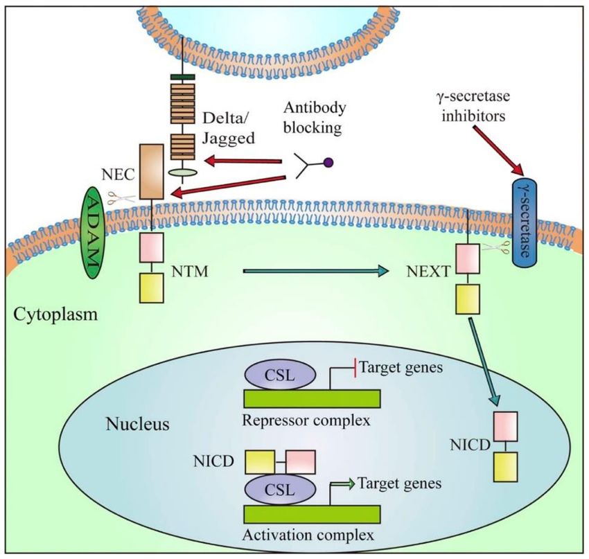
Int. J. Environ. Res. Public Health 2020, 17, 2078 5 of 32
determined yet [44,45]. As a result, there are few drugs authorized by the FDA to date to address this
pathway such as Vismodegib, which is used in basal cell carcinomas [46]. However, more research
is needed for SHH signaling potentially leading to the design of new prevention tools and novel
molecular markers for evaluation of recurrence, survival, and prognosis.
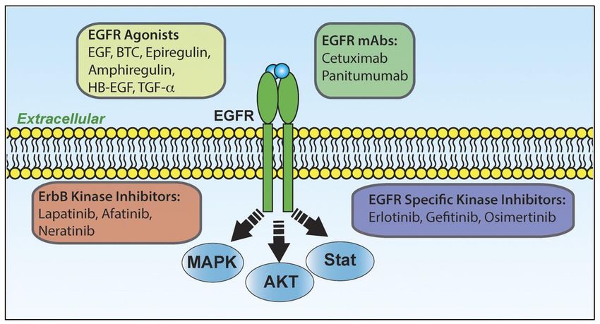
2.3. Wnt/β-Catenin Pathway
Wnt/β-catenin is the most commonly overexpressed pathway leading to transcriptional factor
activation responsible for the stimulation of epithelial to mesenchymal cell (EMT) transitions in CSCs.
Wnt signaling is also dysregulated in both canonical and non-canonical molecules on TNBC [47]. To the
best of our knowledge, there are 19 human Wnts and 10 Frizzled (FZD) receptors and coreceptors [47,48].
Wnt ligands (WNT5A, WNT11, and WNT3A) are pertinent in promoting migration and invasion [49].
FZD6 receptor is the most important representative in TNBC due to its capacity to produce metastasis
by increasing the motility characteristics of the malignant cells in TNBC [50]. Some novel drugs
target Frizzled receptors, for example, OMP-18R5 an antibody targeting Frizzled receptors diminishes
proliferation of tumor cells in the lung, breast, colon, and pancreatic tumors [48]. (Figure 2) Additionally,
overexpression and accumulation of β-catenin protein stimulates cell migration consequently leading
to resistance in TNBC cells [47]. Wnt inhibitors and modulators can eradicate CSC clonal cells and
drug-resistant cells [51], but we need to determine their safety in maintenance of tissue homeostasis
and repair. The activation of Wnt/β signaling pathway is correlated to diminished clinical outcome in
TNBC [52], as it presents the threat of lung and brain metastasis [53]. To date, scientists believe that
pluripotent CSCs play key role in the formation of the primary malignant solid tumors. These CSCs
are also responsible for the formation of drug resistance proteins in breast cancer, and are strongly
implicated in metastasis [54,55].
Figure 2. Canonical Wnt Pathway and Inhibitors of the Wnt/beta-Catenin Signaling Pathway schematic
representation of the Canonical Wnt Pathway and pharmacologic inhibitors of the Wnt/beta-catenin
signaling pathway. Reproduced with permission from Krishnamurthy N, Kurzrock R. Targeting the
Wnt/beta-catenin Pathway in Cancer: Update on Effectors and Inhibitors. Cancer Treatment Reviews.
2018, 62, 50–60 [56].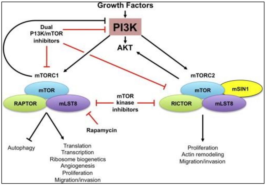
Int. J. Environ. Res. Public Health 2020, 17, 2078                                                 6 of 32

2.4. Poly (ADP-Ribose) Polymerase (PARP) Inhibitors
      The polyadenosine diphosphate-ribose polymerase also called poly (ADP-ribose) polymerase
(PARP) is a superfamily of 18 proteins that effect all the molecular events that leads to recovery of the
cells from DNA damage (participate in DNA base excision repair), gene transcription, apoptosis and
genomic stability [57].
      Roughly, 70% breast cancers evolving in BRCA1 mutation carriers while 23% of breast cancers
evolving in BRCA2 carriers, express a triple negative phenotype [58]. Therefore, PARP inhibitors are
considered perhaps the most important therapeutic drugs under investigation for the BRCA-1 and
BRCA-2 mutations as well as against TNBC. PARP expression in TNBCs is a consequence of exposure
to chemotherapy. PARP-1 and PARP-2 proteins are induced by DNA strand breaks and are associated
in DNA repair processes. PARP synthesized ADP-ribose polymer drives both BER (excision repair
pathway) and single-strand break repair (SSBR) pathways [59] (Figure 3).
      PARP activity, when suppressed, inhibits the ADP-ribose complex formation, so PARP-dependent
DNA-damage repair complexes such as DNA polymerase ε [60] cannot be efficient for repairing
DNA-damage [61]. Trapped PARP-DNA complexes are extremely cytotoxic exhibiting high
anti-proliferative activity (and therefore anticancer activity) [62]. Furthermore, Olaparib (AZD-2281)
and Veliparib (ABT-888) (both are PARP inhibitors) also differed markedly with respect to their catalytic
inhibitory propensities. Thus, the clinical as well as experimental results of each PARP inhibitor
also varies with respect to inhibition [63,64]. Since PARP inhibitors are different with respect to
trapping PARP-DNA complexes [62,65], differences can be seen while comparing the two (Olaparib and
Velipamib) with Velipamib the less dominant drug repressor of PARP1 and PARP2 than Olaparib [62].

      Figure 3. Poly (ADP-ribose) polymerase (PARP) inhibitor treatment of BRCA-1/2-associated and
      sporadic cancers. Reproduced with permission from Leif W. Ellisen. PARP Inhibitors in Cancer
      Therapy: Promise, Progress, and Puzzles. Cancer Cell. 2011, 19(2), 165–167 [66].

2.5. Mammalian Target of Rapamycin (MTOR) Inhibitors
     The erroneous regulation of mammalian target of rapamycin (mTOR) signaling, especially
Phosphoinositide- 3 kinase (PI3K)/Akt/mTOR pathway has a direct relationship with malignancy [67].
The mTor pathway is transformed in TNBC patients, thus is responsible for poor prognosis (aggressive
and tissue invasion) [68].
Int. J. Environ. Res. Public Health 2020, 17, 2078 7 of 32
Phosphorylation reactions, stimulated due to the PI3K/Akt/(mTOR), are responsible for cancer
cell growth, cell proliferation and angiogenesis [69]. Moreover, overexpression of Akt, a protein kinase,
is also correlated with tumor metastasis and invasion [68] The downstream signaling cascade of the
PI3K/Akt pathway is mTOR that is present in two functionally different complexes (mTORC1 and
mTORC2). The mTORC1 pathway promotes mRNA translocation as well as phosphorylates a wide
range of of substrates that accompany many anabolic processes [68] (Figure 4).
Figure 4. Mammalian target of rapamycin (mTOR) signaling pathway. mTOR is a subunit of two
distinct multi-protein complexes, mTORC1 and mTORC2. Both mTORC1 and mTORC2 can be activated
in response to growth-factors stimulation, whereas mTORC2 is a major kinase that phosphorylates
and activates Akt. The importance of mTORC1 and mTORC2 in regulation of multiple cell functions
vital for development of cancer and their strong interaction with oncogenic pathways make mTOR an
attractive target for therapeutic intervention. The mechanisms of action of currently available mTOR
inhibitors are shown. Reproduced with permission from Zaytseva YY, Valentino JD, Gulhati P, Evers
BM. mTOR inhibitors in cancer therapy. Cancer Letters. 2012, 319, 1–7 [68].
There are 6 classes of PI3K/AKT/mTOR network inhibitors: 1. Pan-class I (PI3K blocker)
2. Isoform-selective (PI3K blocker) 3. Rapamycin analogs (Rapalogs: Everolimus, Temsirolimus,
Deforolimus), 4. Active-site (mTOR blocker), 5. Pan-PI3K/mTOR blocker, and 6. AKT blockers [68].
Additionally, mTOR and one PI3K isoform, can be targeted simultaneously to increase the efficiency as
compared to single PI3K inhibition [68].
2.6. Epidermal Growth Factor Receptor (EGFR)
Receptor tyrosine kinase (RTK) targets such as epidermal growth factor receptor (EGFR) expression
are reported in 89% of TNBC cases, and hence considered to be a valid therapeutic target, especially
for BL2-subtype tumors that are augmented in EGFR gene expression [70]. Activation of this gene
stimulates primary tumorigenesis as well as metastasis. Gefitinib (EGFR inhibitor) reduces cancer cell
multiplication and enhances the cytotoxicities of carboplatin and docetaxel (Figure 5) [70,71]. There are a
number of different kinds of EGFR inhibitors trialed against TNBC such as the tyrosine kinase inhibitors
(TKIs)-erlotinib and lapatinib along with the monoclonal antibodies (mAbs) such as cetuximab and
panitumumab [72–75]. The reports of failures of EGFR-TKIs and mAbs, however, inspired combinationInt. J. Environ. Res. Public Health 2020, 17, 2078 8 of 32
therapy that includes mAbs and chemotherapeutics that proved to be a more efficacious. As an example,
cetuximab and carboplatin as well as Cetuximab and cisplatin in advanced TNBC patients, showed
double the efficiency of therapeutic response [76,77]. Moreover, the tri-inhibitors together namely,
gefitinib, carboplatin, and docetaxel synergistically increased the cytotoxicity of TNBC cells [78].
Another drug, cannabidiol caused inhibition of breast cancer metastasis by blocking the EGF/EGFR
signaling pathways and alteration of the tumor milieu [79]. Hence, cannabidiol could potentially be
efficient therapeutic strategy for highly aggressive TNBC [80].
Figure 5. A schematic representation of the activators, inhibitors and outcomes of epidermal growth
factor receptor (EGFR) signaling. EGFR is part of the four-member ErbB superfamily (ErbB1–4). These
receptors form several different homo- and heterodimers (here we only depict the EGFR homodimer).
EGFR is capable of binding several different extracellular ligands that agonize the receptor leading to
activation of several downstream signaling events including, but no limited to those listed. Several
therapeutics have been developed to antagonize EGFR including monoclonal antibodies (mAbs) that
block ligand binding as well as several different kinase inhibitors. In addition to EGFR, some of these
kinase inhibitors also target other ErbB receptors, supporting their use in human epidermal growth
factor receptor-2 (Her2)-amplified breast cancer (BC). All of the listed therapies are Food and Drug
Administration (FDA) approved for various cancers with the exception of Neratinib. Reproduced
with permission from Ali R and Wendt MK. The paradoxical functions of EGFR during breast cancer
progression. Signal Transduction and Targeted Therapy 2017, 2(16042), 1–7 [81].
2.7. Rapalogs
Rapamycin as well as paclitaxel both affect the PI3K/AKT/mTOR pathway, thus playing an
important role in therapeutics of TNBC. The mTOR antibodies coupled with EGFR inhibitors are
more effective as compared to anti-mTOR alone, even though there is no known evidence about the
synergy between anti-EGFR as well as mTOR inhibitors [81]. There is a report in which novel oral
AKT inhibitor Ipatasertip helps in progression-free survival alongwith PI3K/AKT pathway activation
in TNBC patients. But still, there is an urgent need to synthesize novel inhibitors that can target
PI3K/Akt/mTOR pathway for TNBC therapeutics [82].
2.8. TGF-β Signaling Pathway
TGF-β1 belongs to the TGF-β superfamily of cytokines encoding TGF-β1 gene. Human platelets,
which are a 25kDa cytoplasmic fragments have an important role in wound healing as well as inInt. J. Environ. Res. Public Health 2020, 17, 2078 9 of 32
regulation of the immune system. Thus, it inhibits the secretion and activities of different cytokines
such as IFN-gamma, TNF-alpha, and IL-2. TGF-beta 1 has an important activity in breast cancer
stem cells, as they express TGF-β1 and the TGF-β1 receptor exponentially [83,84]. TGF-β inhibitors
can inhibit the growth and multiplication of chemotherapy-resistant tumor-initiating cells (TIC)
in vivo [84] forming the basis for combinatorial chemotherapy for patients suffering from TNBC.
TGF-β stimulates an epithelial-to-mesenchymal transition (EMT) within mammary cells, leading to
an exhibition of tumor-like properties. It is possible to reverse EMT via TGFBR1/2 inhibitors while
stimulating mesenchymal-to-epithelial (MET) differentiation inside mammary epithelial cells [85].
TGF-β is frequently found overexpressed in the TNBC tumor microenvironment, especially in tumor
cells, or by tumor-associated immune and stromal cells. These cells also generate SMAD2/3 and
SMAD4, thus leading to metastasis and angiogenesis. This indicates that the TGF-β inhibitors play an
important role in patients with metastasis [85].
2.9. CSPG4 Protein Signaling Pathway
The CSPG4, which is also known as non-glial antigen or its also known as melanoma chondroitin
sulfate proteoglycan, is a cell-surface proteoglycan exhibited by basal breast carcinoma cells. Inhibition
of CSPG4 is therapeutically effective for breast cancer therapy. This protein leads to the dissemination
of the endothelial basement membrane protein, thus stabilizing the cell-substratum interaction, which
is in similitude to the effects that occur in TNBC. CSPG4 monoclonal antibodies can cause a blockade
of migratory, mitogenic and survival signalling pathways in tumor cells, making CSPG4 a new TNBC
target [86]. Moreover, there is overexpression of CSPG4 in TNBC cell types, resulting in inhibition of
TNBC cells when CSPG4 was targeted in such cells [87].
2.10. Cancer Stem Cells (CSCs) and Autophagy
As mentioned, numerous biochemical pathways in TNBC are relevant to cancer stem cells (CSCs),
thus, efforts are ruining into mAbs, dendritic cells (DC) and pluripotent cells cancer vaccines as well as
adoptive immunotherapy [88].
TNBC cancer stem cells (CSC) feature enhanced proliferative capacity, refractory treatment which
leads to recurrence and metastasis (CD-24, CD-44) [89]. Several biomarkers have been designed to
detect CSCs. However, most biomarkers are also shared by normal stem cells, and therefore these
biomarkers become to unspecific molecules leading to side effects. Chemo-resistance is present in
TNBC stem cells, and they are the ‘generals’ that lead the battle in tumor micro-environments riddled
with hypoxia [90]. Hypoxia is responsible for increasing chemo-resistance of autophagic TNBC stem
cells. Blocking the autophagic cascade network can increase chemo-response [91].
Autophagy is required for cancer stem cells and autophagy processes helps in the maintenance
of cellular homeostasis and, therefore, represents a survival pathway in cells. Unfortunately, cancer
cells can regulate the autophagy pathway to develop resistance to chemotherapy. Therefore, molecular
inhibition of the malignant autophagic pathway could reverse resistance to chemotherapy [91]. More
research needs to be done regarding abnormal stem cells autophagy mechanism, since it may harbor
the key to get a definitive cure not only against TNBC but against many types of cancer.
3. Strategies for TNBC Therapeutics
Despite the discovery of new metabolic and biochemical pathways within tumor
microenvironments, scientists and physicians continue to develop strategies to block network routes
and signals of neovascularization, metastasis, activating apoptosis, and “awakening” the immune
response [92]. This effort is made challenging by the dynamic and chaotic molecular configuration
of tumors, allowing tumors to recruit stromal cells, use valuable resources such as organic metals,
vitamins, and create their own blood supply using aberrant signaling. The standard approach has been
to use cytotoxic therapeutics, the chemotherapies that since the 1970s have been assisting oncology
patients; however, these approaches lack the desired selectivity.Int. J. Environ. Res. Public Health 2020, 17, 2078 10 of 32
Promising unconventional therapeutics are based on materials systems including nanopolymers,
liposomal drug delivery and nanostructured materials [93]. In 1995, the FDA approved Doxil® , the first
liposomal nanodrug part of a novel chemotherapy superior to the conventional [94] and a year later
the FDA also approved Feridex® , nanoparticles for magnetic resonance imaging [95]. In this context,
conventional chemotherapy and diagnostics are embracing the field of nanotechnology, promising to
provide valuable specificity to the treatment of cancer.
3.1. Conventional Therapeutics
3.1.1. Neoadjuvant Therapy
Currently, chemo-resistance is a significant problem for oncologists, with up to 90% of drug failures
in metastatic cancers [96]. TNBC patients initially respond to neoadjuvant treatments. Unfortunately,
there is a possibility of relapse in patients in the first 5 years in comparison with other cancer subtypes [97].
Nevertheless, neoadjuvant chemotherapy is the TNBC gold standard treatment [98]. It is important
to be diligent in TNBC patients, however, with the proper choice of drugs improving prognosis [99].
Additionally, neoadjuvant anthracycline–cyclophosphamide (AC-scheme) chemotherapy appears
to be establishing efficacy, although recently there have been reports on resistance developed for
these drugs [100]. Scheme AC in the presence of BRCA mutations has a pathological complete
response (pCR) rate of 27–30% [101] and consists of: Doxorubicin and Cyclophosphamide for 4 weeks
followed by Paclitaxel for 12 weeks. The prognosis may be improved to 61% if they associate drugs
such as Cisplatin [102]. Other drugs can be used as Carboplatin (CALGB40603 study) or Abraxane
(Nab-Paclitaxel nanoparticles) or immunotherapy using Bevacizumab [103,104]. On the other hand,
after the treatment, the monitoring of the disease should be evaluated by imaging techniques like MRI
which is the most sensitive imaging method for measuring TNBC neoadjuvant response-treatment [105].
Precision medicine strategies identify strategic biomarkers in each oncological patient, providing a
more effective and selective chemotherapy regimen [106]. Within the margins of personalized medicine,
medical research suggests that for TNBC treatment, one of the most useful molecular targets is EGFR
since it is positively expressed (around 60%) in TNBC [107].
Neoadjuvant therapy improves the response rate in patients with TNBC compared to adjuvant
therapy [101,108], as the effectiveness of cisplatin in TNBC is observed in preoperative phase II studies
where BRCA-1 expression is deficient. [109–111]. However, neoadjuvant systemic therapies should be
individualized because tumors with a BRCA-1 mutation are basal, but not all basal cancers express
BRCA-1 mutation. Moreover, cisplatin and bevacizumab, the latest as a molecular target of VEGF, have
shown to be efficient drugs in neoadjuvant therapy against TNBC [101,112] as researchers point out in
different meta-analysis as E2100, AVADO and RIBBON-1 [113]. Taxane-resistance of malignant cells
expressing BRCA-1 mutation is reported in in vivo studies [114], but a clinical trial called “CALGB
9344/INT1048” concluded that the use of paclitaxel reduces cancer recurrence in 17% as well as a
terminal clinical prognosis in 18% of TNBC patients [115].
3.1.2. Adjuvant Therapy
Adjuvant therapy is also a critical strategy to avoid the risk of metastases with concomitant
rapid progression and tumor recurrence activity [116]. The MA5 study showed anthracycline-based
drugs were not effective for treatment when BRCA-1 is expressed in TNBC [117,118], while other
studies show anthracyclines had encouraging results as adjuvant therapeutics [119]. The decision
whether or not to carry out adjuvant therapy must be evaluated for each patient by means of rigorous
analysis of clinical-histopathological staging-conditions and an adequate categorization of genomic
and proteomic profile.
The ability of TNBC to produce metastasis has been mentioned above, and shorter survival
time has been correlated with the presence of extensive tumor stroma. Therefore, it is crucial to
study the chemotherapy regimen in a palliative state because it is key for the clinician to understandInt. J. Environ. Res. Public Health 2020, 17, 2078 11 of 32
which drug is more effective. Many clinical trials are being done to evaluate the best treatment
for TNBC, for example comparing carboplatin efficacy versus docetaxel for metastatic TNBC [120].
Although different doses of taxane have been used in metastatic breast cancer (MBC), there is no
evidence indicating good efficacy in TNBC. For advanced stages (III C) when anthracycline-taxane
scheme resistance is documented, XelodaTM (Capecitabine) combined with TaxotereTM (Docetaxel)
is administered intravenously. (Table 1) Another combination that has shown utility is IxempraTM
(Ixabepilone) plus Capecitabine, although IxempraTM can be used as monotherapy at the same dose.
(Table 1).
Table 1. Conventional treatment of triple negative breast cancer *.
Conventional
Drugs Mechanism Scheme/Dose References
Treatment
Doxorubicin 20 mg/m2 plus
Neoadjuvant Anthracyclines + Taxanes Cyclophospamide 600 mg/m2 4
treatment Early or weeks followed by Paclitaxel 80
Cytotoxicity **
TNBC (Gold Capecitabine + Taxane mg/m2 12 weeks
Stabilization [101]
standard) Ixabepilone monotherapy Capecitabine 1250 mg/m2 14
microtubules
Advanced or or Ixabepilone + days + Docetaxel 75 mg/m2
Metastatic Capecitabine Ixabepilone 40 mg/m2 per 3
weeks
New Adding up standard scheme
Platinums (Carboplatin) Cytotoxicity and CALGB
neoadjuvant Abraxane 125 mg/m2 ,
Bevacizumab, VEGF 40,603 trial
agents (BRCA Carboplatin AUC, Bevacizumab
Nab-paclitaxel. immunotherapy [104]
mutations) 10 mg/kg
Cyclophosphamide 600 mg/m2 +
Adjuvant Anthracyclines and Doxorubicin 20 mg/m2 +
Cytotoxicity [121]
agents Taxanes Docetaxel 75 mg/m2 for q3
weeks 6 cycles.
Surgery: TNBC, surgical treatment is breast preservation.
Radiotherapy: radiation therapy (RT) is often given combined or after chemotherapy. RT also could be useful after
surgery. Probably benefits in BRCA mutations.
* The conventional treatment presently prescribed in hospitals for TNBC (Triple Negative Breast Cancer). It depends
pertinently on the clinical stage of the disease TNM, blood tests, imaging (mammography, ultrasound, CT-Scan, PET),
tolerability to treatment, usually accompanied by corticosteroids (Dexamethasone) and drugs to control symptoms
(Ondansetron, etc.) to reduce adverse effects. **Cytotoxicity: Inhibition of DNA and RNA synthesis. Inhibition of
topoisomerase II enzyme, generation of free oxygen radicals, Induction of histone eviction from chromatin etc.
3.1.3. Surgery
Many studies have been performed to determine the prognostic effects of mastectomy over
lumpectomy [110]. In TNBC, the surgical treatment of choice is the preservation of the breast; this is
because the choice of surgical treatment does not improve the prognosis or the local tumor recurrence,
so patients remain appropriate candidates for breast conservation [122]. A lumpectomy followed by
radiation therapy could be an option (National Comprehensive Cancer Network guidelines). However,
in TNBC, the gold standard is neoadjuvant therapy and is preferred before surgery.
3.1.4. Radiotherapy
Similar to conservative breast surgery, radiotherapy is part of the treatment regimen for TNBC,
albeit with some controversy [123]. However, evidence points out TNBC-BRCA-1 aberrant expression
is highly radiosensitive [124]. TNBC is considered a pathological entity susceptible to radiotherapy.
But unfortunately, like the guidelines in the pharmacological treatment, the use of RT in TNBC does
not have treatment guides [125,126].Int. J. Environ. Res. Public Health 2020, 17, 2078 12 of 32
3.2. Advanced Therapeutics
Chemoresistance is a significant problem in metastatic cancer [127]. Even though chemotherapy
has reached a milestone in the treatment strategies [128], there is a need to reduce the side-effects
of all the therapeutic regimens [129]. Moreover, non-steroidal anti-cancer drugs also possess many
side-effects and they also exhibit severe toxicity towards normal cells apart from cancer cells [130].
There are two main types of strategy for targeting therapeutics to tumor sites:
(1) A passive transport process called “enhanced permeability and retention” (EPR) in which
peripheral blood vessels to the tumor have leaky vasculature that increases nanoparticle
permeability. However, the disadvantage of EPR is that not all tumors possess leaky vasulature.
Therefore an adequate analysis of TNBC tumor biomarkers is required to load the nanoparticles
with a ligand specialized in the search for receptors overexpressed like CXCR4 (folic acid
receptor) [131].
(2) Another approach used by researchers is active transport that is governed by using biomarkers
miRNA (microRNA), proteins, antibodies, as well as therapeutic biomolecules such as siRNA
and aptamers, discussed below.
3.2.1. miRNA
The importance of microRNA (miRNA/miR) related with cancer treatment has recently increased
due to their potential as diagnostic biomarkers [108]. In TNBC, miRNA558 is the one that is
overexpressed [132]. Moreover, several TNBC miRNAs were found in a metanalysis [133]. Detecting
miRNAs promise to be part of the arsenal of oncological studies that will be available in hospitals to
provide better diagnosis and prognosis as powerful biomarkers. The most important study that is
considered to be the first of its kind, focused on primary TNBC as well as normal tissues, was the
microRNA profiling that discovered almost 116 microRNAs that have been deregulated. Among them,
miR-106b, the cluster miR-17/92, miR-200 family (miR-200a, miR-200b and miR-200c), miR-21 and
miR-155 were the highly expressed ones [134]. Furthermore, a second module of mRNA profiling of
TNBC linked to lymph node metastasis showed 6 miRNAs that was expressed differentially in the
lymph node tissues, namely, miR-424, iR-125a-5P, miR-627, miR-579, let-7g, miR-101 [134].
3.2.2. siRNA
Since the discovery of the Caenorhabditis elegans plant’s properties, siRNA has generated a
revolution in the treatment of diseases, with siRNA used to switch off or change the tumor genes
responsible for drug resistance, and in this way increase the efficacy in the treatments [135,136]. The
siRNA screens was performed for plethora of genes in TNBC cell lines and it was found that RSK2 [137].
non-SMC condensin I complex subunit D2 (NCAPD2) [138], Gpx1 (Glutathione peroxidase-1) [139], all
of them act as promising therapeutic targets for the TNBC treatment. The siRNAs that have already
been used in animal models to fight TNBC can be loaded in nanoparticles (non-viral) and viral capsids
or supramolecular complexes, providing gene silencing for proteins that reflect poor prognosis in
oncological medical practice viability [140].
Exosomes also play an important role in delivering siRNA for the suppression of metastasis
of TNBC after operative surgery. Cationic BSA coupled with siS100A4 as well as exosome
membrane covered nanoparticles helps in delivery of SiRNA to inhibit the growth of malignant
TNBC metastasis [141].
3.2.3. Aptamers
Aptamers are molecules made up of nucleotides, generally in a range of 50 DNA or RNA bases, that
are evolved to bind to specific molecular targets. Their small size makes them suitable to reach molecular
targets, therapeutic targets, protein complexes and cancer cells [142]. Engineering of aptamers is based
on a technique called systematic evolution of ligands by exponential enrichment (SELEX). AptamersInt. J. Environ. Res. Public Health 2020, 17, 2078 13 of 32
are easier and cheaper to produce than mAbs, but the degradation of aptamers in the bloodstream
is a clear disadvantage [143]. However, scientists are developing “mirror aptamers” (Spiegelmers)
enantiomers highly resistant to enzymatic degradation through synthetic biology to avoid aptamer
degradation by nucleases [144]. SELEX consists in the amplification of RNA (oligoribonucleotides) or
DNA (oligonucleotides) [145] using PCR (polymerase chain reaction) to subsequently incubate them
with molecular targets (cells, protein complexes, etc.). After five rounds the maximum molecular affinity
is generally obtained [146]. Various aptamers have been developed for therapy in TNBC; for example,
the aptamer 5TR1 pursues the molecular target MUC1 which is a tumor protein in MDA-MB-231,
the researchers have conjugated 5TR1 to Doxorubicin to make it more specific and avoid the known
side effects such as cardiotoxicity [147]. He et al. have reported aptamer-drug conjugate (ApDC),
AS1411-triptolide conjugate (ATC) for TNBC therapeutics with higher efficacies [148]. Nanomedicine
is advancing rapidly, and now researchers have focused on combining all the technologies mentioned
above (siRNA, miRNA, aptamers) by loading them into nanoparticles that pursue the CD-44 receptor
characteristic of TNBC pluripotent cells [149].
3.2.4. Nanomedicine: Armadas for TNBC Therapy
There are a number of clinically approved nanomedicines used in hospitals around the
world, e.g., liposomal doxorubicin (DoxilTM) [150], albumin-bound paclitaxel or Nab-Paclitaxel
(AbraxaneTM) [151] and polyethylene glycol (PEG-1) Asparaginase (OncasparTM) [152]. Additionally,
many nanomaterials have been studied with functions that includes: delivering drugs, aptamers or
microRNA capable of inducing gene or immunological therapy [153]. Some examples of these delivery
vehicles include micelles [154], luminescent carbon nanodots [155], nanodiamonds (NDs) [156], carbon
nanotubes (CNTs) [157,158], Au-nano matryoshkas [159] as well as SPIONs (superparamagnetic iron
oxide nanoparticles (NPs)) [160,161] etc. It is imperative to fabricate nanoparticles with the correct
properties for cancer therapeutics. These properties are dependent on the method of synthesis and
characterization employed. Obviously, nanomaterials for biomedical applications must be non-toxic
and biocompatible. It is also necessary that the synthesis and purification methods for nanoparticles
be reproducible [162], providing uniformity in size and shape; characterization that can be verified
through microscopy tools. Applications in nanomedicine likewise require nanoparticles that are
easy to metabolize by the human body, or be eliminated via renal or hepatobiliary clearance [163].
Nanomedicine provides a potential pathway to solve many of the problems of cytotoxicity and the
lack of tumor specificity of conventional chemotherapies. NPs can also minimize off-target effects. As
an example, lonidamine is an inhibitor of aerobic glycolysis but has failed in clinical trials due to its
intense hepatotoxic activity. Recently, however, NPs have been developed that incorporate lonidamine
together with a monoclonal antibody, providing greater selectivity for malignant cells than for the
healthy cells and reducing undesired systemic side effects [164]. Because there are several numbers of
tumor markers different from the PR, ER and Her-2 neu hormone receptors expressed in TNBC, the NPs
can be of help to achieve greater specificity and efficiency in the treatment, being able to pursue other
molecular objectives [165]. Gold nanorods have been used to carry out siRNA against MDA-MB-231
cells (TNBC), so researchers believe they could be useful for reducing tumoral activity [166]. Therefore,
it is now accepted that nanotechnologies are now part of the oncologist’s therapeutic arsenal including
breast cancer [167]. Nanomedicine has promise for improving the specificity with which drugs
and other molecules are transported using nanoparticles that maximize the therapeutic effect and
decrease the systemic toxicity of conventional chemotherapies. These nanocarriers must first have an
adequate safety profile, the parameters of which must be determined [168]. Therefore, understanding
nano-pharmacokinetics and nanotoxicology is mandatory [168]. Functionalized nanoparticles for
cancer therapeutics and diagnosis can be fabricated from diverse materials such as gold, silver [169],
diamonds [170], copper [171], among others. These materials are used due to their low cytotoxicity, for
example, gold nanoparticles are not cytotoxic making them suitable candidates for nanomedicine [172].
Currently, nanodrugs as DaunoXome® or Doxil® are currently being used in oncology, more researchInt. J. Environ. Res. Public Health 2020, 17, 2078 14 of 32
is needed to switch onto the next pillar-Nanomedicine. Immunotherapy is promising to reduce tumoral
recurrence, improving conventional treatment and reducing side-effects. However, more studies need
to be done. An important challenge in nanomedicine is how to engineer nanoparticles to evade the
immune system. The researchers responded by adding liposome layers, polyethylene glycol PEG
coatings that reduce the recognition of macrophages (evacuation of the reticuloendothelial system),
which increases the bioavailability and half-life of the drug [172]. Another useful coating could be
SDS, CTAB or tween 20 [173]. Even more promising, but not unrealistic, is the concept “Theranostics,”
in which nanoparticles can diagnose and treat at the same time. The nanoparticles can be used to
deliver drugs and generate real-time images,” [174]. Current intrahospital photodynamic therapy
(PDT) consists of administering porphyrins and phthalocyanines that have an affinity for malignant
cells; then, a laser can stimulate the structure and cause the release of reactive oxygen species (ROS).
Nanotechnology takes PDT to a different, enhanced level. PDT based on nanoparticles exploits a
photosensitizing agent creating ROS and apoptosis avoiding healthy tissue damage [175]. Many other
studies were carried out finding utility and efficacy in the joint use of NPs that act as a photosensitizer
to produce PDT [176,177].
There are many different kinds of nanoparticles exploited for cancer therapy which are as follows:
(i) Quantum Dots (QD)
Quantum dots (QDs) were discovered in 1982,and are semiconducting nanocrystals that have
superior light absorbance and high fluorescence intensity [178]. QD-based nanotechnology possesses
wider applications in cancer molecule imaging and quantitative detection [179]. Many studies using
QD technology could substitute immunohistochemistry (IHC) [180], because of its better fluorescent
signaling, performing even more accurate quantitative analyses for evaluating prognosis in TNBC [181].
QDs demonstrate results of molecularly directed images, as well as better quantitative detection of
cancer molecules like Ki67 and EGFR, expressed on TNBC [182].
(ii) Fluorescent nano-diamonds (FNDs)
Current nuclear medicine uses radioisotopes such as strontium-89, iodine-131, samarium-183
and technetium-99. However, nanotechnology proposes the use of non-radioactive materials with
improved sensitivity and specificity. In this technological revolution, we can also find materials such
as fluorescent nano-diamonds (FNDs). Fluorescent nano-diamonds are biocompatible nanomaterials
often used in MDA-MB-231 theranostics [156,183].
(iii) Nano-matryoshkas
Another singular design has been developed as thermal therapeutic, imaging and drug-delivery
nanoparticles. Nano-matryoskka, referred to as a multi-layer nanoparticle reminds of the Russian
doll which can contain many other dolls inside, the application of the hollow nanoparticles capable of
delivering multiple drug loads contained in multilayers that can be designed with different materials
as suggest an MDA-MB-231 murine xenograft study [184].
(iv) Silver nanoparticles (AgNPs)
Silver Nps (AgNP) are another example of Nps that can act against tumor cells in TNBC that
can induce DNA damage as in vivo studies suggest. Silver nanoparticles help in reduction of TNBC
growth and augments radiation therapy [172]. The mechanism of action is physical, it has not been
specifically established. One possibility is that the reaction of silver in the cellular microenvironment
will lead to the release of reactive oxygen species.
(v) Gold nanoparticles
Gold nanoparticles are photothermally tunable since they exhibit plasmonic behavior when
exposed to light, a unique property of matter at the nanoscale. These plasmonic NPs are useful
for producing heat and bringing apoptosis through hyperthermia [185]. Taking advantage of the
near-infrared (NIR) wavelength for medical applications, hyperthermia can kill cells due to reasonably
efficient tissue penetration of NIR radiation. [186]. It is effective when combined with radiotherapy
and chemotherapy [187]. Hyperthermia therapy or photothermal therapy continues to be the subjectInt. J. Environ. Res. Public Health 2020, 17, 2078 15 of 32
of research in nanomedicine, because at less than 100 nm the electromagnetic properties of materials
allow heat generation, prompting innovative treatments and diagnostics (theranostics) [188].
(vi) SPIONs (superparamagnetic iron oxide nanoparticles) and core-shell nanoparticles
The ability of iron oxide NPs to produce strong contrast images in RMI in T1(longitudinal
relaxation—spin-lattice) and T2 (transversal relaxation—spin-spin) has given them a place in the
theranostics of cancer [189,190]. This novel imaging system using iron oxide nanoparticles (IONP)
has been used in several xenograft model [191,192], for MRI diagnostic in TNBC [188]. SPIONs
have higher magnetic properties than paramagnetic materials due to their ability to spin alignment
to an external magnetic field, SPIONs can generate heat inside the tumors producing apoptosis by
hyperthermia [193]. SPIONs’ core-shell are formed by layers: an iron oxide core and a therapeutic
biocompatible coating [185] which can reduce toxic side effects [194]. Hayashi et al. [195] have
shown the advantages of using SPION intravenously for cancer theranostics. Also, SPIONs core-shell
hyperthermia properties, have been the hallmark of this design. Researchers are using lasers [196],
ultrasound [197], radio frequencies [198] or alternating magnetic fields [199] to generate apoptosis.
Moreover, SPIONs are also useful to deliver anti-cancer drugs such as gambogic acid (GA) in TNBC.
Sang et al. developed a GA drug nanoconjugate with an outer layer made up of mono-aminated
poly (ethylene glycol)-grafted hyaluronic acid that can specifically target CD44 receptors on TNBC
(Hyaluronic acid has higher affinity to bind CD44 on TNBC); the middle layer comprises disulfide-linked
hexadecanol (Hex) as well as chitosan oligosaccharide (CSO) that controls the drug release, while the
core layer is made up of SPIONs attached to GA that can increase the enhanced permeation and retention
effect due to magnetic focusing. This complex of mPEG-HA/CSO-SS-Hex/SPION/GA nanosystem led
to efficient delivery of the drug using magnetic guidance to focus in TNBC microenvironment [200].
(vii) Nanocomposites and their advantages over core-shell nanoparticles
The core-shell modality, however, has great challenges, including the negative polarity and the
amphipathic characteristic that makes them an easy target for the immune system. Alternatives in
nanoengineering are the creation of nanocomposites [201] which consist of biphasic or multiphase
materials, respecting the condition that at least one dimension of the material has less than 100 nm [202].
The improved optoelectronic properties allow nanocomposites to be useful candidates for drug
delivery, food packaging [203], sensing devices and their antimicrobial properties are currently being
studied [204]. Nanocomposites’ advantages over core shell design relies on colloidal easy synthesis
and reproducibility, because different matrices of materials can be fused regardless of their polymeric
or porous structure [205]. Administration as colloids would guarantee an adequate renal clearance if
the assemblies are either smaller than ~6 nm or can degrade into components of this size. Nevertheless,
a clear disadvantage in contrast with core-shell design is found in the largest size (100 nm) which is
characteristic of nanostructured materials [206].
(viii) Polymeric nanoparticles
In cancer therapeutics the major drawback of many nanocarriers as well as synaphic moieties
is that they bind non-specifically to many cellular as well as extracellular matrices, thus creating a
barrier for effective drug delivery. There are recent reports in which material scientists have utilized
a nanoparticle drug conjugate formulation in such a way that there is very less interaction with the
blood or other tissue sections which are named DART nanoparticles (poly(lactic-co-glycolic acid)
(PLGA)-polyethylene glycol (PEG)–ITEM4 nanoparticles). ITEM4 or Fn14 monoclonal antibody
binds specifically human as well as murine Fn14 extracellular domain. Paclitaxel loaded-DART
nanoparticles is an FDA-approved nanoformulation for TNBC models as well as an intracranial
model thus indicating that there is TNBC growth which is followed by metastatic propagation to the
brain [207]. Furthermore, Xu et al. developed Hyaluronic acid-coated pH sensitive poly (Beta-amino
ester) nanoparticles for the delivery of both embelin (anti-cancer drug) as well as pTRAIL (tumor
necrosis factor-related apoptosis-inducing ligand (TRAIL) plasmid for anti-TNBC efficacy [208]. All
the above nanotechnologies for health care are presented in Table 2.Int. J. Environ. Res. Public Health 2020, 17, 2078 16 of 32
Table 2. Nanomedicine for triple negative breast cancer theranostics.
Nanoparticle Unique Properties Application Status Evidence
Many studies signs QD technology could
substitute immunohistochemistry (IHC) [178],
Semiconductor nanocrystals they have QD-based nanotechnology possesses wider
because of its better fluorescent signaling, and
Quantum dots (QDs) superior light absorbance and high applications in cancer molecule imaging and Experimental/clinical ongoing
performing even more accurate quantitative
fluorescent intensity [181]. quantitative detection.
analyses for evaluating prognosis for
triple-negative breast cancer cells (TNBCs) [181].
Current nuclear medicine uses radioisotopes such
Tunable-enhanced optoelectronics
Fluorescent as strontium-89, iodine-131, samarium-183 and Fluorescent nano-diamonds (FNDs) are
features allows fluorescent
nano-diamonds technetium-99. FNDs proposes the use of Experimental/clinicalongoing biocompatible nanomaterials often used in
nano-diamonds (FNDs) issuing image
(FNDs) non-radioactive materials for imagining MDA-MB-231 theranostics [183].
signals [156] at low-cost production.
applications enhancing sensitivity and specificity.
Designed by multilayers that can be designed with
Nano-matryoshka, referred to as a
Nano-matryoshka, singular design has been different materials, Nano-matryoshka can exert
multi-layer nanoparticle, hollow
Nano-matryoshkas developed as thermal therapeutic, imaging and Experimental/clinicalongoing several drug medication payloads and inducing
nanoparticles can deliver multiple drug
drug-delivery nanoparticles. hyperthermia as suggest an MDA-MB-231 murine
payloads.
xenograft study [184].
The mechanism of action is physical. Silver NPs (AgNP) are another example of Nps
However, it has not been specifically that can act against tumor cells in TNBC that can
Silver nanoparticles
established. Ag affects cellular Therapeutics by using cytotoxicity. Experimental/clinicalongoing induce DNA damage as in vivo studies suggest.
(AgNPs)
microenvironment will lead to the release Silver nanoparticles help in reduction of TNBC
of reactive oxygen species. growth and augments radiation therapy. [169].
The ability of iron oxide NPs to produce strong
contrast images in MRI in T1(longitudinal This novel imaging system by using IONP has
Iron oxide Tunable-enhanced optoelectronics and
relaxation—spin-lattice) and T2 (transversal Experimental/clinicalongoing been used in several xenograft models [193,194],
nanoparticles (IONP) magnetic features.
relaxation—spin-spin) has given them a place in for MRI diagnostic on TNBC [195].
the theranostic of Cancer [9,90].
SPIONs SPIONs have higher magnetic properties
SPIONs can generate heat inside the tumors SPIONs are often use in human triple-negative
(superparamagnetic than paramagnetic materials due to their
producing apoptosis by using hyperthermia as Experimental/clinicalongoing breast cancer cells (TNBC) MDA-MB-231
iron oxide ability to spin alignment to an external
well as real time images into the tumors [195] therapeutics [209].
nanoparticles) magnetic fieldInt. J. Environ. Res. Public Health 2020, 17, 2078 17 of 32
Table 2. Cont.
Nanoparticle Unique Properties Application Status Evidence
Core shell design has been used for enhancing
SPIONs core-shell are formed by layers: a Enhanced hyperthermia properties, by stimulation
photodynamic, chemotherapy and gene therapy in
Core-shell magnetic iron oxide core and a through lasers [196], ultrasound [197], radio
Experimental/clinicalongoing TNBC [210]. Also Hayashi et al. [195] have shown
nanoparticles therapeutic biocompatible coating [192] frequencies [193] or alternating magnetic field
in advantages of using SPION intravenously for
which can reduce toxic side effects [194]. [199] to generate apoptosis.
cancer theranostics.
Theranostics
Gene Therapy
Enhanced optoelectronics specifically Photodynamics RMI T1- signal magnetic resonance imaging and
Gold nano-stars Experimental/clinicalongoing
T1-signal for RMI. Drug delivery photothermal therapy for TNBC [211].
Hyperthermia
Drug Delivery
Theranostics
Gene Therapy
Capacity to transport and deliver nucleic
Immunotherapy Immunogenic photodynamic therapy with gold
Nanocages acids, peptides and drugs as well as PDT Experimental/clinicalongoing
Photodynamics nanocages on TNBC [212].
properties.
Hyperthermia
Imaging
Theranostics
Gene Therapy
Enhanced magnetic-optoelectronics
Immunotherapy Gold nanorods were developed for delivering
properties according to shape and size.
Nanorods Photodynamics Experimental/clinicalongoing cisplatin and producing photothermal therapy on
Capacity to transport and deliver nucleic
Hyperthermia TNBC [213].
acids, peptides and drugs.
Imaging
Drug Delivery
Theranostics
Enhanced magnetic-optoelectronics Gene Therapy
including plasmon surface resonance Immunotherapy Researchers are experimented on using
Nanocomposites properties. Photodynamics Experimental/clinicalongoing immunotherapy nanocomposites vehicle on TNBC
Nucleic acids, peptides and drug Hyperthermia [214].
releasing with enhanced specificity. Imaging
Drug DeliveryYou can also read