Early Breast Cancer Detection Utilizing Artificial Neural Network
←
→
Page content transcription
If your browser does not render page correctly, please read the page content below
WSEAS TRANSACTIONS on BIOLOGY and BIOMEDICINE                             Zakia Sultana, Md. Ashikur Rahman Khan,
     DOI: 10.37394/23208.2021.18.4                                                                         Nusrat Jahan

    Early Breast Cancer Detection Utilizing Artificial Neural Network

           ZAKIA SULTANA1, 2,*, MD. ASHIKUR RAHMAN KHAN1, NUSRAT JAHAN1
                  1
                    Department of Information and Communication Engineering
                          Noakhali Science and Technology University
                                  Noakhali-3814, BANGLADESH
                              2
                                  Department of Computer Science and Engineering
                                     Daffodil International University, Dhaka
                                                   BANGLADESH

Abstract: - Breast cancer is one of the most dangerous cancer diseases for women in worldwide. A Computer-
aided diagnosis system is very helpful for radiologist for diagnosing micro calcification patterns earlier and faster
than typical screening techniques. Maximum breast cancer cells are eventually form a lump or mass called a
tumor. Moreover, some tumors are cancerous and some are not cancerous. The cancerous tumors are called
malignant and non-cancerous tumors are called benign. The benign tumors are not dangerous to health. But the
unchecked malignant tumors have the ability to spread in other organs of the body. For that early detection of
benign and malignant tumor is important for confining the death of breast cancer. In these research study different
neural networks such as, Multilayer Perceptron (MLP) Neural Network, Jordan/Elman Neural Network, Modular
Neural Network (MNN), Generalized Feed-Forward Neural Network (GFFNN), Self-Organizing Feature Map
(SOFM) Neural Network, Support Vector Machine (SVM) Neural Network, Probabilistic Neural Network (PNN)
and Recurrent Neural Network (RNN) are used for classifying breast cancer tumor. And compare the results of
these networks to find the best neural network for detecting breast cancer. The networks are tested on Wisconsin
breast cancer (WBC) database. Finally, the comparing result showed that Probabilistic Neural Network shows
the best detection result than other networks.

.

Key-Words: - Breast cancer, classification, support vector machine, multilayer perceptron, self-organizing map,
Jordan neural network, Probabilistic Neural Network, Recurrent Neural Network.

Received: January 28, 2021. Revised: March 10, 2021. Accepted: March 12, 2021. Published: March 18, 2021.

1. Introduction                                                  breast. Blood nourishes the cells. The lymph system
Breast cancer is the most common cancer in women.                drains bodily waste products. The lymph vessels
Breast cancer is the second leading cause of death               connect to lymph nodes. Cancer that forms in the
among women. Breast cancer also developed in                     tissues of breast, usually in the ducts (tubes that carry
men, breast cancer in men is rare, an accounting for             milk to the nipple) and in the lobules (glands that
less than 1% of all breast cancers, only about 1 out of          make milk) is called, the breast cancer [1].
1,000 men will get it at some point in their lives. The               Breast cancer can develop when the genetic
breast is made up of different breast tissue, ranging            material in cells changes and causes them to start
from very fatty tissue to very dense tissue. This tissue         multiplying in an uncontrolled way. Lumps and
consists of a network of lobes. Each lobe is made up             nodules then form after some time. Why those
of tiny, tube-like structures called lobules that contain        changes come about, and how the cells develop,
milk glands. Tiny ducts connect the glands, lobules,             depends on many factors that can influence each
and lobes, carrying milk from the lobes to the nipple.           other. The main factors include the woman’s age,
The nipple is located in the middle of the areola,               hormones, and whether there is a higher risk of breast
which is the darker area that surrounds the nipple.              cancer in her family.
Blood and lymph vessels also run throughout the

     E-ISSN: 2224-2902                                      32                                        Volume 18, 2021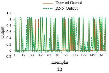
WSEAS TRANSACTIONS on BIOLOGY and BIOMEDICINE                             Zakia Sultana, Md. Ashikur Rahman Khan,
     DOI: 10.37394/23208.2021.18.4                                                                         Nusrat Jahan

     Breast cancer accounts for 1 in 6 female cancer             2. Literature Survey
deaths. The prevention method of the breast is not               Computer aided diagnosis of breast cancer getting
find out until now. Early detection is the main                  famous day by day. Computer aided diagnosis helps
prevention method of breast cancer. The mortality                the radiologists to detect abnormalities earlier than
rate for breast cancer will be reduce if we detect is the        traditional procedures. The first step for CAD is the
early stage of the cancer before spreading into the              ability to identify the abnormal masses in the breast,
other organs of the body. But unfortunately, about 90            while the second step is to diagnosis the masses
present breast cancer is detected in advance stage               detected in the first step. There are many steps in
such as stages III and IV [2]. It is a very bad news for         diagnosis of breast cancer, start from the
breast cancer patient.                                           segmentation and ended by tumor classification. The
It is important to detect breast cancer early to reduce          diagnosis requires precise and reliable diagnosis to
the mortality rate, and this requires accurate and               ensure that doctors can distinguish between benign
reliable diagnoses. When several tests are involved,             and malignant tumors. According to American
the ultimate diagnosis may be difficult to obtain, even          Cancer Society, some studies have shown that CAD
for a medical expert [3]. Diagnosis of breast cancer             can help find cancers that radiologist otherwise might
has improved during the last decades for the                     have missed. The use of classifier systems in medical
development of more effective diagnosis system.                  diagnosis is increasing gradually. There is no doubt
Several Artificial Intelligence (AI) techniques have              that evaluation of data taken from patients and
been intensively applied to radiological assessments             decisions of experts are the most important factors in
to predict the biopsy outcome in breast cancer [4]).             diagnosis. However, expert systems and different
These techniques include many artificial neutral                 artificial intelligence techniques for classification
networks. Moreover Computer-Aided Diagnosis                      also help experts in a great deal.
(CAD) could be beneficial to help radiologists in the                  Automatic diagnosis of breast cancer is an
US based detection of breast cancer, minimizing the              important real world medical problem. Researchers
effect of the operator-dependent nature of US                    made many attempts to efficiently use different
imaging [5]). There are several other classification             neural networks to improve the diagnosis efficiency
techniques to predict and classify breast cancer                 in breast cancer detection.
pattern. Classification systems, helping possible                      A method was implemented using Back
errors that can be done because of fatigued or                   Propagation Algorithm (BAP), Radial Basis
inexperienced expert to be minimized, provide                    Function Networks (RBFN), and Competitive
medical data to be examined in shorter time and in               Learning Network (CLN) and Learning Vector
more detail [6].                                                 Quantization (LVQ) classifier network to detect
In this study, different artificial neural networks such         breast cancer disease on the Wisconsin Breast Cancer
as multilayer Perceptron, Jordan/elman network,                  Dataset (WBCD). After evaluating these four
modular neural network, generalized feed-forward                 networks they found that LVQ is the best classifier
neural network, radial basis function with                       neural network to detect breast cancer. And the order
probabilistic neural network, self-organizing feature            of the accuracy CLN, MLP, RBFN. The accuracy of
map, support vector machine, recurrent network are               the LVQ is 95.82% [7].
used to diagnosis the beaning and malignant cancer                    A system was proposed for breast cancer
cell of breast. And after comparing the diagnosis                detection (classification of benign and malignant)
result of these networks the best networks will be               using back-propagation neural network on the
introduced for the early detection of the breast                 Wisconsin breast cancer dataset (WBCD). This
cancer.                                                          proposed method results were compared with radial
     In this research work, apply different neural               basis function network and found that for detecting
network models for the classification of benign and              breast cancer back-propagation neural network is
malignant cell for the classification of breast cancer.          best. By increasing the number of neurons in hidden
This thesis study main objective is the early detection          layer the accuracy is improved, neural network with
of breast cancer disease through different neural                9 neurons in hidden layer provide 99% accuracy [8].
networks. This thesis classifies benign and malignant            A comparative study of support vector machine
cell using different neural network accurately and               (SVM), Bayesian classifier and other artificial neural
make a comparison among the networks to find the                 network classifier (Back-propagation, learning
best network, which classify more accurately than                vector quantization, and K nearest neighbourhood)
other networks.                                                  was provided on the Wisconsin breast cancer Fine
                                                                 Needle Aspiration (FNA) biopsy data set. The use of
                                                                 machine learning technique cancer cells is detected

     E-ISSN: 2224-2902                                      33                                        Volume 18, 2021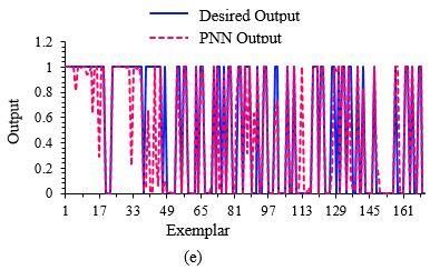
WSEAS TRANSACTIONS on BIOLOGY and BIOMEDICINE                          Zakia Sultana, Md. Ashikur Rahman Khan,
    DOI: 10.37394/23208.2021.18.4                                                                      Nusrat Jahan

early with preciously. By comparing the result K              benign pattern using WBCD dataset was
nearest neighbourhood provide prediction accuracy             implemented. After examination the HENC approach
100%, SVM using the RBF kernel function provide               can obtain better generalization and much lower
98.24% and K2 Bayesian network provide poor                   computational cost than the existing methods. This
accuracy by comparing all the networks evaluated.             proposed approach had an average accuracy more
And the Bayesian network shows a low prediction               than 98%. A CAD method also developed using the
time of 0.07 seconds than other networks evaluated            HENIC approach to make the classifier interesting
[9].                                                          and easy to apply in the real world [4].
     A method was proposed for detecting breast                    A hybrid machine learning method using fuzzy-
cancer using modified imperialist competitive                 artificial immune system with k-nearest neighbour
algorithm (MICA), K-means algorithm and different             algorithm for diagnosing breast cancer was proposed
neural network and working on the Wisconsin Breast            by classifying Wisconsin Breast Cancer Dataset
Cancer database (WBCD). This method includes                  (WBCD). After using this method, the highest
clustering module and classification module. In the           classification accuracy of 99.14%. This classification
clustering module the input data are clustered by the         accuracy was obtained by 10-fold cross validation
combination of modified imperialist competitive               [6].
algorithm (MICA), K-means algorithm and the                         A Computer Aided Diagnosis algorithm was
Euclidean distance of each pattern is examined. In the        developed for identifying breast nodule malignancy
classifier method the multilayer perceptron (MLP),            using multiple ultrasonography (US) feature and
probabilistic neural networks (PNN) and the radial            artificial neural network (ANN) classifier was
basis function neural networks (RBFNN) determines             developed from a database of 584 histologically
the membership of the patterns using examined                 confirmed cases containing 300 benign and
distance. Among various neural networks MLP with              284malignant breast nodules. The features
back-propagation algorithm provide 97.44%                     determining whether a breast nodule is benign or
accuracy of system using only WBC database and                malignant were extracted from US images through
with the Euclidean distance of patters from cluster           digital image processing with a relatively simple
canter which are find from the K-MICA, RBFNN’s                segmentation algorithm applied to the manually
provide the accuracy of system 99.11%. The                    preselected region of interest. An ANN distinguished
performance of classifier also compared by K-mean             malignant nodules in US images based on five
clustering, subtractive clustering and genetic                morphological features representing the shape, edge
algorithm clustering. From the comparison result              characteristics, and darkness of a nodule. The
shows that K-MICA clustering based system                     structure of ANN was selected using fold cross-
achieves higher recognition accuracy of 99.11% [10].          validation method with = 10. The ANN trained with
     A more cost effective and easy to use system was         randomly selected half of breast nodule images
developed for classifying breast cancer disease tumor         showed the normalized area under the receiver
using neural network with feed-forward back-                  operating characteristic curve of 0.95. With the
propagation algorithm to classify the tumor that              trained ANN, 53.3% of biopsies on benign nodules
causes cancer by working on the Wisconsin breast              can be avoided with 99.3% sensitivity. Performance
cancer database (WBCD). Many neural networks                  of the developed classifier was re-examined with new
model were created and trained using different                US mass images in the generalized patient population
number of neurons in hidden layer and by the                  of total 266 (167 benign and 99 malignant) cases
investigation of the networks they find that neural           [13].
network model with 7 node in the hidden layer                      A hybrid system for diagnosis breast cancer
acquire the highest accuracy than others. The result          tumours using Wisconsin breast cancer database
of the feed-forward back-propagation algorithm was            (WBCD) was developed. The system has three main
compared with support vector machine (SVM) and                modules, such as feature extraction module, training
decision tree classifier and showed that feed-forward         module and classifier module. In training module,
back-propagation algorithm is the best classifier than        they used a hybrid bees’ algorithm (BA) - back-
others [11].                                                  propagation (BP) algorithm is proposed to train the
     A hybrid evolutionary neural network classifier           classifier and multi-layer perceptron (MLP) neural
(HENC) combining the evolutionary algorithm,                  network is used for train classification. After
which has a powerful global exploration capability,           analysing this proposed system has high diagnosing
with gradient-based local search method, which can            accuracy than other method and the accuracy
exploit the optimum offspring to develop a diagnostic         95.86%. And for increasing the accuracy of
aid that accurately differentiates malignant from             recommended system, fuzzy feature used as the input

    E-ISSN: 2224-2902                                    34                                       Volume 18, 2021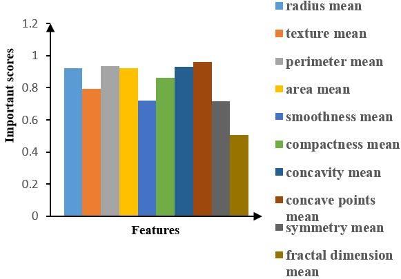
WSEAS TRANSACTIONS on BIOLOGY and BIOMEDICINE                            Zakia Sultana, Md. Ashikur Rahman Khan,
    DOI: 10.37394/23208.2021.18.4                                                                        Nusrat Jahan

of optimized classifier that significantly improves the        It is apparent from previous studies that complex
accuracy to 97.83% [14].                                       neural networks approaches are applied for detecting
     A method was proposed using Support Vector                breast cancer and more research is needed to improve
Machine (SVM) combine with feature section for                 the accuracy of early detection of breast cancer. This
diagnosis breast cancer using Wisconsin breast                 research works for finding early detection of breast
cancer database (WBCD). The performance of this                cancer with more accuracy.
model is evaluated using classification accuracy,
sensitivity, specificity, positive and negative values,        3. Methodology
receiver operating characteristic (ROC). The result            The aim of this research is to define the best artificial
showed that the highest classification accuracy is             neural network (ANN) methods to classify the breast
99.51% is obtained using SVM with these five                   cancer cell for detecting breast cancer in the early
features [15].                                                 stage. Breast cancer microscopic and clinical tests
     The use of deep learning approaches was                   reports are collected. Based on the tests reports the
proposed for breast ultrasound lesion detection and            diagnostic system will predict either Benign (not
investigates three different methods: a Patch-based            cancer) or Malignant (cancer). There are different
LeNet, a U-Net, and a transfer learning approach with          networks like multilayer perceptron (MLP),
a pretrained FCN-AlexNet. Their performance is                 Jordan/Elman network, modular neural network,
compared against four state-of-the-art lesion                  generalized feed forward network, self-organizing
detection algorithms (i.e. Radial Gradient Index,              feature map, support vector machine, recurrent
Multifractal Filtering, Rule-based Region Ranking              network.
and Deformable Part Models). A comparing and
contrasting two conventional ultrasound image                  3.1      Parameters Considered
datasets acquired from two different ultrasound                All the networks were trained, tested and validated
systems. Dataset A comprises 306 (60 malignant and             using a variety of parameters. Each of the networks
246 benign) images and Dataset B comprises 163 (53             was tested using many combinations of hidden node
malignant and 110 benign) images. To overcome the              and epoch number. But the hidden layer, activation
lack of public datasets in this domain, Dataset B will         function and learning rule of all the networks were
be made available for research purposes. After                 same. After a number of experimentations and
analyzing the Transfer Learning FCN-AlexNet                    parameter       alterations     using      theoretical
achieved the best results for Dataset A and the                understandings and best practices, the optimal
proposed Patch-based LeNet obtained the best results           classifier was identified.
for Dataset B in terms of FPs/image and F-measure              The dataset in this research was obtained from the
[5].                                                           UCI Irvine machine learning repository [16]. This
     The statistical neural network structures, radial         dataset was originally created by Dr. Wolberg, Street
basis network (RBF), general regression neural                 and Mangasarian all from University of Wisconsin.
network (GRNN) and probabilistic neural network                Data items in the dataset are composed of ID number,
(PNN) was used on the Wisconsin breast cancer data             the diagnosis which will either be classified as
(WBCD). Then compare the statistical neural                    malignant (M) or benign (B) and numeric shape
networks result with the result of Multilayer                  features of extract cellular nuclei such as radius,
Perceptron (MLP) neural network on the WBCD                    texture, perimeter, area, smoothness, compactness,
database. Finally they found that for the test set             concavity, concave points, and symmetry and fractal
GRNN gives the best classification accuracy [3].               dimension. These data were extracted from
     A new method for detecting the breast cancer              preprocessed Fine Needle Aspiration biopsy image of
more accurately was proposed and in this methods               cell slides. The dataset was composed of a total of
first use the image processing techniques to prepare           569 observations with benign and malignant cases
the mammography images for the feature and pattern             and the number of benign 357 and the number of
extraction process then used this features as input for        malignant 212 observations. And benign will be
the Back Propagation Neural Network (BPNN) and                 detected by “0” and malignant will be detected by “1”
the Logistic Regression (LR) machine learning                  Attribute Information:
supervised algorithm. After analyzing the result of                 1. ID number
this two algorithm they find that, the number of                    2. Diagnosis (M = malignant, B = benign)
features utilized in LR model was much higher
than BPNN. A good regression value using BPNN                  Ten real-valued features are computed for each cell
that exceeded 93% with 204 features [12].                      nucleus:

    E-ISSN: 2224-2902                                     35                                        Volume 18, 2021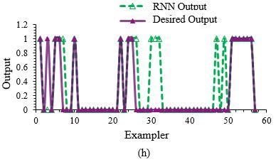
WSEAS TRANSACTIONS on BIOLOGY and BIOMEDICINE                             Zakia Sultana, Md. Ashikur Rahman Khan,
    DOI: 10.37394/23208.2021.18.4                                                                         Nusrat Jahan

    a. radius (mean of distances from center to                  Artificial Number            of             Epoch
         points on the perimeter)                                 Neural    hidden layer and                 number
    b. texture (standard deviation of gray-scale                 Network    elements           /
         values)                                                  Model     Configuration
    c. perimeter                                                Multilayer  one hidden layer                  40,000
    d. area                                                     Perceptron  with          seven
    e. smoothness (local variation in radius                      (MLP)     processing
         lengths)                                                           elements
    f. compactness (perimeter^2 / area - 1.0)                               one hidden layer                  20,000
    g. concavity (severity of concave portions of              Jordan/Elman with           four
         the contour)                                             network   processing
    h. concave points (number of concave portions                           element
         of the contour)                                                    two hidden layer,                 40,000
    i. symmetry                                                   Modular   one hidden layer
    j. fractal           dimension         (“coastline             neural   with             six
         approximation” - 1)                                      network   processing
In the network structures there are ten nodes in the                        element another
input layer, on the basis of breast cancer dataset, j                       hidden layer with
nodes in the hidden layer, and one nodes in the output                      four processing
layer or diagnosis result benign(B) or malignant (M).                       element
Thus, the network structure can be defined as 10-j-1.
                                                                Generalized one hidden layer                  30,000
                                                               feed forward with          seven
                                                                  network   processing
                                                                            element
                                                                    Self-   one hidden layer                    For
                                                                 organizing with           four           unsupervised
                                                                feature map processing                      learning is
                                                                            element. Square                 20,000 and
                                                                            kohonen        with                 for
                                                                            radius of the                   supervised
                                                                            cluster is 1.                  learning the
                                                                                                              epoch
                                                                                                            number is
                                                                                                              40,000
                                                               Support            Compose without             20,000
                                                               vector             regression
                                                               machine
                                                               Recurrent          one hidden layer            50,000
                                                               network            with        four
                                                                                  processing
       Figure 1: Structure of neural network                                      element
                                                               Probabilistic                                     3
3.2      Artificial  Neural     Network      Model             neural
developed                                                      network
Artificial Neural Network models that taken in
consideration are Multilayer perceptron (MLP),                From the above table we can say that the network
Jordan/Elman network, Modular neural network,                 structure of modular neural network is 10-6-4-1 & the
Generalized feed forward network, Self-organizing             network structure is 10-4-1 for jordan/elman, self-
feature map, Support vector machine, Probabilistic &          organizing feature map, and recurrent neural
Recurrent network. For detecting best cancer, the             network. The network structure can be defined as 10-
number of epochs for each model are described                 7-1 for both multilayer perceptron, generalized feed
below, Table 1.                                               forward neural network.
 Table 1 Configuration of Neural Network Models

    E-ISSN: 2224-2902                                    36                                          Volume 18, 2021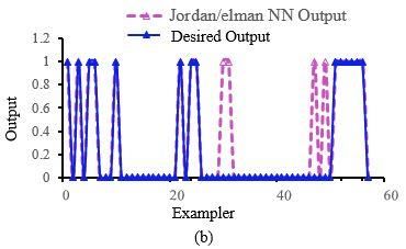
WSEAS TRANSACTIONS on BIOLOGY and BIOMEDICINE                            Zakia Sultana, Md. Ashikur Rahman Khan,
    DOI: 10.37394/23208.2021.18.4                                                                        Nusrat Jahan

3.3      Performance Analysis (Training, Testing
& Validation)
Once a network has been structured for a particular
study, that network has been trained. After the
appropriate training, the selected network has the
ability to interconnect one value of output to a given
input [18]. For starting this process, the initial
weights are chosen randomly and then, the training
or learning begins. For developing an appropriate
network, the networks had been trained many times.
     The study of detecting breast cancer used 60% of
total dataset for training approach, 30% of total
dataset for testing approach, 10% of total dataset for
validation approach.                                           Figure 2: Effect of the various parameters on breast
     Based on the training testing process on the data                                cancer
set, networks will predict the target value or classify
the beaning or malignant. But the prediction of these               Training, testing and validation result for all used
network may contain error. The error is computed               networks are shown in Table 2. It shows the best
with the equation                                              epoch with minimum MSE and the final MSE of the
                                                               training error at last epoch, testing result shows the
                 Error = E = ½ (tT - yo)2          (1)         mean square error (MSE), minimum absolute error
where,                                                         (Min Abs Error), maximum absolute error (Max Abs
        tT = target value                                      Error) and the linear correlation coefficient (r) value.
        yo = the neural network output                         The r value explained the variation between the
        E = error                                              neural network outcomes and desired outcomes. The
    The actual error of the network is calculated by           perfect value of r is 1. The accuracy result for
running all the training cases and testing cases. The          detecting best cancer depends on the validation
performance measure parameters of all networks are             results of the networks.
mean square error (MSE), the r value and the                        From Table 2, it is noticed that probabilistic
diagnosis accuracy of the networks output. Other               neural network shows the minimum training MSE
parameter minimum absolute error (min abs error),              0.002376483. Generalized feed-forward neural
maximum absolute error (max abs error) will be                 network produce the second minimum training MSE
showed in the result section.                                  0.011683897. Multilayer perceptron produces the
                                                               third minimum training MSE 0.016001718 and self-
4. Result Analysis                                             organizing feature map produce the fourth minimum
The aim of the project is to explore the best network          training MSE 0.017059825. From the table 2 it is also
among different neural networks for nearest detection          noticed that recurrent neural network shows the
of breast cancer. The input data can be used to train,         minimum testing MSE 0.053405378 and shows the
test and validate the different neural networks with           height linear correlation coefficient (r) 0.889811535.
the uniform activation function and learning rate.                  Modular neural network produce the second
These yielding 10 input data features have different           minimum testing MSE 0.075174462 and second
impact for detecting cancer. Some features are most            highest correlation coefficient (r) 0.838853385.
useful in predicting malignant or benign cancer.               Probabilistic neural network shows the third lowest
Figure 2 shows the impact of important features with           testing MSE 0.081914184 and third height liner
important scores by applying the random forest                 coefficient 0.830980024. MLP Testing MSE
model [17]. All the networks were run only once.               0.099679093 and linear correlation coefficient is
These networks will be analysed in each training               0.80010834. Self-organizing Feature map produce
testing and validation part and finally the networks           testing MSE 0.103715541 and correlation coefficient
will be discussed and compared for finding the best            is 0.78119486.
network, which accurately detect the breast cancer.

    E-ISSN: 2224-2902                                     37                                        Volume 18, 2021
WSEAS TRANSACTIONS on BIOLOGY and BIOMEDICINE Zakia Sultana, Md. Ashikur Rahman Khan,
DOI: 10.37394/23208.2021.18.4 Nusrat Jahan
Table 2. Training, testing and validation result of
all used neural networks
The detection or diagnosis accuracy of all used
Training Testing result Validation
networks shown in table 2 based on the validation
result Result
Neural Network
result, probabilistic neural network accuracy for
Min. Abs. Error
Max Abs. Error
Final MSE detecting breast cancer is 98.24%. SOMF network
Min. MSE
Diagnosis
Accuracy
accuracy is also 98.24%. The second height accuracy
Epoch
MSE
96.5% of support vector machine. MLP network
r
accuracy for detection is 91.23%. Jordan/Elman and
recurrent neural network accuracy is 93%.
To investigate accuracy of the networks use the line
0.01494327
0.01600171
0.09967909
0.04449310
1.05555556
0.80010834 pattern of the data between the target values and
Perceptron
Multilayer
Network
91.23%
Neural
10930
neural networks outputs. The target values and the
2
8
1
neural networks outputs are generated on the same
graph. In this way the line pattern of all neural
networks are shown in figure 3 and figure 4. Figure 3
Elman Neural
0.019289653
0.035868508
0.093858431
0.011998891
1.055555555
0.815923212
and figure 4 shows that the predicted and
Jordan and
Network
experimental values are how close to each other for
93%
3344
6
all networks in testing and validation step
respectively.
0.02182240
0.02266522
0.07517446
0.01199889
0.97569489
0.83885338
Network
Modular
91.23%
Neural
19651
5
6
1
7
5
Feed-Forward
0.011684549
0.011684549
0.110623939
0.039333689
1.055555556
0.804649659
Generalized
Network
89.47%
30000
0.002376483
0.002376483
0.081914184
0.999998797
0.830980024
Probabilistic
Network
98.24%
Neural
2
0
Self-Organizing
0.015856501
0.017059825
0.103715541
0.031955531
0.968043833
Feature Map
0.78119486
98.24%
37
Support Vector
0.033648098
0.033648126
0.119277819
1.148467175
0.00053024
0.83120825
Machine
96.5%
2983
0.044825823
0.053405378
0.001188856
0.715130689
0.889811535
0.2487822
Recurrent
Network
Neural
93%
757
E-ISSN: 2224-2902 38 Volume 18, 2021WSEAS TRANSACTIONS on BIOLOGY and BIOMEDICINE Zakia Sultana, Md. Ashikur Rahman Khan,
DOI: 10.37394/23208.2021.18.4 Nusrat Jahan
Figure 3: Diagnosis result of testing with respect to
exemplar in different neural network (a) MLP
Network, (b) Jordan/Elman Network, (c) modular
network, (d) GFFNN, (e) PNN, (f) SOFM network,
(g) SVM network, and (h) Recurrent Neural
Network
E-ISSN: 2224-2902 39 Volume 18, 2021WSEAS TRANSACTIONS on BIOLOGY and BIOMEDICINE Zakia Sultana, Md. Ashikur Rahman Khan,
DOI: 10.37394/23208.2021.18.4 Nusrat Jahan
Figure 4: Accuracy result with respect to exemplar
in different neural network (a) MLP Network, (b)
Jordan/Elman Network, (c) modular network, (d)
GFFNN, (e) PNN, (f) SOFM network, (g) SVM
network, and (h) RNN
Based on the result, probabilistic neural network
produce more accurate result than others. The
accuracy of this network is 98.24% and the r value of
this neural network is 0.830980024. SOMF network
accuracy is also 98.24% but the r value is 0.78119486.
Recurrent neural network shows the highest r value
0.889811535 but the accuracy of the network is 93%.
5. Conclusion
Breast cancer is the second leading cause of cancer
deaths worldwide. According to the statistical data of
breast cancer in the world, this cancer disease is
among the most comprehensive cancer types. But the
cancer type is also among the most curable ones if it
E-ISSN: 2224-2902 40 Volume 18, 2021WSEAS TRANSACTIONS on BIOLOGY and BIOMEDICINE Zakia Sultana, Md. Ashikur Rahman Khan,
DOI: 10.37394/23208.2021.18.4 Nusrat Jahan
can be diagnosed early. It is important to detect breast Neural Network Classifier,” (IEEE)., Vol. 3, No.
cancer early to reduce the mortality rate, and this 7, 2014.
requires accurate and reliable diagnoses. [5] Moi Hoon Yap, Member, Gerard Pons, Joan
In this paper, an automatic diagnosis system for Mart´ı, Sergi Ganau, Melcior Sent´ıs, Reyer
early detection of breast cancer based on artificial Zwiggelaar, Adrian K. Davison., “Automated
neural network is proposed. In this paper a number of Breast Ultrasound Lesions Detection using
artificial neural networks was presented for the Convolutional Neural Networks.,”, JOURNAL
classification of benign and malignant. This paper OF L ATEX CLASS FILES, VOL. XX, NO. X,
presented multilayer neural network, Jordan/Elman XXXX 2016.
network, modular neural network, generalized feed- [6] Seral Sahana, Kemal Polata, Halife Kodazb,
forward network, self-organizing feature map Salih Güne, “A new hybrid method based on
network, support vector machine neural network, fuzzy-artificial immune system and KNN
recurrent network, and probabilistic neural network algorithm for breast cancer diagnosis,”
for the classification. Computers in Biology and Medicine., Vol. 37,
Therefore from the overall discussion it showed that 2007.
the probabilistic neural network with 98.24% [7] R. R. Janghel, Anupam Shukla, Ritu Tiwari and
accuracy and 0.830980024 r value emerged the best Rahul Kala, “Breast Cancer Diagnosis using
prediction result for detecting breast cancer than Artificial Neural Network Models,” 3rd
other networks and second best is the Self organizing International Conference on Information
feature map. Sciences and Interaction Sciences (ICIS),
Chengdu, China, pp. 89-94, 23-25 Jun. 2010.
5.1 Future Work [8] Punam S. Pawar and Dharmaraj R. Patil., “Breast
Considering the initial, empirical nature of the work Cancer Detection Using Neural Network
done for this dissertation, the results are also Models.”,2013 International Conference on
informative with respect to potential directions for Communication Systems and Network
future work that are likely to yield valuable results. Technologiesvol, 978-0-7695-4958-3/13, 2013.
The tests focused on the accuracy of disease [9] Haowen You and George Rumbe, “Comparative
detection. However, additional tests on other suitably Study of Classification Techniques on Breast
annotated data sets can reveal the accuracy of the Cancer FNA Biopsy Data,” International Journal
method in detecting each type of tissue. Thus, of Artificial Intelligence and Interactive
reliable information on the distribution of such tissue Multimedia., vol. 1, No. 3, 2004.
and perhaps even a technique to further investigate [10] A. A. Kalteh, Payam Zarbakhsh1, Meysam
such tissue more thoroughly could offer additional Jirabadi2, Jalil Addeh3, “A research about breast
useful diagnosis help. Another direction for future cancer detection using different neural networks
work is, focus on the examination of other type of and K-MICA algorithm,” Journal of Cancer
cancer for the early detection of the cancer using Research and Therapeutics., vol. 9, issue. 3,
neural networks. 2013.
[11] Muhammad Sufyian Bin Mohd Azmi and
References: Zaihisma Che Cob, “Breast Cancer Prediction
[1] Ponraj.N,Jenifer.E,Poongodi,P,Manoharan.S. Based On Backpropagation Algorithm,” In
(2012). "Morphological operations for the Proceedings of 2010 IEEE Student Conference
mammogram image to increase the contrast for on Research and Development (SCOReD 2010),
the efficient detection of breast cancer",European Putrajaya, Malaysia, pp. 164-168, 13 - 14 Dec.
Journal of Scientific Reasrch, (ISSN) 1450-216X 2010.
(68) NO.4(2012).PP.494-505. [12] Moh’d Rasoul Al-hadidi, Abdulsalam
[2] Mohd Anisur Rahman Forazy, “Incidence of Alarabeyyat, Mohannad Alhanahnah, “2016 9th
breast cancer in Bangladesh”,Health Care: International Conference on Developments in
Current Reviews, 2015. eSystems Engineering.,” 978-1-5090-5487-9/17,
[3] Tüba KIYAN and Tülay Yildirim, “Breast Cancer 2017 IEEE.
Diagnosis Using Statistical Neural Networks,” [13] Segyeong Joo, Yoon Seok Yang, Woo
Journal Of Electrical & Electronics Engineering., Kyung Moon, and Hee Chan Kim., “Computer-
vol. 4, 2004, pp. 1149-1153. Aided Diagnosis of Solid Breast Nodules: Use of
[4] R. El hamdi, M. Njah and M. Chtourou, “Breast an Artificial Neural Network Based on Multiple
Cancer Diagnosis Using a Hybrid Evolutionary Sonographic Features”, IEEE TRANSACTIONS
E-ISSN: 2224-2902 41 Volume 18, 2021WSEAS TRANSACTIONS on BIOLOGY and BIOMEDICINE Zakia Sultana, Md. Ashikur Rahman Khan,
DOI: 10.37394/23208.2021.18.4 Nusrat Jahan
ON MEDICAL IMAGING, VOL. 23, NO. 10, Creative Commons Attribution License 4.0
OCTOBER 2004. (Attribution 4.0 International, CC BY 4.0)
[14] Alireza khosravi, Jalil Addeh and Javad
Ganjipour, “Breast Cancer Detection Using BA- This article is published under the terms of the Creative
BP Based Neural Networks and Efficient Commons Attribution License 4.0
Features”, 7th Iranian Conference on Machine https://creativecommons.org/licenses/by/4.0/deed.en_US
Vision and Image Processing, Tehran, Iran ,16-
17 Nov. 2011.
[15] Akay, M., “Support vector machines
combined with feature selection for breast cancer
diagnosis”, Expert systems with applications,
Vol.36, 2009, pp.3240-3247.
[16] Radio!, IJ 2006, 'Neural Network Analysis of
Breast Cancer from Mammographic Evaluation'
Tarbiat Modarres University.
[17] Raul Eulogio, “predicting Breast Cancer
(Wisconsis Data Set) using R,” January 26, 2018
[18] A.P. Markopoulos, D.E. Manolakos and
N.M. Vaxevanidis, “Artificial neural network
models for the prediction of surface roughness in
electrical discharge machining,” J. Intelligent
Manufac., vol. 19, pp. 283–292, 2008
E-ISSN: 2224-2902 42 Volume 18, 2021You can also read