Breast cancer in dense breasts: comparative diagnostic merits of contrast-enhanced mammography and diffusion-weighted breast MRI - Egyptian ...
←
→
Page content transcription
If your browser does not render page correctly, please read the page content below
Anwar et al. Egyptian Journal of Radiology and Nuclear Medicine                  (2021) 52:63
https://doi.org/10.1186/s43055-021-00442-z
                                                                                                                 Egyptian Journal of Radiology
                                                                                                                        and Nuclear Medicine

 RESEARCH                                                                                                                                           Open Access

Breast cancer in dense breasts: comparative
diagnostic merits of contrast-enhanced
mammography and diffusion-weighted
breast MRI
Reham Anwar1*, Mohamed Amr Farouk2, Wafaa Raafat Abdel Hamid3, Amal Amin Abu El Maati3 and Hanan Eissa3

  Abstract
  Background: The study was done to compare the value of contrast-enhanced mammography and diffusion-
  weighted breast MRI in dense breast screening and accurate detection of the breast cancer with correlation of the
  findings to the histopathological results.
  The study included 32 female patients having suspicious breast lesions and underwent digital mammography then
  scheduled for CESM and MRI DW imaging technique. The imaging findings were correlated to the histopathological
  findings.
  Results: The study was conducted on 40 breast lesions in 32 female patients having dense breasts; they were
  classified by the digital mammography into ACR C (59.4%) and ACR D (40.6%). By CESM, there were twenty three
  lesions (57.5%) as mass lesions and thirteen lesions (32.5%) as non-mass lesions. Four lesions (10%) showed no
  contrast enhancement. According to the lesion characteristics in diffusion-weighted imaging, the breast lesions
  were classified into thirty three lesions (82.5%) with restricted diffusion and seven lesions (17.5%) with non-
  restricted diffusion. The study showed a cutoff ADC value to detect the malignant lesions in the dense breasts ≤
  1.1 × 10⁻³ s/mm2 at b value of 1000 s/mm2 with a sensitivity of 96.77%, specificity of 66.67%, PPV of 96.77%, NPV of
  55.55%, and an overall total accuracy of 92.5%.
  On comparing the diagnostic accuracy of the CESM to that of the DW MRI, the sensitivity of DW MRI (96.77%) was
  higher than that of CESM (90.32%). The specificity of DW MRI (66.67%) was higher than that of CESM (33.33%). Total
  accuracy of DW MRI was higher than that of CESM; they were 90% and 77.5%, respectively. Also, PPV and NPV of
  DW MRI were 90.91 and 85.71% as compared with 82.35 and 50.00% in CESM, respectively. When comparing the
  sensitivity of CESM to DW MRI in the detection of multiple breast lesions, they were 88.8 and 100%, respectively.
  Conclusion: CESM is a useful technique in identification of hidden lesions in mammographically dense breasts. DW
  MRI is a fast, unenhanced modality that can be used as a breast cancer screening modality. CESM and DWI
  demonstrated good overall diagnostic accuracy in dense breast patients; however, DW MRI has a higher diagnostic
  accuracy than CESM for the detection of malignant breast lesions and their multiplicity.
  Keywords: Breast cancer, Dense breasts, Contrast-enhanced spectral mammography (CESM), Diffusion-weighted
  MRI (DW MRI), Apparent diffusion coefficient (ADC)

* Correspondence: drrehamanwar@gmail.com
1
 Department of Diagnostic Radiology, Dar Elsalam Cancer Center, Ministry of
Health, Cairo, Egypt
Full list of author information is available at the end of the article

                                       © The Author(s). 2021 Open Access This article is licensed under a Creative Commons Attribution 4.0 International License,
                                       which permits use, sharing, adaptation, distribution and reproduction in any medium or format, as long as you give
                                       appropriate credit to the original author(s) and the source, provide a link to the Creative Commons licence, and indicate if
                                       changes were made. The images or other third party material in this article are included in the article's Creative Commons
                                       licence, unless indicated otherwise in a credit line to the material. If material is not included in the article's Creative Commons
                                       licence and your intended use is not permitted by statutory regulation or exceeds the permitted use, you will need to obtain
                                       permission directly from the copyright holder. To view a copy of this licence, visit http://creativecommons.org/licenses/by/4.0/.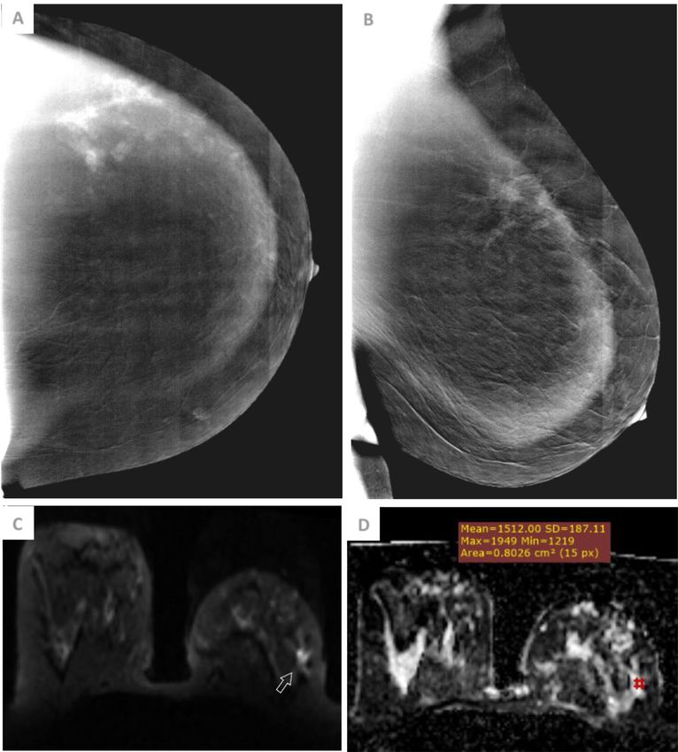
Anwar et al. Egyptian Journal of Radiology and Nuclear Medicine   (2021) 52:63                                         Page 2 of 13

Background                                                            subtracted images with contrast agent uptake informa-
Breast cancer constitutes a major cause of cancer deaths              tion were obtained in mediolateral oblique (MLO) and
in females. Mammographic screening has been shown to                  cranio-caudal (CC) views.
be useful in the reduction of breast cancer mortality;                   The high-energy images were interpreted for the en-
however, the limitations of mammographic screening,                   hancement. Regarding patterns of enhancement seen by
particularly in women with dense or non-involuted                     CESM, the lesions seen in subtracted images were classi-
breasts, are well established [1].                                    fied and described into mass and non-mass enhance-
   Contrast-enhanced spectral mammography (CESM) is                   ment lesions.
a currently established technique in which contrast en-
hancement is used with digital mammography to depict                  MRI diffusion-weighted imaging technique
tumor vascularity. CESM has been confirmed to be more                 MR imaging was performed on 1.5-T GE optima MR
sensitive than mammography for the diagnosis of breast                scanner release 450W GE medical systems and 1.5-T
cancer in dense breasts [2, 3].                                       scanner (Siemens machine Magnetom Aera, Siemens
   CESM has a better resolution of a mammography over                 Medical Systems, Erlangen, Germany) with the patient in
the MRI as it displays a better assessment of microcalci-             prone position, using a double breast coil. All acquisitions
fications with its details. Furthermore, the images are ac-           were performed in the axial plane. T2-weighted fast spin
quired from one breast compression in a couple of                     echo sequences were obtained. DWI was performed using
seconds; therefore, there are no motion artifacts [3].                a diffusion-weighted echo-planar imaging sequence with
   DWI is a fast functional non-contrast technique of the             parallel imaging. Diffusion gradients were applied with b =
breast MR imaging without the costs and toxicity which                0 and 1000 s/mm2. ADC maps were automatically gener-
occur with DCE-MRI. Breast cancers that are hidden                    ated from DW images by the MR software.
mammographically or clinically and identified by DCE                    T2 images were first assessed then diffusion-weighted
MRI are also detected on DWI and can discriminate                     images were reviewed to detect the lesion signal intensity,
them from benign breast lesions by accompanied appar-                 the lesions were considered either nonrestricted diffusion
ent diffusion coefficient (ADC) mapping [4].                          (low- to intermediate-intensity lesions) or restricted diffu-
   Diffusion-weighted (DW) MRI provides a promise in                  sion (high-intensity lesions).
the detection of breast cancers that are mammographically               Signal intensity was interpreted in a qualitative manner
occult and needs more studies to be used as an alternative            using b value of 1000 s/mm2. For apparent diffusion co-
supplemental breast cancer screening technique [5].                   efficient (ADC) calculation, region of interest (ROI) was
                                                                      manually drawn on the ADC maps. Mean ADC for each
Methods                                                               lesion was calculated by the lowest ADC value from all
This is a prospective analytical study which included 40              examined ROIs within the lesion.
breast lesions in 32 female patients having dense breasts;
the age range was 29–72 years with mean age 46 years +
9.93 SD. The study was conducted in Baheya Charity                    CESM and DW MRI image evaluation
Women’s Cancer Hospital and Generalized Air Forces                    Analysis and interpretation of lesions were detected in
Hospital.                                                             both CESM and DW images with determination of the
                                                                      BIRADS category of each lesion and the imaging findings
Subjects                                                              were correlated with the final pathological diagnosis.
Patients included in this study were adult females who
underwent digital mammography which revealed dense                    Statistical analysis
breasts (American College of Radiology (ACR) C or D)                  Data were analyzed using MedCalc© version 18.2.1
and showed indeterminate or suspicious mammographic                   (MedCalc© Software bvba, Ostend, Belgium).
findings (BIRADS 3, 4, and 5) as well as patients had no
detected mammographic abnormality (BIRADS 0) war-                        1. Qualitative data was presented by number and
ranting further CESM/DW MRI assessment (Fig. 1).                            percentage; quantitative data was presented by
                                                                            mean and standard deviation.
Contrast-enhanced spectral mammography technique                         2. ROC curve was used to detect validation of the
Patients were examined by dual-energy CESM which                            diagnostic value of radiological tools.
was performed using Senographe Essential full-field                      3. Inter-method agreement for binary outcomes was
digital mammography machine (GE Healthcare, Chalfont                        examined by calculation of Bennett’s prevalence-
St-Giles, UK). A one-shot intravenous injection of non-                     adjusted bias-adjusted kappa (PABAK).
ionic contrast agent was administered with a dose of 1.5                 4. A P value of less than 0.05 was considered
ml/kg body weight at a rate of 3 ml/s. Then two                             statistically significant.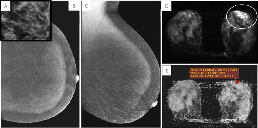
Anwar et al. Egyptian Journal of Radiology and Nuclear Medicine   (2021) 52:63                                              Page 3 of 13

 Fig. 1 A flowchart of the study

Results
The study included 32 female patients; their age range,
main complaints and mammographical ACR classifica-
tion into ACR C (heterogeneously dense breasts) and
ACR D (extremely dense breasts) are demonstrated in
Table 1.                                                              Table 1 Characteristics of the study population
   The study showed 40 different lesions, as we con-                  Variable                                  Frequency    Percentage
sidered cases which had more than one lesion in the                   Age             Age ≤ 45 years            19           59.4%
same or both breasts as separate lesions. Their de-                                   Age > 45 years            13           40.6%
tailed description for the number and percentage of                   Complaint       Mastalgia                 7            21.9%
each pathological breast entity are illustrated in                                    Breast lump               14           43.8%
Table 2.
                                                                                      Nipple discharge          1            3.1%
   From the thirty two cases, four cases (13.8%) had
multifocal breast lesions, and five cases (17.2%) had mul-                            Bleeding per nipple       3            9.4%
ticentric malignant lesions. There were two lesions that                              Nipple erosion            1            3.1%
were missed by CESM; however, all of them were diag-                                  Check up                  4            12.5%
nosed by DW MRI (Figs. 2 and 3). When comparing the                                   Postoperative follow-up   2            6.2%
sensitivity of CESM to DW MRI in the detection of mul-                ACR category    ACR grade C               19           59.4%
tiple breast lesions, they were 88.8 and 100%, respect-
                                                                                      ACR grade D               13           40.6%
ively. The digital mammographic main findings were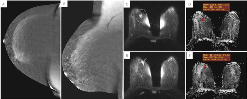
Anwar et al. Egyptian Journal of Radiology and Nuclear Medicine     (2021) 52:63                                         Page 4 of 13

Table 2 Detailed pathological classification of the breast lesions
Pathological diagnosis                                                             Frequency             Percentage
Malignant                   Ductal carcinoma in situ                               1                     2.5%                  77.5%
                            Invasive ductal carcinoma                              17                    42.5%
                            Invasive lobular carcinoma                             6                     15%
                            Mixed ductal and lobular carcinoma                     2                     5%
                            Invasive tubular carcinoma                             2                     5%
                            Invasive carcinoma of no special type                  1                     2.5%
                            Invasive ductal & tubular carcinoma                    2                     5%
Benign                      Fibroadenosis                                          3                     7.5%                  22.5%
                            Fibroadenoma                                           4                     10%
                            Hamartoma                                              1                     2.5%
                            Fibrosis                                               1                     2.5%
                            Total                                                  40                    100%                  100%

correlated to the histopathological results and demon-                  DW MRI findings
strated in Table 3.                                                     The detected breast lesions were classified by T2 signal in-
                                                                        tensity into low T2, isointense T2, and high T2 signal in-
CESM findings                                                           tensities. According to the lesion characteristics in
CESM classified the lesions into twenty three mass lesions              diffusion-weighted image, the breast lesions were classified
and thirteen non-mass lesions, and four lesions showed                  into thirty-three restricted diffusion lesions and seven
no contrast enhancement. CESM findings were correlated                  non-restricted diffusion lesions, then they correlated to
to the histopathological results as illustrated in Table 3.             the histopathological diagnosis as illustrated in Table 7.
The lesions were then described in mass lesions regarding                  ADC value of the lesions were correlated to the histo-
margin of the mass, pattern of enhancement, and intensity               pathological diagnosis, where the mean ADC value of
of enhancement, while non-mass lesions were described                   the benign lesions was 1.5 + 0.42 × 10⁻³ s/mm2 and that
regarding distribution, pattern of enhancement, and inten-              of the malignant lesions was 0.91 + 0.18 × 10⁻³ s/mm2.
sity of enhancement then correlated versus the histo-                      According to the DW MRI characteristics of the breast
pathological diagnosis as illustrated in Table 4.                       lesions, they were categorized according to BIRADS clas-
   There were four lesions that showed no contrast en-                  sification as illustrated in Table 5.
hancement; by digital mammography, they showed asym-                       This study showed one false-negative lesion by DW
metrical density apart from one lesion which showed                     MRI, which pathologically proved to be ILC (Fig. 6) and
pleomorphic microcalcifications (Fig. 4). Then the de-                  three false-positive lesions, one fibroadenoma (Fig. 7)
tected breast lesions were categorized according to BIR-                and two fibroadenosis (Fig. 8).
ADS classification as illustrated in Table 5.                              ROC curve of DW MRI for the probability of malig-
   CESM showed six false-positive lesions; three fibro-                 nancy using BIRADS showed that area under the curve
adenomas, one fibroadenosis, one hamartoma and one                      is about 0.762 where the sensitivity and specificity were
post-operative fibrosis (Fig. 5). Also, it showed three                 96.77 and 66.67%, respectively, total accuracy of 90%,
false-negative lesions—one IDC (Fig. 2) and two lesions                 PPV of 90.91%, and NPV of 85.71% (Table 6).
in the same case were proved pathologically to be inva-                    ROC curve for probability of malignancy using ADC
sive ductal and tubular carcinomas (Fig. 3).                            value showed that area under the curve was about 0.815.
   There were four lesions which showed no contrast en-                 A cutoff value of ≤ 1.1 × 10⁻³ s/mm2 had a sensitivity of
hancement; one lesion proved to be benign (Fig. 7), and                 96.77%, specificity of 66.67%, total accuracy of 92.5%,
three lesions were pathologically proved to be malignant.               PPV of 96.77%, and NPV of 55.55% (Table 6).
They were one DCIS (Fig. 4), one mixed invasive ductal                     Comparing the detected lesions by CESM to that of
and lobular carcinoma, and one invasive ductal and                      DW MRI are demonstrated in Table 8.
tubular carcinoma (Fig. 3).
   ROC curve of CESM for the probability of malignancy                  Discussion
using BIRADS showed area under the curve to be 0.579                    This study presented four multifocal cases; one of them
where the sensitivity and specificity were 90.32%, and                  was missed by CESM, while all of them were diagnosed
33.33%, respectively, total accuracy 77.5%, PPV 82.3%,                  by DW MRI. Furthermore, our study revealed five cases
and NPV of 50% (Table 6).                                               having multicentric malignant lesions; one of them was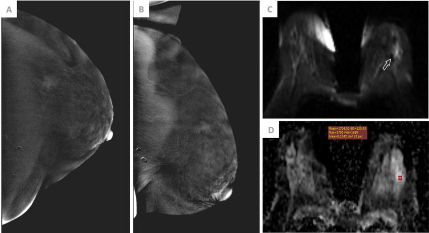
Anwar et al. Egyptian Journal of Radiology and Nuclear Medicine          (2021) 52:63                                                      Page 5 of 13

 Fig. 2 A 62-year-old female patient presented with left breast lump. a and b CESM CC and MLO views of the left breast show central inner region
 (white circles) irregular moderate heterogeneous enhanced mass (BIRADS 4) and UOQ (white arrowheads) lobulated faint homogenous enhanced mass
 (BIRADS 3). c and d DWI and ADC cuts of the left breast show upper central region mass eliciting restricted diffusion (white arrow) with corresponding
 mean ADC value of 0.6 × 10⁻³ s/mm2 (BIRADS 4). e and f DWI and ADC cuts show another similar UOQ mass lesion (black circle) with mean ADC value
 of 0.8 × 10⁻³ s/mm2 (BIRADS 4). Tru-cut biopsy and simple mastectomy of the left breast revealed multicentric invasive ductal carcinoma, grade III
 which matched the DW MRI findings

missed by CESM, and all of them were detected by DW                              CESM mass lesions in this study showed the most com-
MRI. Thus, the sensitivity of DW MRI to detect multiple                       mon features that described malignant lesions were non-
breast lesions was 100% which is higher than that of                          circumscribed margins (speculated or irregular) (69.5%),
CESM 88.8%.                                                                   heterogeneous contrast enhancement (47.9%), and rim en-
  In concordance with Jochelson et al.’s study, they said                     hancement (26.1%). This was matching Schnall et al.’s
that breast cancers are often multifocal and multicentric.                    study which showed that the most important feature of
Additional foci of ipsilateral breast cancer are often mam-                   image interpretation is the characterization of the focal
mographically occult and are identified more frequently                       mass margin. Irregular or speculated margins have a posi-
with MR imaging. On the other hand, Luczyn´ska et al.’s                       tive predictive value (PPV) of 84–91%. Rim-like enhance-
study found that CESM may provide fast and accurate                           ment highly correlates with a cancer diagnosis (PPV,
breast lesion detection and characterization [6, 7].                          84%). Also, intense (43.5%) and moderate contrast
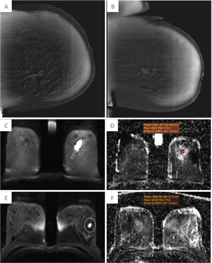
Anwar et al. Egyptian Journal of Radiology and Nuclear Medicine          (2021) 52:63                                                  Page 6 of 13

 Fig. 3 A 44-year-old female patient presented with right breast lump. a and b CESM CC and MLO views of the right breast show LIQ (black circles)
 focal faintly enhanced clumped non-mass lesion (BIRADS 3). c and d DWI and ADC cuts of right breast show LIQ mass eliciting restricted diffusion
 (white circle) with corresponding mean ADC value of 1 × 10⁻³ s/mm2 (BIRADS 4). e and f DWI and ADC cuts show another similar adjacent mass (white
 arrow) with mean ADC value of 1.1 × 10⁻³ s/mm2 (BIRADS 4). Tru-cut biopsy revealed right breast multifocal invasive ductal and tubular carcinoma
 which matched the DW MRI findings

enhancements (30.4%) were common findings of the ma-                            In the study, CESM non-mass lesions showed that the
lignant lesions in this study, and this was in concordance                   most common features that described malignant lesions
with Kaur et al.’s study, which said that in CESM, a mass                    were focal and segmental distribution of non-mass le-
with moderate or intense enhancement is suspicious of                        sions (23.07% for each), regional distribution (15.38%),
malignant transformation [1, 8].                                             clumped contrast enhancement (38.46%), heterogeneous
                                                                             contrast enhancement (30.76%), and intense contrast en-
Table 3 Cross tabulation showing the main classification of                  hancement (38.46%).
digital mammographic and CESM findings versus the                               In contrast to Schnall et al.’s study, a moderate to
histopathological diagnosis                                                  marked non-mass regional enhancement provides a PPV
                                 Histopathological diagnosis      Total      of 59% in malignant lesion detection. Stippled enhance-
                                 Malignant        Benign                     ment was found that it has a low incidence of malignancy
Digital mammographic findings                                                (25%), while clumped, heterogeneous, and homogeneous
  No abnormality detected        6                6               12         enhancements were found to have a 60%, 53%, and 67%
                                 15%              15%             30%        likelihood of cancer, respectively [8].
  Mass lesion                    8                3               11            In the current study, CESM showed six false-positive
                                 20%              7.5%            27.5%      lesions (19.3% false-positive rate), three fibroadenomas,
  Asymmetric density             14               -               14         one fibroadenosis, one hamartoma, and one postopera-
                                 35%                              35%        tive fibrosis.
  Microcalcification             3                -               3             In contrast to Luczyn´ska et al. and Muller et al.’s
                                 7.5%                             7.5%       studies, there was a relatively high rate (20%) of false-
  Total                          31               9               40         positive results and no false-negative findings with
                                 77.5%            22.5%           100%       CESM. Benign lesions, such as fibroadenomas, fibro-
CESM findings                                                                sclerosis, hamartoma, intraductal papillomas, and
  Mass lesion                    19               4               23         phyllodes tumors had shown contrast enhancement
                                 47.5%            10%             57.5%      [7, 9].
  Non-mass lesion                9                4               13            In this study, CESM showed three false-negative le-
                                 22.5%            10%             32.5%      sions (33.3% false-negative rate); one IDC and two le-
  No contrast-enhanced lesions   3                1               4          sions invasive ductal and tubular carcinomas in the
                                 7.5%             2.5%            10%
                                                                             same case. Furthermore, there was one DCIS case
  Total                          31               9               40         which showed no contrast enhancement; however, the
                                 77.5%            22.5%           100%
                                                                             presence of pleomorphic microcalcifications raised the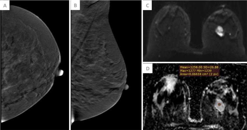
Anwar et al. Egyptian Journal of Radiology and Nuclear Medicine      (2021) 52:63                                       Page 7 of 13

Table 4 Cross tabulation showing CESM morphology descriptors of the mass and non-mass lesions versus the histopathological
diagnosis
CESM findings                                                                          Histopathological diagnosis
                                                                                       Malignant              Benign         Total
Descriptors of mass lesions
  Margin                              Circumscribed (lobulated)                        1                      1              2
                                                                                       4.34%                  4.34%          8.7%
                                      Partial circumscribed                            2                      -              2
                                                                                       8.7%                   -              8.7%
                                      Non-circumscribed (irregular or speculated)      16                     3              19
                                                                                       69.5%                  13%            82.6%
  Pattern of enhancement              Homogenous                                       2                      1              3
                                                                                       8.7%                   4.3%           13%
                                      Heterogeneous                                    11                     3              14
                                                                                       47.9%                  13%            60.9%
                                      Ring enhancement                                 6                      -              6
                                                                                       26.1%                  -              26.1%
  Intensity of enhancement            Faint                                            2                      1              3
                                                                                       8.7%                   4.3%           13%
                                      Moderate                                         7                      2              9
                                                                                       30.4%                  8.7%           39.1%
                                      Intense                                          10                     1              11
                                                                                       43.5%                  4.3%           47.9%
Descriptors of non-mass lesions
  Distribution                        Focal                                            3                      3              6
                                                                                       23.078%                23.078%        46.16%
                                      Segmental                                        3                      -              3
                                                                                       23.07%                 -              23.07%
                                      Regional                                         2                      1              3
                                                                                       15.38%                 7.7%           23.07%
                                      Diffuse                                          1                      -              1
                                                                                       7.7%                   -              7.7%
  Pattern of enhancement              Heterogeneous                                    4                      1              5
                                                                                       30.76%                 7.7%           38.46%
                                      Clumped                                          5                      2              7
                                                                                       38.46%                 15.38%         53.84%
                                      Nodular                                          -                      1              1
                                                                                       -                      7.7%           7.7%
  Intensity of enhancement            Faint                                            2                      -              2
                                                                                       15.38%                 -              15.38%
                                      Moderate                                         2                      3              5
                                                                                       15.38%                 23.07%         38.46%
                                      Intense                                          5                      1              6
                                                                                       38.46%                 7.7%           46.16%

suspicion of the diagnosis. This was in agreement                           In line with Helal et al.’s study, they had nine false-
with Fallenberg et al.’s study which reported that the                    positive cases that were found out in the comparison of
low-energy image of CESM is comparable with stand-                        the CESM diagnoses with the histopathology results.
ard mammography with regard to the visualization of                       These cases were wrongly diagnosed because the operative
microcalcifications [10].                                                 bed showed areas of enhancement, but these enhance-
  CESM in this study presented two postoperative cases;                   ments were caused by benign postoperative sequel [11].
one of them was pathologically proved to be recurrent                       This DW MRI study showed that the mean ADC value
IDC, while the other one showed suspicious contrast en-                   of the benign lesions was 1.5 + 0.42 × 10⁻³ s/mm2 which
hancement by CESM and revealed pathologically to be                       is higher than that of the malignant lesions which was
postoperative fibrosis.                                                   0.91 + 0.18 × 10⁻³ s/mm2. This was in concordance with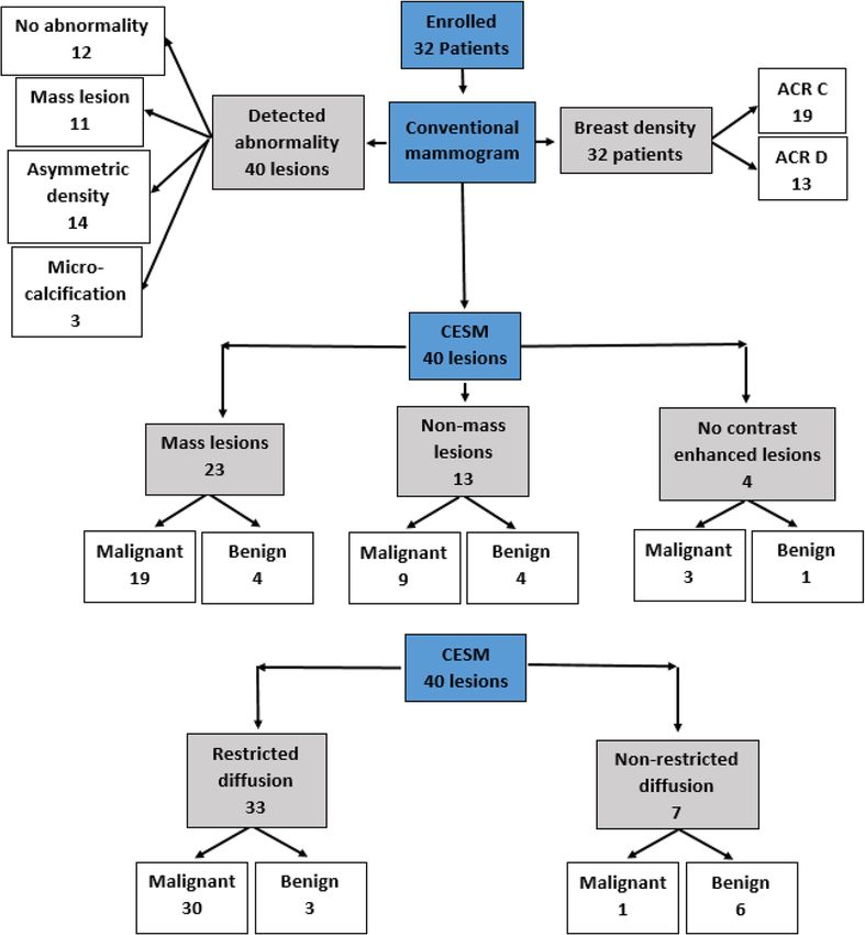
Anwar et al. Egyptian Journal of Radiology and Nuclear Medicine       (2021) 52:63                                                   Page 8 of 13

 Fig. 4 A 31-year-old female patient presented with left breast nipple erosions. a Magnified mammographic image of the left breast shows
 scattered pleomorphic microcalcifications at the UOQ. b and c CC and MLO views of the left breast show no significant contrast enhancement
 (BIRADS 4). d and e DWI and ADC cuts show left breast retroareolar (white circle) linear mass lesion reaching the nipple eliciting restricted
 diffusion with corresponding mean ADC value of 1.1 × 10⁻³ s/mm2 (BIRADS 5). Tru-cut biopsy of the left breast revealed Paget’s disease of the
 nipple with associated high-grade DCIS of the major lactiferous duct which matched the DW MRI findings

Moukhtar and Abo El Maati’s study, where they re-                          55.55%, and with an overall total accuracy of 92.5%. This
ported that the mean ADC value of all benign lesions                       was in concordance with Moukhtar and Abo El Maati’s
was 1.41 ± 0.36 × 10⁻³ s/mm2, which was higher than                        study which showed that the optimal cutoff value to dis-
the mean ADC of all malignant lesions (1.05 ± 0.30 ×                       criminate benign from malignant lesions was 1.25 × 10⁻³
10-3 s/mm2) [12].                                                          s/mm2, with a sensitivity of 82%, a specificity of 68%,
  Our study showed that a cutoff ADC value ≤ 1.1 ×                         and an overall accuracy of 78% [13].
10-3 s/mm2 at b value of 1000 s/mm2 had a sensitivity of                     This study showed that IDCs had a mean ADC value
96.77%, specificity of 66.67%, PPV of 96.77%, NPV of                       (0.92 + 0.14 × 10⁻³ s/mm2) which was slightly lower than

Table 5 Cross tabulation showing the diagnostic accuracy of CESM and DW MRI tested versus histopathological diagnosis as the
gold standard for lesion classification
                                                            Histopathological diagnosis                                                      Total
                                                            Malignant                                 Benign
BIRADS classification by CESM
  Probably benign (BIRADS 1–3)                              3                                         3                                      6
                                                            7.5%                                      7.5%                                   15%
  Probably malignant (BIRADS 4–5)                           28                                        6                                      34
                                                            70%                                       15%                                    85%
  Total                                                     31                                        9                                      40
                                                            77.5%                                     22.5%                                  100%
BIRADS classification by DW MRI
  Probably benign (BIRADS 1–3)                              1                                         6                                      7
                                                            2.5%                                      15%                                    17.5%
  Probably malignant (BIRADS 4–5)                           30                                        3                                      33
                                                            75%                                       7.5%                                   82.5%
  Total                                                     31                                        9                                      40
                                                            77.5%                                     22.5%                                  100%
Anwar et al. Egyptian Journal of Radiology and Nuclear Medicine (2021) 52:63 Page 9 of 13
Fig. 5 A 45-year-old female patient for postoperative follow-up 1 year after left BCS. a and b CESM CC and MLO views of the left breast show
UOQ (white circles) speculated moderately enhanced mass lesion (BIRADS 4). c and d DWI and ADC cuts of the left breast show UOQ (white
arrow) irregularly shaped mass eliciting non-restricted diffusion with corresponding mean ADC value of 1.7 × 10⁻³ s/mm2 (BIRADS 3). Tru-cut
biopsy of the left breast revealed UOQ fibrosis with no atypia or malignancy which matched the DW MRI findings
that of ILCs which showed mean ADC value (0.93 + intensity and lower ADC values than that of benign tu-
0.29 × 10⁻³ s/mm2). Our study had only one DCIS lesion mors and normal breast parenchyma on diffusion-
with ADC value relatively higher than that of the inva- weighted images [12].
sive lesions; it was 1.1 × 10⁻³ s/mm2. In this study, DWI showed one false-negative lesion (2.5%
Woodhams et al.’s study reported that because of their false-negative rate); it was ILC which showed non-restricted
higher cellularity, most of IDCs show higher signal diffusion with high ADC value of 1.5 × 10⁻³ s/mm2.
Table 6 Comparison between the diagnostic accuracy of CESM, Table 7 Cross tabulation showing the diagnostic accuracy of T2
DW MRI and ADC value signal intensity and DW MRI versus histopathological diagnosis
CESM DW MRI ADC value Histopathological diagnosis Total
True positive 28 30 Malignant Benign
70% 75%
T2 signal intensity of the breast lesions
True negative 3 6
7.5% 15% Low T2 signal 18 1 19
45% 2.5% 47.5%
False positive 6 3
15% 7.5% Isointense T2 signal 13 7 20
32.5% 17.5% 50%
False negative 3 1
7.5% 2.5% High T2 signal - 1 1
2.5% 2.5%
Sensitivity 90.32% 96.77% 96.77%
Total 31 9 40
Specificity 33.33% 66.67% 77.78% 77.5% 22.5% 100%
Total accuracy 77.50% 90.00% 92.50% Classification of breast lesions by DW MRI
Disease prevalence 77.50% 77.50% 77.50% Restricted diffusion 30 3 33
Positive predictive value (PPV) 82.35% 90.91% 93.75% 75% 7.5% 82.5%
Negative predictive value (NPV) 50.00% 85.71% 87.50% Non-restricted diffusion 1 6 7
2.5% 15% 17.5%
Positive likelihood ratio (LR+) 1.35 2.90 4.35
Total 31 9 40
Negative likelihood ratio (LR-) 0.29 0.05 0.04 77.5% 22.5% 100%Anwar et al. Egyptian Journal of Radiology and Nuclear Medicine (2021) 52:63 Page 10 of 13 Fig. 6 A 44-year-old female patient presented for check-up. a and b CESM CC and MLO views of the left breast show UOQ (white circles) segmental clumped moderate contrast enhancement non-mass lesion (BIRADS 4). c and d DWI and ADC cuts of the left breast show UOQ (white arrow) mass with non-restricted diffusion and corresponding mean ADC value of 1.5 × 10⁻³ s/mm2 (BIRADS 3). Tru-cut biopsy of the left breast revealed UOQ invasive lobular carcinoma grade II with extensive LCIS which matched the CESM findings According to Woodhams et al.’s study, invasive lobular ADC values. Additionally, fibrocystic disease, which is carcinoma represents a diagnostic challenge at MR im- characterized by considerable degrees of fibrosis and aging as well as its size may be underestimated at proliferation, may have ADC values in the range of ma- diffusion-weighted imaging; this inaccuracy may be due lignant lesions [14]. to the spread of infiltrating cells [12]. This study revealed that the sensitivity and specificity DWI in this study showed three false-positive lesions of the DW MRI to detect malignant lesions were 96.77 (33.3% false-positive rate); one fibroadenoma and two and 66.67%, respectively, PPV of 90.91%, and NPV of fibroadenosis. Fibroadenoma showed restricted diffusion 85.71% with a total accuracy of 90%. with low ADC value 0.8 × 10⁻³ s/mm2. Two fibroadeno- This was in concordance with Partridge and McDo- sis lesions showed restricted diffusion as well as low nald’s study which established that the diagnostic per- ADC value 1.1 and 1.2 × 10⁻³ s/mm2. formance of quantitative breast DWI exhibited pooled According to Pereira et al.’s study, fibroadenomas are sensitivity of 84% and specificity of 79% [4]. supposed to have high rates of diffusion and ADC values In this study, the diagnostic accuracy of DW MRI was owing to their stromal myxoid changes and conse- higher than that of CESM where the sensitivity of DW quently increased mobility of water. However, fibroaden- MRI was 96.77% as compared with 90.32% in CESM. omas with an abundant fibrous component have lower The specificity of DW MRI was 66.67% as compared
Anwar et al. Egyptian Journal of Radiology and Nuclear Medicine (2021) 52:63 Page 11 of 13
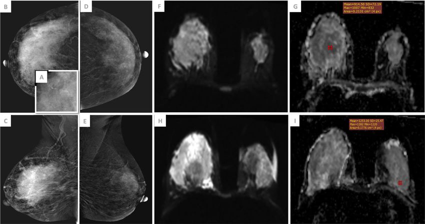
Fig. 7 A 37-year-old female patient presented with right breast lump. a Magnified mammographic image for the pleomorphic microcalcifications at UOQ of
the right breast. b and c CESM CC and MLO views of the right breast show UOQ diffuse non-mass heterogeneous intense enhancement (white arrowheads)
(BIRADS 5). d and e CESM CC and MLO views of the left breast show UOQ (black circles) regional non-mass moderate nodular parenchymal enhancement
(BIRADS 3). f and g DWI and ADC cuts show right breast is nearly totally occupied by a diffuse infiltrative mass lesion (white arrows) eliciting restricted diffusion
with corresponding mean ADC value of 0.9 × 10⁻³ s/mm2 (BIRADS 5). h and i DWI and ADC cuts of the left breast also show UOQ (white circle) similar mass
with mean ADC value of 1.2 × 10⁻³ s/mm2 (BIRADS 4). Tru-cut biopsies revealed right breast invasive lobular carcinoma grade II and left breast fibroadenosis, no
malignancy which matched the CESM findings
with 33.33% in CESM. The total accuracies were 90 and demonstrating lesions that are not visible by standard
77.5%, respectively. Also PPV and NPV of DW MRI mammography. In comparison with MRI, CESM can de-
were 90.91 and 85.71% as compared with 82.35 and tect microcalcifications easily, and there are no limita-
50.00% in CESM, respectively. tions as with MRI in terms of the ferromagnetic effect
This was in concordance with Barra et al.’s study; and machine design.
among the 25 patients who had residual lesions, 19 were DW MRI technique is a diagnostic technique that en-
positive by CESM, and 23 were positive by MRI. Higher ables accurate detection of malignant breast lesions
sensitivity was found by MRI (92%) in contrast to 76% in without need for the contrast media injection, and it
CESM. Also PPV and NPV were higher for MRI com- avoids the irradiation exposure.
parable to CESM; they were 95 and 75% as compared CESM and DWI demonstrated good overall diagnostic
with 92 and 53.8%, respectively [15]. accuracy and correlation in lesion size estimation in
Our study had several limitations. First, our study dense breast patients. However, DW MRI has a higher
population was small. Second, pre- and postmenopausal diagnostic accuracy than CESM for the detection of ma-
women were included and were examined in different lignant breast lesions in dense breasts with a higher sen-
phases of the menstrual cycle. Third, most cases were sitivity, specificity, total accuracy, negative predictive
collected from the cancer breast center, so the negative value, and positive predictive value as well as the detec-
lesions were small in number. Finally, women with no tion of multiple lesions.
suspicious lesions at conventional mammography or
CESM mostly did not undergo DW MRI and thus were Abbreviations
not included in this study. ACR: American college of radiology; ADC: Apparent diffusion coefficient;
BIRADS: Breast imaging reporting and data system; CC: Cranio-caudal;
CESM: Contrast-enhanced spectral mammography; DCE-MRI: Dynamic
Conclusion contrast-enhanced magnetic resonance imaging; DCIS: Ductal carcinoma in
Dual-energy contrast-enhanced digital mammography is situ; DWI: Diffusion-weighted imaging; IDC: Invasive ductal carcinoma;
ILC: Invasive lobular carcinoma; MLO: Mediolateral oblique; NPV: Negative
a useful technique in identification of lesions in mam- predictive value; PPV: Positive predictive value; ROC: Receiver operating
mographically dense breasts and capable of characteristic; ROI: Region of interest; SD: Standard deviationAnwar et al. Egyptian Journal of Radiology and Nuclear Medicine (2021) 52:63 Page 12 of 13
Fig. 8 A 37-year-old female patient presented with left breast lump. a and b CESM CC and MLO views of the left breast show no contrast-enhanced masses. c
and d DWI and ADC cuts of the left breast show UIQ (white circle) well-defined oval-shaped mass eliciting restricted diffusion with corresponding mean ADC
value of 1.2 × 10⁻³ s/mm2 (BIRADS 4). Tru-cut biopsy of the left breast revealed UIQ fibroadenoma
Table 8 Agreement between CESM and MRI as regards lesion classification as probably malignant (BIRADS 4–5) or probably benign
(BIRADS 1–3)
Lesion classification by DW MRI
Probably benign (BIRADS 1–3) Probably malignant (BIRADS 4–5) Total
Lesion classification by CESM
Probably benign (BIRADS 1–3) 1 5 6
2.5% 12.5% 15%
Probably malignant (BIRADS 4–5) 6 28 34
15% 70% 85%
Total 7 33 40
17.5% 82.5% 100%
Bennet’s prevalence- and bias-adjusted kappa (PABAK) 0.45Anwar et al. Egyptian Journal of Radiology and Nuclear Medicine (2021) 52:63 Page 13 of 13
Acknowledgements 8. Schnall MD, Blume J, Bluemke DA, DeAngelis GA, DeBruhl N, Harms S,
This research was carried out at Baheya Charity Women’s Cancer Hospital Gatsonis CA (2006) Diagnostic architectural and dynamic features at breast
and Generalized Air Forces Hospital which are fully equipped by modern MR imaging: multicenter study. Radiology 238(1):42–53
machines for breast cancer diagnosis. We want to thank our colleagues who 9. Muller S, Dromain C, Balleyguier C, Patoureaux F, Puong S, Bouchevreau X,
helped us to do such research work. Katz C (2010) Contrast enhanced digital mammography (CEDM): from
morphological to functional mammography. European society of Radiology
Authors’ contributions ESR/ECR 2010 / C-0300: ECR.
RF wrote the manuscript and was responsible for correspondence to journal. 10. Fallenberg EM, Dromain C, Diekmann F, Engelken F, Krohn M, Singh JM,
SM collected patient data and participated in its design. AA and MF did Ingold-Heppner B, Winzer KJ, Bick U, Renz DM (2013) Contrast-enhanced
image processing and collection of patient’s images. WA participated in the spectral mammography versus MRI: initial results in the detection of breast
design of the study and performed the statistical analysis. HE conceived of cancer and assessment of tumour size. Eur Radiol 24(1):256–264
the study and participated in its design and coordination and helped to 11. Helal MH, Mansour SM, Ahmed HA, Abdel Ghany AF, Kamel OF, Elkholy NG
draft the manuscript. The authors have read and approved the manuscript. (2019) The role of contrast-enhanced spectral mammography in the
evaluation of the postoperative breast cancer. Clinical Radiology. 74:771–781
12. Woodhams R, Ramadan S, Stanwell P, Sakamoto S, Hata H, Ozaki M, Kan S,
Funding Inoue Y (2011) Diffusion-weighted imaging of the breast: principles and
No funding sources clinical applications. RSNA RadioGraphics 31:1060–1082
13. Moukhtar FZ, Abu El Maati AA (2014) Apparent diffusion coefficient values
Availability of data and materials as an adjunct to dynamic contrast enhanced MRI for discriminating benign
The datasets used and analyzed during the current study are available from and malignant breast lesions presenting as mass and non-mass like
the corresponding author on reasonable request. enhancement. Egypt J Radiol Nucl Med 45:597–604
14. Pereira FPA, Martins G, de Oliveira RDVC (2011) Diffusion magnetic resonance
imaging of the breast. Magn Reson Imaging Clin N Am 19(1):95–110
Ethics approval and consent to participate
15. Barra FR, Sobrinho AB, Barra RR, Magalhaes MT, Aguiar LR, Lins De
The study was approved by the ethical committee of “Research Ethics
Albuqurque GF, Costa RP, Farage L, Pratesi R (2018) Contrast-enhanced
Committee at the Faculty of Medicine, Ain Shams University” with ethical
mammography (CEM) for detecting residual disease after neoadjuvant
committee approval number FMASU M D 193/2018 and approval date 22/
chemotherapy: a comparison with breast magnetic resonance imaging
07/2018. An informed written consent was taken from all subjects.
(MRI). BioMed Res Int 2018:1–9
Consent for publication
All patients included in this research gave written informed consent to Publisher’s Note
publish the data contained within this study. Springer Nature remains neutral with regard to jurisdictional claims in
published maps and institutional affiliations.
Competing interests
No financial or non-financial competing interests.
Author details
1
Department of Diagnostic Radiology, Dar Elsalam Cancer Center, Ministry of
Health, Cairo, Egypt. 2Egyptian Military Medical Academy, Cairo, Egypt.
3
Department of Diagnostic and Interventional Radiology, Ain Shams
University, Cairo, Egypt.
Received: 3 December 2020 Accepted: 10 February 2021
References
1. Kuhl CK, Strobel K, Bieling H, Leutner C, Schild HH, Schrading S (2017)
Supplemental breast MR imaging screening of women with average risk of
breast cancer. Radiology 283(2):361–370
2. Sogani J, Morris EA, Kaplan JB, D’Alessio D, Goldman D, Moskowitz CS,
Jochleson MS (2017) Comparison of background parenchymal
enhancement at contrast-enhanced spectral mammography and breast MR
imaging. Radiology 282(1):63–73
3. Yousef AF, Khater HM, Jameel LM (2018) Contrast-enhanced spectral
mammography versus magnetic resonance imaging in the assessment of
breast masses. Benha Med J 35:5–12
4. Partridge SC, McDonald ES (2013) Diffusion weighted magnetic resonance
imaging of the breast protocol optimization, interpretation, and clinical
applications. Magn Reson Imaging Clin N Am 21(3):601–624
5. Amornsiripanitch N, Bickelhaupt S, Shin HJ, Dang M, Rahbar H, Pinker K,
Partridge S (2019) C: Diffusion-weighted MRI for unenhanced breast cancer
screening. Radiology 9:1–17
6. Jochelson MS, Dershaw DD, Sung JS, Heerdt AS, Thornton C, Moskowitz CS,
Ferrara J, Morris EA (2013) Bilateral contrast-enhanced dual-energy digital
mammography: feasibility and comparison with conventional digital
mammography and MR imaging in women with known breast carcinoma.
Radiology 266:743–751
7. Luczyńska E, Heinze-Paluchowska S, Dyczek S, Blecharz P, Rys J, Reinfuss M
(2014) Contrast-enhanced spectral mammography: comparison with
conventional mammography and histopathology in 152 women. Korean J
Radiol 15(6):689–696You can also read