ViewDEX 3.0-RECENT DEVELOPMENT OF A SOFTWARE APPLICATION FACILITATING ASSESSMENT OF IMAGE QUALITY AND OBSERVER PERFORMANCE
←
→
Page content transcription
If your browser does not render page correctly, please read the page content below
Radiation Protection Dosimetry (2021), pp. 1–6                                                                            doi:10.1093/rpd/ncab014

ViewDEX 3.0—RECENT DEVELOPMENT OF A SOFTWARE
APPLICATION FACILITATING ASSESSMENT OF IMAGE
QUALITY AND OBSERVER PERFORMANCE

                                                                                                                                                        Downloaded from https://academic.oup.com/rpd/advance-article/doi/10.1093/rpd/ncab014/6158056 by guest on 18 June 2021
Angelica Svalkvist 1,2, *, Sune Svensson1 , Tommy Hagberg1 and Magnus Båth 1,2
1
  Department of Medical Physics and Biomedical Engineering, Sahlgrenska University Hospital, Gothenburg
SE-413 45, Sweden
2
  Department of Radiation Physics, Institute of Clinical Sciences, The Sahlgrenska Academy, University of
Gothenburg Gothenburg SE-413 45, Sweden

*Corresponding author: angelica.svalkvist@radfys.gu.se
Received 2 November 2020; revised 2 November 2020; editorial decision 13 January 2021; accepted 13 January
2021

ViewDEX (Viewer for Digital Evaluation of X-ray Images) is an image viewer compatible with Digital Imaging and Communi-
cations in Medicine (DICOM) that has been especially designed to facilitate image perception and observer performance studies
within medical imaging. The software was first released in 2004 and since then a continuous development has been ongoing. One
of the major drawbacks of previous versions of ViewDEX has been that they have lacked functionality enabling the possibility to
evaluate multiple images and/or image stacks simultaneously. This functionality is especially requested by researchers working
with modalities, where an image acquisition can result in multiple image stacks (e.g. axial, coronal and sagittal reformations in
computed tomography). In ViewDEX 3.0 this functionality has been added and it is now possible to perform image evaluations
of multiple images and/or image stacks simultaneously, by using multiple monitors and/or multiple image canvases in monitors.
Additionally, some of the previously available functionality has been updated and improved. This paper describes the recent
developments of ViewDEX 3.0.

INTRODUCTION                                                                over 40 countries and had received over 230 citations
The introduction of digital radiography in the                              in scientific publications. ViewDEX has for example
late 1900s resulted in new challenges regarding                             been used for studies in conventional projection
the optimization of image quality and radiation                             radiography(6) , tomosynthesis(7) , computed tomog-
dose. Digital technology enables the possibility to                         raphy (CT)(8) , mammography(9) , magnetic resonance
change the appearance of a collected radiograph                             imaging (MRI)(10) , nuclear medicine imaging(11) ,
not only by changing the acquisition parameters,                            interventional imaging(12) and ultrasound(13) .
but also by adjusting the image processing. Hence,                              The development of ViewDEX has always been
the task of optimising the examinations became                              focused on keeping up with the technological progress
more challenging as more variables were added to                            within the field of medical imaging. The goal of the
the equation. At many radiological departments one                          development is to create a software that enables the
limiting factor in the optimization process is finding                      observers to review study images in a surrounding
time for the radiologists to review and evaluate                            that resembles the real clinical situation. The largest
the radiological images. In order to facilitate the                         limitation with previous versions of ViewDEX is that
image evaluation process, the software ViewDEX                              they only support the possibility to include one single
(Viewer for Digital Evaluation of X-ray Images)                             image or image stack in each case. Hence, the clinical
was developed(1) . The software was first released                          benefits obtained by e.g. multi-planar reconstructions
in 2004 and the prerequisites for the development                           (MPRs) cannot be fully utilised during image review
were that the software should be DICOM compatible,                          using ViewDEX, as the different reconstructions can-
easy to use, freely available and be suitable for both                      not be shown to the observer side-by-side. Addi-
imaging research and clinical optimisation. Since the                       tionally, performing alternative forced choice (AFC)
first release of the software, continuous development                       studies(14) is cumbersome, as each set of images has
has been ongoing and additional papers describing                           to be manually created before the study is configured
the developments have been published(2–5) . The                             in ViewDEX. Other limitations with ViewDEX 2.0
ViewDEX software can be downloaded free of charge                           include that the log file may be difficult to work with
from www.vgregion.se/sas/viewdex, and in the end of                         and that data from physical measurements cannot
October 2020 ViewDEX had been downloaded from                               automatically be saved in the log file.

© The Author(s) 2021. Published by Oxford University Press.
This is an Open Access article distributed under the terms of the Creative Commons Attribution License (http://creativecommons.org/licenses/by/4.0/),
which permits unrestricted reuse, distribution, and reproduction in any medium, provided the original work is properly cited.
A. SVALKVIST ET AL.
In this paper, ViewDEX 3.0 will be presented.
This new version of the software has been adapted
to modern medical imaging, e.g. by the development
of a new architecture that enables the possibility to
simultaneously review multiple images/image stacks.
Downloaded from https://academic.oup.com/rpd/advance-article/doi/10.1093/rpd/ncab014/6158056 by guest on 18 June 2021
GENERAL FUNCTIONALITY IN ViewDEX Figure 1: Schematic illustration of the configuration of the
In Svalkvist et al.(5) the history of the development of image database in ViewDEX 2.0. In ViewDEX 2.0 each case
can consist of only one image or image stack.
ViewDEX since the first release in 2004 is presented,
together with a detailed description of the available
functionality of ViewDEX 2.0. To summarise,
ViewDEX 2.0 can handle DICOM images from
different modalities, for example conventional pro- technologies result in examinations including several
jection radiography, CT, single-photon emission CT, different image stacks, e.g. MPRs in CT and MRI.
positron emission tomography, MRI and ultrasound. In the clinical situation, all the image/image stacks
The cases included in a study can be displayed included in the examination contribute with informa-
in a unique randomised order for each observer. tion that is important for diagnosis. Hence, in order
During image review the observers are able to alter to resemble the clinical situation and take advantage
the image display properties e.g. by changing the of the increased amount of information obtained
window settings or zoom and pan the images. The when multiple reconstructions, reformations and/or
observers can also perform physical measurements projections are available for diagnosis, it should be
in the images, such as distance measurements, area possible for each case included in a ViewDEX study
measurements and measurements of mean pixel value to consist of more than one image or image stack, e.g.
in specific locations of an image. If a study is based as illustrated in Figure 2.
on localisation of pathology the observers can make In order to facilitate the possibility to review multi-
localization markings in the images and also answer ple images simultaneously in ViewDEX, the architec-
questions connected to the markings made. It is ture of the software has been changed. In ViewDEX
also possible for the observer to write general notes 3.0, it is possible to review images using up to four
regarding specific cases included in the study. The different monitors. Additionally, each monitor can
person responsible for study setup has full control be divided into four separate image canvases, which
over the conditions for image review and also has means that a total of 16 different images/image stacks
the possibility to log into a study and review (‘show can be included in each case and reviewed simulta-
mode’) or edit (‘edit mode’) the responses from each neously. Possible canvas setup on each monitor is
specific observer. 1 × 1 (full-screen), 1 × 2 or 2 × 1 (two canvases
vertically or horizontally oriented) or 2 × 2 (four
canvases). If three image/image stacks are included in
ViewDEX 3.0 each study and only one monitor is used, the canvas
setup is 2 × 2 with one of the four canvases empty
ViewDEX 3.0 is written in the Java programming (black). Each image canvas will function as a separate
language version 8 (but also works on later versions) display, which means that all functionality that was
and the code can run on any desktop system. The available in ViewDEX 2.0 is implemented in each
software is compatible with Windows 64, Linux 64 canvas separately in ViewDEX 3.0. This means that
and MacOS 64. Compared to ViewDEX 2.0, version the observer for example can change window settings
3.0 is optimised with respect to performance, leading and use zoom, pan and cine-loop individually in each
to for example shorter execution times. image canvas. Additionally, the observers can mark
two or more canvases, i.e. perform a multi-select of
Multiple monitors and multiple canvases canvases. If more than one canvas is selected the
scroll function (if image stacks are reviewed) and all
on each monitor
changes of image properties (e.g. window settings,
In the previous version of ViewDEX only one zoom and pan) are altered in the selected canvases
image/image stack at a time could be displayed to simultaneously.
the observer. As a consequence, each case included During the setup of a study the default configura-
in a study could only consist of one image/image tion of number of monitors and number of canvases
stack (Figure ure1). Already in the past this limitation in each monitor is determined. During image review
caused problems as many conventional X-ray exam- the observer has the possibility to temporarily change
inations could include images from several different the image setup, e.g. to review one image/image stack
projection angles. Today more advanced imaging in full-scale format on one of the monitors (1 × 1) or
2VIEWDEX 3.0
Downloaded from https://academic.oup.com/rpd/advance-article/doi/10.1093/rpd/ncab014/6158056 by guest on 18 June 2021
Figure 2: Schematic illustration of the configuration of the image database in ViewDEX 3.0. In ViewDEX 3.0 each case can
consist of several different image series, each including either a single image or an image stack.
to review two out of four images on one monitor side- the functionality. Up until now, the only way to store
by-side (i.e. in canvas setup 1 × 2 or 2 × 1 instead of results from physical measurements in ViewDEX has
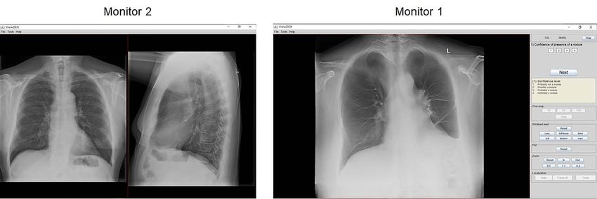
2 × 2). In Figure 3, an example of a study setup using been to manually type the result in the notes panel.
two monitors is presented. The case displayed con- This solution comes with a number of disadvantages.
sists of one chest tomosynthesis examination (image For example, the risk for incorrect registration due
stack) and two projections from a conventional chest to typing errors increases and if several different
examination (frontal + lateral). The tomosynthesis measurements are performed in each case, it may be
examination is displayed in full-screen format (1 × 1) difficult to distinguish the registered measurements
on Monitor 1, and the two projection images from the from each other.
conventional chest examination are displayed side- In ViewDEX 3.0, the observers can choose which
by-side (1 × 2) on Monitor 2. measurements should be stored in the log file. A phys-
During study setup, the person responsible for ical measurement is made by holding down specific
study design specifies the location at which each buttons on the keyboard while clicking and dragging
image series should be displayed (monitor and the mouse pointer over the image. By using keyboard
canvas). However, it is also possible to choose that short commando, the result from the measurement
the location of each image series included in a case is stored and incorporated into the log file. For a
is randomised for each case and observer. This distance measurement the distance will be stored in
functionality enables the possibility to conduct AFC millimetres (mm). Additionally, the coordinates for
studies without any need to process the images in the starting point and end point of the measured
advance. line will be given. For area measurements using a
circular ROI or an ROI of variable shape, the mea-
sured area will be stored in cubic millimetres (mm2 )
Save results from physical measurements and the mean pixel value will be given. The actual
Functionality enabling the possibility to perform locations of the stored measurements can be reviewed
physical measurements in images is a valuable tool in retrospect by logging in to the study in show mode.
in many image evaluation studies. Also in the clinic,
measurements of different kinds are valuable in order
Restructured log file
to make a correct diagnosis. Already in ViewDEX 2.0
measurements such as distance, area and mean pixel The log file resulting from an image review study in
values were enabled. This functionality has been used ViewDEX is a text file including both information
in many studies conducted using ViewDEX(15–20) . regarding the cases and the ratings of the observers
However, the fact that it is not possible to automat- for the tasks connected to the different cases included
ically store the results from physical measurements in the study. The information in the text file is sepa-
performed in ViewDEX 2.0 limits the usefulness of rated using different delimiters. Unfortunately, many
3A. SVALKVIST ET AL.
Downloaded from https://academic.oup.com/rpd/advance-article/doi/10.1093/rpd/ncab014/6158056 by guest on 18 June 2021
Figure 3: Screenshots of a study setup in ViewDEX 3.0. In this example, two monitors are used. The study consist of one
chest tomosynthesis examination displayed on monitor 1 in full-screen (1 × 1) format (right) and two projections from a
conventional chest examination (frontal + lateral) displayed side-by-side (1 × 2) on monitor 2 (left).
different delimiters are used in order to separate the setup, it can be determined which functionality the
data in the log file in ViewDEX 2.0. As a consequence, observers should be able to use during the review of
a lot of manual handling is necessary in order to for the images. This can be of importance for example
example extract the data to separate columns in e.g. in situations, where the study design requires that the
Microsoft Excel. Due to both the new architecture images are evaluated e.g. with a fixed window setting
in ViewDEX 3.0 and the possibility to store results or a fixed zoom level. In these cases, the possibility
from physical measurements, the log file needed a new for the observers to change window settings or zoom
structure. First, the amount of information stored in level can be prohibited in the property file. Other
the log file is increased as each image/image stack functionality that strengthens the outcome of a study
included in one case may contain information stored is the fact that the cases included in a study can
during image review, e.g. localization markings or be presented in a unique randomised order for each
stored physical measurements. Therefore each case observer and that the observers are unable to return
needs multiple lines in the log file in order to clearly to a previous case at a later time during image review.
display the resulting data. Second, the data included This prohibits the risk for biases due to e.g. changes
in the log file are now separated by only two types of in the observers’ thresholds or due to a reduction in
delimiters, which makes it easier to separate the data diagnostic accuracy because of fatigue during image
into columns in e.g. Microsoft Excel. review(21) .
It is difficult to determine the reading times using
ViewDEX as these are affected by e.g. type of study,
number of images in each case (single images or image
DISCUSSION
stacks) and number of tasks for the observer. In
In order to thoroughly evaluate and/or optimise an estimation of reviewing times using ViewDEX,
medical images, it is important to account for all Håkansson et al.(4) found that a receiver operating
aspects of an examination. One important part is to characteristics (ROC) study(22) with single images is
include all available information in the evaluation. If reviewed with a rate of ∼150 cases/hour, whereas a
an examination results in more than one image/image free-response ROC study(23) with image stacks of 60
stack all of these should be reviewed together. In images/case is reviewed with a rate of ∼20 cases/hour.
previous versions of ViewDEX only one image/image One reason for the relatively high image review rates
stack at a time could be reviewed. In order to is the fact that both image display and the registration
customise ViewDEX to modern medical imaging, of answers are made using the same software. The
the architecture of the software has been updated. observer can concentrate solely on image evaluation,
In ViewDEX 3.0, it is possible to review more than while the software automatically logs the answers
one image/image stack at a time by using multiple connected to the current case. If separate systems
monitors and/or multiple canvases on each monitor. would be used for image display and for recording
In ViewDEX, the person responsible for study answers, the observers themselves would need to keep
setup has full control over the design of the study and track of the recorded answers in order to certify that
can accommodate the viewing condition as appropri- the answers are registered for the correct case.
ate. The study setup is customised by editing a study Even though the development of ViewDEX 3.0
property file, which is a simple text file. During study has led to a more modern and general software, there
4VIEWDEX 3.0
is still room for further improvements. The devel- e.g. type of monitor, monitor calibration and sur-
opment of ViewDEX has been focused on reducing rounding light environment, which also might lead to
the effort needed by the observers reviewing a study, biases in the results from a study.
because one of the most common limiting factors for
conducting image review studies is finding time for
the observers to review the images. However, study CONCLUSION
setup in ViewDEX is still quite cumbersome. Tech-
Downloaded from https://academic.oup.com/rpd/advance-article/doi/10.1093/rpd/ncab014/6158056 by guest on 18 June 2021
nically, study setup is easy, as it only requires editing ViewDEX 3.0 is a more modern version of the well-
of the property files that are simple text files. Nev- established and frequently used image evaluation
ertheless, as more and more functionality is added software ViewDEX. The software has now been con-
to the software the property files become longer and figured to facilitate review of multiple images/image
study setup more time consuming. Also, small errors stacks simultaneously, enabling the possibility to use
in e.g. spelling or data that accidentally was left out ViewDEX for evaluations of more complex imaging
in the editing of the property files might lead to modalities.
the fact that the study cannot be run in ViewDEX.
Finding the error in order to correct the input in
the property file is time consuming and often leads FUNDING
to frustration. Future releases of ViewDEX 3.0 will
This work was supported by grants from the Swedish
include the development of a semi-automatic configu-
state under the agreement between the Swedish
ration menu in which the details concerning the study
government and the county councils, the ALF-
setup can be customised. The configuration menu will
agreement [ALFGBG-718111]; The Healthcare
require less manual texting in order to edit the prop-
Committee, Region Västra Götaland (Hälso- och
erty file. Instead, the menu will consist of different
sjukvårdsstyrelsen) [VGFOUREG-932018]; and the
headings under which available editing alternatives
Euratom research and training programme 2014–18
are presented, e.g. as check boxes. Another future
under grant agreement No. 755523 (MEDIRAD).
development may include making the software even
more general regarding the configuration of moni-
tors and canvases in each monitor. In the current
release of ViewDEX 3.0, there is a limitation regard- REFERENCES
ing the maximum number of simultaneous monitors 1. Börjesson, S. et al. A software tool for increased effi-
and canvases/monitor (maximum four monitors and ciency in observer performance studies in radiology.
four canvases/monitor), leading to a maximum of 16 Radiat. Prot. Dosimetry 114, 45–52 (2005).
images/image stacks that can be reviewed simultane- 2. Håkansson, M., Svensson, S., Båth, M. and Månsson,
ously. L. G. ViewDEX - a Java-based software for presentation
and evaluation of medical images in observer performance
The fact that ViewDEX is not server based leads studies. Proc. SPIE 6509, 65091R1–65091R8 (2007).
to limitations regarding the possibilities to easily con- 3. Håkansson, M., Svensson, S., Zachrisson, S., Svalkvist,
duct a ViewDEX study with observers from differ- A., Båth, M. and Månsson, L. G. VIEWDEX: an effi-
ent sites. Today, the only solution in this situation cient and easy-to-use software for observer performance
is to distribute one copy of the study to each site. studies. Radiat. Prot. Dosimetry 139, 42–51 (2010).
Another limitation is that ViewDEX cannot commu- 4. Håkansson, M., Svensson, S., Zachrisson, S., Svalkvist,
nicate with the Picture archiving and communication A., Båth, M. and Månsson, L. G. ViewDEX 2.0: a Java-
system (PACS), which means that the person design- based DICOM-compatible software for observer perfor-
ing the study needs to copy the images that should be mance studies. Proc. of SPIE 7263, 72631G1–72631G10
(2010).
included in the study from the PACS system and insert 5. Svalkvist, A., Svensson, S., Håkansson, M., Båth, M.
them into a separate folder, which will be working and Månsson, L. G. Viewdex: a status report. Radiat.
as the image database for the study. This work can Prot. Dosimetry 169, 38–45 (2016).
be quite cumbersome and time consuming. There 6. Moorman, L., Precht, H., Jensen, J., Svalastoga, E.,
are, however, several reasons why the development Nielsen, D. H., Proschowsky, H. F. and McEvoy, F.
of ViewDEX has not been focused on creating a J. Assessment of image quality in digital radiographs
server-based software. For example, such a solution submitted for hip dysplasia screening. Front Vet Sci 6,
might require a higher level of data security in order 428 (2019).
to follow the recommendations of the General Data 7. Jadidi, M., Båth, M. and Nyrén, S. Dependency of image
quality on acquisition protocol and image processing
Protection Regulation. Additionally, if a study can in chest tomosynthesis-a visual grading study based on
be reviewed online, the observers have full access to clinical data. Br. J. Radiol. 91, 20170683 (2018).
the study from an arbitrary number of locations. This 8. Precht, H., Thygesen, J., Gerke, O., Egstrup, K.,
might limit the possibilities for the person responsible Waaler, D. and Lambrechtsen, J. Influence of adaptive
for the study to control the image reading conditions, statistical iterative reconstruction algorithm on image
5A. SVALKVIST ET AL.
quality in coronary computed tomography angiography. 16. Söderman, C., Johnsson, A. A., Vikgren, J., Norrlund,
Acta Radiol Open 5, 1–9 (2016). R. R., Molnar, D., Mirzai, M., Svalkvist, A., Månsson,
9. Salvagnini, E., Bosmans, H., Van Ongeval, C., Van L. G. and Båth, M. Detection of pulmonary nodule
Steen, A., Michielsen, K., Cockmartin, L., Struelens, growth with chest tomosynthesis: a human observer study
L. and Marshall, N. W. Impact of compressed breast using simulated nodules. Acad. Radiol. 26, 508–518
thickness and dose on lesion detectability in digital (2019).
mammography: FROC study with simulated lesions in 17. Söderman, C., Johnsson, Å. A., Vikgren, J., Nor-
Downloaded from https://academic.oup.com/rpd/advance-article/doi/10.1093/rpd/ncab014/6158056 by guest on 18 June 2021
real mammograms. Med. Phys. 43, 5104–5116 (2016). rlund, R. R., Molnar, D., Svalkvist, A., Månsson,
10. Zarb, F., McNulty, J., Gatt, A., Formosa, C., Chock- L. G. and Båth, M. Effect of radiation dose level on
alingam, N., Evanoff, M. G. and Rainford, L. Compar- accuracy and precision of manual size measurements
ison of in vivo vs. frozen vs. Thiel cadaver specimens in in chest tomosynthesis evaluated using simulated pul-
visualisation of anatomical structures of the ankle on pro- monary nodule. Radiat. Prot. Dosimetry 169, 188–198
ton density magnetic resonance imaging (MRI) through (2016).
a visual grading analysis (VGA) study. Radiography 18. Söderman, C., Johnsson, A., Vikgren, J., Norrlund, R.
(Lond) 23, 117–124 (2017). R., Molnar, D., Mirzai, M., Svalkvist, A., Månsson,
11. Swedish Radiation Protection Authority. Kartläggning L. G. and Båth, M. Detection of pulmonary nodule
av bildkvalitet vid myokardscintigrafi: en nationell studie. growth with dose reduced chest tomosynthesis: a human
SSI Rapport 2008, 16 (2008). observer study using simulated nodules. Proc. of SPIE
12. Eloot, L., Thierens, H., Taeymans, Y., Drieghe, B., De 9787, 97870P1–97870P11 (2016).
Pooter, J., Van Peteghem, S., Buytaert, D., Gijs, T., 19. Söderman, C., Johnsson, A. A., Vikgren, J., Norrlund,
Lapere, R. and Bacher, K. Novel x-ray imaging tech- R. R., Molnar, D., Svalkvist, A., Månsson, L. G. and
nology enables significant patient dose reduction in inter- Båth, M. Evaluation of accuracy and precision of manual
ventional cardiology while maintaining diagnostic image size measurements in chest tomosynthesis using simu-
quality. Catheter. Cardiovasc. Interv. 86, E205–E212 lated pulmonary nodules. Acad. Radiol. 22, 496–504
(2015). (2015).
13. Lorentsson, R., Hosseini, N., Johansson, J. O., Rosen- 20. Johnsson, A. A., Fagman, E., Vikgren, J., Fisichella, V.
berg, W., Stenborg, B., Månsson, L. G. and Båth, M. A., Boijsen, M., Flinck, A., Kheddache, S., Svalkvist,
Comparison of the low-contrast detectability of two ultra- A. and Båth, M. Pulmonary nodule size evaluation
sound systems using a grayscale phantom. J. Appl. Clin. with chest Tomosynthesis. Radiology 265, 273–282
Med. Phys. 17, 366–378 (2016). (2012).
14. Burgess, A. E. Visual perception studies and observer 21. Krupinski, E. A., Berbaum, K. S., Caldwell, R. T.,
models in medical imaging. Semin. Nucl. Med. 41, Schartz, K. M. and Kim, J. Long radiology workdays
419–436 (2011). reduce detection and accommodation accuracy. J. Am.
15. Meltzer, C., Fagman, E., Vikgren, J., Molnar, D., Coll. Radiol. 7, 698–704 (2010).
Borna, E., Beni, M. M., Brandberg, J., Bergman, B., 22. Metz, C. E. Basic principles of ROC analysis. Semin.
Båth, M. and Johnsson, A. A. Surveillance of small, Nucl. Med. 8, 283–298 (1978).
solid pulmonary nodules at digital chest tomosynthe- 23. Bunch, P. C. A free-response approach to the
sis: data from a cohort of the pilot Swedish CAr- measurement and characterization of radiographic-
dioPulmonary bioImage Study (SCAPIS). Acta Radiol. observer performance. J Appl. Photogr Eng 4, 156–171
(2020). doi: 10.1177/0284185120923106. (1978).
6You can also read