Dental Side Effects of Long-Term Obstructive Sleep Apnea Therapy: A Comparison of Three Therapeutic Modalities
←
→
Page content transcription
If your browser does not render page correctly, please read the page content below
JDSM
ORIGINAL ARTICLES                                                                                        http://dx.doi.org/10.15331/jdsm.7022

Dental Side Effects of Long-Term Obstructive Sleep Apnea
Therapy: A Comparison of Three Therapeutic Modalities
Julia A.M. Uniken Venema, DMD1,2,3; Cornelis Stellingsma, DMD, PhD1; Michiel H.J. Doff, MD, DMD, PhD1;
Aarnoud Hoekema, MD, DMD, PhD1,2,3,4
Department of Oral and Maxillofacial Surgery, University Medical Center Groningen, University of Groningen, Groningen, The Netherlands;
1

Department of Oral Kinesiology, Academic Centre for Dentistry Amsterdam (ACTA), MOVE Research Institute Amsterdam, University of
2

Amsterdam and VU University Amsterdam, Amsterdam, The Netherlands; 3Department of Oral and Maxillofacial Surgery, Academic Medical
Center (AMC), Amsterdam, The Netherlands; 4Department of Oral and Maxillofacial Surgery, Tjongerschans Hospital, Heerenveen, The
Netherlands

     Study Objectives: Obstructive sleep apnea (OSA) is a sleep-related breathing disorder characterized by repetitive obstruction of
     the upper airway during sleep. Patients are often treated with either continuous positive airway pressure (CPAP) or a mandibular
     advancement device (MAD). The objective of this study was to evaluate changes in dental occlusion, associated with long-term MAD
     and CPAP therapy.
     Methods: Patients with OSA who used a bilateral thrust MAD (n = 31) were matched with a patient group from a previous randomized
     trial evaluating the dental side effects of an anterior traction MAD and CPAP therapy. Changes in dental occlusion were analyzed
     from dental plaster casts taken at baseline and after 2 years of treatment.
     Results: The number of occlusal contact points in the (pre)molar region significantly decreased in all treatment groups (MAD
     groups; P < .01) (CPAP group; P = .03). The changes in overbite and anterior-posterior movement was significantly different between
     the anterior traction MAD and CPAP group (P < .01) and between both MAD groups (overbite; P = .01, anterior-posterior movement;
     P < .01). The anterior traction MAD group was associated with more pronounced occlusal changes when compared with the bilateral
     thrust MAD group.
     Conclusions: Significant changes in dental occlusion are seen following 2 years with both MAD and CPAP therapy. Specific features
     in oral appliance design may affect the extent of changes in dental occlusion.
     Keywords: continuous positive airway pressure, mandibular advancement devices, obstructive sleep apnea, side effects
     Citation: Venema JA, Stellingsma C, Doff MH, Hoekema A. Dental side effects of long-term obstructive sleep apnea therapy: a
     comparison of three therapeutic modalities. Journal of Dental Sleep Medicine. 2018;5(2):39–46.

                         INTRODUCTION                                     CPAP is usually very effective in reducing the number of
                                                                          apneas, but may be complicated by suboptimal acceptance and
Obstructive sleep apnea (OSA) is a sleep-related breathing                adherence in a relatively high proportion of patients.8,16–18
disorder characterized by repetitive obstructions of the upper               A MAD is possibly a more patient friendly alternative to
airway during sleep.1–3 During sleep the muscle tone of the               CPAP, especially in patients with mild to moderate disease.
upper airway decreases and the airway trembles or collapses.              In order to prevent upper airway obstructions, a MAD is
OSA can be diagnosed when patients have five or more partial              designed to advance the mandible in a more forward posi-
obstructions (hypopneas) and/or complete obstructions                     tion. A MAD improves upper airway patency by pulling the
(apneas) of the upper airway per hour of sleep. The number                tongue base, epiglottis and soft palate forward. In addition,
of apneas and hypopneas per hour of sleep are quantified by               MAD therapy has been shown to stimulate the muscula-
the apnea-hypopnea index (AHI).3–6 OSA is a sleep-related                 ture of the palate, tongue base and pharynx, resulting in a
breathing disorder associated with excessive daytime sleepi-              decreased upper airway resistance.19,20 Acceptance of and
ness and an increased risk of cardiovascular disease that                 therapeutic outcome with a MAD is favorable in many
usually requires a lifelong treatment.7–10                                patients, especially in the treatment of mild to moderate
   The precise treatment of OSA depends on the severity of                OSA.6,8,21–23 However, mild and transient side effects have
symptoms and disease, and the patient’s anatomical character-             been reported in the initial period of therapy. These may
istics and health status.11 There are several treatment options           include tooth pain, myofascial pain, temporomandibular
for OSA, including lifestyle changes, continuous positive                 joint pain, excessive salivation or a dry mouth, and gum
airway pressure (CPAP), a mandibular advancement device                   irritation. 21,24–28 Long-term MAD use is associated with
(MAD), and upper airway surgery.12                                        changes in craniofacial morphology26,27 as well as changes in
   CPAP is generally applied through a nasal mask. As a result            dental occlusion, including a reduction in overjet, overbite,
of this positive pressure, the upper airway is pneumatically              and the number of occlusal contact points.20,21,25,27,29,30 The
splinted and obstructed breathing events are prevented.13–15              amount of mandibular protrusion with the MAD has been

Journal of Dental Sleep Medicine                                    39                                                      Vol. 5, No. 2, 2018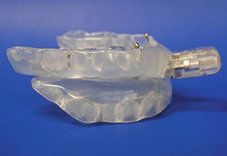
Dental Side Effects of Long-Term OSA Treatment—Venema et al.
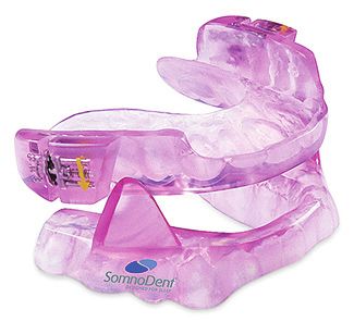
Figure 1— Thornton Adjustable Positioner. Figure 2—SomnoDent appliance.
positively correlated with its efficacy.8,31,32 However, the more
the mandible is positioned anteriorly, the more likely that
(dental) side effects will occur.8,33,34
The current study focuses on the effects of MAD and CPAP
therapy on dental occlusion. To date it is unknown to what
extent different types of MADs cause adverse changes in dental years or older and when they had used a SomnoDent appliance
occlusion and how these changes relate to the effects of CPAP for a 2-to 3-year period for at least 5 nights/wk and 5 h/night.
therapy on dental occlusion. The objective of the current study Patients were excluded when they had previous treatment for
is to evaluate the changes in dental occlusion from a bilat- their OSA (eg, CPAP or upper airway surgery). The number
eral thrust and anterior traction MAD and to compare these of patients who met the required follow-up time was 164.
outcomes with the dental effects of CPAP therapy. All these patients were checked for the remaining inclusion
criteria and invited for participation in this current study. Due
METHODS to mismatching the inclusion criteria or not responding to the
invitation, eventually 31 patients were recruited in the Somno-
Patient Selection Dent group. The patients in the TAP group (n = 29) and CPAP
In a previous randomized controlled trial, Doff et al. evalu- group (n = 34) from the study by Doff et al. were recruited
ated the dental side effects of MAD therapy and compared through the Department of Home Mechanical Ventilation of
them with CPAP therapy after a 2-year treatment period.29 the University Medical Center Groningen - the Netherlands.29
The CPAP system evaluated in this study applied pressure The two treatment modalities in this study were randomly
via a nasal mask.29 The MAD evaluated in this study was the assigned and followed for a 2-year treatment period. Inclu-
Thornton Adjustable Positioner (TAP) (Airway Management sion and exclusion criteria were similar for all three treatment
Inc., Dallas, Texas, United States), an anterior traction MAD.29 groups.29 All patients using an oral appliance in the current
In the current study the results from this previous study were study had been treated by the same clinician.
compared with a third patient group that was retrospectively
recruited and used a different type of MAD. The MAD evalu- Study Design
ated for this purpose was the SomnoDent appliance (Somno- At baseline all patients had been subjected to a polysomno-
Dent, Somnomed AG, Australia), a bilateral thrust MAD. The graphic evaluation. The data in the SomnoDent group were
TAP (Figure 1) and SomnoDent (Figure 2) appliance differ in longitudinally and retrospectively obtained. The data in the
the way in which the mandible is protruded. With the TAP this TAP and CPAP group were longitudinally and prospectively
is accomplished by a fixed screw in the front of the appliance, obtained.
whereas with the SomnoDent this is accomplished with two The degree of protrusion in both MAD groups at base-
screw mechanisms in the molar region of the appliance. line was set at 50% to 70% of the patient’s maximum protru-
Patients in the SomnoDent group were recruited through the sion. Patients using the SomnoDent could change the degree
Department of Oral and Maxillofacial Surgery of the Univer- of protrusion by means of two propulsion screws in the (pre)
sity Medical Center Groningen - The Netherlands and from the molar region in the upper part of the appliance. Patients using
Department of Oral and Maxillofacial Surgery of the Tjonger- the TAP could adjust mandibular protrusion by means of a
schans Hospital Heerenveen - The Netherlands. In accordance screw mechanism incorporated in the anterior part of the
with the American Academy of Sleep Medicine recommenda- upper appliance. If patients still experienced subjective OSA
tion, OSA was defined by an AHI higher than 5 and subjective complaints, they were instructed to advance the mandible.
complaints.10 In addition, patients were eligible when aged 20 Patients were instructed to advance their mandible until
Journal of Dental Sleep Medicine 40 Vol. 5, No. 2, 2018Dental Side Effects of Long-Term OSA Treatment—Venema et al.
symptoms abated or until further protrusion of the mandible Anterior-posterior movement was measured by evaluating
resulted in discomfort while wearing the appliance. the changes in distance between the buccal groove of the
After an 8-week habituation and adjustment period in all mandibular and the maxillary first molars (pretherapeutic
three treatment groups a polysomnographic evaluation study values minus posttherapeutic values). Negative values were
was performed to evaluate treatment efficacy. If this study related to a mesial shift of the occlusion and positive values to
showed a residual OSA, treatment was further adjusted if a distal shift. If first molars were missing, secondary molars
possible. Subsequently, after a 4-week period, another poly- were used.29
somnographic evaluation was performed. The Angle classification of malocclusion was used to clas-
Treatment was considered successful if AHI was less than 5 sify molar and cuspid occlusion and was recorded as Class I/
or showed a 50% reduction from the baseline value to a value neutro-occlusion, Class II/disto-occlusion, or Class III/mesio-
less than 20 in a patient who had no symptoms while using the occlusion.35 Angle classification was classified as unchanged
therapy. If treatment was not successful an alternative therapy or changed. The classification was determined at baseline and
was offered.29 follow-up at the right and left first molar and cuspid posi-
tion. When cuspid or molar teeth were missing or damaged,
Study Model Analysis the patient was listed as indefinable. The change of the Angle
At baseline and after a 2- to 3-year treatment period, algi- classification was defined as favorable if it changed toward a
nate impressions of the upper and lower dental arches were neutro-occlusion.29
obtained from each patient. Also, a bite registration in Maxillary and mandibular teeth in the (pre)molar region
maximum occlusion was obtained by using a vinyl polysi- were used to determine the transversal relation. This relation
loxane registration material (Exabyte II NDS, GC America was determined as normal, end-to-end or crossbite. If the
Inc, Alsip, Illinois, United States). From these impressions, transversal relation had changed at one or more teeth at the
dental plaster cast study models (GC FUJIROCK EP, GC (pre)molar region, this was recorded.29
America Inc, Alsip, Illinois, United States) were made that Crowding was visually defined as “increased,” “decreased”
were set into maximum occlusion in an articulator (Artex, or “no change” in space in the alveolar arch. Evaluation of
Girrlach Dental, Koblach, Germany) with bilateral sagittal diastemas was performed and classified as “unchanged,”
condylar inclination of 32.5 and Bennett angle of 17.5. “increased” or “decreased.” If teeth were extracted during
Measurements on the plaster casts were performed using the follow-up period, they were not counted as interproximal
a digital sliding caliper with a 0.01-mm resolution. The spaces. Dummies were not counted as present permanent
degree of mandibular protrusion in which the MAD was teeth, but were included in all analyses.29
set was measured with the digital sliding caliper when the A skilled and trained professional performed all measure-
MAD was fixed in the patient’s mouth. Subsequently, the ments. Measurements were all performed three times by the
percentage of mandibular protrusion in which the appli- same observer who was blinded for the patient’s treatment.
ance was set was calculated. All measurements were done The mean of the second and third measurement was used for
twice by the same observer. For continuous variables the further analysis.
mean of both measurements was used for further analysis.
When models were damaged or if there were not enough Statistical Analysis
teeth for adequate classification, the variable was listed as The Statistical Package for the Social Sciences (SPSS Statistics
indefinable. 29 version 22; IBM Corp., Armonk, New York, United States) was
Occlusal contact points were determined by using vinyl used to perform the analyses. A 95% confidence interval and
polysiloxane registration material (Exabite II NDSTM, GC the level of significance α was set at 0.05. The changes in dental
America Inc, Alsip, Illinois, United States) directly on the occlusion in the SomnoDent group were compared with results
cast models in maximum occlusion. Biting force or the size of from the TAP and CPAP group from the study by Doff et al.29
the contact points were not recorded. The number of occlusal To compare pretreatment and posttreatment variables,
contact points was obtained from the maxillary model in both paired Student t tests were performed. To compare variables
the cuspid-incisor region and in the (pre)molar region.29 between the three intervention groups, an analysis of variance
Anterior overjet and overbite were measured at both maxil- was done. For comparison of variables that were not normally
lary central incisors, the mean of both was used for further distributed the Mann-Whitney U test and the Kruskal-Wallis
calculations. Anterior overjet was defined as the horizontal test was used. For categorical variables within and between the
distance from the labial plane of the lower central incisor to three groups, chi-square tests were performed. Finally, linear
the mesial end of the incisal edge of the upper central incisor. regression analysis, the Pearson and Spearman correlation test,
Anterior overbite was measured as the vertical distance from was used to determine confounders, the relationship between
the incisal edge of the lower central incisor to the incisal possible dental side effects, and other therapy- or patient-
edge of the upper central incisor. The difference between related variables during the follow-up.
the pretreatment and posttreatment overjet and overbite This study was approved by the appropriate ethics committee
was called delta overjet and overbite. Negative values for and has been performed in accordance with the ethical stan-
delta overjet were defined as a mesial shift of the occlusion, dards laid down in the 1964 Declaration of Helsinki and
and positive values were defined as a distal shift of the of its later amendments. All persons have given their written
the occlusion.29 informed consent prior to inclusion in this study.
Journal of Dental Sleep Medicine 41 Vol. 5, No. 2, 2018Dental Side Effects of Long-Term OSA Treatment—Venema et al.
RESULTS In both MAD groups no differences were observed in
crowding of the upper and lower dental arches. In the CPAP
Patient Characteristics group crowding in the upper arch decreased in 1 patient and in
A total of 31 patients were included in the SomnoDent group. both arches in another patient, whereas crowding in the lower
In the study by Doff et al. 29 patients were included in the TAP arch increased in a third patient. In the SomnoDent group, a
group and 34 patients in the CPAP group.29 The mean ± stan- decrease in interproximal space in the upper arch was observed
dard deviation follow-up period in the SomnoDent group was in 1 patient (3%) and an increase in interproximal space in the
2.5 ± 0.3 years (range 2.0 to 3.3), in the TAP group 2.3 ± 0.2 upper arch was observed in 2 patients (6%). A decrease in inter-
years (range 2.1 to 3.1) and in the CPAP group 2.4 ± 0.3 years proximal space in the lower arch was observed in 3 patients
(range 2.1 to 3.2). The mean follow-up time was not signifi- (9%). In the TAP group, a decrease in interproximal space in
cantly different between the three groups.29 The mean degree of the upper arch was observed in 2 patients (7%), and an increase
protrusion in de SomnoDent group was 76.7% ± 11.2% (n = 21) in interproximal space in the upper arch was observed in 2
and in the TAP group 79.3% ± 19.3% (n = 29). The mean degree patients (7%). An increase in interproximal space in the lower
of protrusion was not significantly different between the two arch was observed in 7 patients (23%). In the CPAP group, an
groups. increase in interproximal space in the upper arch was observed
In the SomnoDent group the variables baseline AHI, body in 1 patient (3%), and an increase in interproximal space in the
mass index, nights/wk use and the number of occlusal (pre) lower arch was observed in 4 patients (12%).
molar contact points at baseline, were not normally distrib-
uted. A significant difference was observed in body mass DISCUSSION
index between the SomnoDent (30.3 ± 5.6) and CPAP group
(33.7 ± 5.7) (P < .01) (Table 1). Also, a significant difference was In this study we compared long-term side effects on dental
observed in baseline AHI between the SomnoDent (17.5 ± 13.2) occlusion between different treatment modalities in patients
and TAP group (35.6 ± 22.3) (P < .01), and between the Somno- with OSA. We observed that CPAP and both MADs resulted
Dent and CPAP group (44.2 ± 5.7) (P < .01) 29 (Table 1). Finally, in significant dental changes with long-term use. However,
a significant difference was observed in AHI after 2 to 3 the changes in overjet and anterior-posterior movement in the
months of treatment between the SomnoDent (6.9 ± 5.9) and SomnoDent and CPAP group were less pronounced than the
TAP group (2.9 ± 3.7) (P < .01), and between the SomnoDent changes observed in the TAP group.
and CPAP group (2.1 ± 4.0) (P < .01) (Table 1). When evaluating baseline characteristics, a distinct differ-
ence is observed in the AHI between the different treatment
Study Model Analysis groups. Because Doff et al.29 also included patients with more
When evaluating the number of occlusal contact points, a severe OSA, a significant difference was observed between the
significant decrease was observed in the (pre)molar region from SomnoDent (17.5 ± 13.2) and TAP group (35.6 ± 22.3) and
baseline to follow-up in all three treatment groups (Table 1). between the SomnoDent and CPAP group (44.2 ± 5.7), respec-
A significant reduction in overbite was observed when the tively. These significant differences may influence the outcomes
treatment groups were compared. A significant difference was of this study. Previous studies have reported that the higher the
observed between the SomnoDent (−0.6 ± 0.5) and TAP group baseline AHI, the higher percentage mandibular protrusion
(−1.2 ± 1.1) (P = .01), between the SomnoDent and CPAP group required for a successful treatment outcome.8,28,31,32 However,
(−0.1 ± 0.6) (P = .02), and between the TAP and CPAP group when evaluating the percentage of protrusion in both MAD
(P < .01). With respect to the decrease in overjet, a significant groups, no significant differences were observed between the
difference was observed between the SomnoDent (−0.6 ± 0.7) TAP and SomnoDent group.
and TAP group (−1.5 ± 1.5) (P < .01) and between the TAP Also, a significant difference in follow-up AHI after 2 to 3
and CPAP group (−0.2 ± 0.7) (P < .01) (Table 1). The anterior- months was observed between the SomnoDent versus the TAP
posterior movement was significantly different between the group. This phenomenon may have resulted from the difference
SomnoDent (−0.5 ± 0.6) and TAP group (−1.3 ± 1.5) (P < .01), in oral appliance design. The TAP appliance has a fixed screw
and between the TAP and CPAP group (−0.1 ± 0.6) (P < .01) mechanism in the front that is associated with minimal mouth
(Table 1). opening possibility. The SomnoDent appliance consists of two
No significant association was observed between the amount separate parts that allow for mouth opening as a result of auto-
of mandibular protrusion and the change in overbite and rotation of the mandible during sleep. This phenomenon may
overjet in the SomnoDent group. In the TAP group; however, explain why the AHI after 2 to 3 months was on average signifi-
a significant correlation was observed between the change in cantly higher in the SomnoDent group. In addition, this may
overbite and the amount of mandibular protrusion (β = −0.02; also explain why fewer dental side effects were observed in the
95% confidence interval −0.04 to 0.00) (Table 1). SomnoDent group. The autorotation of the mandible during
When evaluating cuspid and molar occlusion, it was sleep could have resulted in less force transmitted to the teeth,
observed that in most patients occlusion did not change. In resulting in fewer dental side effects.
addition, no significant differences were found between the In the current study a significant decrease in the number
three groups. When evaluating the direction of the occlusal of occlusal contact points was seen in the (pre)molar region.
shift, in most cases a mesial direct movement of mandibular This phenomenon was not only observed in both MAD groups,
teeth was observed (Table 2). but also in the CPAP group. As demonstrated previously, a
Journal of Dental Sleep Medicine 42 Vol. 5, No. 2, 2018Dental Side Effects of Long-Term OSA Treatment—Venema et al.
Table 1—Baseline characteristics, therapeutic use, and cast analysis of the SomnoDent, TAP, and CPAP groups.
SomnoDent TAP CPAP
Variable (n = 31) (n = 29)* (n = 34)* Difference (P )
Age (years) 50.7 ± 12.1 49.7 ± 8.9 50.6 ± 10.1 NS a
Male/female ratio 23/8 22/7 32/2 NS b
< .01 c
SmnDt versus TAP: NS
BMI 30.3 ± 5.6 # 31.4 ± 5.7 33.7 ± 5.7
SmnDt versus CPAP: < .01
TAP versus CPAP: NS
< .01 c
SmnDt versus TAP: < .01
Baseline AHI (events/h) 17.5 ± 13.2 # 35.6 ± 22.3 44.2 ± 5.7
SmnDt versus CPAP: < .01
TAP versus CPAP: NS
< .01 a
SmnDt versus TAP: < .01
Follow-up AHI (events/h) (after 2–3 months) 6.9 ± 5.9 2.9 ± 3.7 2.1 ± 4.0
SmnDt versus CPAP: < .01
TAP versus CPAP: NS
76.7 ± 11.2 79.3 ± 19.3
Mandibular protrusion (%) NS d
(n = 21) (n = 29)
Therapeutic use
nights/wk 6.6 ± 0.8 # 6.9 ± 0.4 6.7 ± 1.1 NS c
h/night 6.7 ± 1.1 7.1 ± 0.8 6.7 ± 1.3 NS a
Number of teeth
Upper arch 13.5 ± 1.7 12.7 ± 1.5 13.1 ± 1.8 NS a
Lower arch 13.4 ± 1.5 13.1 ± 1.4 13.0 ± 1.5 NS a
Occlusal contact points cuspid-incisor region (no.)
Baseline 2.23 ± 1.6 2.50 ± 1.7 3.24 ± 2.0 NS a
Follow-up 2.19 ± 1.8 2.21 ± 1.8 3.03 ± 1.9 NS a
Difference (P) NS e
NS e
NS e
Delta pre-post treatment contact points cuspid-incisor region
0.0 ± 1.2 0.3 ± 1.5 0.2 ± 1.4 NS a
(no.)
Occlusal contact points (pre)molar region (no.)
Baseline 7.19 ± 2.0 # 6.75 ± 2.6 6.91 ± 2.60 NS c
Follow-up 6.23 ± 2.1 5.14 ± 2.3 6.44 ± 2.3 NS a
Difference (P) < .01 f
< .01 e
.03 e
Delta pre-post treatment contact points (pre)molar region (no.) 0.9 ± 1.3 1.6 ± 2.7 0.5 ± 1.2 NS a
< .01 a
SmnDt versus TAP: .01
Delta overbite (mm) −0.6 ± 0.5 −1.2 ± 1.1 −0.1 ± 0.6
SmnDt versus CPAP: .02
TAP versus CPAP: < .01
< .01 a
SmnDt versus TAP: < .01
Delta overjet (mm) −0.6 ± 0.7 −1.5 ± 1.5 −0.2 ± 0.7
SmnDt versus CPAP: NS
TAP versus CPAP: < .01
< .01 a
SmnDt versus TAP: < .01
Anterior-posterior movement (mm) −0.5 ± 0.6 −1.3 ± 1.5 −0.1 ± 0.6
SmnDt versus CPAP: NS
TAP versus CPAP: < .01
Values are presented as mean ± standard deviation. * = from Doff et al.29 # = not normally distributed values. Superscript letters indicate the
following tests: a = analysis of variance, b = χ2, c = Mann-Whitney U and Kruskal-Wallis tests, d = independent t test, e = paired samples t test,
f = Wilcoxon signed-rank test. AHI = apnea-hypopnea index, BMI = body mass index, CPAP = continuous positive airway pressure, NS = not
significant, SmnDt = SomnoDent, TAP = Thornton Adjustable Positioner.
Journal of Dental Sleep Medicine 43 Vol. 5, No. 2, 2018Dental Side Effects of Long-Term OSA Treatment—Venema et al.
Table 2—Angle classification change.
Left Side Angle Classification Right Side Angle Classification
Number of Patients Number of Patients
Patient Group Occlusion Unchanged Mesial Shift Unchanged Mesial Shift
Cuspid 26 out of 30 (87%) 3 out of 4 (75%) 28 out of 31 (90%) 3 out of 3 (100%)
SomnoDent (n = 31)
Molar 28 out of 30 (93%) 2 out of 2 (100%) 24 out of 27 (89%) 3 out of 3 (100%)
Cuspid 21 out of 29 (72%) 8 out of 8 (100%) 22 out of 28 (79%) 6 out of 6 (100%)
TAP (n = 29)
Molar 16 out of 20 (80%) 3 out of 4 (100%) 17 out of 21 (81%) 4 out of 4 (100%)
Cuspid 28 out of 30 (93%) 1 out of 2 (50%) 29 out of 33 (88%) 3 out of 4 (75%)
CPAP (n = 34)
Molar 20 out of 23 (87%) 1 out of 3 (33%) 25 out of 28 (89%) 1 out of 3 (33%)
CPAP = continuous positive airway pressure, TAP = Thornton Adjustable Positioner.
decrease in overjet and overbite may result from long-term observed with anterior-posterior movement. From these obser-
MAD therapy.20,21,25,27,29,30 As a result of this phenomenon fewer vations it appears that the three treatment modalities result in
contact points may occur with long-term MAD use.25,29,36 changes in the overjet and anterior posterior movement of the
However, this does not explain the difference in occlusal mandible, which are less pronounced in the SomnoDent and
contact points with long-term CPAP therapy. CPAP therapy CPAP group. Furthermore, a more pronounced mesial move-
does not protrude the mandible. However, changes in the ment in occlusion was observed in the SomnoDent and the
number of occlusal contact points in the CPAP group may also TAP groups, respectively, when compared to the CPAP group.
occur as a result of a tight fitting and therefore large pressure In the MAD group, more patients shifted from an Angle Class
of the nasal mask on the frontal part of the maxilla, which may I to III occlusion or from an Angle Class II to I or III occlu-
result in a retroinclination of the maxillary incisors.37 Because sion. It must be mentioned that data on mandibular protrusion
of this phenomenon a slight autorotation of the mandible may were missing in a substantial proportion of the patients in the
occur, which explains the alteration in number of occlusal SomnoDent group. This fact hampers robust conclusions on
contact points.20,21,25,27,29,30 this phenomenon.
All analyses in the current study were manually done by When the results from the current study are compared
the same observer on dental plaster casts. We used this meth- with those of previous studies, similar changes in dental
odology because it was in line with the previously performed occlusion, as a result of long-term MAD therapy, have been
study by Doff et al.29 All measurements were done three times observed. Some studies describe a change in overbite and
by the same observer. Mean values of the second and third overjet,20,21,25,27,30,39 whereas others observe a change in anterior-
measurement were used for further analysis. A previously posterior movement.33,40 However, not all studies found signifi-
performed randomized clinical trial has shown that manual cant changes in overbite and overjet with long-term MAD
model analysis has the same methodological accuracy as therapy.39 These differences in changes in overbite and overjet
digital model analysis.38 We believe these aspects have mini- between different studies might, in addition to other variables,
malized the margin of error in the current study. be explained by differences in MAD design. The MADs evalu-
MAD and CPAP therapy is usually considered a lifelong ated in the current study had a full arch coverage, whereas in
requisite. With that in mind side effects are important to the study of Ringqvist et al.39 the MAD only covered the (pre)
monitor. In the current study a significant decrease is seen molars. This might result in less force on the frontal part of
in a number of occlusal contact points, overbite, overjet, and the dental arches, resulting in fewer changes in overbite and
anterior-posterior movement, not only in the MAD groups overjet.
but also in the CPAP group. In the TAP group an associa- The observed differences between the TAP and Somno-
tion was observed between the decrease in overbite and the Dent group could be explained by the difference in mecha-
percentage of mandibular protrusion. This correlation was nism contacting the upper and lower jaw. With the TAP, a
not observed in the SomnoDent group. In the TAP group the fixed screw in the frontal part of the upper appliance connects
percentage of mandibular protrusion was obtained from every both parts of the appliance. In the case of the SomnoDent, this
patient, whereas in the SomnoDent group, in one-third of the is accomplished by two stabilizing blocks in the (pre)molar
patients, the data on mandibular protrusion were missing. This region of the upper appliance. It could be hypothesized that
may explain why a significant correlation, between changes in with the TAP relatively more force is applied to the frontal part
dental occlusion and the percentage mandibular protrusion in of the upper and lower dental arches. This phenomenon may
the SomnoDent group, was not observed. partly explain the difference in delta overbite and delta overjet
When the delta overjet was compared between the different and anterior-posterior movement between the two groups. In
treatment modalities, a significant difference was observed addition, the materials from which the appliances are made
between the TAP and SomnoDent group and between the TAP may also explain the observed differences in delta overbite and
and CPAP group, respectively. A similar phenomenon was delta overjet between both appliances. The TAP was made of
Journal of Dental Sleep Medicine 44 Vol. 5, No. 2, 2018Dental Side Effects of Long-Term OSA Treatment—Venema et al.
hard acrylic, whereas the SomnoDent consisted of hard acrylic occlusion should always be preferred over “favorable” changes
on the outside with a soft lining on the inside of the appliance. in dental occlusion.
It could be hypothesized that as a result of the latter feature, Finally, it is important to use a strict selection procedure
less force is transferred to the individual teeth resulting in when selecting patients for a MAD. First, dental health has
less changes in overbite and overjet. However, because data to be adequate and, second, enough healthy teeth have to
on mandibular protrusion were missing in a substantial be present in order to stabilize the appliance. Therefore the
proportion of patients in the SomnoDent group, differences possible dental changes will be kept to a minimum. It is impor-
between both MAD groups in this variable may also explain tant that dental practitioners pay attention to possible dental
the observed difference in dental changes. Definite conclusions changes with MAD therapy. If these changes are unfavorable
on the effect of appliance design on changes in dental occlu- or too prominent, an alternative intervention must be consid-
sion can therefore not be drawn based on the data from this ered. Conversely, it should also be noted that CPAP, over the
study. In addition, other factors such as periodontal health and long term, may also result in similar changes in the dental
vertical skeletal characteristics were not taken into account occlusion.
and could also be responsible for the observed differences. It must be kept in mind that OSA is a sleep-related breathing
The correlation we observed between the baseline AHI and disorder that usually requires lifelong treatment. Therefore, it
the amount of mandibular protrusion following titration of the is important to pay attention to the therapeutic side effects
appliances has been described before. A higher baseline AHI with long-term CPAP or MAD use. Within the limitations of
requires more mandibular protrusion to reach a successful the current study we observed that TAP and SomnoDent as
treatment outcome.8,28,31,32 However, it is likely that an increased well as CPAP therapy resulted in significant dental changes
amount of mandibular protrusion will also result in more with long-term use. Changes in overjet and anterior-posterior
dental side effects.8,24,33 In this study a significant difference movement as a result of the SomnoDent and CPAP therapy
was observed in baseline AHI between the TAP and Somno- appear to be less pronounced when compared with the TAP
Dent appliance, with the TAP group having a higher baseline group. Specific features in oral appliance design may there-
AHI. However, the percentage of mandibular protrusion did fore affect the extent of changes in dental occlusion. However,
not correlate with the baseline AHI in both MAD groups. This other factors such as periodontal health and vertical skeletal
may have been the result of the relative small sample size of characteristics were not taken into account and could also be
both MAD groups. responsible for the observed differences. It should also be noted
The changes in transverse relation were not pronounced that AHI reductions in the SomnoDent group were also signif-
in the current study in all three treatment groups. Therefore, icantly less than in the other two groups. Nevertheless, it is
MAD or CPAP therapy does not appear to have a significant important to pay attention to possible dental side effects during
effect on the transverse relationships in the maxillary and MAD therapy. Before treatment, patients should be informed
mandibular dental arches. This may be explained by the fact about the possible dental changes and they should be subjected
that the force of both a MAD and a CPAP mask is mainly to a strict examination.
applied on the frontal aspect of the dental arches, which has
few effects on transverse relationships. REFERENCES
Another aspect requiring discussion is the effect of treat- 1. Jenkinson C, Davies RJ, Mullins R, Straddling JR. Comparison of
ment on the Angle classification. In the previous study by therapeutic and subtherapeutic nasal continuous positive airway
Doff et al., changes in Angle classification were observed.29 pressure for obstructive sleep apnoea: a randomised prospective
parallel trial. Lancet. 1999;353(9170):2100–2105.
A mesial shift of the lower dental arch relative to the upper
2. Sullivan CE, Issa FG. Obstructive sleep apnea. Clin Chest Med.
dental arch was generally observed. However, not all occlusal 1985;6(4):633–650.
changes should be regarded as negative changes. The most 3. Epstein LJ, Kristo D, Strollo PJ, et al. Clinical guideline for the
favorable occlusal pattern for patients is an Angle Class I evaluation, management and long-term care of obstructive sleep
occlusion.35 It could be unfavorable to change an Angle Class apnea in adults. J Clin Sleep Med. 2009;5(3):263–276.
I occlusion into an Angle Class III occlusion. However, when 4. de Britto Teixeira A, Abi-Ramia L, de Oliveira Almeida M. Treatment
of obstructive sleep apnea with oral appliances. Prog Orthod.
a patient starts with an Angle Class II occlusion and the lower 2013;14:10.
arch shifts forward resulting in an Angle Class I occlusion, 5. Flemons WW. Clinical practice. Obstructive sleep apnea. N Engl J
these changes may be regarded as favorable. Previous research Med. 2002;347(7):498–504.
regarding long-term side effects of a MAD observed that the 6. Hoekema A, Stegenga B, Wijkstra P, van der Hoeven J, Meinesz
orthodontic side effects could be classified as “favorable” in A, de Bont L. Obstructive sleep apnea therapy. J Dent Res.
2008;87(9):882–887.
41% of patients.30 In the current study no Angle Class change
7. Marin JM, Carrizo SJ, Vicente E, Agusti AG. Long-term
was observed in 91% of the molar measurements and in cardiovascular outcomes in men with obstructive sleep apnoea-
89% of the cuspid measurements of the SomnoDent group. hypopnoea with or without treatment with continuous positive airway
However, in the study of Almeida et al., including 70 patients, pressure: an observational study. Lancet. 2005;365(9464):1046–1053.
no changes in the Angle Class were observed only in 14% of 8. Ferguson KA, Cartwright R, Rogers R, Schmidt-Nowara W. Oral
appliances for snoring and obstructive sleep apnea: a review. Sleep.
patients.30 These differences may be explained by the follow- 2006;29(2):244–262.
up period which was 2 years in the current study and 7.4 years 9. Bradley T, Floras J. Obstructive sleep apnoea and its cardiovascular
in the study by Almeida et al.30 In addition, because we do not consequences. Lancet. 2009;373(9657):82–93.
know how progressive these changes are, no changes in dental
Journal of Dental Sleep Medicine 45 Vol. 5, No. 2, 2018Dental Side Effects of Long-Term OSA Treatment—Venema et al.
10. Sleep-related breathing disorders in adults: recommendations 30. Almeida FR, Lowe AA, Otsuka R, Fastlicht S, Farbood M, Tsuiki S.
for syndrome definition and measurement techniques in clinical Long-term sequellae of oral appliance therapy in obstructive sleep
research. The Report of an American Academy of Sleep Medicine Task apnea patients: Part 2. Study-model analysis. Am J Orthod Dentofacial
Force. Sleep. 1999;22(5):667–689. Orthop. 2006;129(2):205–213.
11. Kushida CA, Morgenthaler TI, Littner MR, et al. Practice parameters 31. Robertson C, Herbison P, Harkness M. Dental and occlusal changes
for the treatment of snoring and obstructive sleep apnea with oral during mandibular advancement splint therapy in sleep disordered
appliances: an update for 2005. Sleep. 2006;29(2):240–243. patients. Eur J Orthod. 2003;25(4):371–376.
12. Malhotra A, White DP. Obstructive sleep apnoea. Lancet. 32. Kato J, Isono S, Tanaka A, et al. Dose-dependent effects of
2002;360:(9328)237–245. mandibular advancement on pharyngeal mechanics and nocturnal
13. Hoekema A, Wijkstra PJ, Buiter CT, van der Hoeven JH, Meinesz AF, oxygenation in patients with sleep-disordered breathing. Chest.
de Bont LGM. Treatment of the obstructive sleep-apnea syndrome in 2000;117(4):1065–1072.
adults. Ned Tijdschr Geneeskd. 2003;147(49):2407–2412. 33. Aarab G, Lobbezoo F, Hamburger HL, Naeije M. Effects of an
14. Sullivan CE, Issa F, Berthon-Jones M, Eves L. Reversal of obstructive oral appliance with different mandibular protrusion positions at a
sleep apnoea by continuous positive airway pressure applied through constant vertical dimension on obstructive sleep apnea. Clin Oral
the nares. Lancet. 1981;1(8225):862–865. Investig. 2010;14(3):339–345.
15. Giles TL, Lasserson TJ, Smith BJ, White J, Wright J, Cates CJ. 34. Marklund M, Franklin K, Persson M. Orthodontic side-effects of
Continuous positive airways pressure for obstructive sleep apnoea in mandibular advancement devices during treatment of snoring and
adults. Cochrane Database Syst Rev. 2006;CD001106. sleep apnoea. Eur J Orthod. 2001;23(2):135–144.
16. Doff MH, Hoekema A, Wijkstra PJ, et al. Oral appliance versus 35. Angle EH. Treatment of Malocclusion of the Teeth. Angle’s System.
continuous positive airway pressure in obstructive sleep apnea 7th ed. Philadelphia, PA: The S.S. White Dental Manufacturing
syndrome: a 2-year follow-up. Sleep. 2013;36(9):1289–1296. Company; 1907.
17. Sawyer AM, Gooneratne NS, Marcus CL, Ofer D, Richards KC, 36. Otsuka R, Almeida F, Lowe A. The effects of oral appliance therapy
Weaver TE. A systematic review of CPAP adherence across age on occlusal function in patients with obstructive sleep apnea: a
groups: clinical and empiric insights for developing CPAP adherence short-term prospective study. Am J Orthod Dentofacial Orthop.
interventions. Sleep Med Rev. 2011;15(6):343–356. 2007;131(2):176–183.
18. Lim J, Lasserson TJ, Fleetham J, Wright J. Oral appliances 37. Doff MH, Hoekema A, Pruim GJ, Huddleston Slater JJ, Stegenga
for obstructive sleep apnoea. Cochrane Database Syst Rev. B. Long-term oral-appliance therapy in obstructive sleep
2006;CD004435. apnea: a cephalometric study of craniofacial changes. J Dent.
2010;38(12):1010–1018.
19. Chan AS, Lee RW, Cistulli PA. Dental appliance treatment for
obstructive sleep apnea. Chest. 2007;132(2):693–699. 38. Lippold C, Kirschneck C, Schreiber K, et al. Methodological accuracy
of digital and manual model analysis in orthodontics – a retrospective
20. Akssam G, Irmtrud EJ, Edmund CR. Dental side effects of clinical study. Comput Biol Med. 2015;62:103–109.
mandibular advancement appliances – a 2-year follow-up. J Orofac
Orthop. 2008;69(6):437. 39. Ringqvist M, Walker-Engström ML, Tegelberg A, Ringqvist I. Dental
and skeletal changes after 4 years of obstructive sleep apnea treatment
21. Hoffstein V. Review of oral appliances for treatment of sleep- with a mandibular advancement device: a prospective, randomized
disordered breathing. Sleep Breath. 2007;11(1):1–22. study. Am J Orthod Dentofacial Orthop. 2003;124(1):53–60.
22. Hoekema A, Stegenga B, De Bont LG. Efficacy and co-morbidity of 40. Chen H, Lowe AA, de Almeida FR, Fleetham JA, Wang B. Three-
oral appliances in the treatment of obstructive sleep apnea-hypopnea: dimensional computer-assisted study model analysis of long-term
a systematic review. Crit Rev Oral Biol Med. 2004;15(3):137–155. oral-appliance wear. Part 2. Side effects of oral appliances in
23. Sutherland K, Vanderveken OM, Tsuda H, et al. Oral appliance obstructive sleep apnea patients. Am J Orthod Dentofacial Orthop.
treatment for obstructive sleep apnea: an update. J Clin Sleep Med. 2008;134(3):408–417.
2014;10(2):215–227.
24. Doff MH, Veldhuis SK, Hoekema A, et al. Long-term oral appliance
therapy in obstructive sleep apnea syndrome: a controlled
SUBMISSION & CORRESPONDENCE
study on temporomandibular side effects. Clin Oral Investig. INFORMATION
2012;16(3):689–697.
Submitted for publication June 23, 2017
25. Martínez-Gomis J, Willaert E, Nogues L, Pascual M, Somoza M, Submitted in final revised form September 28, 2017
Monasterio C. Five years of sleep apnea treatment with a mandibular Accepted for publication October 10, 2017
advancement device. Side effects and technical complications. Angle Address correspondence to: J.A.M. Uniken Venema, Department of
Orthod. 2010;80(1):30–36. Oral Kinesiology, Academic Centre for Dentistry Amsterdam (ACTA),
26. Fritsch KM, Iseli A, Russi EW, Bloch KE. Side effects of mandibular University of Amsterdam and VU University Amsterdam, MOVE
advancement devices for sleep apnea treatment. Am J Respir Crit Care Research Institute Amsterdam, Amsterdam, The Netherlands; Tel: +3120
Med. 2001;164(5):813–818. 5980380; Fax: +3120 5980333; Email: j.a.m.unikenvenema@acta.nl
27. Hammond RJ, Gotsopoulos H, Shen G, Petocz P, Cistulli PA,
Darendeliler MA. A follow-up study of dental and skeletal changes DISCLOSURE STATEMENT
associated with mandibular advancement splint use in obstructive
sleep apnea. Am J Orthod Dentofacial Orthop. 2007;132(6):806–814. Work for this study was performed at the University Medical Center
28. Walker-Engström ML, Ringqvist I, Vestling O, Wilhelmsson B, Groningen. The authors report no conflicts of interest.
Tegelberg A. A prospective randomized study comparing two
different degrees of mandibular advancement with a dental appliance
in treatment of severe obstructive sleep apnea. Sleep Breath.
2003;7(3):119–130.
29. Doff MH, Finnema KJ, Hoekema A, Wijkstra PJ, de Bont LG, Stegenga
B. Long-term oral appliance therapy in obstructive sleep apnea
syndrome: a controlled study on dental side effects. Clin Oral Investig.
2013;17(2):475–482.
Journal of Dental Sleep Medicine 46 Vol. 5, No. 2, 2018You can also read