Nocturnal One-Hour Lighting Stimulates Gonadal Development and Lowers Fat Deposition in Male Mule Ducks - MDPI
←
→
Page content transcription
If your browser does not render page correctly, please read the page content below
animals
Article
Nocturnal One-Hour Lighting Stimulates Gonadal Development
and Lowers Fat Deposition in Male Mule Ducks
Tz-Chuen Ju             , Kai-Chien Tsao, Tzu-Yu Liu and Shyi-Kuen Yang *

                                          Department of Animal Science and Biotechnology, Tunghai University, Taichung 40704, Taiwan;
                                          tzchuen@thu.edu.tw (T.-C.J.); S06610224@thu.edu.tw (K.-C.T.); jane890325@gmail.com (T.-Y.L.)
                                          * Correspondence: skyang@thu.edu.tw

                                          Simple Summary: Photoperiods can affect sexual maturity, body weight, and body composition. In
                                          this work, we provided male mule ducks with a one-hour lighting from 20:00 to 21:00, in addition
                                          to the natural photoperiod, and evaluated its effects on their body weight, organ mass, gonadal
                                          function, and plasma levels of metabolites. The results indicate that the nocturnal lighting stimulated
                                          gonadal development and function and reduced fat deposition. This implies that nocturnal lighting
                                          is able to shorten the feeding period for the marketing of mature ducks.

                                          Abstract: In this study, the effects of a nocturnal light pulse on body weight, organ mass, gonadal
                                          function, and plasma levels of metabolites were determined in male mule ducks. In total, 32 15-week-
                                          old mule ducks were randomly allocated to either Group C (control group) or L+ (lighting group).
                                          Group C was exposed to the natural photoperiod, whereas Group L+ was provided with a 1-h
                                          lighting over 20:00–21:00 every day, in addition to the natural photoperiod. At the end of the 42-day
                                          experiment, Group L+ had significantly lower relative weights (% of live weight) of the digestive

                                   tract and abdominal fat and higher relative weights of the breast meat and testes than Group C.
Citation: Ju, T.-C.; Tsao, K.-C.; Liu,
                                          Moreover, Group L+ had significantly higher plasma testosterone and lower plasma glucose levels.
T.-Y.; Yang, S.-K. Nocturnal One-Hour     However, no between-group differences were observed in the triacylglycerol and uric acid levels.
Lighting Stimulates Gonadal               Histological examination demonstrated that the seminiferous tubule diameter was larger in Group
Development and Lowers Fat                L+ than in Group C. Moreover, the meiosis stage in spermatogenesis had begun in Group L+ but not
Deposition in Male Mule Ducks.            in Group C. In conclusion, the supplemented 1-h lighting at 20:00 stimulated gonadal development
Animals 2021, 11, 614. https://           and function and reduced fat deposition.
doi.org/10.3390/ani11030614

                                          Keywords: fat deposition; gonadal development; mule duck; photoperiod
Academic Editor: Ilias Giannenas

Received: 1 February 2021
Accepted: 20 February 2021
                                          1. Introduction
Published: 26 February 2021
                                               The mule duck (or mulard) is a sterile hybrid between a Muscovy drake (Cairina moschata)
Publisher’s Note: MDPI stays neutral
                                          and a mallard duck (Anas platyrhynchos). Muscovy ducks are large ducks, native to Mex-
with regard to jurisdictional claims in
                                          ico and Central and South American countries, which breed in the spring and summer.
published maps and institutional affil-   Domestic ducks are believed to be derived from wild mallard ducks. Wild mallard ducks
iations.                                  are migratory birds, which breed in the northern breeding range and winter in the south.
                                          Before migration, the feed intake and fat deposition increase in many bird species [1].
                                          Most birds, including migratory birds, demonstrate a seasonal reproductive pattern for the
                                          maximum survival rates of their offspring. The annual cycle of the photoperiod is the most
Copyright: © 2021 by the authors.
                                          crucial zeitgeber of the circannual rhythms, such as fat deposition, molt, reproduction, and
Licensee MDPI, Basel, Switzerland.
                                          migration, in almost all bird species living at high altitudes [2–4], even the temperate zone
This article is an open access article
                                          species which are exposed to near-equatorial photoperiods [5] and the subtropical species
distributed under the terms and           which are exposed to programmed photoperiodic schedules [6].
conditions of the Creative Commons             Domestication may have reduced but not repressed the seasonality of these birds. For
Attribution (CC BY) license (https://     instance, the captive undomesticated Muscovy ducks lay from February to September in
creativecommons.org/licenses/by/          England (in the Northern Hemisphere) [7], and the domesticated Muscovy ducks typi-
4.0/).                                    cally lay from August to April or May in Mozambique (in the Southern Hemisphere) [8].

Animals 2021, 11, 614. https://doi.org/10.3390/ani11030614                                                 https://www.mdpi.com/journal/animals
Animals 2021, 11, 614 2 of 9
The gonadal recrudescence and maturation of Muscovy ducks are controlled by the pho-
toperiod [9,10]. In Pekin ducks, which were domesticated from the wild mallard, the
seasonality of reproduction is retained, and their gonadal recrudescence is photosensitive
to the increasing day length [11]. The photoperiod also controls changes in body weight,
fat deposition, and feed intake. In general, shorter days promote fat deposition and longer
days stimulate protein accretion in domestic animals [12].
Mule ducks are the most common species of duck consumed for meat in Taiwan. They
grow rapidly, attaining near-mature body weight early in life, and thus are sold at 10 weeks
of age. However, they may also be sold at >14 weeks of age as replacement for mature male
Muscovy ducks, which are used for cooking ginger duck stew, a main course consumed
in winters. Considering their rapid growth early in life, understanding the effects of the
photoperiod on fat deposition in mule ducks may be more helpful than understanding
those on weight gain or protein accretion after the conventional fattening period. In this
study, we investigated the effects of supplemented light in the scotophase (i.e., skeleton
long photoperiod) during short days on the body weight, body composition, and testis
development in male mule ducks. Plasma levels of metabolites associated with nutrient
metabolism were also measured.
2. Materials and Methods
2.1. Animals
The experimental animals used in this study were three-way crossbred male mule
ducks (Muscovy × [Pekin × Tsaiya]), which were originally purchased from a commercial
hatchery. They were grown on an elevated plastic mesh floor without litter and exposed
to a natural photoperiod in another barn of the experimental farm of Tunghai University
before the experiment. They were transferred to the experimental barn and assigned to the
experiment when they were 105 days old.
2.2. Estimation of Sample Size
Based on the deduction from the independent two-sample t-test, the sample size per
group (n) for a 5% level of significance was calculated by the formula below:
n = 2 × (t0.05 )2 × (SD/ES)2
where t0.05 (14) = 1.76; t0.05 (30) = 1.70.
SD is the predicted standard deviation; ES is the predetermined effect size. Since a
previous study showed that the standard deviation of testicular weights in ducks, which is
the most important parameter in this study, was small (ranged from o.1 to 3.1 g) [13], and
we anticipated that the photoperiodic effect would be large, ES was anticipated to be equal
to or larger than SD. Therefore, 7 ducks per group should be enough for the comparison in
testicular weight. The photoperiodic effect on the weight of abdominal fat, an important
parameter in this study, was also anticipated to be large, and 7 ducks per group should
be enough for the comparison between groups. Therefore, 8 ducks per group were killed
for the collection of related data. However, the SD of body weight ranged from 222 to
232 g [14], and we anticipated that the photoperiodic effect would be moderate; therefore,
ES was anticipated to be 60% of SD. Therefore, 16 ducks per group were required.
2.3. Experimental Design
We allocated 32 male mule ducks to either Group C (control group) or Group L+
(lighting group) on 3 October. Each group was kept in a separated room comprising four
125 × 180 cm pens, with each pen containing four ducks. Group C was exposed to the
natural photoperiod, which was decreasing in day length, whereas Group L+ was provided
an artificial lighting using fluorescent tubes over 20:00–21:00 every day, in addition to the
natural photoperiod. The intensity of the light on the floor during the lighting period
ranged from 55 to 60 lux. During the experimental period (from 3 October to 13 November),Animals 2021, 11, 614 3 of 9
the length of the day (civil dawn to civil dusk) of the natural photoperiod decreased from
12.6 to 11.8 h, and the daily mean ambient temperature fluctuated between 21.1 and 28.6 ◦ C.
2.4. Management and Data Collection
During the experimental period, the ducks were raised on concrete floors without
litter, and the floor was cleaned once daily. They were fed ad libitum on a fattening
diet (Fwusow, Taichung, Taiwan), mainly composed of corn meal and soybean meal and
containing >14% CP, >3% fat,Animals 2021, 11, 614 4 of 9
than in Group C (Table 4). However, fasting plasma triacylglycerol and uric acid levels
were not affected by the photoperiod. Plasma testosterone levels were higher in Group L+
than in Group C (Table 4).
Table 1. Effects of the nocturnal 1-h lighting on the body weight, neck circumference, and feed intake
in male mule ducks.
Items Group C # Group L+ # SEM p-Value
Body Weight (g)
15 weeks of age 3523.8 3523.8 14.4 1.000
21 weeks of age 3404.4 3205.0 57.0 0.074
Weight Loss 119.4 318.8 63.5 0.121
Daily Feed Intake (g) 143.2 141.3 3.3 0.784
Neck Circumference (cm)
18 weeks of age 10.88 10.97 0.101 0.649
21 weeks of age 11.03 11.31 0.076 0.065
# Group C was subjected to natural photoperiod only, and Group L+ was supplemented with 1-h artificial light
from 20:00 to 21:00, in addition to the natural photoperiod.
Table 2. Effects of the nocturnal 1-h lighting on the organ weights of male mule ducks.
Items Group C # Group L+ # SEM p-Value
Live body weight (g) 3415.0 3175.0 52.186 0.015
Liver (g) 34.0 33.4 0.776 0.707
Heart (g) 26.2 28.5 1.044 0.280
Digestive tract (g) 177.0 151.4 4.526 0.001
Abdominal fat (g) 23.0 6.4 2.741Animals 2021, 11, 614 5 of 9
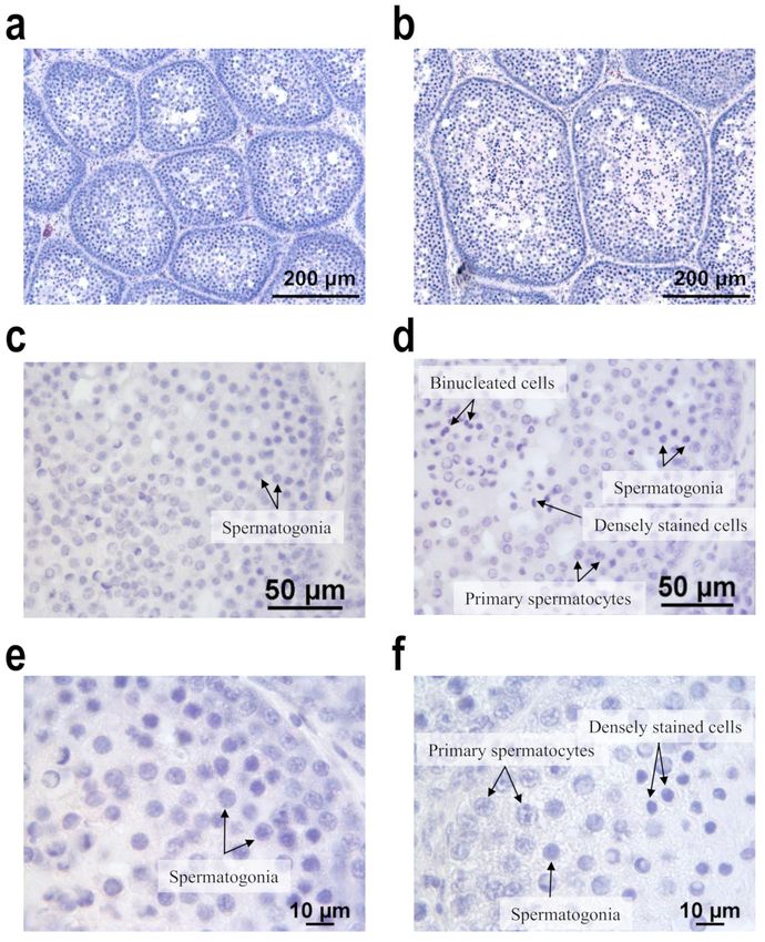
cells were observed in the central areas of the tubules, and lumen had started developing
(Figure 1f). However, Group C demonstrated only spermatogonia because spermatoge-
nesis had not commenced (Figure 1c,e). No spermatozoa were observed in either group
(Figure 1). Moreover, at 126 days of age, 9 of the 16 ducks in Group L+ exhibited ac-
tive mounting behavior, but no Group C duck exhibited this behavior (data not shown).
At 119 days of age, the bills of 14 of the 16 ducks in Group L+ had started to change color
from pale pink to red at the caudal border, whereas the bills of only 7 of the 16 ducks in
Group C had begun turning orange. By the final week, the color of the bills in both groups
was changing to red or orange; however, empirically, the bills had more area becoming red
in Group L+ than in Group C.
Figure 1. Effects of the nocturnal 1-h lighting on the histological characteristics of testes of our mule
ducks. (a) Section from the testis of a mule duck in Group C displays thin seminiferous tubules.
(b) Section from Group L+ reveals thick seminiferous tubules. (c,e) Section from Group C does not
exhibit spermatogenesis, and only spermatogonia exist. (d,f) Section from Group L+ displays active
spermatogenesis, and primary spermatocytes, as well as spermatogonia, are present. In addition,
some small deeply stained cells and binucleated cells are present in the central area of the tubule in
sections from Group B. No spermatozoa are observed in any sections. Group C was subjected to the
natural photoperiod, and Group L+ was supplemented with 1-h artificial light from 20:00 to 21:00, in
addition to the natural photoperiod. Hematoxylin and eosin stain.
4. Discussion
The growth curve of a mule duck is sigmoid in shape, with an inflection point between
25 and 30.5 days for male mule ducks [15,16]. Their body weights peak at 12 weeks ofAnimals 2021, 11, 614 6 of 9
age and decrease thereafter [16]. Therefore, at the beginning of this experiment, the body
weights of ducks had attained somatic maturity. In this study, mule ducks in both groups
lost their body mass during the first week of the experiment. The weight loss may be
attributed to the stress caused by the change in environment or experimental operation.
The final body weights in both groups did not recover to the initial weights. In this study,
the body mass was not significantly affected by the photoperiod regimen.
In this study, Group L+ had higher relative heart weights than Group C, even though
there was no difference in the absolute weights between groups. The higher relative heart
weight was consistent with the higher breast meat weight (Table 2). In sparrows, the breast
meat mass is positively correlated with the heart mass [17]. The heart mass may reflect the
metabolic demand. In general, skeletal muscles are more metabolically active than other
tissues, particularly the adipose tissue. Here, Group L+ demonstrated lighter digestive
tracts than Group C. Since their daily feed intake did not differ from that of Group C and
their relative digestive tract weight was lower than that of Group C, the food passage
rate in the digestive tract in Group L+ was speculated to be higher than that in Group C.
Therefore, our results demonstrate that a nocturnal 1-h lighting resulted in high relative
weights of the breast meat and heart and a low relative weight of the digestive tract and
implied that the photoperiodic regimen increased the food passage rate.
In this study, the precocious testis development in Group L+ was accompanied by
active mounting behavior and high plasma testosterone levels. High plasma testosterone
levels might contribute to the active mounting behavior. Higher testosterone levels might
result in higher relative breast meat weight and lower fat deposition in Group L+ compared
with Group C. In general, testosterone promotes aggressive and sexual behaviors, protein
accretion, and decreases in fat deposition. In domestic animals such as cattle, sheep, and
pigs, castration increases the ratio of fat mass-to-lean mass [18]. In chickens, caponization
enhances hepatic lipogenesis, whereas testosterone implantation reduces hepatic lipogen-
esis and lipid accumulation [19,20]. An in vitro study reported that androgens stimulate
myogenic differentiation and inhibit adipogenesis in mesenchymal pluripotent cells via an
androgen receptor-mediated pathway [21]. Here, the nocturnal 1-h lighting possibly caused
a decrease in the relative weight of abdominal fat and an increase in the relative weight
of the breast meat, possibly because of increased testosterone levels. By contrast, [22]
demonstrated that testosterone had no direct effect on muscle mass in sparrows.
Although the feed intakes fluctuated in both groups, there were no between-group
differences in average daily feed intake during the entire experimental period. Therefore,
the feed intake of mule ducks was not affected by the photoperiod substantially. Silky
starlings exposed to short days had higher feed intakes than those exposed to long days [23].
The feed intake in mature Chinese geese subjected to nocturnal 15-min lighting periods
at 2-h intervals was similar to that in control geese subjected to 12 h of light and dark per
day (12L/12D) [24]. In broilers, feed intake increased with the photoperiod during the first
21 days but was not affected later by photoperiods longer than 6 h [25]. The effect of the
photoperiod on feed intake may be dependent on the species, age, or length of the day.
In Germany, migratory garden warblers (Sylvia borin) were exposed to 12L/12D
from September to May and kept in outdoor aviaries from June to August; their basal
plasma levels of triacylglycerols and glucose exhibited seasonal fluctuations, with the
highest levels being between September and January and the lowest levels being between
February and April [26]. The current study demonstrates that the nocturnal lighting
lowered the fasting plasma glucose in mule ducks. This may be partially attributed to the
high plasma testosterone levels, as well as direct effects of the photoperiod. Testosterone
replacement therapy could reduce fasting glucose and glycated hemoglobin levels in
patients with type 2 diabetes mellitus with hypogonadism [27]. The effect of androgen
on the plasma triacylglycerol levels is not conclusive. A review suggested that androgen
administration either increased or had no effect on plasma triacylglycerol levels in aging
men with hypogonadism [28]. Our study demonstrates that the nocturnal light pulse
did not affect the fasting plasma triacylglycerol levels in mule ducks. Uric acid is theAnimals 2021, 11, 614 7 of 9
main nitrogenous waste in birds, and plasma uric acid levels reflect the state of protein
catabolism. The plasma level of uric acid was not influenced by the photoperiodic regimen
in this study. Regardless of the mechanism, nocturnal 1-h lighting reduced fasting plasma
glucose levels but did not affect the fasting plasma triacylglycerol and uric acid levels.
Most birds are long-day breeders: they enter the breeding season when the length of
the day increases, where the signal of the long photoperiod is conveyed to hypothalamus
to stimulate the secretion of GnRH, and hence to stimulate the pituitary to secrete go-
nadotropins and consequently to enhance the gonadal functions. Our study demonstrated
that the weight of the testes and plasma testosterone levels were significantly higher in
Group L+ than in Group C, and histological examination revealed that spermatogenesis
had begun in Group L+ but not in Group C. The red bill, a sexual characteristic of Mus-
covy drakes, started to appear in the male mule ducks exposed to the nocturnal lighting.
Mounting behavior was also actively exhibited by the ducks exposed to the 1-h nocturnal
lighting. Our results indicate that the nocturnal lighting substantially enhanced gonadal
development and function in our male mule ducks. Moreover, the length of the day (civil
dawn to civil dusk) at the beginning of (12.6 h) or early in the experiment period (12.6 and
11.8 h) is short enough to reset or induce photosensitivity in mule ducks. Chickens ex-
posed to photoperiods increasing from 6L/18D to 23L/1D demonstrated larger testes and
higher plasma testosterone levels at 8 weeks than those under the constant photoperiod
(23L/1D) [29]. In addition, a long-day response is believed to result from light extending
into the photoinducible phase of the circadian photoperiodic rhythm (CPR), and the CPR is
entrained by dawn. If the light is separated into two parts, the beginning of the long light
pulse entrains the CPR and the short light pulse determines the physiological response.
When the short light pulse falls in the photoinducible phase, the photoperiod is read as a
“long day”; when it does not extend into the photoinducible phase, it is read as a “short
day” [30]. In the current study, the supplemented lighting began from 14.2 to 14.5 h after
dawn and led to a long photoperiodic effect. Therefore, the supplemented lighting is
speculated to have extended into the photoinducible phase of mule ducks in this study.
Our histological examination also found that the seminiferous tubule diameter was larger
in Group L+ than in Group C. The histological examination also revealed that spermato-
genesis did not begin in Group C ducks, but ducks in Group L+ had reached the meiosis
stage in spermatogenesis. These results demonstrate that a 1-h lighting supplemented
at 20:00 caused long-day effects (enhancing gonadal development and function) in mule
ducks, and this regimen could be called skeleton long photoperiods.
In Group L+, the testis sections of mule ducks were observed and some small cells
with a deeply stained nucleus as well as some binucleated and multinucleated cells in
the central part of the seminiferous tubule were found. Snapir et al. reported that the
sections of mule duck testes demonstrate organized seminiferous tubules with secondary
spermatocytes as well as spermatogonia and primary spermatocytes [13]. The authors
also mentioned that spermatids were rarely noted, with some having several nuclei in a
single cell. This finding is similar to our current observation. However, [31] suggested that
the meiosis stage in spermatogenesis in the primary spermatocytes of mule ducks enters
pachytene but fails to progress beyond diakinesis-metaphase I. Nevertheless, this and
previous studies were consistently unable to observe any mature spermatozoa in the mule
duck testes. Therefore, the 1-h lighting supplemented at 20:00 promoted spermatogenesis
in male mule ducks.
5. Conclusions
A 1-h lighting over 20:00–21:00, in addition to the natural photoperiod, caused long-
day effects in mule ducks. In brief, it enhanced gonadal development and function and
increased the relative weight of the breast meat but reduced the body weight and relative
weight of the digestive tract and abdominal fat.
Author Contributions: T.-C.J. and S.-K.Y. conceived and designed the experiment. T.-C.J., K.-C.T.,
and T.-Y.L. performed the experiment and analyzed the data. S.-K.Y. wrote the paper. T.-C.J. con-Animals 2021, 11, 614 8 of 9
tributed to critical revision and editing of the manuscript. T.-C.J. and S.-K.Y. provided the grant. All
authors have read and agreed to the published version of the manuscript.
Funding: This work was supported by grants from the Tunghai University industry-academia
collaboration (No. 109168, 109167 and No. 109654), Taiwan. These were used in the design and
execution of the experiment.
Institutional Review Board Statement: The experimental protocols used in the present study were
approved by Tunghai University Experimental Animal Care and Use Committee.
Data Availability Statement: All data of this study are included in the manuscript.
Conflicts of Interest: The authors have no conflicts of interest to declare.
References
1. King, J.R.; Farner, D.S. Studies of fat deposition in migratory birds. Ann. N. Y. Acad. Sci. 1965, 131, 422–440. [CrossRef]
2. Gwinner, E. Circannual rhythms in birds. Curr. Opin. Neurobiol. 2003, 13, 770–778. [CrossRef]
3. Dawson, A.; King, V.M.; Bentley, G.E.; Ball, G.F. Photoperiodic control of seasonality in birds. J. Biol. Rhythm. 2001, 16, 365–380.
[CrossRef]
4. Wikelski, M.; Martin, L.B.; Scheuerlein, A.; Robinson, M.T.; Robinsin, N.D.; Helm, B.; Hau, M.; Gwinner, E. Avian circannual
clocks: Adaptive significance and possible involvement of energy turnover in their proximate control. Philos. Trans. R. Soc. B
2008, 363, 411–423. [CrossRef] [PubMed]
5. Dawson, A. Seasonality in a temperate zone bird can be entrained by near equatorial photoperiods. Proc. R. Soc. B 2007, 274,
721–725. [CrossRef] [PubMed]
6. Dixit, A.S.; Singh, N.S. Photoperiod as a proximate factor in control of seasonality in the subtropical male tree sparrow. Passer
Montanus. Front. Zool. 2011, 8, 1–12. [CrossRef] [PubMed]
7. Murton, R.K.; Kear, J. Photoperiodism in waterfowl: Phasing of breeding cycles and zoogeography. J. Zool. 1978, 186, 243–283.
[CrossRef]
8. Harun, M.A.S.; Veeneklaas, R.J.; Van Kampen, M.; Masasso, M. Breeding biology of Muscovy duck Cairina moschata in natural
incubation: The effect of nesting behavior on hatchability. Poult. Sci. 1998, 77, 1280–1286. [CrossRef]
9. Jacquet, J.M.; Sauveur, B. Photoperiodic control of sexual maturation in Muscovy drakes. Domest. Anim. Endocrinol. 1995, 12,
189–195. [CrossRef]
10. Jacquet, J.M. Photorefractory period of the Muscovy duck (Cairina moschata): Endocrine and neuroendocrine responses to day
length after a full reproductive cycle. Br. Poult. Sci. 1997, 38, 209–216. [CrossRef]
11. Cherry, O.; Morris, T.R. The maintenance requirement of domestic drakes. Br. Poult. Sci. 2005, 46, 725–727. [CrossRef] [PubMed]
12. Tucker, H.A.; Petitclerc, D.; Zinn, S.A. The influence of photoperiod on body weight gain, body composition, nutrient intake and
hormone secretion. J. Anim. Sci. 1984, 59, 1610–1620. [CrossRef] [PubMed]
13. Snapir, N.; Rulf, J.; Meltzer, A.; Gvaryahu, G.; Rozenboim, I.; Robinzon, B. Testosterone concentrations, testes weight and
morphology of mule drakes (Muscovy drake X Khaki Campbell). Br. Poutry Sci. 1998, 39, 572–574. [CrossRef]
14. Su, C.-H.; Lin, T.-A.; Tseng, T.-F.; Cheng, C.-H.; Huang, J.-F.; Liu, H.-C.; Lin, J.-H. The effect of different rearing environment on
two-way crossbred mule duck’s growth performances and carcass traits. Taiwan Livestock Res. 2018, 51, 68–74.
15. Gille, U.; Salomon, F.V. Heart and body growth in ducks. Growth Dev. Aging 1994, 58, 75–81.
16. Chen, Y.-H.; Roan, S.-W. Growth curve of white mule ducks. J. Chin. Soc. Anim. Sci. 2005, 34, 31–37.
17. Chappell, M.A.; Bech, C.; Buttemer, W.A. The relationships of central and peripheral organ masses to aerobic performance
variation in house sparrows. J. Exp. Biol. 1999, 202, 2269–2279.
18. Field, R.A. Effect of castration on meat quality and quantity. J. Anim. Sci. 1971, 32, 849–858. [CrossRef] [PubMed]
19. Chen, K.L.; Chi, W.T.; Chen, R.S.; Chiou, P.W.S. Effect of caponization and testosterone implantation on hepatic lipids and
lipogenic enzymes in male chickens. Poult. Sci. 2007, 86, 1754–1759. [CrossRef]
20. Duan, J.L.; Shao, F.; Shao, Y.; Li, J.; Ling, Y.; Tenk, K.; Li, H.; Wu, C. Androgen inhibits abdominal fat accumulation and negatively
regulates the PCK1 gene in male chickens. PLoS ONE 2013, 8, e59636. [CrossRef] [PubMed]
21. Rajan, S.; Jorge, N.A.; Wayne, E.T.; Nestor, F.G.; Shalender, B. Androgens stimulate myogenic differentiation and inhibit
adipogenesis in C3H 10T1/2 pluripotent cells through an androgen receptor-mediated pathway. Endocrinology 2003, 144,
5081–5088.
22. Buttemer, W.A.; Warne, S.; Bech, C.; Astheimer, L.B. Testosterone effects on avian basal metabolic rate and aerobic performance:
Facts and artefacts. Comp. Biochem. Physiol. Part A Mol. Integr. Physiol. 2008, 150, 204–210. [CrossRef]
23. Wang, J.-Q.; Wang, J.-J.; Wu, X.-J.; Zheng, W.-H.; Liu, J.-S. Short photoperiod increases energy intake, metabolic thermogenesis
and organ mass in silky starlings Sturnus sericeus. Dongwuxue Yanjiu 2016, 37, 75–83.
24. Huang, D.-J.; Yang, S.-K. Nocturnal light pulses lower carbon dioxide production rate without affecting feed intake in geese.
Asian Australas. J. Anim. Sci. 2016, 29, 390–395. [CrossRef]
25. Lewis, P.D.; Danisman, R.; Gous, R.M. Photoperiodic responses of broilers. I. Growth, feeding behavior, breast meat yield, and
testicular growth. Br. Poult. Sci. 2009, 50, 657–666. [CrossRef]Animals 2021, 11, 614 9 of 9
26. Lugaresi, E.; Montagna, P.; Tinuper, P.; Plazzi, G.; Gallassi, R.; Wang, T.C.; Markey, S.P.; Rothstein, J.D. Endozepine stupor.
Recurring stupor linked to endozepine-4 accumulation. Brain 1998, 121, 127–133. [CrossRef] [PubMed]
27. Harada, N. Role of androgens in energy metabolism affecting on body composition, metabolic syndrome, type 2 diabetes,
cardiovascular disease, and longevity: Lessons from a meta-analysis and rodent studies. Biosci. Biotechnol. Biochem. 2018, 82,
1667–1682. [CrossRef] [PubMed]
28. Schleich, F.; Legros, J.J. Effects of androgen substitution on lipid profile in the adult and aging hypogonadal male. Eur. J.
Endocrinol. 2004, 151, 415–424. [CrossRef]
29. Charles, R.G.; Robinsin, F.E.; Hardin, R.T.; Yu, M.W.; Feddes, J.; Classen, H.L. Growth, body composition, and plasma androgen
concentration of male broiler chickens subjected to different regimens of photoperiod and light intensity. Poult. Sci. 1992, 71,
1595–1605. [CrossRef] [PubMed]
30. Nicholls, T.J.; Goldsmith, A.R.; Dawson, D. Photorefractoriness in birds and comparison with mammals. Physiol. Rev. 1988, 68,
133–176. [CrossRef] [PubMed]
31. Islam, F.B.; Ishishita, S.; Uno, Y.; Mollah, M.B.R.; Srikulnath, K.; Matsuda, Y. Male hybrid sterility in the mule duck is associated
with meiotic arrest in primary spermatocytes. J. Poult. Sci. 2013, 50, 311–320. [CrossRef]You can also read