Multimodality Review of Imaging Features Following Breast Reduction Surgery
←
→
Page content transcription
If your browser does not render page correctly, please read the page content below

Goudreau et al. Breast Reduction

      Review                                                                                                         Eur J Breast Health 2021; 17(3): 206-213
                                                                                                                     DOI: 10.4274/ejbh.galenos.2021.2021-1-3

      Multimodality Review of Imaging Features Following
      Breast Reduction Surgery
          Sally H. Goudreau1,          Meghan A. Woughter2,              Stephen J. Seiler1
      Department of Radiology, UT Southwestern Medical Center, Texas, USA
      1

      Department of Radiology, Radiology Partners, Texas, USA
      2

          ABSTRACT
          Reduction mammoplasty is a common surgical procedure that removes a significant portion of the breast, and the resulting changes to the breast parenchyma
          are frequently seen on breast imaging studies. Any radiologist who interprets breast imaging studies must be able to recognize these changes in order to avoid
          unnecessary recall from screening and/or breast biopsy. The surgical techniques used in reduction mammoplasty are discussed in order to provide relevant
          background information for understanding the resulting imaging features. These imaging characteristics are presented for the most common breast imaging
          modalities, including mammography, ultrasound, and magnetic resonance imaging. Additionally, tips for distinguishing malignancy from postsurgical
          change are provided, as are potential pitfalls in imaging interpretation. To avoid unnecessary patient morbidity, it is critical to differentiate between the
          classic, benign imaging appearance of the breast after reduction mammoplasty and findings that indicate a potential malignancy.
          Keywords: Breast cancer, breast imaging, breast reconstruction, mastopexy

          Cite this article as: Goudreau SH, Woughter MA, Seiler SJ. Multimodality Review of Imaging Features Following Breast Reduction Surgery.
          Eur J Breast Health 2021; 17(3): 206-213

          Key Points
          •    All reduction procedures involve the removal and displacement of varying amounts of parenchymal tissue and skin, largely from the inferior breast.
          •    This tissue and skin removal alters the normal distribution of fibroglandular tissue and can result in architectural distortion, focal asymmetries, and
               regions of fat necrosis with or without dystrophic calcifications.
          •    Fat necrosis may be difficult to interpret by ultrasound interrogation alone. A spot tangential mammographic view may assist in the visualization of
               internal fat within the mass to clench the diagnosis of fat necrosis.
          •    Preoperative imaging with mammography is recommended to assess for occult malignancy for average-risk women 40 years of age and older as well as
               women of any age who are at high-risk of developing breast cancer.

      Introduction
      Reduction mammoplasty is a common non-oncologic surgical procedure that accounts for 18% of all breast surgeries (1). In patients with
      symptomatic macromastia, the surgery aims to remove excess fat, glandular tissue, and skin from the breast to create a smaller, more aesthetically
      pleasing breast shape (2). Moreover, chronic back, neck, or shoulder pain, kyphosis, chronic intertrigo involving the inframammary folds,
      limitation of physical activity, or other problems associated with overly large breasts are common indications. Following reduction surgery,
      preoperative symptoms have been shown to be significantly reduced (3). In addition, some patients have reduction mammoplasty to improve
      symmetry following a contralateral partial mastectomy or total mastectomy with reconstruction. Several similar surgical techniques are used to
      alleviate patient symptoms while producing aesthetically pleasing results. Disruption of breast tissue following these surgical procedures can
      appear perplexing to those who are unfamiliar with these classic changes. Once the technical aspects of these procedures are conceptualized, the
      imaging appearance of the breast frequently becomes apparent. Even an experienced interpreting radiologist may have difficulty distinguishing
      between postsurgical changes after reduction mammoplasty and the imaging appearance of malignancy on occasion. Ultimately, if malignancy
      is suspected, a biopsy will be required for a definitive histopathologic diagnosis.

      Corresponding Author:                                                                                                                           Received: 22.01.2021
206   Sally H. Goudreau; sally.goudreau@utsouthwestern.edu                                                                                            Accepted: 16.03.2021

      ©Copyright   2021 by the the Turkish Federation of Breast Diseases Societies / European Journal of Breast Health published by Galenos Publishing House.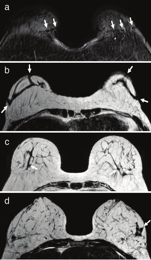
Goudreau et al. Breast Reduction

Surgical techniques                                                          swirled or with an upward sweeping configuration (Figure 2b) (5).
                                                                             Mammographically, such inferior pole changes are evident in nearly
There are several reduction mammoplasty techniques available, the            all patients (6, 7).
majority of which involve removing parenchymal tissue and skin from
the inferior breast while elevating the nipple-areolar complex (NAC).        Typical post-reduction scar patterns also include fibrotic bands that run
The most common surgical techniques for reduction mammoplasty                parallel to the skin scar line and are easier to identify in non-anatomic
involve a circumferential incision around the NAC, followed by a             orientations. Vertical scarring is common in the inferior breast, and
vertical incision extending to the inframammary fold, resulting in a
keyhole scar (Figure 1a). This technique enables superior repositioning
of the NAC as well as removal and repositioning/lifting of the inferior
breast tissue (mastopexy). By using this keyhole skin flap pattern, the
conical breast shape is preserved because the skin flaps are opposed,
resulting in a skin brassiere. To achieve an aesthetic result, the NAC
can be transposed with the underlying ducts and vascular pedicle
intact using the inferior pedicle technique (Figure 1b) or transplanted
with a full-thickness free nipple-areolar graft. With the inferior pedicle
technique, the blood supply to the nipple and areola originates from the
relatively large surface of the inframammary fold, which contributes to
the preservation of nipple, areola, and skin sensations (4). Because it
has a lower risk of NAC avascular necrosis than NAC transposition,
free nipple-areolar graft transplantation is more common in women
with large, pendulous breasts and those past childbearing age (5). The
resulting post-reduction skin scar usually has a keyhole or lollipop
appearance, with a subtle periareolar scar, a vertical scar extending
inferiorly from the nipple to the inframammary fold, and a transverse
scar curving along the fold (Figure 1c).

Breast reduction can also be accomplished solely through liposuction.
This is the least invasive technique, resulting in only minor volume
reduction and omitting the benefit of mastopexy. There are some
non-specific imaging findings associated with this technique, such
as developing asymmetries in regions where fat has been removed.
Obtaining a history of the plastic surgical intervention would be
beneficial in avoiding a biopsy of the new imaging finding, which
would otherwise be required.

Mammography
Architectural distortion
When evaluating architectural distortion in the breast, the presence
of surgical clips is often the first indication that a patient has had a
prior surgical procedure, though this does not always mean reduction
mammoplasty. While some surgical clips are left behind in reduced
breasts, most surgeons do not. When mammographically evaluating
what is thought to be postsurgical architectural distortion, the clinical
history is confirmatory.

All reduction procedures involve the removal and displacement of
varying amount amounts of parenchymal tissue and skin, which
alters the normal distribution of fibroglandular tissue and can result
in focal asymmetries (Figure 2a). In one early study of post-reduction
mammographic changes, these asymmetries developed in roughly half
of the women postoperatively, either persisting or gradually decreasing
over time (6). With the appropriate clinical history and knowledge
of this typical mammographic appearance, unnecessary recall from
                                                                             Figure 1. Surgical incisions for reduction mammoplasty. (a) An
screening and/or biopsy can be avoided.
                                                                             incision is made around the nipple areolar complex along with
                                                                             a vertical incision extending to a curvilinear inframammary fold
During reduction mammoplasty, most breast tissue is usually
                                                                             incision. (b) The breast tissue is removed inferiorly, and the nipple
excised from the inferior aspect of the breast. The remaining tissue         is transposed superiorly, maintaining the vascular pedicle. (c)
is gathered together, often with rearrangement. This technique               Appearance of the breast before (right breast) and after (left breast)
produces a distinctive pattern of architectural distortion that appears      reduction mammoplasty using the keyhole technique                           207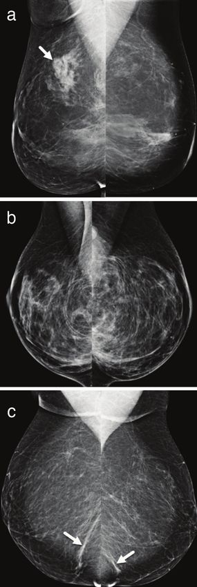
Eur J Breast Health 2021; 17(3): 206-213

      transversely oriented scars can be seen posteriorly in the breast (8).    from normal breast parenchyma. Parenchymal bands, which can
      This non-anatomic scarring pattern may result in linear bands that        extend from the chest wall to the NAC, can also be relatively thick.
      mammographically resemble skin folds (Figure 2c). These bands can         The most common cause of non-malignant architectural distortion
      be subtle, thinning over time and becoming difficult to distinguish       is postsurgical scarring. However, as with any type of breast surgery,
                                                                                parenchymal surgical scarring should diminish or stabilize over time.
                                                                                New or increasing architectural distortion, even scarred regions, is
                                                                                suspicious and warrants further investigation (Figure 3).

                                                                                Figure 3. Malignancy developing in a reduction scar. (a) Sequential
                                                                                right craniocaudal images taken one year apart as part of screening
                                                                                examinations in a 57-year-old woman demonstrate interval
                                                                                development of architectural distortion at the reduction scar
                                                                                (arrow). (b) Craniocaudal right spot compression image taken
                                                                                as part of a diagnostic work-up shows persistent architectural
                                                                                distortion (arrow). Sonography at that time was non-contributory,
      Figure 2. Rearrangement of the fibroglandular breast tissue
                                                                                only revealing scarring in the area of concern. Stereotactic core
      after reduction mammoplasty. (a) Bilateral mediolateral oblique
                                                                                biopsy of the architectural distortion (not shown) confirmed invasive
      digital mammographic images show asymmetric tissue in the right
                                                                                ductal carcinoma. (c) Enhanced MRI was subsequently performed
      posterior superior breast resulting from tissue rearrangement
                                                                                to evaluate the extent of disease which demonstrated extensive
      following mammoplasty. (b) Bilateral mediolateral oblique digital
                                                                                multifocal/multicentric abnormal enhancement, including the area
      mammograms show the characteristic sweeping parenchymal
                                                                                of developing architectural distortion (arrow) seen on this sagittal
      pattern seen after breast reduction. (c) Bilateral mediolateral oblique
                                                                                image of the right breast
      digital mammograms demonstrate non-anatomic post-reduction
208   linear fibrotic bands which mimic skin folds (arrows)                     MRI: Magnetic resonance imaging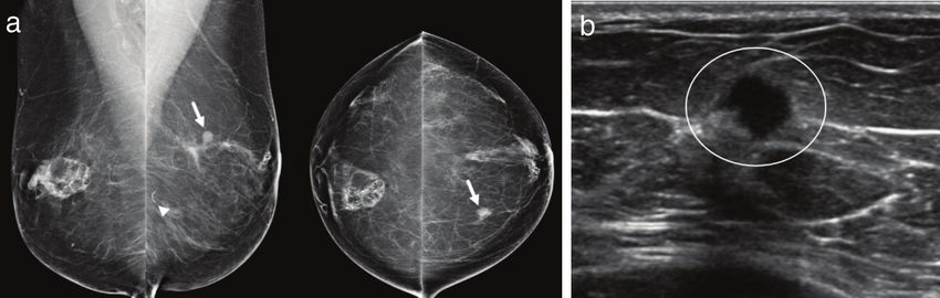
Goudreau et al. Breast Reduction

Changes of the NAC                                                          Breast surgery frequently results in areas of benign fat necrosis
                                                                            manifesting as oil cysts. These masses are well-defined, are round or
Mastopexy is used in various types of reduction mammoplasty
                                                                            oval in shape, and contain fat, with or without rim calcification (5,
procedures, with the NAC being relocated superiorly, as seen on
                                                                            11). Oil cysts of varying sizes are seen in nearly 20% of patients and
the mediolateral oblique views. To create a better periareolar scar,
                                                                            may resolve or shrink in size over time (7). As a result of oil cysts,
permanent suture material may be used to secure the nipple complex
                                                                            dystrophic calcifications can develop. These calcifications may be
(Figure 4). Along the periareolar margin, scarring with or without
                                                                            difficult to interpret accurately at first, but they frequently coarsen
calcifications may be visible (9). Danikas et al. (7) demonstrated that
                                                                            over time.
these periareolar alterations can be seen on mammography in 85% of
women postoperatively.                                                      Fat necrosis can also manifest as a nonspecific mass or a focal
                                                                            architectural distortion with or without calcifications. Moreover,
Calcifications and fat necrosis                                             postoperative changes and fat necrosis can easily be attributed to
                                                                            fat-containing masses with or without associated coarse or rim
Benign calcifications are a common postoperative mammographic
                                                                            calcifications. However, dystrophic calcifications associated with
finding, though they appear later than other mammographic features.
                                                                            fat necrosis that appear in the early postoperative stage infrequently
According to one study, calcifications were found in only 3% of
                                                                            may have a questionable morphology and/or distribution (12). The
mammograms performed within the first 12 months after reduction,
                                                                            risk of malignancy is still very low in these cases, and calcifications
compared to 53% of mammograms performed 24 months or later
                                                                            associated with fat necrosis should gradually evolve, assuming a more
after surgery (6). Furthermore, skin calcifications with lucent centers
                                                                            classic dystrophic appearance and confirming the benign etiology
are more common at anastomotic sites.
                                                                            (12). Therefore, reporting these calcifications in the Breast Imaging
Breast reduction surgery usually entails extensive manipulation of          Reporting and Database System 3 (BI-RADS 3) category (0% to
the breast parenchyma. Fat necrosis is caused by major trauma to an         ≤2% likelihood of malignancy) with a recommendation for a short-
area of adipose tissue, which results in cellular death of the adipocytes
and the subsequent appearance of residual oil/fat material and
dystrophic calcification. As a result, fat necrosis is often encountered
postoperatively and is a common cause of palpable abnormality in
the postoperative breast (10). Because of internal fat at the palpable
site that correlates well with the clinical history, a new palpable area
of fat necrosis is often easily diagnosed mammographically, whereas
the corresponding sonographic appearance can be indeterminate and
potentially suspicious (Figure 5).

                                                                            Figure 5. Post-reduction fat necrosis presenting as an area of
                                                                            palpable concern. (a) Sonography shows a nonspecific, solid,
                                                                            irregularly shaped heterogeneous mass (circle) in the region of a
Figure 4. Mammographic appearance of the nipple areolar complex             new, palpable lump. (b) Left mediolateral oblique and craniocaudal
after reduction mammoplasty. Permanent sutures are seen                     digital mammogram images subsequently performed demonstrate
(arrows) in the peri-areolar regions on bilateral craniocaudal digital      post-reduction changes with a fatty mass (circles) in the 7:00 position,
mammographic images                                                         confirming benign fat necrosis                                             209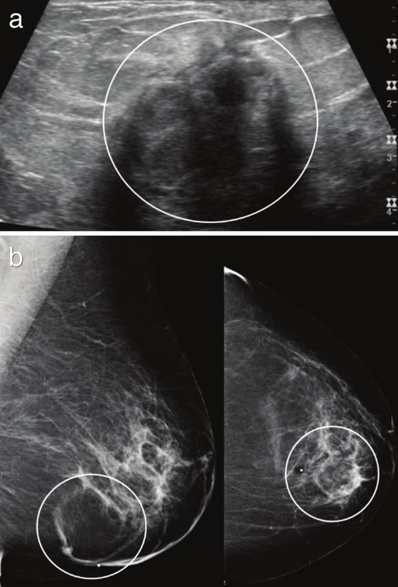
Eur J Breast Health 2021; 17(3): 206-213

      term close interval follow-up to document morphologic stability is          Furthermore, architectural distortion can occur anywhere within
      appropriate. Stereotactic biopsy should be reserved for cases that do       the breast parenchyma in post-reduction patients if significant tissue
      not show early morphology and/or distribution of fat necrosis, or for       rearrangement has occurred. Sonographically, this distortion appears
      cases that become suspicious after a short interval of follow-up.           as vague hypoechogenicity, disruption of the normal fascial planes, and
                                                                                  posterior acoustic shadowing, which is most prominent in the inferior
      Sonography                                                                  breast and inframammary fold region. This sonographic appearance
                                                                                  may be identical to cancer, necessitating a biopsy.
      Physical examination of the breast, along with sonography, will
      reveal the typical scarring pattern associated with prior reduction         Magnetic resonance imaging
      mammoplasty. A periareolar skin scar (which may be very subtle), an
      inframammary fold scar, and a radially oriented scar in the 6 o’clock       Because of its high sensitivity and negative predictive value of
      position will all be visible patterns. Depending on the type of surgical    malignancy, breast magnetic resonance imaging is a valuable diagnostic
      procedure performed, patients may have a variable combination of            tool for detecting breast cancer. There are a number of distinct post-
      these scars.                                                                reduction mammoplasty findings seen with magnetic resonance
                                                                                  imaging (13).
      The appearance of post-reduction fat necrosis on imaging varies greatly
      depending on its stage of evolution, particularly with sonography.          Signal voids corresponding to suture material and/or surgical clips
      Sonography is frequently used to investigate new palpable findings;         are seen in a linear fashion along the inframammary fold, encircle
      however, fat necrosis is can be difficult to interpret with ultrasound      the NAC, and may be scattered throughout the breast parenchyma
                                                                                  (Figure 7a). Similarly, post-contrast imaging can easily detect dermal
      alone. If the etiology of a palpable mass cannot be determined after an
                                                                                  scarring and keloids around the NAC, inframammary fold, and 6
      initial ultrasound interrogation, a spot tangential mammographic view
                                                                                  o’clock radiant (Figure 7b). During surgery, fibroglandular tissue is
      can be obtained easily in order to visualize fat within the mass (Figure
                                                                                  rearranged, resulting in architectural distortion, parenchymal bands
      6). One of the most useful features in classifying the mass as benign fat
                                                                                  (Figure 7c), and islands of breast tissue (Figure 7d) similar to those
      necrosis is the discovery of internal fat.
                                                                                  seen on mammography. The imaging characteristics of these islands
                                                                                  should be similar to other areas of benign fibroglandular tissue found
                                                                                  in the breast.

                                                                                  Fat necrosis can occur anywhere in the reconstructed breast, but it is
                                                                                  most common along the inferior aspect of the breast, where distortion
                                                                                  is most common. Fat necrosis produces an isointense signal to the rest
                                                                                  of the fatty breast tissue, with varying degrees of rim enhancement
                                                                                  depending on its current stage of evolution and the degree of
                                                                                  inflammation and granulation tissue. Although a thin enhancing rim
                                                                                  is common with fat necrosis, a thickened and irregular enhancing
                                                                                  rim that can be mistaken for malignancy may be present. The kinetic
                                                                                  analysis of fat necrosis is nonspecific, encompassing both benign and
                                                                                  malignant enhancement patterns (13). T1-weighted images with and
                                                                                  without fat saturation are frequently used to determine the presence
                                                                                  of fat within a mass or area of architectural distortion, assisting in
                                                                                  the confirmation of the presence of benign fat necrosis (Figure 8).
                                                                                  Fat necrosis may also be characterized by enhancing internal thin
                                                                                  septations. The T1 signal from fat necrosis is typically isointense to
                                                                                  other fats in the breast; however, fat necrosis may sometimes appear
                                                                                  to have a slightly darker T1 signal due to hemosiderin deposition
                                                                                  or chronic inflammatory changes. Mammographic correlation is
                                                                                  recommended because the presence of oil cysts or coarse calcifications
                                                                                  within the region of interest may provide further supporting evidence
                                                                                  of fat necrosis.

                                                                                  Cancer detection
                                                                                  Prior to undergoing elective reduction mammoplasty, preoperative
                                                                                  imaging to assess for occult malignancy is recommended for average-
                                                                                  risk women 40 years of age and older, as well as women of any age
      Figure 6. A palpable mass seen sonographically is confirmed to be           who are at high-risk of developing breast cancer (14). Mammography
      fat necrosis with mammography. (a) Sonographic image shows                  is the most cost-effective and widely available screening method in
      an irregularly shaped, heterogeneous mass (circle) at the site of a         eligible patients. Any suspicious lesions detected during preoperative
      patient’s palpable abnormality after reduction mammoplasty. (b)
      Corresponding spot tangential digital mammogram of the palpable
                                                                                  imaging will require tissue diagnosis prior to surgery. Infrequently
      finding demonstrates a fat containing mass (circle), confirming fat         (0.8%), malignancy is discovered during reduction mammoplasty,
210   necrosis                                                                    posing a diagnostic dilemma if no preoperative imaging was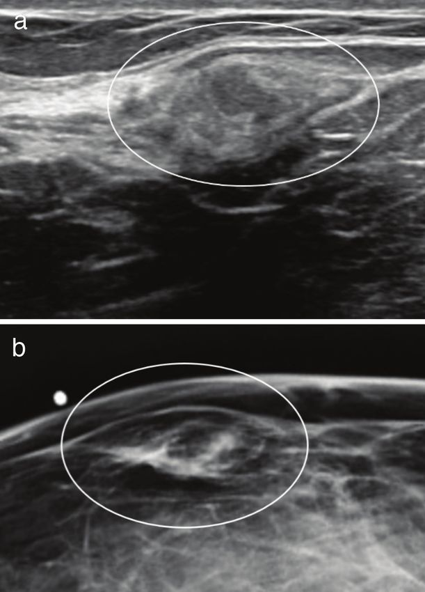
Goudreau et al. Breast Reduction

obtained (15). In the event of a postoperative diagnosis of breast      residual cancer and masking from postsurgical changes (15). At our
cancer, the patient will need additional diagnostic evaluation;         institution, we have observed similar limitations in the immediate
however, the sensitivity of breast magnetic resonance imaging for       postoperative period and therefore recommend that the patient wait
malignancy is reduced in the immediate post-reduction breast due        at least 6 weeks after surgery before undergoing magnetic resonance
to the expected postoperative enhancement of inflamed and healing       imaging to evaluate for residual disease in order to lessen these
tissue. In a large cohort of 4,804 women examined by Tang et al.        postoperative changes. After treatment for the primary malignancy,
(15), 48% of patients with an incidental diagnosis of malignancy at     if mastectomy is not performed, repeat bilateral breast magnetic
mammoplasty had postoperative breast magnetic resonance imaging         resonance imaging in 6 months could be considered for reevaluation
to assess the extent of disease. The majority of the initial cancers    after the postsurgical changes have resolved. Cancer detection and
discovered were low grade and small. In fact, 8% of invasive cancers    recall rates in breasts that have undergone reduction mammoplasty
and 72 % of DCIS were grade 1 or 2, and 94% of invasive cancers         have been reported to be comparable to native breast (16). For
were stage T1. The authors found that postoperative magnetic            average-risk women, routine annual or biennial mammographic
resonance imaging had limited sensitivity for detecting any residual    screening following reduction mammoplasty is the appropriate
malignancy, hypothesizing that this was due to the small size of any    recommendation. The postoperative baseline mammogram is
                                                                        usually the most difficult to interpret because significant changes
                                                                        in the configuration of the breast parenchyma have occurred in
                                                                        addition to the expected interval postoperative changes (7). Any
                                                                        interval changes, such as a developing asymmetry or mass, after
                                                                        this baseline mammogram should be viewed with caution, and
                                                                        an appropriate diagnostic evaluation should be recommended
                                                                        (Figure 9).

                                                                        Differentiating fat necrosis from residual or new cancer can
                                                                        sometimes be difficult with any breast imaging modality, especially if

Figure 7. Post-surgical magnetic resonance appearance after
reduction mammoplasty. (a) Signal void along the bilateral
inframammary folds on a fat saturated T1-weighted axial image
corresponds to surgical staples and/or sutures (arrows). (b) Scarring
is represented by dark bands (arrows) on a T1-weighted axial image
along the inframammary fold region bilaterally. (c) A T1-weighted       Figure 8. Fat necrosis seen with magnetic resonance imaging. (a)
axial image shows parenchymal bands (arrows) similar to those seen      Rim enhancement (arrow) in the medial left breast on post-contrast
mammographically. (d) An axial post-contrast image shows an island      T1-weighted image corresponds with (b) a fat-containing lesion
of non-enhancing fibroglandular tissue (arrow) in the lateral left      (arrow) seen on T1-weighted pre-contrast image, both confirming fat
breast                                                                  necrosis                                                                 211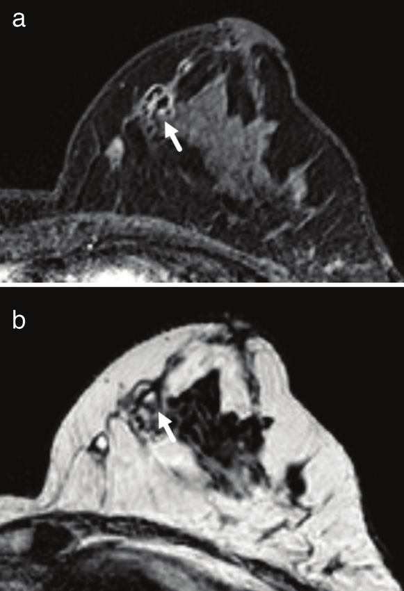
Eur J Breast Health 2021; 17(3): 206-213

      Figure 9. Interval cancer development following reduction mammoplasty. (a) This 58-year-old woman was found to have a new mass (arrows)
      in the 11 o’clock position of the left breast, middle depth, on routine screening mammography. Reduction changes, including areas of fat
      necrosis, are seen bilaterally. A suture needle (arrowhead) from the prior reduction surgery is noted posteriorly on the left mediolateral
      oblique projection. (b) Ultrasound imaging confirmed an irregularly shaped hypoechoic mass with indistinct margins (circle), with subsequent
      ultrasound-guided biopsy yielding triple-negative invasive ductal carcinoma

      there is excessive degree of tissue fibrosis and no discernible internal    Conclusion
      fat. The enhancement kinetics for fat necrosis are highly variable,
      with a wide range of kinetic curves reported in the literature (13).        All common breast imaging modalities reliably predict the imaging
      Depending on the degree of diagnostic certainty, some lesions may           features of reduction mammoplasty. To avoid confusion with
      be allowed short-term follow-up. However, if there is moderate to           developing malignancy, radiologists interpreting these studies should
      high clinical suspicion of malignancy, percutaneous sampling should         be able to recognize the described patterns of distortion, scarring, and
      be recommended.                                                             calcification. When there are indeterminate lesions, a biopsy or close
                                                                                  short-term follow-up should be performed.
      Calcifications in the postoperative breast may also pose a diagnostic
      dilemma. Over time, benign calcifications caused by surgical changes
                                                                                  Peer-review: Externally and internally peer-reviewed.
      tend to coarsen and become more dystrophic. As calcifications begin
      to form, they may appear amorphous and coarse heterogeneous within          Author Contributions
      the BI-RADS 4b intermediate suspicion category (17). Moreover,
                                                                                  Surgical and Medical Practices: S.H.G., M.A.W., S.J.S.; Conception: S.H.G.,
      calcifications with a high likelihood of benignity (≤2% risk of             M.A.W., S.J.S.; Design: S.H.G., M.A.W., S.J.S.; Analysis and/or Interpretation:
      malignancy) may be classified as probably benign and followed as a          S.H.G., M.A.W., S.J.S.; Literature Search: S.H.G., M.A.W., S.J.S.; Writing:
      precautionary measure. Any calcifications that remain suspicious            S.H.G., M.A.W., S.J.S.
      after thorough mammographic work-up and/or follow-up should
      be subjected to stereotactic biopsy for histopathologic diagnosis           Conflict of Interest: The authors have no conflicts of interest to declare.
      (Figure 10).
                                                                                  Financial Disclosure: The authors declared that this study has received no
                                                                                  financial support.

                                                                                  References
                                                                                  1.   American Society of Plastic Surgeons. 2018 Plastic Surgery Statistics
                                                                                       Report 2019. Last Accessed Date: 21.04.2020. Available from: https://
                                                                                       www.plasticsurgery.org/documents/News/Statistics/2018/plastic-surgery-
                                                                                       statistics-full-report-2018.pdf [Crossref ]
                                                                                  2.   Davis GM, Ringler SL, Short K, Sherrick D, Bengtson BP. Reduction
                                                                                       mammaplasty: long-term efficacy, morbidity, and patient satisfaction.
                                                                                       Plast Reconstr Surg 1995; 96: 1106-1110. (PMID: 7568486). [Crossref ]
                                                                                  3.   Glatt BS, Sarwer DB, O'Hara DE, Hamori C, Bucky LP, LaRossa D.
                                                                                       A retrospective study of changes in physical symptoms and body image
                                                                                       after reduction mammaplasty. Plast Reconstr Surg 1999; 103: 76-82;
                                                                                       discussion 3-5. (PMID: 9915166). [Crossref ]
      Figure 10. Developing malignant calcifications after reduction              4.   Azurin DJ, Fisher J, Maxwell GP. Mastopexy. In: Weinzweig J, editor.
      mammoplasty. This 66-year-old patient developed grouped,                         Plastic Surgery Secrets Plus. Chicago, IL: Mosby/Elsevier; 2010. p. 453-
      amorphous calcifications (circles) in the 3 o’clock position of the right        457.
      breast 11 years after reduction mammoplasty, demonstrated on
      spot magnification diagnostic mammogram                                     5.   Miller CL, Feig SA, Fox JWt. Mammographic changes after reduction
                                                                                       mammoplasty. AJR Am J Roentgenol 1987; 149: 35-38. (PMID:
                                                                                       3495989). [Crossref ]
212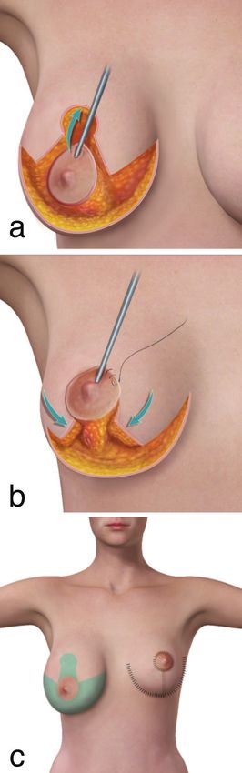
Goudreau et al. Breast Reduction

6.   Brown FE, Sargent SK, Cohen SR, Morain WD. Mammographic                    12. Cakir M, Kucukkartallar T, Tekin A, Selimoglu N, Poyraz N, Belviranli
     changes following reduction mammaplasty. Plast Reconstr Surg 1987; 80:         MM, et al. Comparison of mammography sensitivity after reduction
     691-698. (PMID: 3671561). [Crossref ]                                          mammoplasty targeting the glandular and fat tissue. Ulus Cerrahi Derg
7.   Danikas D, Theodorou SJ, Kokkalis G, Vasiou K, Kyriakopoulou K.                2015; 31: 68-71. (PMID: 26170752). [Crossref ]
     Mammographic findings following reduction mammoplasty. Aesthetic           13. Daly CP, Jaeger B, Sill DS. Variable appearances of fat necrosis on breast
     Plast Surg 2001; 25: 283-285. (PMID: 11568832). [Crossref ]                    MRI. AJR Am J Roentgenol 2008; 191: 1374-1380. (PMID: 18941072).
8.   Margolis NE, Morley C, Lotfi P, Shaylor SD, Palestrant S, Moy L, et al.        [Crossref ]
     Update on imaging of the postsurgical breast. Radiographics 2014; 34:      14. Kerrigan CL, Slezak SS. Evidence-based medicine: reduction
     642-660. (PMID: 24819786). [Crossref ]
                                                                                    mammaplasty. Plast Reconstr Surg 2013; 132: 1670-1683. (PMID:
9.   Burk KS, Seiler SJ, Porembka JH. Diagnosis, management, and                    24281593). [Crossref ]
     percutaneous sampling of nipple-areolar calcifications: how radiologists
                                                                                15. Tang R, Acevedo F, Lanahan C, Coopey SB, Yala A, Barzilay R, et al.
     can help patients avoid the operating room. AJR Am J Roentgenol 2021;
                                                                                    Incidental breast carcinoma: incidence, management, and outcomes in
     216: 48-56. (PMID: 33170739). [Crossref ]
                                                                                    4804 bilateral reduction mammoplasties. Breast Cancer Res Treat 2019;
10. Hogge JP, Robinson RE, Magnant CM, Zuurbier RA. The mammographic
                                                                                    177: 741-748. (PMID: 31317348). [Crossref ]
    spectrum of fat necrosis of the breast. RadioGraphics 1995; 15: 1347-
    1356. (PMID: 8577961). [Crossref ]                                          16. Muir TM, Tresham J, Fritschi L, Wylie E. Screening for breast cancer
                                                                                    post reduction mammoplasty. Clin Radiol 2010; 65: 198-205. (PMID:
11. Shaheen R, Schimmelpenninck CA, Stoddart L, Raymond H, Slanetz
                                                                                    20152275). [Crossref ]
    PJ. Spectrum of diseases presenting as architectural distortion on
    mammography: multimodality radiologic imaging with pathologic               17. Tan PH, Lai LM, Carrington EV, Opaluwa AS, Ravikumar KH, Chetty
    correlation. Semin Ultrasound CT MR 2011; 32: 351-362. (PMID:                   N, et al. Fat necrosis of the breast--a review. Breast 2006; 15: 313-318.
    21782125). [Crossref ]                                                          (PMID: 16198567). [Crossref ]

                                                                                                                                                                 213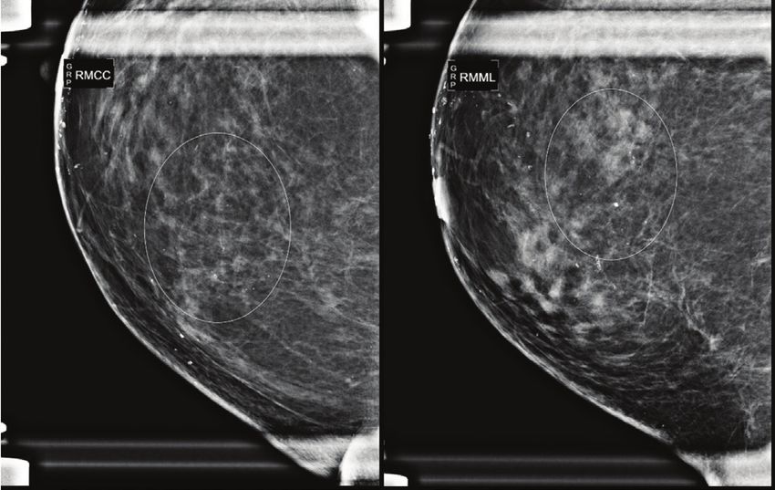
You can also read


