CLINICAL PRACTICE GUIDELINES
←
→
Page content transcription
If your browser does not render page correctly, please read the page content below
CLINICAL PRACTICE GUIDELINES
                                   MOH/P/PAK/209.10 (GU)

 MINISTRY OF HEALTH MALAYSIA        ACADEMY OF MEDICINE MALAYSIA

                               1
These are Clinical Practice Guidelines on Management of Dengue
  Infection in Adults (Revised 2nd Edition) 2010. The CPG supersede the
  previous CPG on Management of Dengue Infection in Adults (2nd Edition) 2008.

These guidelines are meant to be guides for clinical practice,
based on the best available evidence at the time of develoment.
Adherence to these guidelines may not necessary guarantee
the best outcome in every case. Every healthcare provider is
responsible for the management of his/her unique patient
based on the clinical picture presented by the patient and
the management options available locally.

Review of the Guidelines
These guidelines were issued in 2010 and will be reviewed in
2014 or sooner if new evidence becomes available.

CPG Secretariat
Health Technology Assessment Section
Medical Development Division
Ministry of Health Malaysia
4th Floor, Block E1, Parcel E
62590 Putrajaya.

Electronic version available on the following websites :
http://www.moh.gov.my
http://www.acadmed.org.my

                                      2
GUIDELINES DEVELOPMENT AND OBJECTIVE
GUIDELINES DEVELOPMENT

The development group for these guidelines consisted of a family medicine
specialist, an emergency medicine specialist, a general physician, infectious
disease physicians, intensivists, haematologists, public health physicians,
a virologist and a nursing sister from the Ministry of Health and Ministry of
Higher Education, Malaysia. During the process of development of these
guidelines, there was active involvement of a review committee.

The previous edition of CPG (2003) was used as the basis for the
development of these present guidelines.

These guidelines provide:

a. A detailed description of the clinical course of dengue illness which
   reflects the dynamism and systemic nature of dengue that have crucial
   bearing on the patient’s management.

b. A detailed description of the basic pathophysiological changes of
   severe dengue (i.e. plasma leakage and hypovolemia/shock) and
   provide guidance on the recognition of these changes and appropriate
   action of management.

c. A brief discussion on WHO Classification (1997) and its limitations.

d. Some useful guides on the differential diagnoses that can be confused
   with dengue or vice versa; they were described according to the stage
   of disease.

e. A more focused guide on the disease monitoring in accordance with
   the dynamic changes as the disease progresses.

f.   Emphasis on the importance of monitoring the plasma leakage (clinical
     signs of plasma leakage and haematocrit (HCT) and haemodynamic
     status of the patients.

g. Clearer algorithm on fluid management in severe dengue.

h. Emphasis on the importance of recognising or suspecting significant
   occult bleed with some useful guides.

i.   A more systematic approach on the recognition of signs of recovery.

Literature search was carried out at the following electronic databases:
International Health Technology Assessment Website, PUBMED, Cochrane
Database of Systemic Reviews (CDSR), Journal full text via OVID search
engine, Comprehensive; Database of Abstracts of Reviews of Effectiveness,

                                      i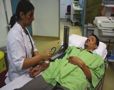
Cochrane Controlled Trials Registered, CINAHL via EBSCO search
engine. In addition, the reference lists of all relevant articles retrieved were
searched to identify further studies. Reference was also made to other
guidelines – WHO Dengue Haemorrhagic Fever: Diagnosis, Treatment,
Prevention and Control, WHO Geneva, 1997; Guidelines, Guidelines for
DHF Case Management, Bangkok, Thailand 2002; Guidelines on Clinical
Management Of Dengue Fever / Dengue Haemorrhagic Fever 2005 Sri
Lanka; WHO Regional Publication SEARO, 1999; Guidelines for Treatment
of Dengue Fever/Dengue Hemorrhagic Fever in Small Hospitals, WHO
Regional Office for SE Asia, New Delhi, 1999. There were very few studies
carried out on dengue patients in the adult population. Many of the studies
included in these guidelines are based upon the management of dengue
in children. The findings of these studies were then extrapolated on to the
adult population, taking into consideration our local practices.

The clinical questions were divided into major subgroups and members
of the development group were assigned individual topics within these
subgroups. The group members met a total of 15 times throughout the
development of the guidelines. All literature retrieved were appraised by
at least two members and presented in the form of evidence tables and
discussed during group meetings. All statements and recommendations
formulated were agreed by both the development group and review
committee. Where the evidence was insufficient the recommendations were
derived by consensus of the development group and review committee.

The articles were graded using the modified version of the criteria used
by the Catalonia Agency for Health Technology Assessment and Research
(CAHTAR) Spain, while the grading of recommendation in this guideline
was modified from the Scottish Intercollegiate Guidelines Network (SIGN).

The draft guidelines was posted on both the Ministry of Health Malaysia
and Academy of Medicine, Malaysia websites for comment and feedback.
These guidelines had also been presented to the Technical Advisory
Committee for Clinical Practice Guidelines, and the Health Technology
Assessment and Clinical Practice Guidelines Council, Ministry of Health
Malaysia for review and approval.

                                       ii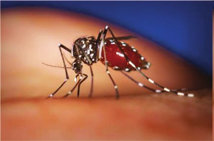
OBJECTIVES
GENERAL OBJECTIVES
To provide evidence-based guidance in the management of dengue
infection in adult patients

SPECIFIC OBJECTIVES
• To improve recognition and diagnosis of dengue cases and provide
  appropriate care to the patients

• To identify severe dengue and carry out more focused close monitoring
  and prompt appropriate management

• To provide guidance on appropriate and timely fluid management and
  the use of blood and blood products

• To improve on early and accurate notification of dengue cases for
  prompt public health intervention

CLINICAL QUESTIONS
Please refer to Appendix 6

TARGET POPULATION
Adult patients with dengue fever, dengue haemorrhagic fever or dengue
shock syndrome and other forms of severe dengue.

TARGET GROUP/USER
These guidelines are applicable to primary care doctors, public health
personnel, nurses, assistant medical officers, physicians and critical care
providers involved in treating adult patients with dengue fever, dengue
haemorrhagic fever or dengue shock syndrome and other forms of severe
dengue.

HEALTHCARE SETTINGS
Both outpatient and inpatient settings

                                    iii
clinical indicators for quality management

Primary indicators
i.    Case fatality rate (DF & DHF)
      Numerator: No of DF & DHF/DSS death
      Denominator: No of DF & DHF cases (clinically diagnosed)
      National target (9th Malaysian Plan):< 0.2%

ii. DHF fatality rate
      Numerator: No of DHF/ DSS death
      Denominator: No of DHF/ DSS cases (clinically diagnosed)
      National target (9th Malaysian Plan):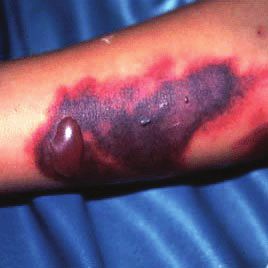
GUIDELINES DEVELOPMENT GROUP

CHAIRPERSON
Dr. Mahiran Mustafa
Senior Consultant Infectious Disease Physician
Hospital Raja Perempuan Zainab II
Kota Bharu Kelantan

MEMBERS (alphabetical order)
 Dr. Abdul Hamid Jaafar                           Dr. Norita Ahmad
 Assistant Director                               Consultant Infectious Disease Physician
 Communicable Disease Control Division            Hospital Raja Perempuan Zainab II
 Ministry of Health                               Kelantan

 Dr. Ainul Nadziha Mohd. Hanafiah                 Dr. Salmah Idris
 Assistant Director                               Consultant Pathologist
 Health Technology Assessment Section             Hospital Sungai Buloh
 Medical Development Division, MOH                Selangor

 Dr. Chow Ting Soo                                Dr. Sheamini Sivasampu
 Consultant Infectious Disease Physician          Principal Assistant Director
 Hospital Pulau Pinang                            Health Technology Assessment Section
 Pulau Pinang                                     Medical Development Division MOH
 Dr. Faisal Salikin                               Ms Sin Lian Thye
 Emergency Medicine Specialist                    Nursing Sister
 Hospital Kuala Lumpur                            Health Technology Assessment Section
 Kuala Lumpur                                     Medical Development Division MOH

 Dato’ Dr. Faraizah Abdul Karim                   Dato’ Dr. K. Sree Raman
 Deputy Director                                  Senior Consultant Physician
 National Blood Centre Kuala Lumpur               Hospital Tuanku Ja’afar
                                                  Negeri Sembilan
 Dr. Ho Bee Kiau                                  Dr. Suresh Kumar
 Family Medicine Specialist                       Consultant Infectious Disease Physician
 Bukit Kuda Health Clinic                         Hospital Sungai Buloh
 Selangor                                         Selangor

 Dr Mohamad Ikhsan Selamat                        Dr. Tan Cheng Cheng
 Principal Assistant Director                     Senior Consultant Intensivist and
 Communicable Disease Control Division            Anaesthesiologist
 Ministry of Health                               Hospital Sultanah Aminah Johor
 Dr. Jameela Sathar                               Dr. Tan Lian Huat
 Senior Consultant Haematologist                  Lecturer and Infectious Disease Physician
 Hospital Ampang Selangor                         University Malaya Medical Centre
                                                  Selangor

 Dr. Lim Chew Har
 Consultant Intensivist & Anaesthesiologist
 Hospital Pulau Pinang
 Pulau Pinang

                                              v
REVIEW COMMITTEE (alphabetical order)
The draft guidelines was reviewed by a panel of independent expert referees
from both public and private sectors, who were asked to comment primarily
on the comprehensiveness and accuracy of interpretation of the evidence
supporting the recommendations in the guideline.
Dr. Christopher Lee
Senior Consultant Infectious Disease Physician
Hospital Sungai Buloh
Selangor
Professor Lucy Lum Chai See
Professor of Paediatrics
University Malaya Medical Centre
Selangor
Datin Paduka Dr. Santha Kumari
Senior Consultant Physician
Hospital Tengku Ampuan Rahimah
Selangor
Dr. Radhakrishnan Sothiratnam
Consultant Physician
Columbia Asia Medical Centre
Negeri Sembilan
Dr. Rudy Yeoh Seok Ching
Consultant Haematologist
S. C. Yeoh Haemotology Consultancy Sdn Bhd
Kuala Lumpur
Datin Dr. Rugayah Bakri
Deputy Director
Health Technology Assessment Section
Medical Development Division
Ministry of Health
Dr. Tai Li Ling
Senior Consultant Intensivist & Anaesthesiologist
Hospital Kuala Lumpur
Kuala Lumpur
viEXTERNAL REVIEWERS (alphabetical order)
The following external reviewers provided feedback on the draft
Dr. Alan Teh Dr. Maimunah Mahmud
Consultant Physician & Haematologist Family Medicine Specialist
Subang Jaya Medical Centre Klinik Kesihatan Jinjang
Selangor Kuala Lumpur
Dr. Chua Kaw Beng Dato’ Dr. Ravindran Jegasothy
Consultant Virologist Head of Department and Senior
National Public Health Laboratory Consultant O&G
Ministry of Health Hospital Kuala Lumpur
Sungai Buloh, Selangor Kuala Lumpur
Dr. Jeyaram Menon Dr. Rashidi Ahmad
Senior Consultant Gastroenterologist Emergency Physician/Lecturer
& Head of Department Hospital Universiti Sains Malaysia
Hospital Queen Elizabeth Kelantan
Sabah
Dato’ Dr. ST Kew Assoc. Prof. Dr. Shaiful Bahari Ismail
Senior Consultant Physician Lecturer and Family Medicine Specialist
International Medical University Hospital Universiti Sains Malaysia
Kuala Lumpur Kelantan
Dr. G. R. Letchuman Ramanathan Dr. Tan It
Senior Consultant Physician Consultant Anaesthetist
Hospital Taiping Sunway Medical Centre
Perak Selangor
Dato’ Dr. Lim Yu Hoe Dr. S Visalachy Purushothaman
Senior Consultant Physician Senior Consultant Haematologist
Hospital Pulau Pinang Hospital Ampang
Pulau Pinang Selangor
Dr. Mahathar Abdul Wahab Dr. Yoong Kar Yaw
Emergency Medicine Specialist Consultant Physician
Hospital Kuala Lumpur Hospital Sultan Ismail
Kuala Lumpur Johor
viiTABLE OF CONTENTS
GUIDELINES DEVELOPMENT AND OBJECTIVE i
GUIDELINES DEVELOPMENT COMMITTEE v
REVIEW COMMITTEE vi
EXTERNAL REVIEWERS vii
TABLE OF CONTENT viii
1. EPIDEMIOLOGY 1
2. VIROLOGY 3
3. CLINICAL MANIFESTATIONS AND PATHOPHYSIOLOGY 3
3.1 SPECTRUM OF DENGUE INFECTION 3
3.2 CLINICAL COURSE OF DENGUE INFECTION 4
i.Febrile Phase 4
ii.Critical Phase 4
iii.Recovery Phase 5
3.3 PATHOPHYSIOLOGY OF PLASMA LEAKAGE IN DENGUE 6
HAEMORRHAGIC FEVER (DHF) / DENGUE SHOCK SYNDROME (DSS)
3.4 TOURNIQUET TEST 8
3.5 WHO DENGUE CLASSIFICATION 8
3.5.1 Limitations of WHO classification 8
3.5.2 Suggested WHO Classification 2009 9
3.6 OTHER IMPORTANT MANIFESTATIONS 9
3.7 DIAGNOSTIC CHALLENGES 10
4. DISEASE NOTIFICATION 11
5. LABORATORY INVESTIGATIONS 12
5.1 DISEASE MONITORING LABORATORY TESTS 12
5.2 DIAGNOSTIC TESTS 13
5.2.1 Dengue Serology Tests 13
5.2.2 Virus Isolation 15
5.2.3 Polymerase Chain Reaction (PCR) 15
5.2.4 Non-structural Protein-1 (NS1 Antigen) 15
6. INVESTIGATION OF POST MORTEM CASE 16
7. MANAGEMENT OF DENGUE INFECTION 16
7.1 OUTPATIENT MANAGEMENT 16
7.2 PATIENT TRIAGING AT EMERGENCY AND 18
OUTPATIENT DEPARTMENTS
7.3 CRITERIA FOR HOSPITAL REFERRAL / ADMISSION 19
7.3.1 Referral from Primary Care Providers to Hospital 19
7.3.2 Referral from Hospitals Without Specialist to Hospital with 19
Specialists
viii7.4 DISEASE MONITORING 20
7.4.1 Principles of Disease Monitoring 20
7.4.2 Outpatient Disease Monitoring 20
7.4.3 Inpatient Disease Monitoring 20
7.5 FLUID MANAGEMENT 23
7.5.1 Dengue with Warning Signs 23
7.5.2 Non-shock Patients (DHF Grade I & II) 24
7.5.3 Dengue Shock Syndrome (DSS) (DHF Grade III &IV)
ALGORITHM A - FLUID MANAGEMENT IN COMPENSATED SHOCK 27
ALGORITHM B - FLUID MANAGEMENT IN DECOMPENSATED SHOCK 28
7.6 MANAGEMENT OF BLEEDING/HAEMOSTASIS 29
7.6.1 Haemostatic Abnormalities in Dengue Infection 29
7.6.2 How to Recognize Significant Bleeding? 29
7.6.3 Management of Bleeding in Dengue 29
7.6.4 Management of Upper Gastrointestinal Bleeding (UGIT) 30
7.6.5 The Role of Prophylactic Transfusions in Dengue 30
7.6.6 The Role of Adjunctive Therapy in Dengue 30
7.7 INTENSIVE CARE MANAGEMENT 31
7.7.1 Indications for Respiratory Support (Non-invasive and 31
Invasive Ventilation)
7.7.2 Indications for Haemodynamic Support 31
7.7.3 Guide on Safety and Risk of Invasive Procedures 32
8. DISCHARGE CRITERIA 33
9. PREVENTION OF DENGUE TRANSMISSION IN HOSPITALS 33
10. VACCINATION 34
11. DENGUE IN PREGNANCY 34
REFERENCES 36
APPENDIX 1 - WORLD HEALTH ORGANIZATION CLASSIFICATION 46
OF DF AND DHF (1997)
APPENDIX 2 - Methods of Sample Collection 48
APPENDIX 3 - Home Care Advice Leaflet 49
APPENDIX 4 - Disease Monitoring Card 50
APPENDIX 5 - Dengue Monitoring Chart 51
APPENDIX 6 - Clinical Questions 52
APPENDIX 7 - Search Strategy 54
LIST OF ABBREVIATIONS 55
ACKNOWLEDGEMENT 56
DISCLOSURE STATEMENT 56
SOURCES OF FUNDING 56
LEVELS OF EVIDENCE & GRADES OF RECOMMENDATION
ix1. EPIDEMIOLOGY
Dengue is one of the most important arthropod-borne viral diseases in
terms of human morbidity and mortality. Dengue has become an important
public health problem. It affects tropical and subtropical regions around the
world, predominantly in urban and semi urban areas.
The number of reported dengue fever (DF) and dengue haemorrhagic
fever (DHF) cases in Malaysia shows an increasing trend (Figure 1). The
incidence rate also shows an upward trend from 44.3 cases/100,000
population in 1999 to 181 cases/100,000 population in 2007 (Figure 2).
This exceeds the national target for the incidence rate of DF and DHF
which is less than 50 cases/100,000 population. Dengue fever accounts
for almost 95% of all reported cases. The serologically confirmed cases
are approximately 40-50% of these cases at the time of notification. This
relatively low percentage of seropositivity is due to lack of convalescent
samples (second blood specimen) being sent for confirmation.
The incidence rate is higher in the age group of 15 years and above (Figure
2). The highest incidence rate is among the working and school-going age
groups. An increase of dengue deaths in the adult population has been
observed since 2002 (Figure 3). The case fatality rates for both DF and DHF
however remain well below 0.3% since 2002 (Figure 4).
Most of the dengue cases reported were from urban areas (70 – 80%)
where there is a high density of its population and rapid development
activities factors which favour dengue transmission.
Figure 1 : Number of Dengue Cases, Malaysia 1995-2007
60,000 100
90.0
46,542
90
50,000 79.7 79.6 80.049,173
39,654 80
38,556
70.0
Serology Confirmed (%)
39,654 70
33,895 38,556
40,000 32,767
Serology Positve (%)
31,545 60.0
33,895 60
32,767
No of cases
27,381 52.5
52.4 52.9
53.0 31,545
50.2
50.2
48.9 49.0 50.0 48.7
30,000 46.5
46.2 47.3 47.3 47.3 47.3 50
27,381
43.0
42.5 42.5 42.7
40.9 40.9
19,429 39.6 39.6 40.0
40
19,429 16,368
20,000 14,255 29.5 30.0
16,368 30
14,255
10,146
10,146 20.0
7,103 20
10,000 6,543
7,103
6,543
10.0 10
0
0 0.0 0
1995 1996 1997 1998 1999 2000 2001 2002 2003 2004 2005 2006 2007
Year
Total
Total (Clinical) DF
DF DHF
DHF Serology Positive
Serologically (%) (%)
Confirmed
1Figure 2 : Dengue Incidence Rate by Age Group in Malaysia, 1999-2007
Incidence (per 100,000) 250
200 181
151.9
144.7
150 133.6
125.9 132.5
100
68.2
44.3
50 30.2
0
Year 1999 2000 2001 2002 2003 2004 2005 2006 2007
Population 0 - 14 Years > 15 Years
Figure 3 : Dengue Deaths by Age Group in Malaysia, 1999-2007
90
80
70
No Of Death
60
50
40
30
20
10
0
Year 1999 2000 2001 2002 2003 2004 2005 2006 2007
0 - 14 Years (IR) > 15 Years (IR)
Figure 4 : Dengue Case Fatality Rate (CFR) by Age in Malaysia, 1999-2007
1.40
Incidence (per 100,000)
1.20
1.00
0.80
0.63
0.60
0.36
0.40 0.31 0.30 0.30 0.28
0.23 0.23
0.20
0.20
0.00
Year 1999 2000 2001 2002 2003 2004 2005 2006 2007
Population (CFR) 0 - 14 Years (CFR) > 15 Years (CFR)
22. VIROLOGY
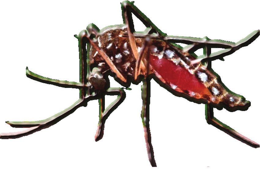
Dengue infection is caused by dengue virus which is a mosquito-borne
flavivirus. It is transmitted by Aedes aegypti and Aedes albopictus. There
are four distinct serotypes, DEN-1, 2, 3 and 4. Each episode of infection
induces a life-long protective immunity to the homologous serotype but
confers only partial and transient protection against subsequent infection
by the other three serotypes. Secondary infection is a major risk factor
for DHF due to antibody-dependent enhancement. Other important
contributing factors for DHF are viral virulence, host genetic background,
T-cell activation, viral load and auto-antibodies.
All four serotypes can be isolated at any one time but the predominant
circulating dengue virus will show a sinusoidal pattern (Figure 5). For
example, DEN-3 was the predominant serotype in the early 90s with a peak
in 1993, and then subsequently declined. It then re-emerged, reaching the
peak in 2001. Other serotypes had been observed to be co-circulating at
the same time
Figure 5 : Percentage of Dengue Serotype in Malaysia, 1991-2007
100
90
80
70
60
% of Serotype
50
40
30
20
10
0
Den 1 (%) Den 2 (%) Den 3 (%) Den 4 (%)
3. CLINICAL MANIFESTATIONS AND PATHOPHYSIOLOGY
3.1 SPECTRUM OF DENGUE INFECTION
The incubation period for dengue infection is 4-7 days (range 3-14).2 It
may be asymptomatic or may result in a spectrum of illness ranging from
undifferentiated mild febrile illness to severe disease, with or without
plasma leakage and organ impairment. Symptomatic dengue infection is a
systemic and dynamic disease with clinical, haematological and serological
profiles changing from day to day. These changes accelerate by the hour
or even minutes during the critical phase, particularly in those with plasma
leakage (refer to section 3.3).
3Understanding the systemic and dynamic nature of dengue disease as well
as its pathophysiological changes during each phase of the disease will
produce a rational approach in the management of dengue
3.2 CLINICAL COURSE OF DENGUE INFECTION
Dengue infection is a dynamic disease. Its clinical course changes as the
disease progresses. After the incubation period, the illness begins abruptly
and will be followed by 3 phases: febrile, critical and recovery phase (refer
Figure 6). 3, 4
i. Febrile Phase
Typically, patients develop high grade fever suddenly. This acute febrile
phase usually lasts 2-7 days and often accompanied by facial flushing,
skin erythema, generalised body ache, myalgia, arthralgia and headache.3,4
Some patients may have sore throat, injected pharynx and conjunctival
injection. Anorexia, nausea and vomiting are common. These clinical
features are indistinguishable between DF and DHF.5
Mild haemorrhagic manifestations like positive tourniquet test or petechiae
and mucosal membrane bleeding may be seen in DF and DHF.5,6 Per
vaginal bleeding is common among young adult females. Massive vaginal
bleeding and gastrointestinal bleeding may occur during this phase but
are not common.7, 6 The findings of an enlarged and tender liver are more
suggestive of DHF.5
The earliest abnormality in the full blood count is a progressive decrease
in total white cell count. This should alert the physician to a high index
of suspicion of dengue especially when there is positive history of
neighborhood dengue. This disease should be notified as early as possible
to prevent disease from assuming epidemic proportion.
ii. Critical Phase
The critical phase occurs towards the late febrile phase (often after 3rd day
of fever) or around defervescence (usually between 3rd to 5th day of illness
but may go up to 7th day) when a rapid drop in temperature may coincide
with an increase in capillary permeability in some patients. In other viral
infections, the patient’s condition improves as the temperature subsides,
but the contrary happens in DHF. At this point the patient will either become
better if no or minimal plasma leak occurs, or worse if a critical volume of
plasma is lost.3, 4, 8, 9
The critical phase lasts about 24-48 hours. (refer Figure 6) Varying
circulatory disturbances (refer to Table 1) can develop. In less severe cases,
these changes are minimal and transient. Many of these patients recover
spontaneously, or after a short period of fluid or electrolyte therapy. In
more severe forms of plasma leakage, the patients may sweat, become
restless, have cool extremities and prolonged capillary refill time. The
pulse rate increases, diastolic blood pressure increases and the pulse
pressure narrows. Abdominal pain, persistent vomiting, restlessness,
4REVISED
JULY 2010
altered conscious level, clinical fluid accumulation, mucosal bleed or
tender enlarged liver are the clinical warning signs of severe dengue or high
possibility of rapid progression to shock.9, 10, 11 The patient can progress
rapidly to profound shock and death if prompt fluid resuscitation is not
instituted.
It is important to note that thrombocytopaenia and haemoconcentration
(evidenced by a raised haemotocrit (HCT) from baseline or a drop in HCT
after rehydration) are usually detectable before the subsidence of fever
and the onset of shock. Refer to 3.5.1 for further details. The HCT level
correlates well with plasma volume loss and disease severity. However, the
levels of HCT may be equivocal when there is frank haemorrhage, early and
excessive fluid replacement or untimely HCT determinations.
Leucopaenia with relative lymphocytosis, clotting abnormalities, elevation
of transminases [typically the level of aspartate aminotransaminase
(AST) is about 2-3 times the level of alanine aminotransaminase (ALT)],
hypoproteinaemia and hypoalbuminaemia are usually observed.3, 4, 5
iii. Recovery Phase
After 24-48 hours of defervescence, plasma leakage stops and is followed
by reabsorption of extravascular fluid. Patients’ general well being improves,
appetite returns, gastrointestinal symptoms abate, haemodynamic status
stabilises and diuresis ensues. Some patients may have a classical rash
of “isles of white in the sea of red”.3 Some may experience generalised
pruritus. Bradycardia and electrocardiographic changes are not uncommon
during this stage. It is important to note that during this phase, HCT level
stabilises or drops further due to haemodilution following reabsorption of
extravascular fluid. The recovery of platelet count is typically preceded by
recovery of white cell count (WCC).
Figure 6 : CLINICAL COURSE OF DHF12
Course of
dengue illness FEBRILE CRITICAL RECOVERY
Days of illness 1 2 3 4 5 6 7 8 9 1 0
Temperature 40
Potential Shock / Bleeding Reabsorption / Fluid overload
Dehydration
clinical issues
Organ Impairment
Laboratory Platelet
changes
Hematocrit
Viraemia IgM/IgG
Serology
and virology
Note : Onset of defervescence usually occurs between day 3 to day 5 of illness
5• Clinical deterioration often occurs during the critical phase
(plasma leakage) and it is therefore crucial to recognise the
onset of this phase.
• The onset of critical phase is marked by plasma leakage and
usually occurs around the onset of defervescence.
• Evidence of plasma leakage includes raised HCT (early marker),
haemodynamic instability, fluid accumulation in extravascular
space (rather late marker) or hypoproteinemia.
• Abdominal pain, persistent vomiting, restlessness, altered
REVISED conscious level, clinical fluid accumulation, tender enlarged
JULY 2010
liver or mucosal bleed are the clinical warning signs of severe
dengue or high possibility of rapid progression to shock.
3.3 PATHOPHYSIOLOGY OF PLASMA LEAKAGE IN DENGUE
HAEMORRHAGIC FEVER (DHF)/DENGUE SHOCK SYNDROME (DSS)
The primary pathophysiological abnormality seen in DHF and DSS is an
acute increase in vascular permeability that leads to leakage of plasma
into the extravascular compartment, resulting in haemoconcentration and
hypovolaemia or shock.13,3,4 Hypovolaemia leads to reflex tachycardia and
generalised vasoconstriction due to increased sympathetic output.14,15
Clinical manifestations of vasoconstriction in various systems are as
follows :
a. Skin - coolness, pallor and delayed capillary refill time
b. Cardiovascular system - raised diastolic blood pressure and a narrowing
of pulse pressure
c. Renal system - reducing urine output
d. Gastrointestinal system - vomiting and abdominal pain
e. Central nervous system – lethargy, restlessness, apprehension, reduced
level of consciousness
f. Respiratory system – tachypnoea (respiratory rate >20/min)
In patients whose consciousness is not obtunded, intense thirst is another
prominent symptom. At the same time, the inadequate perfusion of the
tissue leads to increased anaerobic glycolysis and lactic acidosis. If the
hypovolaemia is not corrected promptly, the patient will progress to a
refractory shock state. By then, the tissue perfusion would not respond to
vasopressor drugs, even if the blood pressure and intravascular volume
were to be restored and cardiac output would remain depressed. The
resultant lactic acidosis further depresses the myocardium and worsens
the hypotension.15 The common late complications of prolonged shock
are massive bleeding, disseminated intravascular coagulopathy (DIC) and
multi-organ failure which are often fatal.
The following table is the summary of the continuum of various
pathophysiological changes in a patient who progresses from normal
circulatory state to hypovolaemic shock.
6Table 1 : A continuum of pathophysiological changes from normal circulation to
compensated and decompensated/ hypotensive shock (Adapted from15 )
Decompensated /
Normal Circulation Compensated shock
Hypotensive shock
Clear consciousness – shock can be Change of mental state – restless,
Clear consciousness
missed if you do not touch the patient combative or lethargy
Mottled skin, very prolonged
Brisk capillary refill time (2 sec)
capillary refill time
Warm and pink extremities Cool extremities Cold, clammy extremities
Good volume peripheral pulses Weak & thready peripheral pulses Feeble or absent peripheral pulses
Severe tachycardia with
Normal heart rate for age Tachycardia
bradycardia in late shock
Normal systolic pressure with
Normal blood pressure for age raised diastolic pressure Hypotension/unrecordable BP
Postural hypotension
Narrowed pulse pressure
Normal pulse pressure for age Narrowing pulse pressure
(3.4 TOURNIQUET TEST
In DHF grade 1, a positive tourniquet test serves as the only indicator of
haemorrhagic tendency. The sensitivity of the test varies widely from as low
as 0% to 57%, depending on the phase of illness the test was done and
how often the test was repeated, if negative. In addition 5-21% of patients
with dengue like illness had positive tourniquet test but subsequently have
negative dengue serology.22, Level 1
A recent study demonstrated that there was 95.3% positive preditive
value if fever, positive tourniquet test, leucopenia/ thrombocytopaenia/
haemoconcentration were used as screening criteria.23, Level 8
The tourniquet test may be useful as an additional tool when the diagnosis
is in doubt, especially when the platelet count is still relatively normal.
How to perform tourniquet test
Inflate the blood pressure cuff on the upper arm to a point midway
between the systolic and diastolic pressures for 5 minutes.
A positive test is when 20 or more petechiae per 2.5 cm (1 inch)
square are observed.
Recommendation
The tourniquet test may be helpful in the early febrile phase (less
than three days) in differentiating dengue from other febrile illnesses.
(Grade C)
3.5 WHO DENGUE CLASSIFICATION
Based on current WHO dengue classification scheme (refer Appendix 1), the
key differentiating feature between DF and DHF is the presence of plasma
leakage in DHF. However, in the early febrile phase of dengue infection,
the symptoms can overlap and one cannot differentiate DF and DHF.
DHF is further classified as mild (grades I and II) or severe (grades III and
IV), the presence of shock being the main difference. Grades III and IV are
classified as Dengue Shock Syndrome (refer Appendix 1).
(Note : The existing WHO dengue classification is being reviewed and revised)
3.5.1 Limitations of WHO classification 22, Level 1
It has been observed that the existing WHO classification scheme has
several limitations as the disease has spread to new regions and infected
older age groups. For example:
1. Dengue with shock without fulfilling all the 4 criteria for DHF
There have been many case reports of patients with severe dengue with shock
who do not fulfil all the 4 criteria for DHF. These patients would have been
classified as dengue fever if the WHO criteria are to be strictly applied.
2. Severe organ impairment
Patients with severe organ impairment such as liver, respiratory,
cardiac and brain dysfunction are not captured as having severe
disease based on the existing classification.
83. Plasma leakage in DHF
The requirement of 20% increase in HCT as one of the evidence of
plasma leakage is difficult to fulfill due to several issues:
a. Baseline HCT is not available in most patients and therefore, the
interpretation of plasma leak can only be made retrospectively
b. Early fluid administration may affect the level of HCT
c. Bleeding will affect the HCT level
4. The existing classification scheme is often not useful for disease
management because the correct disease classification can only be
made towards the end of the illness.
Patients can present with severe dengue without fulfilling ALL the
4 criteria (refer Appendix 1) for DHF/DSS.
3.5.2 Suggested WHO Classification 2009
The classification into levels of severity has a high potential for being of
practical use in the clinicians’ decision as to where and how intensively
the patient should be observed and treated (i.e. triage, which is particularly
useful in outbreaks).
Figure 7: Suggested Dengue Classification and Level of Severity
REVISED
JULY 2010
Figure 1.4 Suggested dengue case classification and levels of serverity
DENGUE + WARNING SIGNS SEVERE DENGUE
1. Severe plasma leakage
with warning signs 2. Severe haemorrhage
without 3. Severe organ impairment
CRITERIA FOR DENGUE + WARNING SIGNS CRITERIA FOR SEVERE DENGUE
Probable dengue Warning signs* Severe plasma leakage leading to :
live in/ travel to dengue endemic area. l Abdominal pain or tenderness l Shock (DSS)
Fever and 2 of the following criteria: Persistent vomiting
l
l Fluid accumulation with respiratory distress
l Nausea, vomiting l Clinical fluid accumulation
l Rash Severe bleeding
Mucosal bleed
Aches and pains
l
l as evaluated by clinician
l Torniquet test positive l Lethargy, restlessness
l Leukopenia l Liver enlargment > 2cm Severe organ involvement
l Any warning sign l Laboratory: increase in HCT concurrent Liver : AST or ALT > = 1000
with rapid decrease in platelet count CNS : Impaired consciousness
Laboratory-confirmed dengue
(important when no sign of plasma leakage) * (requiring strict observation and medical intervention) Heart and other organs
Source: World Health Organization. Dengue Guidelines for Diagnosis, Treatment,
Prevention and Control - New Edition 2009. WHO: Geneva; 2009
3.6 OTHER IMPORTANT MANIFESTATIONS
Severe bleeding or organ impairment might occur without plasma leakage.
The Following manifestations are important in dengue infection but are
often under- recognised or misdiagnosed:
91. Acute abdomen :
Acute abdominal pain is a common symptom in dengue infection. It can
be due to hepatitis, acalculous cholecystitis and shock, and occasionally
misdiagnosed as acute appendicitis.24, Level 8; 25, Level 8 The history of
onset of fever before the abdominal pain, and laboratory findings of
leucopenia, thrombocytopenia, or prolonged APTT with normal PT
help to differentiate acute abdominal pain due to dengue infection from
other surgical causes.24, Level 8 Furthermore, in patients with shock, the
abdominal pain is relieved by intravenous fluid therapy.
2. Hepatitis and liver failure :
Hepatitis is common in patients with DF/DHF and may be mild or
severe regardless of the degree of plasma leakage. In some cases,
liver failure may occur.22, Level 1 The patients with liver failure have a high
propensity to bleed, especially gastrointestinal bleeding. 26, Level 8; 11, Level 8
3. Neurological manifestation :
Patients with dengue infection may have neurological manifestations,
(FEBRILE PHASE
Table 2 : Differential diagnoses for dengue illness during febrile phase
Clinical syndrome Differential diagnoses
Influenza
Measles
Chikungunya
Flu-like syndrome
Adenovirus
Infectious mononucleosis
Acute HIV seroconversion illness
Rubella
Measles
Scarlet fever
Rash
Meningococcal infection
Chikungunya
Drug
Rotavirus
Diarrhea
Food poisoning
Meningoencephalitis
Neurological manifestation
Febrile seizures
CRITICAL PHASE
Table 3: Differential diagnoses for dengue illness during critical phase
Clinical syndrome Differential diagnoses
Acute appendicitis
Acute cholecystitis
Acute abdomen Perforated viscus
Viral hepatitis
Diabetic ketoacidosis
Shock Septic shock
Diabetic ketoacidosis
Respiratory distress
Renal failure
(Kussmaul’s breathing)
Lactic acidosis
Acute leukaemia
Immune thrombocytopaenia purpura
Thrombotic Thrombocytopenic purpura
Leucopaenia &
Malaria / Leptospirosis / Typhoid / Typhus
thrombocytopenia ± bleeding
Bacterial sepsis
SLE
Acute HIV seroconversion illness
4. DISEASE NOTIFICATION
All suspected dengue cases must be notified by telephone to the nearest
health office within 24 hours of diagnosis, followed by written notification
within a week using the standard notification format.29 Any delay in notification
will increase the risk of dengue transmission in the locality of the residence.
In 2007, 98.4% of dengue cases were notified by public and private hospitals
with only 1.6% from the government and private clinics. The average day of
illness at the time of notification was about 4-5 days after the onset of illness
even though patients might have presented themselves to the healthcare
facilities at day 1-3 day of fever.
11Notification should be done as soon as a clinical diagnosis of dengue is
suspected; serological confirmation is not necessary. Notified cases will be
followed up by the health authorities for the verification of case definition
and preventive measures. It is also important to note that re-notification
has to be done if the diagnosis has been changed from DF to DHF or DF
to other diagnosis.
Failure to notify is liable to be compounded under the Prevention and
Control of Infectious Diseases Act, 1988 (Act 342).30
5. LABORATORY INVESTIGATIONS
5.1 DISEASE MONITORING LABORATORY TESTS
Full Blood Count (FBC)
1. White cell count (WCC) :
In the early febrile phase WCC is usually normal but will decrease
rapidly as the disease progresses.5, Level 8 This trend of leucopenia
should raise the suspicion of possible dengue infection.
2. Haematocrit (HCT) :
A rising HCT is a marker of plasma leakage in dengue infection and
helps to differentiate between DF and DHF but it can be masked in
patients with concurrent significant bleeding and in those who receive
early fluid replacement.22, Level 1 Setting the patient’s baseline HCT in the
early febrile phase of disease will be very useful in the recognition of
a rising HCT level.
3. Thrombocytopaenia :
Thrombocytopaenia is commonly seen in dengue infection.22, Level 1 In
the early febrile phase, platelet count is usually within normal range
but it will decrease rapidly as the disease progresses to the late febrile
phase or at defervescence and it may continue to remain low for the
first few days of recovery.
There is a significant negative correlation between disease severity
and platelet count 3, Level 9; 31 Level 8 but it is not predictive of bleeding.32,
Level 8; 33, Level 1; 34, Level 6; 35, Level 8; 36, Level 8
Liver Function Test
Elevated liver enzymes is common and is characterised by greater elevation of
the AST as compared to the ALT.37, Level 8 The frequency and degree of elevation
of the liver enzymes are higher with DHF compared to DF.38, Level 8; 37, Level 8
12Leucopaenia followed by progressive thrombocytopaenia is
suggestive of dengue infection.
A rising HCT accompanying progressive thrombocytopaenia is
suggestive of DHF.
There is no local data available on the normal range of HCT in adults.
In the absence of a baseline HCT level, a HCT value of >40% in
female adults and >46% in male adults should raise the suspicion
of plasma leakage.
Recommendation
The baseline HCT and WCC should be established as early as
possible in all patients with suspected dengue. (Grade A)
Serial FBC and HCT must be monitored as the disease progresses.
(Grade A)
5.2 DIAGNOSTIC TESTS
Definitive diagnosis of dengue infection can only be confirmed in the
laboratory. However, the interpretation of laboratory diagnostic results
should be done in the clinical context. Laboratory confirmatory tests include
antibody detection (serology), virus isolation, detection of virus genetic
materials (polymerase chain reaction -PCR) and detection of dengue virus
protein (NS1 antigen).
5.2.1 DENGUE SEROLOGY TESTS
Haemagglutination Inhibition Test
The haemagglutination Inhibition (HI) test has been the gold standard for
serological diagnosis. However, because it is labour intensive and requires
paired samples for interpretation, this test is now being used mainly for
research purposes to differentiate between primary and secondary dengue
infections.
Dengue IgM test
The IgM capture enzyme-linked immunosorbent assay (ELISA) is the most
widely used serological test. This antibody titre is significantly higher in
primary infections, compared to secondary infections. Once the IgM is
detectable, it rises quickly and peaks at about 2 weeks after the onset of
symptoms, and it wanes to undetectable levels by 60 days. However in
some patients, it may persist for more than 90 days. A positive result thus
has to be intepreted and correlated cautiously with the clinical picture. If
the dengue IgM test is the only available diagnostic test in the hospital,
then establishing a negative IgM early in the illness, and demonstrating a
positive serology later will be essential to exclude false negative results.
13In one study, IgM was detected in only 55% of patients with primary dengue
infections between day 4-7 onset of fever, and it became positive in 100%
of the patients after day 7. However, in secondary dengue infections, IgM
was detected in only 78% of patients after day 7.39, Level 7. In another study,
28% of secondary dengue infections were undiagnosed when IgM was the
only test performed.4, Level 9; 40, Level 8; 41, Level 8
Indirect IgG ELISA test
In primary and secondary dengue infection, dengue IgG was detected in
100% of patients after day 7 of onset of fever. Therefore dengue IgG is
recommended if dengue IgM is still negative after day 7 with the negative
IgG in the initial test sample.39, level 7; 40, level 8; , level 8
Recommendation
In order to establish serological confirmation of dengue illness a
seroconversion of dengue IgM needs to be demonstrated. Therefore
a dengue IgM should be taken as soon as the disease is suspected.
(Grade C)
Dengue IgM is usually positive after day 5-7 of illness. Therefore a
negative IgM taken before day 5-7 of illness does not exclude dengue
infection. (Grade B)
If dengue IgM is negative before day 7, a repeat sample must be taken
in recovery phase. (Grade B)
If dengue
Please referIgM is still negative
to Appendix after day of
2 for methods 7 with negative
sample IgG tested at less
collection
then 7 days, dengue IgG is recommended for diagnostic confirmation.
(GradeRapid
Dengue C) tests
Simple rapid tests such as the strip assays (immunochromatography test)
are available for qualitative detection of dengue IgM and IgG (e.g. Pan Bio
Dengue IgM ELISA and Dengue IgM Dot Enzyme Immunoassay).
The yield of rapid tests was shown to be higher when samples were
collected later in the convalescent phase of infection, with good specificity
and could be used when ELISA test were not available 43, Level 1 But the
result had to be interpreted in the clinical context because of false positive
and negative results.44, Level 8; 45, Level 8; 41, Level 8; 46, Level 8 It is recommended that
the dengue IgM Capture ELISA test be done after a rapid test, to confirm
the status.44, Level 8
Note : False positive dengue serology
Serological tests for dengue have been shown to cross-react with:
• other flavivirus – Japanese Encephalitis.47, Level 9; 41, Level 8
• non-flavivirus – malaria, leptospirosis, toxoplasmosis, syphilis48, Level ; 45, Level 8
• connective tissue diseases – rheumatoid arthritis44, Level 8
145.2.2 VIRUS ISOLATION
Virus isolation is the most definitive test for dengue infection. It can only be
performed in the lab equipped with tissue culture and other virus isolation
facilities. It is useful only at the early phase of the illness. Generally, blood
should be collected before day 5 of illness; i.e. before the formation of
neutralizing antibodies.
During the febrile illness, dengue virus can be isolated from serum, plasma
and leucocytes. It can also be isolated from post mortem specimens. The
monoclonal antibody immunofluorescence test is the method of choice for
identification of dengue virus. It may take up to two weeks to complete the
test and it is expensive.
Note: Virus isolation has a poor yield if compared with molecular tests. It is most probably due
to the viability of the virus and the quality of the samples.49, Level 8
5.2.3 POLYMERASE CHAIN REACTION (PCR)
Molecular tests such as the reverse transcriptase – ploymerase chain
reaction (RT- PCR) are useful for the diagnosis of dengue infection in the
early phase (< 5 days of illness). It was shown to have a sensitivity of 100%
in the first 5 days of disease, but reduced to about 70% by day 6, following
the disappearance of the viraemia.50, Level 8; 42, Level 8; 51, Level 8
An additional advantage of RT- PCR is the ability to determine dengue
serotypes 52, Level 7; 42, Level 8; 53, Level 8; 49, Level 8; 54, Level 8
Limitations of RT- PCR are:
a) This test is only available in a few centres with facilities and trained
personnel (e.g. IMR, HKL, National Public Health Laboratory and University Malaya
Medical Centre).
b) The test is expensive
c) The specimen requires special storage temperatures and short
transportation, time between collection and extraction (refer Appendix 1)
In view of these limitations, the use of RT- PCR should only be considered
for in-patients who present with diagnostic challenges in the early phase
of illness.
5.2.4 NON-STRUCTURAL PROTEIN-1 (NS1 Antigen)
NS1 antigen is a highly conserved glycoprotein that seems to be essential
for virus viability. Secretion of the NS1 protein is a hallmark of flavivirus
infecting mammalian cells and can be found in dengue infection as well
as in yellow fever and West Nile virus infection. This antigen is present in
high concentrations in the sera of dengue infected patients during the early
phase of the disease.55, Level 8; 56, Level 8
The detection rate is much better in acute sera of primary infection (75%-
97.3%) when compared to the acute sera of secondary infection (60% -
70%).57, Level 8; 58, Level 8; 59, Level 8; 60, Level 8 The sensitivity of NS1 antigen detection
drops from day 4-5 of illness onwards and usually becomes undetectable
in the convalescence phase.61, Level 8; 60, Level 8; 58, Level 8; 59, Level 8
15Recommendation
PCR can be used as a diagnostic tool in early dengue infection (Grade
B). It is not recommended as a routine diagnostic test due to limited
availability and cost. (Grade C)
NS1 Ag is a new diagnostic tool that may be useful in the early phase of
dengue infection. It is not useful in the convalescence phase. However,
this test is still undergoing evaluation. (Grade C)
Please refer to Appendix 2 for methods of sample collection for dianostic tests
6. INVESTIGATION OF POST MORTEM CASE
Suitable samples for viral isolation and PCR should be obtained from the
liver, lung, thymus, spleen, lymph nodes, CSF, pleural fluid and brain tissues
in a patient suspected to have died of DF/DHF.2, Level 9; 62,Level 9 However PCR
is a more sensitive method.62 Level 9 ; 63, Level 7 ; 64, Level 8
For serological confirmation of dengue illness a seroconversion of dengue
IgM needs to be demonstrated. In a patient who has died suspected of
dengue, a repeat dengue serology together with the samples mentioned
above should be obtained.
Caution : Massive blood transfusion may affect the test results mentioned above.
Recommendation
A repeat dengue serology should be obtained at the time of death.
(Grade C)
Suitable specimens for virus isolation and/ or RT-PCR and/ or antigen
detection are recommended for confirmation of diagnosis. (Grade C)
Please refer to Appendix 2 for methods of sample collection.
7. MANAGEMENT OF DENGUE INFECTION
7.1 OUTPATIENT MANAGEMENT
The management of dengue infection is symptomatic and supportive.
A stepwise approach as suggested in Table 4 can be useful.
Dengue is a dynamic disease and management issues vary according
to the 3 phases of the clinical course (refer to section 7.4). It is crucial to
recognise plasma leakage, shock early or severe organ impairment. This
can be achieved by frequent clinical and laboratory monitoring.
Dengue patients who are managed in the outpatient setting should be
provided with a disease monitoring record (refer to Appendix 4) to ensure
that all relevant information is available to all health care providers.
Primary care providers with no immediate haematocrit facilities should
refer patient to the nearest health facility for further management.
16Table 4 : A Stepwise Approach On Outpatient Management of Dengue Infection
It is important to evaluate every patient in a stepwise manner as in the following :
Step 1: Overall assessment
1. History
• Date of onset of fever/ illness
• Oral intake
• Assess for alarm signs – refer to Table 5
• Diarrhoea
• Bleeding
• Change in mental state/seizure/dizziness
• Urine output (frequency, volume and time of last voiding)
• Other important relevant histories:
- Family or neighbourhood history of dengue
- Jungle trekking and swimming in waterfall (consider leptospirosis, typhus, malaria)
- Recent travel
- Recent unprotected sexual or drug use behaviour (consider acute HIV seroconversion illness)
- Co-morbidities (consider sepsis particularly in patients with diabetes mellitus)
2. Physical examination
i. Assess mental state and Glasgow Coma Scale (GCS) score
ii. Assess hydration status
iii. Assess haemodynamic status
- Skin colour
- Cold/ warm extremities
- Capillary filling time (normalTable 5 : Warning signs 8, level 8, 9, level 8
• Abdominal pain or tenderness
• Persistent vomiting
• Clinical fluid accumulation (pleural effusion, ascites)
• Mucosal bleed
• Restlessness or lethargy
REVISED
JULY 2010 • Tender enlarged liver
• Laboratory : Increase in HCT concurrent with rapid decrease in platelet
Table 6 : Clinical and Laboratory Criteria for Patients Who Can be Treated at Home
1. Able to tolerate orally well, good urine output and no history of bleeding
2. Absence of clinical alarm signals (refer Table 5)
3. Physical examination:
• Haemodynamically stable
- pink, warm extremities
- normal capillary filling time (normal 20mmHg)
- no disproportionate tachycardia
• No tachypnoea or acidotic breathing
• No hepatomegaly or abdominal tenderness
• No bleeding manifestation
• No sign of pleural effusion ascites
• No alterations in mental state and full GCS score
4. Investigation:
• Stable serial HCT
• In the absence of a baseline HCT level, a HCT value of >40% in female adults
and >46% in male adults should raise the suspicion of plasma leakage.
Therefore admission may be required
Adapted from 65, Level 9; 66, Level 9; 67, Level 9
7.2 PATIENT TRIAGING AT EMERGENCY & TRAUMA / OUTPATIENT DEPARTMENT
The purpose of triaging patients is to determine whether they require urgent
attention. This is to avoid critically ill patients being missed upon arrival.
68, Level 9; 65 Level 9; 69, Level 9; 70, Level 9
Triage Checklist:
1. History of fever
2. Abdominal pain
3. Vomiting
4. Dizziness/ fainting
5. Bleeding
Vital parameters to be taken :
Mental state,blood pressure, pulse, temperature, cold or warm peripheries
187.3 CRITERIA FOR HOSPITAL REFERRAL / ADMISSION
7.3.1 Referral from primary care providers to hospital
The decision for referral and admission must not be based on a single clinical
parameter but should depend on the Total Assessment of the patient.
Referral from primary care providers to hospital
1. Symptoms :
• Alarm signals (refer to Table 5)
• Bleeding manifestations
• Inability to tolerate oral fluids
• Reduced urine output
• Seizure
2. Signs :
• Dehydration
• Shock (refer to Table 1)
• Bleeding
• Any organ failure
3. Special Situations :
• Patients with co-morbidity e.g.Diabetes, Hypertension, Ischaemic
Heart Disease, Coagulopathies, Morbid Obesity, Renal Failure,
Chronic Liver disease, COPD,
• Elderly (more than 65 years old)
• Pregnancy
• Social factors that limit follow-up e.g. living far from health
facility, no transport, patient living alone, etc
4. Laboratory Criteria: Rising HCT accompanied by reducing platelet count
7.3.2 Referral From hospitals without specialist to hospitals with specialists
Early consultation with the nearest physician should be initiated for ALL
DHF or DF with organ dysfunction/ bleeding.
Prerequisites for transfer
1. All efforts must be taken to optimise the patient’s condition before
and during transfer.
2. The Emergency Department and/or Medical Department of the receiving
hospital must be informed prior to transfer.
3. Adequate and essential information must be sent together with the
patients that includes fluid chart, monitoring chart and investigation
results.
197.4 DISEASE MONITORING
7.4.1 Principles of Disease Monitoring
1. Dengue is a systemic and dynamic disease. Therefore disease
monitoring is governed by different phases of the disease.
2. The critical phase (plasma leakage) may last for 24-48 hours. Monitoring
needs to be intensified and frequent adjustments in the fluid regime
may be required.
3. Recognition of onset of reabsorption phase is also important
because intravenous fluid regime needs to be progressively reduced/
discontinued at this stage.
7.4.2 Outpatient Disease Monitoring
Every patient suspected of dengue attending the outpatient/ emergency
and trauma department should be assessed in stepwise manner as
recommended in Table 4.
Daily or more frequent follow up is necessary especially from day 3 of
illness, until the patient becomes afebrile for at least 24- 48 hours without
antipyretics. A disease monitoring record has been developed and it is
recommended to be used for outpatient care (refer to Appendix 4.).
7.4.3 Inpatient Disease Monitoring
Immediately after admission every patient with suspected dengue should
be reviewed thoroughly similar to the stepwise approach in outpatient (refer
to Table 4). The plan of management and monitoring should be based on
the phase of the disease and the haemodynamic status of the patient.
Table 8 summarises the parameters and frequency of monitoring according
to the different phases of the illness.
20Table 7: Issues of Monitoring According to Different Phases Of Dengue Illness
Phases of illness Issues :
- Differentiation of dengue illness from other febrile
illnesses.
Febrile
- Not possible to differentiate DF from DHF.
- Plasma leakage occurs as patient progresses to
late febrile phase or as temperature begins to
defervescence (T < 38.0 °C).
- Clinical deterioration occurs during this phase due to
plasma leakage.
- Plasma leakage results in haemoconcentration and
hypovolemia/ shock.
Critical
- Excessive plasma leakage due, in part, to intravenous
fluid therapy may cause respiratory distress.
- Bleeding can be precipitated by prolonged shock and
shock can be perpetuated by bleeding.
- May mimic acute abdomen of other causes.
- May be confused with septic shock or other forms
of shock.
- Cessation of plasma leakage.
- Reabsorption of fluid from extravascular compartment.
Reabsorption
- Haemodilution occurs following fluid reabsorption.
- Hypervolaemia and pulmonary oedema if intravenous
fluid therapy is continued.
21Table 8 : Parameters and Frequency of Monitoring According to Different
Phases of Dengue Illness
Frequency of monitoring
Parameters for monitoring
Febrile phase Critical phase Recovery phase
Clinical Parameters
General well being
At least twice a
Appetite/ oral intake Daily or more Daily or more
day and more
Warning signs frequently towards frequently as
frequently as
Symptoms of bleeding late febrile phase indicated
indicated
Neurological/ mental state
Haemodynamic status
• Pink/ cyanosis
• Extremities (cold/warm) 2-4 hourly
• Capillary refill time depending on
clinical status
• Pulse volume
4-6 hourly
• PR
depending on In shock 4-6 hourly
• BP
clinical status Every 15-30
• Pulse pressure
minutes till
stable then 1-2
Respiratory status hourly
• RR
• SpO2
At least twice a
Signs of bleeding, abdominal Daily or more Daily or more
day and more
tenderness, ascites and frequently towards frequently as
frequently as
pleural effusion late febrile phase indicated
indicated
2-4 hourly
Urine output 4 hourly In shock 4-6 hourly
Hourly
Frequency of monitoring
Parameters for monitoring
Febrile phase Critical phase Recovery phase
Clinical Parameters
4-12 hourly
depending on
clinical status
In shock
Daily or more Repeated
FBC + HCT frequently if before and Daily
indicated after each
attempt of fluid
resuscitation
and as
indicated
At least daily or
more frequently
BUSE/ Creatinine
as indicated
LFT
RBS As indicated In shock As indicated
Coagulation profile Crucial to
monitor acid-
HCO3 / TCO2 / Lactate
base balance/
ABG closely
Adapted from 2, Level 9; 65, Level 9
227.5 FLUID MANAGEMENT
7.5.1 Dengue with Warning Signs (refer to Table 5)
Recognising and monitoring for warning signs are crucial in identifying
patients who may deteriorate into severe dengue.
• All patients with warning signs should be considered for monitoring
in hospitals.
• Common pitfalls in fluid therapy:
* Treating patient with unnecessary fluid bolus based on raised
* HCT as the sole parameter without considering other clinical
parameters
* Excessive and prolonged fixed fluid regime in stable patients
* Infrequent monitoring and adjustment of infusion rate
* Continuation of intravenous fluid during the recovery phase
Cases of dengue with warning signs will probably recover with early
intravenous rehydration. Some cases will deteriorate to severe dengue. If
the patient has dengue with warning signs, the action plan should be as
in Table 9a.
Table 9a: Action Plan for Patient who has Dengue with Warning Signs
• Obtain a baseline HCT before fluid therapy.
REVISED
JULY 2010
• Give crystalloids solution (such as 0.9% saline).
• Start with 5-7 ml/kg/hour for 1-2 hours, then reduce to 3-5 ml/kg/hr for 2-4 hrs
and then reduce to 2-3 ml/kg/hr or less according to the clinica response.
• If the clinical parameters are worsening and HCT is rising, increase the rate of
infusion.
• Reassess the clinical status, repeat the HCT and review fluid infusion rates
accordingly
7.5.2 Non-Shock Patients (DHF Grade I & II)
There are no studies that have looked at fluid therapy in non shock dengue
patients. Increased oral fluid intake may be sufficient in some patients who are
haemodynamically stable and not vomiting. However IV fluid (0.9% saline is
recommended) is indicated in patients with increasing HCT (indicating on-going
plasma leakage) despite increased oral intake. IV fluid therapy should also be
considered in patients who are vomiting and not tolerating orally.71, level 9; 65, level 9
The normal maintenance requirement for IV fluid therapy in such patients
could be calculated based on the formula in Table 9b. Frequent adjustment
of maintenance fluid regime is often needed during the critical phase.
Often 1.2-1.5 times the normal maintenance will be required during the
critical phase. If the fluid infusion rate exceeds more than the maintenance
requirement, the infusion rate should be reviewed within 4 to 6 hours.
A rising HCT AND/ OR haemodynamic instability indicates on-going
plasma leakage and will require an increase in the IV fluid infusion rate. If
patients deteriorate and progress to shock, fluid resuscitation is indicated
(refer to the section on 7.5.3).71, level 9; 65, level 9
Reduce or consider discontinuation of IV fluid therapy when patients begin
to show signs of recovery (usually after 24-48 hours of defervescence, or
the HCT drops in a stable patient).
23You can also read